DOCTORAL SCHOOL OF BIOLOGY
Section “Biomolecular and cellular”
XXIV cycle
Role of AMBRA1 in nervous tissue homeostasis
and in neurodegeneration.
Ruolo di AMBRA1 nell’omeostasi del tessuto
nervoso e in processi neurodegenerativi.
Candidate: Sara Sepe
Tutors: Dr. Sandra Moreno, University of “Roma Tre
Dr. Pier Giorgio Mastroberardino, Erasmus Medical Center
Coordinator: Prof. Paolo Mariottini
Contents
Abstract i
Riassunto iv
Section I: Introduction and Obj
ectives1. Chapter 1 :Ambra1, autophagy and the nervous system 1
1.1. Ambra1 1
1.2. Autophagy 3
1.3. Autophagy in the nervous system 6
1.3.1. Autophagy and neurodegenerative disorder 8
2. Chapter 2 : Objectives 12
Section II: Results
3. Chapter 3: Distribution of AMBRA1 in mouse brain 15 3.1. Immunohistochemical and immunofluorescence analysis 15
3.2. Ultrastructural analysis 23
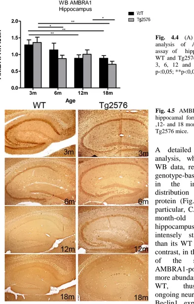
4. Chapter 4: AMBRA1 expression in physiological and Alzheimer-like ageing 24
4.1. Expression of AMBRA1 in the ageing neocortex 24
4.2. Expression of AMBRA1 in the ageing hippocampus 26
5. Chapter 5: AMBRA1 in parkinsonian neurodegeneration 28 5.1. AMBRA1 in a pharmacological model of pre-symptomatic PD 28
5.2. AMBRA1 in a genetic model of pre-symptomatic PD 30
Section III: Discussion and Conclusions
6. Chapter 6: Discussion 33
6.1. Distribution of AMBRA1 in mouse brain 33
6.2. AMBRA1 in neurodegeneration 39
6.2.1. AMBRA1 in physiological and Alzheimer–like ageing 6.2.2. AMBRA1 in mouse of model of Parkinson disease
7. Chapter 7: Conclusions and future perspectives 44
i
Abstract
My PhD project focused on the role of AMBRA1 in mouse brain in physiological and pathological conditions. Ambra1 (“Activating Molecule in Beclin 1 Regulated-Autophagy”) is a recently identified gene encoding for a large protein (around 130 kDa), with an N-terminal WD40 domain (Fimia et al., 2007). Ambra1 gene is highly conserved among vertebrates, and is expressed in different splicing isoforms. In the developing mouse, AMBRA1 protein shows abundant expression in the central and peripheral nervous system. At embryonic day 8.5 (E8.5), AMBRA1 is present in the neuroepithelium, while at E11.5 protein expression is localized to the ventral-most part of the spinal cord, the encephalic vesicles, the neural retina, and the dorsal root ganglia. This distribution pattern suggests that AMBRA1 is centrally involved in the proper development of the nervous system. Indeed, Ambra1gt/gt mice, obtained by gene trapping technique, and displaying a deficient expression of the protein, show early and severe neuropathological features, including neuroepithelial hyperplasia and defective neural tube closure, leading to embryonic death (Fimia et al., 2007; Cecconi et al., 2008). These disturbances appear to derive from dysfunctions in the autophagic process, also resulting in an imbalance of apoptotic cell death and cell proliferation. These data, together with in vitro evidence, allowed proving a regulatory role of AMBRA1 in autophagy activation in vertebrates (Fimia et a., 2007). Autophagy is involved in the intracellular turnover of proteins and cell organelles, and has an important role in regulating cell fate in response to stress (Levine, 2005). Three types of autophagy have been described: microautophagy, chaperone-mediated autophagy and macroautophagy. The last one is a bulk degradation pathway and the only intracellular mechanism potentially capable of degrading large protein aggregates or damaged organelles. A cup-shaped isolation membrane forms around cytosolic components, eventually fusing to form a double membrane bound vesicle, the so-called “autophagic vacuole” (AV) (Mizushima et al., 2002). In thi respect AMBRA1 is essential in the induction of AV formation (Di Bartolomeo et al, 2010). Recent studies show that macroautophagy is constitutively active in healthy neurons and is vital to cell survival (Bolland and Nixon, 2006). Mice lacking either Atg5 or Atg7 genes exhibit motor and behavioral deficits as well as degeneration of specific neuronal subtypes (Hara et al., 2006; Komatsu et al., 2006). Diffuse protein aggregates appear in surviving neurons within several brain regions, culminating in the formation of toxic inclusion bodies. This supports an essential role of autophagy is for neuronal health. The evidence so far presented and the severe neuropathological phenotype of Ambra1gt/gt mice prompted us to investigate the expression of AMBRA1 protein in adult mouse CNS, in physiological and pathological conditions.
The main results obtained from the research and described in this thesis can be summarized as follows:
ii
1. I provided the first neuroanatomical/histological/ ultrastructural map of the distribution of AMBRA1 expression in mouse brain taking advantage of immunohistochemical, immunofluorescence and immunoelectron microscopy approaches. Wide presence of the protein in the forebrain, midbrain, and hindbrain was observed, demonstrating prevalent expression in neurons, even though astrocytes and microglial cells also contain moderate levels of AMBRA1. This suggests that in physiological conditions AMBRA1 is crucial for neural tissue homeostasis. Ultrastructural analysis revealing association of with the endoplasmic reticulum strongly supports AMBRA1 activity in regulating basal autophagy, essential for neuronal survival. Detailed examination of different brain territories along the rostro-caudal axis allowed me to show that AMBRA1 content varies among brain regions, neuronal populations, and subtypes. The concentration of neuronal AMBRA1 appears at least partially related to cell volume. Neurons featuring a large soma, highly brached dendrites and long axons generally display higher immunoreactivity, compared to smaller cells. Among these, mitral cells in the olfactory bulb, giant pyramidal neurons of the neocortex, motor neurons of the brainstem, Purkinje cells of the cerebellar cortex are paradigmatic. Some of these giant neurons have also been described to contain other pro-autophagic molecules (Tamura et al., 2010), suggesting that their high metabolic and turnover rates are likely to involve large amounts of autophagy regulators, such as AMBRA1. Even more interestingly, we also found some correlation between AMBRA1 content and other parameters, namely susceptibility to damage. In particular, some neuronal subsets, which are selectively spared by neurodegenerative insults, demonstrably contain high levels of the protein. Representative examples of this concept are cholinergic interneurons in the corpus striatum, virtually unaffected by Huntington’s Disease (HD), and CA3 pyramidal neurons of the hippocampus, resistant to Alzheimer’s Disease (AD) and to ischemic injury. Importantly, this pathology is suggested to involve inefficient regulation of autophagic processes (Jaeger et al., 2009)). However, in some cases, neurons showing relatively high AMBRA1 content are target of specific neurodegenerative disease. For instance, Purkinje and mitral cells are both affected by the so-called “Purkinje cell death” (pcd) pathology. Remarkably, in this syndrome neuronal cell loss which probably occurs through the autophagic, rather than apoptotic, pathway, indicating that dysregulation of autophagy, possibly involving also AMBRA1 expression, may participate to some neuropathologies.
2. I highlighted important age-related variations in AMBRA1 expression in regions prone to neurodegeneration, such as the neocortex and hippocampus during normal and Alzheimer-like ageing. For this part of the project, I used a WT and transgenic strain for AD (Tg2576) at different time points.
In the neocortex, the high levels of AMBRA1 in normal young mouse led me to hypothesize that AMBRA1 is essential for a faultless maturation of neocortical neurons. Interestingly, Tg2576 mice show lower AMBRA1 content at the very
iii
onset of disease, when most of the histopathological hallmarks are still undetectable, thus suggesting the possible involvement of AMBRA1 down-regulation in the impaired plasticity of neuronal circuits. During normal adulthood, we found an overall decrease of AMBRA1 content which may reflect a relatively balanced cell activity, with a stable regulation of biosynthesis and degradation pathways. The novel increase of the protein in the aged neocortex, which is independent of the genotype, may instead suggest that AMBRA1 is up-regulated in this critical period, since brain ageing likely requires a higher rate of basal autophagy also to counteract oxidative stress and consequent accumulation of damaged organelles. Differences in AMBRA1 expression are not only related to age, but even to the specific brain region, since the the neocortex and the hippocampus show different behavior during ageing and pathological progression. In particular, overall levels in young mouse hippocampal formation are similar in the two genotypes. However, a different distribution in hippocampal subregions of Tg2576 animals compared to WT was detected. In fact, the neurogenic region of dentate gyrus is more intensely AMBRA1 immunoreactive in the transgenic animals than in WT, thus suggesting that in the pathological condition an early response involving enhanced neurogenesis may occur to cope the first deficits. Then we found a decrease in AMBRA1 level during adulthood and ageing in both genotypes, even though, at 18 months, the transgenic hippocampus shows a further drop in AMBRA1 reactivity, possibly contributing to neurodegeneration.
3. I showed that AMBRA1 expression can be induced in dopaminergic neurons by a mild 1-Methyl-4-phenyl-1,2,3,6-tetrahydropyridine (MPTP) treatment, producing a pharmacological model of early PD. Importantly, similar effects are also observed in a genetic model of genomic instability (ERCC1 mutants) which is associated with an early PD-like phenotype. These findings support the involvement of AMBRA1 in cellular response against oxidative imbalance in dopaminergic neurons that are closely related to PD onset. Surprisingly, AMBRA1 levels increase in both the substantia nigra (SN) and ventral tegmental area (VTA), the former of which turns to cell death in PD while the latter is spared. One could envision that the activation of the autophagic pathway by AMBRA1 up-regulation, can be defensive in some cases and detrimental in others, strictly depending on the specific neuronal population. To this respect, it is worth noting that the features of neuronal cell death in PD are still debated, because it seems to bring back to autophagic cell death. In conclusion, the results obtained in my PhD project suggest that AMBRA1 is a fundamental molecule in the nervous tissue, given the abundant content in neurons, although to different degree with respect to the brain area and neuronal population. In addition, modulation of AMBRA1 expression may be critical for establishing, promoting or counteracting neurodegenerating processes. Thus, our study opens the way to further investigations aimed to defining the precise contribution of AMBRA1 to nervous tissue development, homeostasis and response to acute or chronic injury.
iv
Riassunto
L’attività di ricerca svolta nei tre anni di dottorato si è focalizzata sul ruolo della proteina AMBRA1 nel cervello di topo in condizioni fisiologiche e patologiche. Il gene Ambra1, recentemente identificato, codifica per una proteina di grandi dimensioni (circa 130 kDa), contente un dominio WD40 nella porzione ammino-terminale (Fimia et al., 2007). Durante lo sviluppo embrionale AMBRA1 è altamente espressa nel sistema nervoso centrale e periferico. Inoltre gli embrioni omozigoti per la mutazione nel gene Ambra1 muoiono durante lo sviluppo embrionale prima del sedicesimo giorno e mostrano una severa compromissione nella chiusura del tubo neurale caratterizzata da iperplasia del neuroepitelio ed esencefalia (Fimia et al., 2007; Cecconi et al., 2008). Questo fenotipo sembra associato a un mancato equilibrio tra apoptosi e proliferazione cellulare ed a una disfunzione nella regolazione dell’autofagia. Il coinvolgimento di AMBRA1 nella regolazione dell’autofagia è suggerito dalla sua interazione con Beclin1, il cui ruolo nel meccanismo autofagico è stato ben caratterizzato. In particolare, AMBRA1 sembra svolgere un ruolo essenziale per stabilizzare il legame molecolare tra Beclin1 and VPS34, interazione fondamentale affinché venga indotta la formazione dell’vacuolo autofagico (o autofagosoma). Quest’ultimo è costituito da una doppia membrana all’interno della quale si accumula il materiale che verrà degradato dagli enzimi lisosomiali, in seguito alla fusione dell’autofagosoma con il lisosoma (Fimia et al., 2007, Mizushima et al., 2002). L’autofagia è un sistema di degradazione in cui porzioni di citoplasma e organelli danneggiati vengono degradati all’interno dei lisosomi (Wang e Klionsky, 2004). Le vie autofagiche maggiormente caratterizzate nel sistema nervoso sono l’autofagia mediata da chaperoni e la macroautofagia. Quest’ultima è attiva costitutivamente nei neuroni e, studi recenti, hanno dimostrato che è un meccanismo cellulare essenziale per l’omeostasi neuronale. Infatti topi mutanti per geni autofagici del gruppo degli Atg, come ad esempio Atg5 o Atg7, mostrano disturbi nel comportamento e dell’attività motoria associati a neurodegenerazione, con perdita delle cellule del Purkinje nel cervelletto e dei neuroni piramidali dell’ippocampo. I neuroni che sopravvivono nei mutanti presentano aggregati proteici con formazione di inclusioni tossiche (Hara et al., 2006; Komatzu et al., 2006). Il ruolo essenziale del meccanismo autofagico per la sopravvivenza neuronale, e dunque il ruolo di AMBRA1 nella regolazione di tale meccanismo, ci hanno spinto ad analizzare l’espressione di AMBRA1 nel cervello di topo adulto in condizioni fisiologiche e patologiche.
I principali risultati ottenuti durante la mia attività di ricerca e descritti in questa tesi possono essere riassunti come segue:
1. Con questo studio è stata ottenuta la prima mappa neuronanatomica, istologica e ultrastrutturale dell’espressione della proteina AMBRA1 nel cervello di topo. Per raggiungere tale risultato ci siamo avvalsi di un approccio morfologico utilizzando tecniche di immunoistochimica, immunofluorescenza e immunolocalizzazione
v
ultrastrutturale. Dallo studio effettuato è emerso che AMBRA1 è ampiamente espressa in tutto il cervello, mostrando una localizzazione prevalentemente neuronale, infatti astrociti e microglia mostrano livelli più bassi della proteina, suggerendo che AMBRA1 sia essenziale per l’omeostasi neuronale. L’analisi della localizzazione intracellulare ha mostrato come all’interno del neurone la proteina sia associata al reticolo endoplasmatico, coerentemente con i dati descritti in letteratura (Di Bartolomeo et al., 2010). L’analisi dettagliata delle differenti regioni cerebrali ha rivelato che l’espressione di AMBRA1 può variare nelle diverse regioni cerebrali, nelle popolazioni e nei sottotipi neuronali. La concentrazione della proteina può essere correlata parzialmente al volume cellulare, infatti i neuroni caratterizzati da un corpo cellulare grande, dendriti ramificati e lunghi assoni mostrano alti livelli di AMBRA1 rispetto a neuroni di dimensioni inferiori. Tra le cellule di grandi dimensioni, le cellule mitrali nel bulbo olfattivo, i neuroni piramidali giganti nella corteccia, i neuroni motori del tronco encefalico e le cellule di Purkinje rappresentano chiari esempi. Inoltre diversi studi hanno dimostrato un’elevata espressione di molecole pro-autofagiche nei su citati tipi cellulari (Tamura et al., 2010), suggerendo che le loro richieste metaboliche implichino alti livelli di autofagia. Inoltre possiamo ipotizzare che il contenuto di AMBRA1 sia correlato ad altri parametri come la suscettibilità dei neuroni ad insulti cronici e acuti. In particolare alcune popolazioni neuronali che vengono risparmiate durante certi insulti neurodegenerativi contengono alti livelli di AMBRA1 in condizioni fisiologiche. Esempi rappresentativi possono essere considerati gli interneuroni colinergici nel corpo striato, preservati nella malattia di Huntington (HD) e i neuroni dello strati piramidale del CA3 nell’ippocampo resistenti alla malattia di Alzheimer (AD) e al danno ischemico. E’ importante notare come in queste patologie sia stato descritto un’inefficiente regolazione del meccanismo autofagico. Comunque in alcuni casi neuroni contenenti alti livelli di AMBRA1 vengono danneggiati durante alcune patologie neurodegenerative, come ad esempio le cellule mitrali e le cellule di Purkinje negli animali pcd (Purkinje cell death). In questa sindrome si assiste ad una morte cellulare caratterizzata da figure autofagiche, probabilmente legata ad una disfunzione dell’autofagia che coinvolge direttamente AMBRA1.
2. Nella seconda parte della mia attività di ricerca mi sono focalizzata sullo studio dell’espressione di AMBRA1 durante l’invecchiamento fisiologico e patologico, utilizzando un modello transgenico per la malattia di Alzheimer (Tg2576), considerando in particolare le aree della neocorteccia e della formazione ippocampale, zone fortemente suscettibili agli insulti neurodegenerativi.
I risultati ottenuti hanno evidenziato delle differenze nell’espressione di AMBRA1 correlate all’età considerata, al genotipo e alla regione cerebrale. In dettaglio nella neocorteccia i livelli di proteina osservati a 3 mesi di età nell’animale WT sono più alti rispetto alla controparte transgenica. Questo dato fa pensare che AMBRA1 sia essenziale per la corretta maturazione dei neuroni neocorticali e che AMBRA1 possa essere rilevante all’esordio della patologia, dati i livelli relativamente più
vi
bassi nella neocorteccia dell’animale Tg2576 in questa fase in cui sopraggiungono i primi deficit neuronali, legati ad una down-regolazione della plasticità sinaptica. Nelle età successive nel genotipo transgenico i livelli di AMBRA1 si mantengono piuttosto stabili, mentre nella condizione fisiologica, si assiste ad una diminuzione della proteina, che riflette una attività cellulare bilanciata, caratterizzata da una stabile regolazione dei meccanismi di sintesi e di degradazione. Il successivo incremento nei soggetti anziani, indipendentemente dal genotipo, indica un induzione dell’espressione di AMBRA1 durante l’invecchiamento cerebrale caratterizzato da modificazioni cellulari legate all’aumentare dello stress ossidativo e al conseguente accumulo di organelli danneggiati. Inoltre sono state individuate differenze nell’espressione di AMBRA1 relative alla regione considerata. Infatti nell’ippocampo si assiste ad un generale decremento con l’età in tutti e due i genotipi, anche se nell’ippocampo dell’animale transgenico anziano si assiste ad un decremento più accentuato rispetto alla controparte WT, suggerendo una maggiore suscettibilità di questa zona durante l’invecchiamento patologico. Un dato rilevante risulta dall’analisi morfologica della formazione ippocampale degli animali giovani, in cui nonostante l’analisi biochimica non abbia mostrato differenze tra i livelli di espressione nei due genotipi, la distribuzione di AMBRA1 è differente nelle sottoregioni ippocampali, in particolare la regione neurogenica del giro dentato è più immunoreattiva nel Tg2576 rispetto al WT, suggerendo un coinvolgimento di AMBRA1 nella proliferazione e nel differenziamento cellulare come risposta ai deficit iniziali.
3. Lo studio è stato esteso all’analisi dell’espressione di AMBRA1 mediante approccio morfologico-quantitaitivo in modelli di malattia di Parkinson presintomatico. In particolare sono stati utilizzati due modelli: i) un modello farmacologico in cui abbiamo utilizzato una molecola tossica (1-Methyl-4-phenyl-1,2,3,6-tetrahydropyridine, MPTP) specifica per i neuroni dopaminergici; ii) un modello genetico che presenta una mutazione in un gene appartenente al sistema di riparo del DNA definito Nuclear Excition Repair (NER). In entrambi i modelli l’insulto tossico induce un aumento dell’espressione di AMBRA1 nei neuroni dopaminergici. Interessante è che l’induzione avviene nei neuroni dopaminergici sia nelle aree colpite dalla patologia (Substantia Nigra, SN) sia in quelle risparmiate (Ventral tegmental area). Questo dato suggerisce che l’induzione dell’autofagia nelle due zone porta a risoluzioni diverse, dal momento che la morte cellulare dei neuroni dopaminergici nella zona affetta (SN) sembra, secondo recenti studi, avere caratteristiche autofagiche.
In conclusione i risultati ottenuti suggeriscono che AMBRA1 sia una molecola fondamentale nell’omeostasi del tessuto nervoso, data la sua abbondante espressione nelle aree cerebrali. Inoltre possiamo concludere che la modulazione dell’espressione può essere critica per l’onset, la progressione e la risposta cellulare durante i processi neurodegenerativi.
1
Chapter 1
Ambra1, autophagy and the nervous system
1.1 Ambra1Ambra1 (“Activating Molecule in Beclin 1 Regulated-Autophagy”) is a recently
identified gene encoding for a large protein (around 130 kDa), with an N-terminal WD40 domain (Fimia et al., 2007). Ambra1 is localized on chromosome 1 in mouse and on chromosome 11 in humans, it is highly conserved among vertebrates, and is expressed in different splicing isoforms.
In the developing mouse, AMBRA1 protein shows abundant expression in the central and peripheral nervous system. At embryonic day 8.5 (E8.5), AMBRA1 is present in the neuroepithelium, while at E11.5 protein expression is localized to the ventral-most part of the spinal cord, the encephalic vesicles, the neural retina, the limbs and the dorsal root ganglia (Fig.1.1). This distribution pattern suggests that AMBRA1 is centrally involved in cell proliferation and differentiation in the developing nervous system. Indeed, Ambra1gt/gt mice, which was obtained by gene trapping
technique and displays a deficient expression of the protein, show early and severe neuropathological features, including neuroepithelial hyperplasia and defective neural tube closure, leading to embryonic death (Fig.1.2) (Fimia et al., 2007; Cecconi et al., 2008).
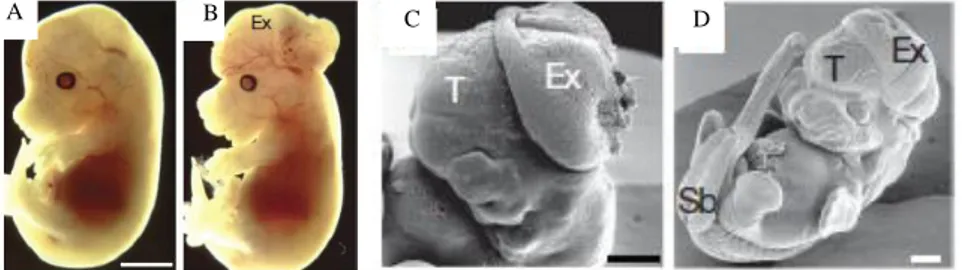
A B C D
Fig.1.2 Anatomical features of Ambra1 deficient mice. A, B, Wild-type (A) and Ambra1gt/gt (B) embryos at E14.5 are characterized by prominent exencephaly (Ex) C, D, Scanning electron
microscopic analysis of E11.5 (C) and E12.5 (D) Ambra1gt/gt embryos. Note the failure of the
neural tube closure, the extensive midbrain/hindbrain exencephaly (Ex) with a closed telencephalon (T), and the lumbosacral spina bifida (Sb) (Fimia et al., 2007).(A), (B) 2 mm; (C),
(D) 500 m.
Fig. 1.1 Expression of Ambra1 in the mouse embryonic midbrain (Mb), forebrain (Fb) and
hidebrain (Hb). -gal staining on whole-mount Ambra1+/gtmouse embryos at E8.5 (upper picture) and at E11.5 (lower picture) (Fimia et al., 2007). Bar 10 m.
2
The reported disturbances to the nervous system in Ambra1gt/gt mice appear to
derive to dysfunctions in the autophagic process, also resulting in an imbalance of apoptotic cell death and cell proliferation. These data, together with other in vitro evidence, allowed proving a regulatory role of AMBRA1 in autophagy activation in vertebrates (Fimia et a., 2007).
It has been demonstrated that AMBRA1 interacts with Beclin1 (Bcl2 intarcting protein), promoting its binding to lipid kinase Vps34, thus mediating autophagosome nucleation (Fimia et al, 2007). Taken together, AMBRA1, Beclin1, and Vps34 have been defined as the autophagy core-complex (He and Levine, 2010).
Autophagosome formation is primed by AMBRA1 release from the cytoskeleton. In fact, upon autophagy induction, AMBRA1-“dynein light chain 1” (DLC1) complex translocates from the microtubules to the endoplasmic reticulum (ER), thus enabling autophagosome nucleation (Di Bartolomeo et al, 2010). Dissociation of AMBRA1-DLC1 from the dynein complex requires phosphorylation of AMBRA1 by the serine/threonine kinase ULK1 (Di Bartolomeo et al., 2010) (Fig.1.3).
Beclin1/Vps34-mediated autophagy is negatively regulated through a interaction between Beclin1 and BCL-2. In this context, it is worth mentioning that recent studies showed that AMBRA1 is a new partner for BCL-2 in mammals and that their binding is independent of Beclin1 (Strapazzon et al., 2011).
As AMBRA1 and BCL-2 bind Beclin1 on the same site, the two proteins could be competitors. In addition, after autophagy induction, AMBRA1/Beclin1 interaction
Fig.1.3 Proposed model of AMBRA1 dynamic interaction with the dynein motor complex during autophagy induction (Di Bartolomeo et al., 2010).
3
increases, whereas the AMBRA1/BCL-2 interaction is disrupted. Altogether, these results led to propose a model in which, under normal conditions, a pool of AMBRA1 associates with BCL-2 in proximity of mitochondria, inhibiting its autophagic function; after autophagy induction, this mitochondrial pool of AMBRA1 separates from mito-BCL-2 and increases its binding to Beclin1 in order to favor the autophagic program (Strapazzon et al., 2011).
To this respect, it is worth mentioning that a recent study demonstrates an interaction between AMBRA1 and Parkin in dopaminergic neurons, occurring on the mitochondrial surface and leading to mitophagy induction (Van Humbeeck et al., 2011).
1.2 Autophagy
Cells have a constant need for the building blocks of life: amino acids, lipids, carbohydrates, and nucleic acids. To sustain this catabolic and anabolic needs, they rely on uptake and recycling. While nutrient uptake is important, different degradation systems are in place to efficiently degrade recyclable intracellular material and provide quality control.
The main pathways for protein degradation and recycling are the ubiquitin/proteasome pathway (for degrading short-lived cytosolic and nuclear proteins), the lysosomal pathway (for cytosolic proteolysis), and autophagy (for bulk cytosolic degradation and organelle recycling) (Rubinsztein et al., 2006). Deficits in any of these recycling routes can result in uncontrolled accumulation of cellular debris or severe deficiencies in metabolic productivity, ultimately causing cell death. The importance of these pathways is emphasized by a group of human metabolic disorders, often referred to as accumulation diseases, caused by impaired degradation machineries (Bi et al., 2010).
Autophagy is involved in the intracellular turnover of proteins and cell organelles, and has an important role in regulating cell fate in response to stress (Levine, 2005). It is a highly conserved process that occurs in all species and cell types studied thus far.
The term autophagy was coined in 1963 by Christian de Duve to establish a nomenclature for different cellular pathways and compartments in the endosomal-lysosomal pathway (Klionsky, 2008). Early studies performed in rat liver allowed to define the autophagic process as a physiological response to starvation, in order to degrade and recycle non-essential intracellular macromolecules (Deter et al., 1969). Later, the molecular mechanisms underlying autophagy were identified in yeast (Takeshige et al., 1992; Tsukada et al., 1993). Subsequent identification of the mammalian homologues enhanced the interest of several research groups towards the investigation of the role of autophagy in tissue and organ homeostasis. Two main types of mammalian autophagy have been identified: macroautophagy and chaperone-mediated autophagy (CMA) (Fig. 1.4).
4
Macroautophagy is a bulk degradation pathway and the only intracellular mechanism potentially capable of degrading large protein aggregates or damaged organelles. It is a well-understood process in yeast, but details about the exact sequence of events and the proteins involved are still uncertain in mammals. A cup-shaped isolation membrane forms around cytosolic components, eventually fusing to form a double membrane bound vesicle, the so-called “autophagic vacuole” (AV) (Mizushima et al., 2002).
The origin of the membrane material for the formation of the isolation membrane is still under investigation, but recent evidence suggests that it might derive from the ER (Axe et al., 2008). The protein MAP1LC3 is docked via conjugated phosphatidyl ethanolamine (MAP1LC3-II) to the isolation membrane and is a specific marker for the so-called autophagosome (Mizushima et al., 2004). The autophagosome undergoes several microtubule (Jahreiss et al., 2008) and dynein-dependent maturation events (Kimura et al., 2008). For successful degradation of
Fig.1.4 Steps in macroautophagy and chaperone-mediated autophagy.
Macroautophagy: 1.) Nucleation. A class III PI3K complex consisting of at least BECN1, PIK3C3, PIK3R4, UVRAG, and AMBRA1 is required for PAS (Pre-autophagosomal structure) formation and MAP1LC3 is anchored to the membrane via a phosphoethanolamine (PE) anchor (LC3-II). 2.) Expansion. This stage is also called "isolation membrane". More membrane and LC3-II is being recruited to the developing vacuole. 3.) Maturation. The exact nature and sequence of this maturation, and whether these steps are always required is currently unknown. The autophagosomal lumen becomes more acidified during this maturation. 4.) Docking and fusion. During docking and fusion the inner membrane compartment together with its content gets released into the lysosome/autolysosome and is being degraded by lysosomal hydrolases.
Chaperone-mediated autophagy: 5.) Recognition and binding. The HSC70 chaperone complex recognizes unfolded proteins with the KFERQ sequence and moves them to the lysosome. 6.) Translocation. LAMP2A and a lysosomal form of HSC70 (l-HSC70) translocate the substrate protein across the lysosomal membrane into the lumen for degradation. (Jaeger and Wyss-Coray, 2008).
5
the autophagosomal content, autophagosomes need to migrate from their site of formation to lysosome rich perinuclear regions (Fass et al., 2006). After fusion with the lysosome, the outer autophagosome membrane can be reused, while lysosomal enzymes degrade the inner membrane and its cytosolic content, enabling the recycling of macromolecules (Kimura, 2007).
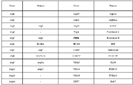
Large number of autophagy-related genes (ATG) have been identified over time and their nomenclature has been unified (Klionsky et al., 2003). Most of the gene products function at the step of autophagosome formation. Since the membrane dynamics of autophagy in yeast is quite similar to that in mammalian cells, these discoveries prompted the researchers to look for mammalian homologues of yeast ATG genes (Fig. 1.5).
This allowed manipulations of autophagy (by knocking down or overexpressing ATG genes) to elucidate its contribution to different physiological and pathological processes. These molecules can be used as specific probes for autophagy (Mizushima, 2004).
Chaperone-mediated autophagy (CMA) differs from macroautophagy in that no vesicular trafficking is involved (Fig. 1.4). Instead, a pentapeptide motif in substrate proteins allows their specific translocation through the lysosome membrane (Majeski and Dice, 2004). Thus, CMA degrades only proteins with the motif KFERQ or a biochemically related sequence, which is present in about 30% of all cytosolic proteins (Chiang and Dice, 1988).
Fig. 1.5. Homologue genes in Homo sapiens of autophagic genes discovered in yeast (modified from Levine and Klionsky 2005).
6
1.3 Autophagy in the nervous system.
The two main types of autophagic processes, i.e., CMA and macroautophagy, importantly contribute to degradation of cytosolic components in the nervous tissue. The selective nature of CMA makes it the ideal system for removing misfolded proteins when refolding is not possible; in fact oxidized and misfolded CMA substrates are more readily degraded through this autophagic pathway (Finkbeiner et al., 2006). CMA declines with age because of a decrease in the levels of lysosome-associated membrane protein (LAMP) type 2A, a lysosomal receptor for this pathway. This favours the accumulation of CMA substrates, possibly causing damage to the neural tissue (Massey et al., 2006).
Macroautophagy components are expressed in neurons and neuronal cell lines. While the function of autophagy-related proteins has been described for some, it is still unknown for others. (Jaeger et al, 2009).
Recent studies show that macroautophagy is constitutively active in healthy neurons and is vital to cell survival (Bolland and Nixon, 2006). For this reason and for the relatively non-specific nature of this process, responsible for bulk cytoplasmic turnover, the term macroautophagy will be referred to as autophagy.
Fig.1.6 Ubiquitin-positive inclusion in Atg5-deficient neurons. Immunoistochemistry of brain sections from
control (Atg5flox/+; nestin-Cre) and Atg5flox/flox; nestin-Cre (Atg5flox/flox) mice at six weeks of age (Hara et a.,
7
Mice lacking either Atg5 or Atg7 genes exhibit motor and behavioral deficits as well as degeneration and loss of Purkinje cell neurons in the cerebellum and pyramidal neurons in the hippocampus (Hara et al., 2006; Komatsu et al., 2006). Diffuse protein aggregates appear in surviving neurons within these brain regions and others, culminating in the formation of toxic inclusion bodies.
Because neuronal proteasome activity is not reduced in these mouse models, the results imply that autophagy is normally responsible for clearing aggregated proteins that are not degraded by the proteasome (Rideout et al., 2004). Moreover, constitutive clearance of cytosolic proteins by low-level basal autophagy has an important cytoprotective function, particularly in neurons, as evidenced by accumulation of ubiquitinated proteinaceous deposits (Fig. 1.6) and development of neurodegeneration in mice deficient for basal autophagy (Hara et al., 2006; Komatsu et al., 2006).
Further insights into the dynamic and essential mechanisms of autophagic process in neurons came when several studies characterized autophagosome-related compartments, their retrograde transport, and progressive maturation and fusion with lysosomes along neuritic processes (Hollenbeck, 1993). These investigations led to robust evidence showing that constitutive neuronal autophagy may serve as both a key mechanism for remodeling neurite and growth cone structure during neurite extension, as well as a neuroprotective mechanism by removing damaged proteins and organelles that may otherwise accumulate within axons (Hollenbeck, 1993). More recent papers suggest that autophagy might contribute to the homeostasis and maintenance of the axons (Gumy et al., 2010). Neuronal homeostasis essentially depends on balanced, bidirectional trafficking of intracellular constituents between distal neurites and the cell soma. In neurons, autophagosomes and endosomes that fuse in the distal axon must be retrogradely transported to the soma, often over great distances, in order to fuse with lysosomes and degrade their contents (Yue, 2007). Thus, subtle disruptions of autophagosome formation, maturation, or trafficking would be predicted to have dire consequences for autophagic flux and neuronal homeostasis.
The fundamental role of autophagy in cell homeostasis explains its importance in development. Ambra1gt/gt mutants show letal neural tube defects, as mentioned earlier (Section 1.1). These phenotypes have been linked to defective neural progenitor death, supporting the function of autophagy as a cell death pathway (Clarke, 1990).
During ageing, the autophagy-lysosome system undergoes striking changes. Ageing leads to reduction in autophagosome formation and autophagosome-lysosome fusion, both of which are consistent with decreased autophagy. There are also notable changes in lysosomes, such as increased volume, decreased stability, altered activity of hydrolases, and accumulation of the indigested material in the form of lipofuscin (Terman and Brunk, 2004). The precise molecular defects remain unknown, but these changes correlate with a decrease in the total capacity
8
for degradation of long-lived proteins in nervous tissue of aged animals (Donati et al., 2001).
Autophagic dysfunction may also lead to defective turnover of mitochondria, which results in the accumulation of older mitochondria, which generate increased levels of ROS, especially in the microglia. In turn, ROS activate redox-dependent transduction cascades and transcription factors, which induce the expression of inflammatory genes and exacerabate the consequences. Therefore, “microglia-ageing” could function as a major driver for brain ageing. These evidence led to hypothesize that prevention of lysosomal autophagic dysfunction and mitochondrial DNA damage in microglia may be a potential novel therapeutic targets against brain ageing (Nakanishi and Wu, 2009).
In neurons, the consequences of age-related decline in autophagy are diminished turnover of intracellular components and reduced ability of cells to adapt to changes in the extracellular environment (Ward, 2002). Compromised clearance of old and/or damaged organelles (as mitochondria or peroxisomes) by autophagy coupled with a reduced turnover of long-lived proteins likely contribute to the intracellular accumulation of oxidized proteins in aged organisms (Kim et al., 2007). This age-related decline in autophagy may be particularly detrimental to the nervous system, as postmitotic cells such as neurons are vulnerable to the accumulation of undegraded metabolic products over the lifetime of the organism (Terman, 1995). Indeed, evidence is mounting that integrity of the autophagosomal-lysosomal network appears to be critical in the progression of ageing, indeed it is now largely accepted that autophagy affects several cellular activities crucial for longevity and healthy ageing (eulongevity), particularly in the context of neurodegenerative disease states.
1.3.1 Autophagy and neurodegenerative disorders.
Abnormal autophagy may be involved in the pathology of both acute brain injuries and chronic nervous system disorders, including proteinopathies, as Alzheimer’s and Parkinson’s diseases (Fig.1.7).
Alzheimer’s disease (AD) is one of the most common age-related neurodegenerative disorders. It is the prevalent form of dementia, characterized by progressive cognitive dysfunction, together with behavioral and neuro-psychiatric disturbances. The pathogenesis of AD is highly complex, involving both genetic and environmental factors. From a neuropathological point of view, AD features progressive loss of neurons and synapses, intracellular neurofibrillary tangles, composed of hyperphosphorylated Tau protein, extracellular deposition of of senile plaques and cerebral amyloid angiopathy. The main constituents of senile plaques are amyloid peptides (A), which are generated from amyloid precursor protein (APP) by sequential proteolytic cleavage, mediated by - and -secretases (Bianchi et al., 2011).
9
Recent evidence has shown that A is generated during autophagic turnover of APP-rich organelles supplied by both autophagy and endocytosis.
A generated during normal autophagy is subsequently degraded by lysosomes. In AD, the maturation of autophagolysosomes and their retrograde transport are impeded, leading to a massive accumulation of ‘autophagy intermediates
(namely, AVs, see section 1.2) within large swellings along dystrophic and degenerating neuritis (Fig.1.7). The combination of increased autophagy
induction and defective clearance of Acontaining AVs creates conditions favorable for A accumulation in AD (Nixon, 2007).
PD is the most common neurodegenerative movement disorder and the second most common neurodegenerative disease. Most of the PD cases are sporadic, although familial PD with autosomal dominant or autosomal recessive mutations also account for about 5% of all PD cases.
10
PD patients suffer from resting tremor, bradykinesia, muscle rigidity and postural instability. The deterioration of motor functions observed in PD patients is predominantly attributable to the degeneration of dopaminergic neurons in the substantia nigra, showing that the nigrostriatal circuit is involved in neurodegeneration (Zelda and Nancy, 2009).
On a cellular level, neuronal loss is accompanied by neurite degeneration and the presence of cytoplasmic inclusions known as Lewy bodies, involving -synuclein aggregation. In PD, the autophagic pathway participates in the degradation of -synuclein, as well as in the turnover of damaged mitochondria, a hallmark of disease (Zelda and Nancy, 2009). Indeed, a striking increase of oxidative stress related to dysfunction of mitochondrial metabolism is known to occur. This last feature is crucial for dopaminergic neurons, because these cells have distinct redox properties, that make them particularly keen to oxidation, even in normal conditions, compared to other neuronal subtypes (Horowitz et al., 2011).
Despite mounting observations that are in support of a protective role of autophagy in various models of PD, some essential issues remain unsolved. While it is perceivable that insufficient autophagy activation would impair clearance of protein aggregates and dysfunctional mitochondria, whether excessive activation of autophagy occurs in PD, and whether it plays a role in PD pathology remains unknown. This question is particularly important since excessive activation of autophagy is associated with neuronal loss (Bredesen et al., 2006; Zelda and Nancy, 2009).
Huntington’s disease (HD) is an age-related neurodegenerative disorder, characterized by motor and cognitive impairment, and caused by mutations in the gene encoding for huntingtin protein (htt). The mutated protein contains abnormally long sequences of polyglutamine, resulting in the formation of intranuclear ubiquitinated inclusions. Mutated htt also accumulates in autophagic compartments, in amounts proportionate to the length of its polyglutamine tract, suggesting that its degradation by autophagy is impeded (Kegel et al., 2000). Importantly, stimulating autophagy by rapamycin treatment reduces htt accumulation and neurodegeneration in cell and fly models of polyglutamine disease and reduces neurological deficits and htt aggregation in a mouse model of HD (Ravikumar et al., 2004). By contrast, inhibiting the formation of autophagosomes or impeding their fusion with lysosomes increases htt aggregation in cells in vitro and in vivo (Ravikumar et al., 2005).
Under certain conditions, induction of autophagy can be even detrimental in neurons. In some paradigms, massive activation of autophagy can result in self-digestion of the whole cell, leading to cell death. This process has been termed type II programmed cell death, in order for it to be distinguished from apoptotic pathways (Bursch, 2001; Gozuacik and Kimchi, 2004).
In the Lurcher mouse model of cerebellar degeneration, autophagic neuronal death was implicated as mediating the pathological effects of mutations in the glutamate receptor subunit GluR2 (Fig. 1.9; Florez-McClure et al., 2004). These findings may have broad implications regarding the possibility that autophagy may be a common
11
mediator of cell death initiated by excitotoxicity, but this remains to be established (Orr, 2002).
However, a general role for autophagy in neuronal death remains speculative. In most instances in which autophagic morphology has been found to accompany neuronal cell death, it remains indeterminate whether autophagy is the culprit, or is induced secondarily to facilitate the removal of cellular components, or is induced as a cytoprotective response to cellular stress. It is quite possible that autophagic induction has the potential to be either protective or destructive, and the influence of autophagy depends on the type, degree and duration of the inciting cellular stress. (McCray and Taylor, 2008).
Fig.1.9 Trasmission electron microscopy of Purkinje dying neurons, showing ultrastructural features of autophagy. Autophagic vacuoles are indicated by open arrows (Florez-McClure et al., 2004).
12
Chapter 2
Objectives
AMBRA1 is a recently discovered protein, centrally involved in nervous system development, regulating cell proliferation and autophagy (Fimia et al., 2007). Indeed Ambra1 deficient embryos shows severe abnormalities, including neural tube defects, leading to early embryonic death.
These findings prompted us to investigate the expression of AMBRA1 in adult mouse CNS, in physiological and pathological conditions.
The primary aim of this study was to analyse the distribution of AMBRA1 protein in the brain, with special reference to the cerebral region considered, to the cell type, to the neuronal population, and to the intracellular compartment. These issues were addressed by morphological approaches, namely immunohistochemistry, immunofluorescence, and immunoelectron microscopy, which in our view are especially suitable to the study of the nervous tissue, for its highly complex organization and heterogeneous composition. As a preliminary step, we screened a panel of custom and commercial antibodies against AMBRA1 protein, to identify the most efficient one for morphological localization purposes. We then performed a systematic histological examination of AMBRA1 immunoreactivity in serial, coronal and sagittal sections of normal adult mouse brain. This approach allowed us to obtain a detailed map of the protein in the different brain territories along the rostro-caudal axis.
Based on this overview, showing some highly immunoreactive areas and others weakly AMBRA1-positive, our next aim was to examine, within each region, protein expression in the different neuronal populations, in the attempt to correlate AMBRA1 content with special features of specific neural cells. To this purpose, different neuronal markers, including tyrosine hydroxylase (TH) for dopaminergic neurons, glutamic acid decarboxylase (GAD) for GABAergic neurons, choline acetyl transferase (ChAT) for cholinergic neurons, were used in combination with AMBRA1, to perform double immunofluorescence experiments. In addition to these light and confocal microscopy studies we selected performed immunoelectron microscopy to characterize the intraneuronal localization of AMBRA1 with ultrastructural resolution to gather crucial information about the function of this protein.
Parallel to this scrutiny, the presence of AMBRA1-immunoreactive glial cells was investigated in different brain areas, using glial fibrillary acidic protein (GFAP), as an astroglial marker, and ionized calcium-binding adaptor molecule1 (Iba1), as a microglial marker.
According to our preliminary interpretation of the above results, heterogeneous expression of AMBRA1 in the various areas of forebrain, midbrain, and hindbrain could be related to the different cell metabolic and turnover request, as well as to susceptibility/resistance of specific neuronal subsets to chronic or acute injury. To
13
further investigate this hypothesis, we chose a number of neurodegeneration paradigms, including normal ageing, Alzheimer’s disease (AD), Parkinson’s disease (PD), and genomic instability mouse models. Importantly, in all these physiological and pathological conditions, autophagy, which is demonstrably regulated by AMBRA1, acts as a protective or detrimental process.
Concerning the study of possible age-related variations in AMBRA1 levels, we focused on the neocortex and hippocampal formation, i.e., the primarily affected areas in the ageing brain. To this aim, we analyzed 3-, 6-, 12- and 18-month-old mouse brain by immunoblotting and immunohistochemistry, as means for obtaining both quantitative data and analytical information on protein expression. Parallel to this investigation, we analyzed by the same approaches the AD-like ageing model, at the same time points. For this part of the project, we chose a transgenic mouse strain (Tg2576), overexpressing the human isoform of amyloid precursor protein, carrying the Swedish mutation. This model was generated by Hsiao et al (1996) and was selected because it closely recapitulates human pathology in time-dependent manner (Jacobsen et al., 2006). In this model, neuronal dysfunction occurs before the accumulation of -amyloid-containing plaques and patent neurodegeneration. Indeed neuronal deficits in Tg2576 mice can be temporally clustered into early deficits observed in 3 to 5-month-old animal and late deficits observed in animals older than 12 months (D’Amelio et al., 2010; Jacobsen et al., 2006). For a further understanding of age-, genotype-, and brain region-dependent variations in AMBRA1 levels, we extended the study by analyzing the expression pattern of its main interactor, i.e. Beclin1, whose function in autophagy regulation is well established.
We then addressed the issue of a putative role of AMBRA1 in PD, given the close relationship between the disease and autophagy mechanisms, particularly in the degradation of -synuclein and the turnover of mitochondria. We were particularly interested in early PD stages, to get an insight into the contribution of the protein at the onset of disease. To this aim, we utilized a pharmacological murine model of PD based on the administration of the neurotoxin 1-methyl-4-phenyl-1,2,3,6-tetrahydropyridine (MPTP) (Khun et al., 2003). To analyze AMBRA1 expression in relation to the pathological phenotype, we took advantage of a quantitative-morphological approach to measure TH and AMBRA1 levels in nigrostriatal circuits.
To obtain further insights into the role of AMBRA1 during the neurodegenerative processes in the dopaminergic system associated with ageing – which constitutes the major risk factor for PD - we extended our investigations to mouse models with defective DNA Nucleotide Excision Repair (NER), which corrects a broad spectrum of distortion in the DNA double helix. Defects in the NER repair system have been associated with accelerated ageing, with a phenotype that correlates with the severity of the mutation. In our studies, we used a mutant strain with a truncation in the gene coding for the Excision Repair Cross Complementation Group 1 (ERCC1) protein. We demonstrated that in this particular model, which
14
exhibits only a mild phenotype, dopaminergic neurons of the nigrostriatal circuits show alterations resembling those observed in the MPTP model during the early stages of the pathogenesis. For these reasons, we examined expression of AMBRA1 protein in dopaminergic neurons from ERCC1/+ mouse mutants.
15
Chapter 3
Distribution of AMBRA1 in mouse brain
In order to examine AMBRA1 protein distribution in adult mouse brain we took advantage of several morphological techniques, including immunohistochemistry (IHC), immunofluorescence (IF) and pre-embedding immunoelectron microscopy (IEM) on the brain sections obtained from adult mouse
3.1Immunohistochemical and immunofluorescence analysis.
As a first step, we investigated AMBRA1 presence in neuronal and glial cell populations. Double IF experiments, performed in different brain areas, using anti-AMBRA1 in combination with either anti-neuronal nuclei (NeuN, neuronal marker), or glial fibrillar acid protein (GFAP, astrocytic marker), or ionized calcium-binding adaptor molecule 1 (Iba1, microglia marker), show that AMBRA is more abundant in neurons, with respect to glial cells (Fig. 3.1). Interestingly, while the vast majority of neurons demonstrably contains AMBRA1, the protein is not distributed at the same levels in NeuN positive cells, prompting us to further investigate its distribution in different brain areas and neuronal subsets.
Fig.3.1 Double IF using anti-AMBRA1 (green signal) in combination with neural cell markers (red signal). The protein is abundantly expressed in neurons, although few NeuN-positive cells appear as AMBRA1-negative (arrows). GFAP-positive astrocytes and Iba1-positive microglia are mostly devoid of AMBRA1. (A) Neocortex; (B) Hippocampus; (C) Cerebellar cortex.
AMBRA1 is widely expressed in the forebrain, midbrain and hindbrain, with a generally higher concentration in the cortical area and in the caudal-most nuclei. The protein is present in most neurons of the archicortex (hippocampal formation), paleocortex (piriform cortex) and neocortex, even though the signal intensity varies among different brain regions, and within each area. A detailed description of AMBRA1 IHC distribution along the brain rostro-caudal axis is given below. In the olfactory bulb, mitral cells, representing the major source of afferent input to the olfactory cortex, are strongly immunoreactive in their cytoplasm (Fig. 3.2A).
16
Other output neurons, namely tufted cells in the external plexiform layer (EPL), considered the smaller version of mitral cell (Shepherd, 1990) show a lower immunostaining degree. Interestingly, the targets of olfactory bulb, including the piriform cortex (especially pyramidal layer II), the olfactory tubercle and the insular cortex show remarkable staining (Fig. 3.2B-D).
In the neocortex, pyramidal cells of layer V of the motor cortex show the strongest immunoreactivity (Fig. 3.3A-B). AMBRA1 richness in these giant spiny neurons, which are excitatory in function, could account for the presumed high organelle turnover, due to their size and plasticity. In the basal ganglia, the caudate putamen shows a differential AMBRA1 positivity in its various neuronal populations (Fig. 3.3C). While giant, aspiny interneurons are densely stained in their somata, medium-sized spiny projection neurons show faint immunoreactivity (Fig.3.3D).
Fig.3.2 AMBRA1 IHC in coronal sections of mouse rhinencephalon. (A) Olfactory bulb, showing intense staining in mitral cell layer (ML) and weaker immunoreactivity in tufted cells (arrows). (B) Layer II of the piriform cortex, showing positive pyramidal neurons.
17
Fig. 3.3 AMBRA1 IHC in coronal sections of mouse telencephalon. (A) Overview of the neocortex, showing numerous positive neurons throughout the layers (I-VI). (B) Pyramidal cells of layer V of the motor cortex are strongly immunoreactive (arrows). (C) Differential staining in the caudate-putamen (CP) and globus pallidus (GP), showing immunoreactivity in both neurons and fiber bundles. (D) Giant aspiny neurons (black arrow) are intensely positive, while medium-sized spiny neurons are weakly immunostained (white arrow).
Even in these neurons, the presence of high protein levels is likely linked to their intense cytoplasmic turnover. Interestingly, axon bundles crossing the whole corpus striatum, representing part of the corticostriatal circuit, are intensely stained, suggesting association of a pool of AMBRA1 protein with axonal cytoskeletal components.
Fig.3.4 AMBRA1(green)/ChAT (red) double IF in the caudate putamen. AMBRA1 signal is especially intense in ChAT positive interneurons (arrows), while ChAT negative spiny neurons are only mildly AMBRA1-positive.
IF analysis of caudate putamen using choline acetyltransferase (ChAT), as a marker for cholinergic neurons, demonstrates high AMBRA1 expression in ChAT positive interneurons (Fig. 3.4), suggesting that in physiological conditions AMBRA1 regulated processes are fundamental for the health of these cells. The hippocampal formation appears dyshomogeneously AMBRA1-positive (Fig. 3.5A). Interest in this structure arises from its involvement in major cognitive functions, and from its susceptibility to neurodegeneration.
18
IHC analysis demonstrates moderate staining levels in the pyramidal layer of the hippocampus proper. AMBRA1 pattern seems to follow gradual changes in morphology of pyramidal layers around the hippocampus. Indeed, proceeding from region CA1, where cell bodies are relatively small, to region CA3, where giant neurons are found, AMBRA1 immunoreactivity in pyramidal cells becomes more and more intense (Fig. 3.5B-C). Concerning interneurons, basket cells located in the stratum pyramidale/stratum oriens border are weakly positive, while other cells dispersed in the stratum radiatum and stratum lacunosum-moleculare are more densely labelled. Interestingly, the dentate gyrus (DG) is characterized by a gradient of AMBRA1 immunostaining, moving from the polymorphic layer/ granular layer border to the granular layer/molecular layer border (Fig. 3.5D).
The richest population may include neural stem cells, based on their morphology and distribution. In this view, the observed gradual changes in AMBRA1 content may reflect the cell differentiation status, that in this
Fig. 3.5 AMBRA1 IHC in coronal sections of mouse telencephalon and diencephalon. (A) Overview of the hippocampal formation showing varying immunreactivity degrees in different fields and layers. (B) CA1 hippocampal region. Or, stratum oriens; Pyr, pyramidal layer; Rad, stratum radiatum. (C) CA3 hippocampal region. Or, stratum oriens; Pyr, pyramidal layer; Rad, stratum radiatum, LMol, stratum lacunosum-moleculare. (D) DG of the hippocampal formation. PoDG, polymorphic layer; GL, granule cell layer; ML, molecular layer. Arrow indicated a positive mossy cell. (E) Septal complex; inset shows AMBRA1 positive neurons and fibers. (F) Anterior thalamic nucleus.
19
neurogenic region is known to proceed in an inner-to-outer manner, from neural progenitors, to neuroblasts, to mature neurons (Bonfanti et al., 2011). Since inputs of the hippocampal formation include terminations of septum and diagonal band (which preferentially project to CA3 and DG), we examined AMBRA1 distribution in the septal complex. Indeed, neurons in this area are stained in both their somata and their processes (Fig. 3.5E). Another central structure involved in learning and memory processes is the anterior thalamic nucleus. It is worth noting that this region receives inputs from the hippocampus, so being part of the hippocampo-diencephalic circuit. Interestingly, neurons belonging to the thalamic region and, markedly to anterior nucleus express high levels of AMBRA1 (Fig 3.5F).
The results of IF in the thalamus, using the glutamic acid decarboxylase (GAD67) as a marker for gabaergic neurons, shows colocalization with AMBRA1, even though the pro-autophagic protein is more widely expressed (Fig. 3.6).
Fig.3.6 AMBRA1(green)/GAD67(red) double IF in the thalamus. Several AMBRA1-positive cells are observed, some of which are also GAD67-positive (arrows).
In the mesencephalon, select regions contain several immunoreactive neurons (Fig. 3.7A). Among these, the deep gray layer of the superior colliculus, red nucleus, locus ceruleus, and oculomotor nucleus (Fig. 3.7B-E).
20
On the other hand, important mesencephalic centers, such as the substantia nigra and ventral tegmental area (VTA), are weakly AMBRA1 immunoreactive. Indeed double immunofluorescence confirms low levels of AMBRA1 signal in dopaminergic neurons, identified by anti-tyrosine hydroxylase (TH), (Fig. 3.8)
Fig.3.8AMBRA1(green)/TH(red) IF in VTA. Scarce colocalization of the markers is detected. Fig.3.7AMBRA1 IHC in coronal sections of mouse mesencephalon. (A) Overview of the distribution of the protein in mesencephalic territories. (B) Superior colliculus, showing intense staining in the deep gray layer (DpG). (C) Red nucleus, magnocellular part (D)Locus coeruleus, (E) Oculomotor nucleus.
21
The brainstem is especially rich in AMBRA1, as shown in Fig. 3.9, illustrating the distribution of the protein in several sensory and motor nuclei.
AMBRA1 is widely expressed in cerebellum (Fig. 3.10A) at different levels depending on the considered area and neuronal subtype.
Fig.3.9 AMBRA IHC in coronal sections of mouse brainstem. (A) Overview of positive centers, including abducens nucleus (6N), mesencephalic trigeminal nucleus (Me5), and medial vestibular nucleus (MVe). (B) Pontine reticular nucleus (C) Raphe nucleus.
In the deep cerebellar nuclei, the large excitatory glutamatergic neurons are strongly immunoreactive (Fig. 3.10A). The magnocellular projections of these nuclei reach the red nucleus and the thalamic region in which AMBRA1 is strongly expressed.
IHC analysis of the cerebellar cortex shows a remarkable immunoreactivity in Purkinje cell layer (PL), while the granule cell layer (GL) and stellate and basket neurons in the molecular layer (ML) are only mildly stained (Fig. 3.10B).
Close-up view of the intracellular distribution of AMBRA1 signal in Purkinje cells demonstrates polarized and particulate immunoreaction product, suggesting association of the protein to specific cytoplasmic compartments (Fig. 3.10C). Notably, even within the PL, different degrees of immunoreactivity can be recognised. An inner-to-outer gradient of immunoreactivity in each cerebellar lobule is observed. Even more intriguingly, Purkinje neurons of the whole lobule 10 and of the ventral part of lobule 9 are strikingly negative.
22
Fig. 3.10 AMBRA1 IHC in sagittal sections of mouse cerebellum.
(A) Overview of the cerebellum showing a different expression in the lobules (2-10 Cb) and staining in the medial cerebellar nucleus (Med); (B) Purkinje cell layer (PL) shows a stronger immunoreactivity than granule (GL) and molecular (ML) layers; (C) detail of the PL showing particulate staining concentrated at the apical cell pole.
23
3.2 Ultrastructural analysis of AMBRA1
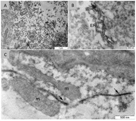
To the aim of investigating the intracellular distribution of AMBRA1 we performed pre-embbedding immunolabelling of brain sections. The IEM analysis revealed an exclusively cytoplasmic localization of the protein (Fig 3.11A), that results to be mostly associated to the endoplasmic reticulum (Fig. 3.11B-C), consistent with its role in regulating autophagosome formation.
Fig. 3.11. AMBRA1 Pre-embedding IEM of pyramidal hippocampal neurons.
(A) Low-power micrograph showing that immunodeposits are present in the cytoplasm, while absent from the nucleus (n). (B) Higher magnification showing association of immunoprecipitates with ER. (C) AMBRA1- immunoreactivity is found around and inside ER cisternae (arrows), and, to a lesser extent close to mitochondria (m).
A
B
C
A
B
24
Chapter 4
AMBRA1 expression in physiological and Alzheimer-like
ageing
The suggested correlation between brain ageing and decreased autophagic efficiency prompted us to analyze age-dependent variations in AMBRA1 expression, focussing on the neocortex and hippocampus, i.e., the most damaged areas during ageing (Moreno et al, 2011)
AMBRA1 expression was investigated in normal and pathological ageing, utilizing a transgenic mouse model (Tg2576, Hsiao et al., 1996) of Alzheimer’s disease (AD). The ages selected for this study, i.e., 3, 6, 12 and 18 months, correspond to the onset and progression of disease in the transgenic strain (Jacobsen et al., 2006; D’Amelio et al.,2011).
Moreover, in order to evaluate and interpret our data in view of the role of AMBRA1 in regulating the autophagic process, we also analysed the expression of Beclin1, which is centrally involved in autophagy and considered the main interacting molecule of AMBRA1 (Fimia et al., 2007).
4.1 Expression of AMBRA1 in the ageing neocortex
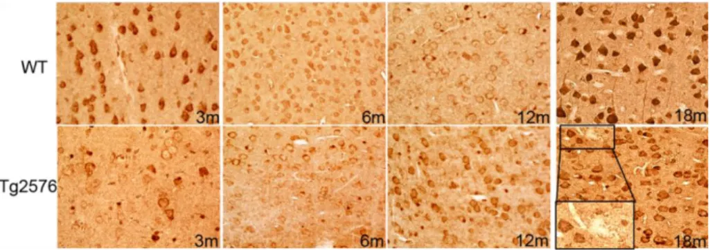
In the neocortex, western blotting (WB) analysis in wild-type animals (WT) (Fig. 4.1, black columns) shows a progressive decrease in AMBRA1 levels until 12 months of age, followed by an increase at the oldest age considered. While the general trend of AMBRA1 expression during ageing appears similar in the two genotypes, Tg2576 neocortex differs in some respects to its WT counterpart (Fig. 4.1, gray columns). At the starting time point (3 months), AMBRA1 levels are lower in transgenic than in WT mice, but remain relatively stable at subsequent ages. Indeed, the only age-related variation that reaches statistical significance is observed at 18 months, when an increase of AMBRA1 levels is detected. The morphological analysis of the neocortex shows a predominantly neuronal immunostaining and confirms WB results, as age- and genotype-dependent differences in AMBRA1 immunoreactivity are 3m 6m 12m 18m 0.0 0.5 1.0 1.5 * ** * WB AMBRA Neocortex ** A * Age A M BR A 1 /A c ti n
Fig.4.1 Densitometric analysis of AMBRA1WB assay of neocortex from WT and Tg2576 mice, ageing 3, 6, 12 and 18 months. *, p<0.05; **, p<0.01.
25
observed (Fig.4.2). Interestingly, in Tg2576 neocortex, a higher heterogeneity degree of AMBRA1 expression is detected, compared to its WT counterpart. Specifically, in 3-month-old transgenic brain, most pyramidal neurons express hardly detectable AMBRA1 levels, while only few display more intense immunoreactivity. This situation dramatically changes at 18 months, when Tg2576 neocortex shows most pyramidal neurons remarkably stained in their somata and apical dendrites. In addition, at this advanced AD stage, the neocortex appears partially disrupted in its cytoarchitecture, due to the appearance of amyloid plaques, surrounded by dystrophic neurites and AMBRA1-positive glial cells (Fig. 4.2, inset). By contrast, in the WT neocortex rather uniform AMBRA1 staining is found throughout the neocortex. In particular, both 3- and 18-month-old animals show high levels of AMBRA1 immunostaining in the vast majority of pyramidal neurons.
Fig.4.2AMBRA1 IHC in the neocortex of WT and Tg2576 mice ageing 3, 6, 12 and 18 months. Inset shows immunoreactive glial cell bodies surrounding an amyloid plaque.fare inset
Fig. 4.3 shows Beclin1 protein levels in the neocortex during normal and Alzheimer-like ageing. Fairly stable expression during the earliest time points is observed, independent of the genotype. However, a significant increase of protein levels at 12 months of age is detected in both WT and Tg2576 animals. The pathological genotype shows further increment of Beclin1 at 18 months, suggesting autophagy induction.
Fig.4.3 Densitometric analysis of Beclin1 WB assay of
neocortex from WT and
Tg2576 mice, ageing 3, 6, 12 and 18 months. *, p<0.05; ***, p<0.001 3m 6m 12m 18m 0.0 0.5 1.0 1.5 2.0 WT Tg2576 * * *** WB Beclin1 Neocortex Age B e c li n 1 /A c ti n