1 11
UNIVERSITÀ DEGLI STUDI DI SASSARI
Co-tutelle of Thesis
between
PhD school in Biomedical sciences University of Sassari (ITALY) And
PhD school in Biotecnology University of nautal resources and life science Vienna (AUSTRIA)
STUDY OF THE MOLECULAR PATTERNING IN
STEM CELLS EXPOSED TO BIOPHYSICAL STIMULI:
A NEW VISION OF REGENERATIVE MEDICINE
Italian Tutor: Prof.Margherita Maioli, PhD Austrian Tutor: Prof.Regina Grillari-Voglauer, PhD PhD student: Valentina Basoli, MSc
UNIVERSITÀ DEGLI STUDI DI SASSARI
CORSO DI DOTTORATO DI RICERCA IN SCIENZE BIOMEDICHE
Coordinatore del Corso: Prof. Andrea Fausto Piana
CURRICULUM IN GENETICA MEDICA XXIX CICLO
STUDY OF THE MOLECULAR PATTERNING IN
STEM CELLS EXPOSED TO BIOPHYSICAL STIMULI:
A NEW VISION OF REGENERATIVE MEDICINE
Anno Accademico 2015 - 2016
La presente tesi è stata prodotta nell’ambito del Corso di Dottorato in Scienze Biomediche dell’Università degli Studi di Sassari, a.a. 2013/2014 – XXIX ciclo, con il supporto di una borsa di studio finanziata con le risorse del P.O.R. SARDEGNA F.S.E. 2007-2013 - Obiettivo competitività regionale e occupazione, Asse IV Capitale umano, Linea di Attività l.3.1.
Coordinatore: Prof. Andrea Fausto Piana Italian Tutor: Prof.Margherita Maioli Austrian Tutor: Prof.Regina Grillari-Voglauer Tesi di dottorato di: Dott.ssa Valentina Basoli
Università degli Studi di Sassari Corso di Dottorato di ricerca in GENETICA MEDICA La presente tesi è stata prodotta durante la frequenza del corso di dottorato in genetica medica dell’Università degli Studi di Sassari, a.a. 2014/2017 - XXIX ciclo, con il sostegno di una borsa di studio cofinanziata con le risorse del P.O.R. SARDEGNA F.S.E. 2007-2013 - Obiettivo competitività regionale e occupazione, Asse IV Capitale umano, Linea di Attività l.3.1 “Finanziamento di corsi di dottorato finalizzati alla formazione di capitale umano altamente specializzato, in particolare per i settori dell’ICT, delle nanotecnologie e delle biotecnologie, dell'energia e dello sviluppo sostenibile, dell'agroalimentare e dei materiali tradizionali”. La tesi è stata prodotta, altresì, grazie al contributo della Fondazione di Sardegna.
Έτσι, δεν γνωρίζω
(Ἀπολογία Σωκράτους,VI- Πλάτων 399 BC)Summary
Abstract ... 7
Introduction ... 8
Regenerative medicine ... 8
Historical background ... 8
Strategies used in regenerative medicine ... 11
1.Cell-based therapy ... 11
Classification of Stem cells ... 13
The role of cell niche in regenerative medicine ... 20
Activation of stem cells and cells ... 22
Cell cycle ... 23
Cell polarity ... 27
Concept of plasticity ... 29
Problems with stem cells ... 36
2.Use of materials able to increase the repair processes and cell growth and migration ... 43
3.3D printing cells ... 44
Principle aim and challenges in regenerative medicine ... 45
4.Use of biophysical stimuli on cells or tissue ... 46
Physical devices used in regenerative medicine ... 47
Radio Electric Assimetric Conveyor (REAC) ... 49
Extra corporeal shock Waves therapy (ESWT) ... 50
AIM OF THE PROJECT ... 54
REAC SECTION ... 57
RESULTS ... 65
DISCUSSION REAC AND SENESCENCE ... 81
STEM CELL DIFFERENTIATION AND REAC ... 87
REAC AND PC12 NEURONAL DIFFERENTIATION ... 88
MATERIALS AND METHODS ... 89
RESULTS ... 93
REAC AND CARDIAC COMMITMENT ON UIPSCs ... 100
RESULTS ... 105
DISCUSSION ... 114
EXTRA CORPOREAL SHOCK WAVE THERAPY (ECSWT) SECTION ... 117
EXTRA CORPOREAL SHOCK WAVE THERAPY AND CELL PROLIFERATION ... 117
MATERIAL AND METHODS ... 119
RESULTS ... 124
SHOCK WAVE TREATMENT AND CELL ACTIVATION BY PRO-INFLAMMATORY INDUCTION ... 132 DISCUSSION ... 142 BIBLIOGRAPHY ... 149 Acknowledgment ... 181
Abstract
Regenerative medicine is a novel applied field of medical management based on the application of stem cell technology and tissue engineering in order to replace or regenerate human tissues and organs and restore their functions. The regenerative medicine has the prospective to help scientists and clinicians while planning early-intervention treatments for traumatic injury or degenerative diseases, by regrowth or replacement of cells or tissues.
However, the challenges in this field are still countless, as (1) which type of cells are suitable (2) which is the best protocol to commit all the stem cells toward a specific phenotype with the 100% of yield, (3) drop cell manipulation.
At the same time, physical energy were found capable to interact with cell physiology and increase their performance. In the clinical practice physical therapies enhance the quality and time of spontaneous tissues regeneration.
Here, I focused on the effect of physical stimuli by Radio electric asymmetric conveyor (REAC) and Extra corporeal shock wave (SW) and their interaction with cells or stem cell behaviour.
In particular, during the modulation of the main molecular patterning controlling cellular senescence, differentiation toward specific phenotypes and proliferation, in order to figure out the targets of action, to enhance their performance for future therapeutic applications.
Introduction
Regenerative medicine Historical background
Regenerative medicine is an applied field of medical treatment which is based on the application of stem cell technology and tissue engineering in order to replace or regenerate human tissues and organs and restore their functions [1].The term “regenerative medicine” was coined by William Haseltine in 1999 during a conference on Como Lake, describing an emerging field, deriving from different subjects: tissue engineering (TE), cell transplantation, stem cell biology, biomechanics prosthetics, nanotechnology, biochemistry [2]. Historically, this term was found for the first time in a 1992 paper by Leland Kaiser, who listed the technologies which would impact the future of hospitals [3].
The regenerative medicine has the potential to help scientists and clinicians while planning early-intervention treatments for traumatic injury or degenerative diseases, by regrowth or replacement of cells or tissues. This branch of medicine is highly cross-disciplinary and serves as a bridge between basic science and clinical medicine, however the idea of the production of perfect tissues and organs in a dish is still a big challenge. Several studies and approaches were applied to follow the regeneration of tissues and organs, like growth factors in the damaged site, in order to stimulate the cells to regenerate the tissue, or alternatively the development of biomaterials for tissue engineering, as biomimetic polymers and bioactive three-dimensional scaffolds, capable of inducing specific cellular responses and direct the formation of new tissues to be implanted in vivo.
But, the idea of human regeneration potential was already known in ancient times since the old Greek culture, as demonstrated by the myth of Prometheus, whose liver was eaten by an eagle during the day and it completely regenerated itself overnight.
In Nature, the regeneration of body parts is rather common in reptiles, it is known that the salamander can regenerate an amputated limb in several days, or humans have this “ability” as well to regenerate fingertip but they lose it over the years[1]. Actually the absolute and applicable solution for the regeneration of tissues and organs is really long and it upon small steps that will allow the achievement of amazing discoveries.
In 1954 the transplantation of the first kidney substituted in a human between identical twins, in 1967 it happened the first heart transplant by Christiaan Bernand but without success, the patient pasted away after 18 days. However, a lot of programs directed on organ transplantation started during these years, paving the way to the idea that transplantation of organs and tissues was not absolutely an utopia, but a relevant solution to save patients. In 1968 the first immune deficient patient with the sibling’s bone marrow[2] was successful transplanted. Currently, the organ transplant is widespread and practice which deeply altered medicine, improving the patients' life expectancy.
However, finding compatible donors is not easy and due to the progressively aging population, transplantations will progressively represent a need to replace end-stage diseased organs injured by age-related diseases. That reason encouraged the researchers to direct their force to find a parallel solution besides the organ transplantation and regeneration of damaged tissues.
Relatively at the same time, in 1978 stem cells were isolated for the first time in human cord blood [3], opening a novel chance for patients with leukemia and anemia.
In 1981 for the first time a stem cell line (embryonic) from mice was cultivated in vitro[4]. That pioneering works, paved the way to the world of regenerative medicine and its application with the principal aim to find a solution to organs replacements from donors. The first “product” of application of cells for regenerative medicine was published in 1981 by Burk which using cells seeded on biomaterial composed by collagen, created the first “acceptable bilayer artificial skin” for the treatment of extensive burn injury. Moreover in 1998, Thompson isolated the first human embryonic stem cells from blastocysts showing the ability of these cells to differentiate in the three germs layers[5]. In 1999, in a paper published on the Lancet, the implantation of a laboratory-grown bladder implanted in a patient suffering from myelomengicocele [6] using as scaffolds homologous decellularised bladder submucosa with muscle cells isolated from the same patient, was described.
In 2004, the group directed by Raya-Rivera performed the first study on reconstruction and implantation of urine in five boys, showing that already after 3 months after implantation the engineered grafts developed a normal architecture with normal physiological functions in patients[7], strongly supporting tissue engineering as an effective solution for recovering lost physiological conditions.
In 2006, Takahashi and Yamanaka announced the discovery of the a induction of pluripotency in adult unipotent fribroblast from mouse, recovering an embryonic-like state by introducing four factors, Oct3/4, Sox2, c-Myc, and Klf4. The IPS (induced pluripotent stem) exhibit the
a revolutionary discovery for the applied research in regenerative medicine, introducing the concept of reprogramming, otherwise the possibility to set stem cells back to the embryonic state, by using a few defined factors.
Unlimited studies regard the approaches and methodologies applied to regenerative medicine have been made, like the suitable types of cells to be used, how to isolate and manipulate them, how to obtain the organized tissues, which materials are safe for the patient, which is the best material on for seeding cells, which cells are ethically feasible, a number of questions that still do not have a clear and dogmatic answer. However, in regenerative medicine many questions are still endless.
Strategies used in regenerative medicine
There are different strategies used in regenerative medicine 1. Cell-based therapy
2. Use of materials able to increase repairing processes, cell growth and migration 3. 3D printing cells
4. Use of biophysical stimuli on cells or tissue 1.Cell-based therapy
The multicellular organisms are organized by several types of cells specialized in particular functions, originate from a single cell called “zygote”; during development, all the cells differentiate progressively and acquire specific phenotype, losing their capability to
differentiate into other cells. The ability to differentiate into other cell types is defined as “cell potency or plasticity” and is typical particular cells called “stem cells”.
For this reason, stem cells represent the main tool in regenerative medicine due to their extensive ability to self-renew and to generate differentiated progeny.
Stem cells (SC) can divide and differentiate into different specialized cell types and can self-renew to produce more stem cells. They are capable to develop into organized tissues in the body during early life, and to handle tissue growth and homeostasis during the all life. When a stem cell divides, each new cell has the potential either to remain a stem cell or become another type of specialized and unipotent cell. In particular, stem cells can be identified by special properties that characterized them, as self-renewal, unspecialized nature and differentiation. They can replicate many times, or proliferate. This particular type of division is called “asymmetric division[8].
Classification of Stem cells
Fig.1 Hierarchic representation of stem cell organization
Classification based on potency
We could classify the different stem cells basically on the ability to differentiate into other cell types. Etymologically, the potency is taken from the Latin term "potens" which means "having power”, due to this physiological property, the stem cells are the main actors in the theatre of regenerative medicine.
cell called zygous. The totipotent stem cells give rise to somatic stem/progenitor cells and primitive germ-line stem cells[9]
• Pluripotent stem cells are the descendants of totipotent cells and can differentiate into nearly all cells derived from any of the three germ layers, but not in foetal annexes. These pluripotent cells are characterized by self-renewal and a differentiation potential for all the cell types of the adult organism[10].
• Multipotent stem cells cells can differentiate into a number of cells, but only those of a closely related family of cells. These are true stem cells but can only differentiate into a limited number of types. For example, the bone marrow contains multipotent stem cells that give rise to all the cells of the blood but not to other types of cells. Adult Haematopoeitic Stem Cells are multipotent as well the Adipose tissue is a source of multipotent stem cells.
• Oligopotent stem cells can differentiate into only a few kind of cells, such as lymphoid or myeloid stem cells. For examples the corneal epithelium is a squamous epithelium that is constantly renewing, because is constantly exposed to damages, and is Oligopotent [11].
• Unipotent cells can produce only one cell type, their own, but have the property of self-renewal, which distinguishes them from non-stem cells. Most epithelial tissues self-renew throughout adult life due to the presence of unipotent progenitor cells[12]. It is even possible, classify stem cell by their origin:
Types of stem cells
The embryonic stem (ES) derived from embryos at an early developmental stage (day 5–8,
called blastocysts) after implantation [5], are considered as totipotent [13] because they have the ability to differentiate into all cell types in the body give rise to cells of all the three embryonic germ layers like ectoderm, mesoderm and endoderm, even after being grown in culture for a long time.
The human ES cell lines (SSEA)–3, SSEA-4, TRA-l-60, TRA-1- 81, and alkaline phosphatase expressed cell surface markers, and are able to produce teratomas after injection [5], this is even an important parameter for testing the potency status. It is known that ESs express high levels of telomerase activity, a ribonucleoprotein involved in maintaining telomere length, which plays an important role in replicative life-span and senescence.
The ES pre-implantation epiblast have an active epigenetic regulation program, which is essential for maintaining pluripotency. They define the cellular epigenetic mechanisms that produce persistent effects in the biological system, without, however, altering the genome sequence. The epigenetic hence enables the modulation of the expression of DNA or structures associated with it by means of special processes, such as DNA methylation and the associated action of Methyl Binding Proteins (Mbds), the modification of histones with proteins belonging the Polycomb Group (PcG) and Trithorax Group (TrxG), the chromatin remodeling [14]. Specifically, the extra- and intra-cellular signals can stimulate the specific signal "propagators"inside the cells, which, can in turn activate defined pathways, producing
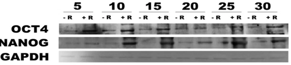
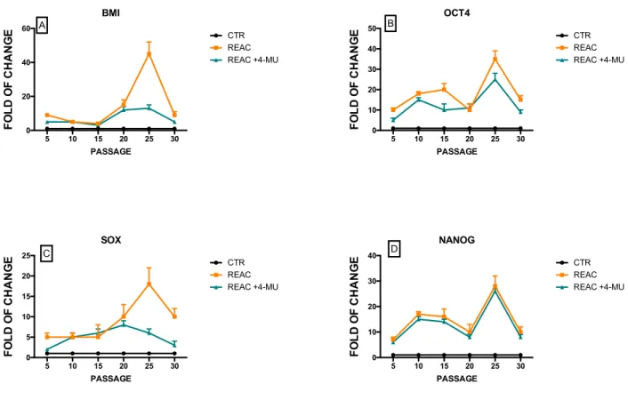
long lasting effects, although not altering the genomic sequence. The chromatin structure of mouse ES has been demonstrated to be hyperdynamic, with increased global levels of trimethylated lysine 9 H3 (TriMeK9 H3), a heterochromatic histone modification linked to gene repression, and decreased levels of acetylated histones H3 and H4 (AcH3 and AcH4), modifications linked to euchromatin and the permissively of gene expression. Analyses of global histone modification patterns in ESC has previously suggested that the ESC genome is subject to generalised histone acetylation and lysine 4 H3 methylation[15]. ESC have to act not only to silence such genes in order to maintain totipotency, but also to allow them to remain poised for transcription, so they can be rapidly activated upon the differentiation- induced silencing of NANOG, SOX2 and OCT4. These genes able to interact with epigenetic modifiers, including Polycomb Group (PcG), moreover it was recently discovered that ESCs have a high rate of methylation at CpG site level in Low density CpG promoters (LCPs), usually associated with tissue-specific gene expression. The regulation of these genes occurs upon methylation / demethylation of distal regulatory elements, enhancers or silencers, during the dynamic process of differentiation of ESCs in different phenotypes by the action of active DNA methyltransferases (DNMTs): DNMT1, which is largely responsible for the maintenance of DNA methylation over replication, and DNMT3A and DNMT3B, which generally perform de novo methylation of either unmethylated DNA or hemimethylated DNA to assist in maintenance[16].
However, the overexpression of the pluripotent pathway of Oct-3/4 a master regulator of lineage commitment with SOX2 and Nanog [13], combined with the absence in vitro of LIF (Leukemia Inhibitor Factor)[17] a member of the interleukin-6 family of cytokines that bind
to the gp130 receptor, by activating the Jak/Stat and Ras/MAPK signal transduction pathways of FGF-5 and BMP4 increase the differentiation toward the germinal layers.
Foetal Stem Cells
Foetal stem cells are primitive cell types found in the organs of foetus. The Foetal stem cells can be isolated from foetal blood and bone marrow as well as from other foetal tissues, including liver and kidney[18] The developing baby is referred to as a foetus from approximately 10 weeks of gestation. Most tissues in a foetus contain stem cells that are pluripotent and drive the rapid growth and development of the organs. Foetal blood is a rich source of haemopoietic stem cells, which proliferate more rapidly than those in cord blood or adult bone marrow[19]. Like adult stem cells, fetal stem cells are generally tissue-specific, and generate the mature cell types within the particular tissue or organ in which they are found.
The adult stem cells are Mesenchymal stem cells (MSCs) are adult stem cells which can be
isolated from several sources such as bone marrow, adipose tissue[20][21], amniotic fluid[22], endometrium, dental pulp and ligament, umbilical cord, Wharton’s jelly, epidermis, liver and intestine[23], most of these sources are considered wasting materials after clinical practice but a treasure for regenerative purposes. Adult stem cell are resident in specific zone of the body called “niche”, highly dynamic, mainly implicated in the regeneration and homeostasis of the tissues, and have been best characterized in tissues that have a rapid rate of cell turnover.
The easily isolation, selection, characterization, expansion in vitro and the relative easily capability to differentiate into mesodermal lineage such as osteocytes, adipocytes and chondrocytes as well as ectodermal (neurocytes) and endodermal lineages (hepatocytes)[23] endorsed them to become a main candidate for regenerative medicine.
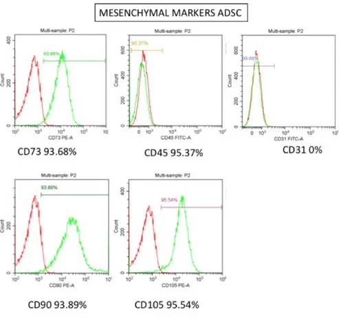
The International Society for Cellular Therapy has proposed minimum criteria to define MSCs, These cells should exhibits plastic adherence, possess specific set of cell surface markers like (CD)73, D90, CD105 and lack expression of CD14, CD34, CD45 characteristic for hematopoietic linages, as human leucocyte antigen-DR (HLA-DR) and must have the ability to differentiate in vitro into adipocyte, chondrocyte and osteoblast. Probably the best efficient population of MSCs has been reported with these features is from bone marrow[24], however the isolation process from the iliac crest is absolutely invasive and comprise several risks for the patient.
The adult stem cell are able to differentiate into adipocytes, osteocytes and chondrocytes, confirmed in vitro, by production of oil droplet, formation of mineralized matrices and expression of type II collagen.
Induced pluripotent Stem cells
The study of adult stem cells and mostly the embryonic stem cell, in term of transcriptomic and gene expression allowed the achievement a revolutionary discovery in the field of regenerative medicine, Takahashi and Yamanaka in 2006 announced the discovery of the combination of a “cocktail of genes” involved in cell reprogramming in adult fibroblast from mice[25].
Induced pluripotent Stem cells (iPSC) are created by inducing the expression of genes that are usually present in embryonic stem cells and that control cell functions. IPS cells are a powerful method for creating patient- and disease-specific cell lines for research and maybe for future application in regenerative medicine. These are not adult stem cells, but rather reprogrammed cells with pluripotent capabilities. Using genetic reprogramming [25][26] cells through the introduction of Oct3/4, Sox2, c-Myc, and Klf4[25], involved in maintaining cell pluripotency at that time by retrovirus[27], actually with episomes[28] or miRNA[29], pluripotent stem cells equivalent to embryonic stem cells have been derived from human adult tissues. A new frontier using this methodology pave the way to the optimization of the use of wasting biological material like urine [30] or from easily isolation material like frozen blood samples[31], opening a new avenue for suitable application. iPSCs are useful tools for drug development and modeling of diseases, and scientists hope to use them in transplantation medicine, however the capability to derive in all cells even to teratomas need to be carful[32]. iPSCs are derived from somatic cells, epigenetically reprogrammed to lose tissue-specific features and gain pluripotency. Similar to hESCs, they can theoretically differentiate into any type of cells[33]. The concept of induced pluripotent stem cells remains an important area of focus for future research and has serious implications for the stem cell cancer theory [32]. The minimal criteria to define a IPs is the expression of pluripotency-related factors, endogenous Oct3/4, Nanog, FoxD3, Rex1, Dnmt3b, and Abcg2[34] as well markers as the protein antigens CD9, Thy1 (CD90), tissue-nonspecific alkalinephosphatase (Tra-2-49 and Tra-2-54), class-1 human leukocyte antigen, and podocalyxin (GCTM2), the globoseries glycosphingolipid antigens stage-specific embryonic antigen (SSEA)-3 and SSEA-4, and the
81[35][26]. IPS must be able to generate teratome in vivo, and develop all cell types. The creation of pluripotent stem cells from adult cells by the introduction of reprogramming transcription factors absolutely raised new hope for future applications like in the production of new disease models and in drug development as well as in transplantation medicine.
Cell in the body or cell in the dish?
The role of cell niche in regenerative medicine
The term ‘niche’ was first used by Schofield in 1978 to explain the variation in the self- renewal capability of an apparently pure populations of HSCs following transplantation in mice. He hypothesized that the capability of stem cells to self-renew and retain their identity depends on the environment provided by neighbouring, components of the niche, including direct interactions between stem cells and neighbouring cells, secreted factors, inflammation and scarring, extracellular matrix (ECM), physical parameters such as shear stress and tissue stiffness, and environmental signals such as hypoxia and ROS. The niche have been described in a variety of adult tissues, including skin[36], intestine[37] and nervous system[38]. In many adult tissues, the stem cell niche contains a variety of cell types, each with a distinct function. Communication between stem cells and niche cells is either direct, through physical interactions, or indirect, through secreted factors that mediate communication between cells that are not in direct contact by cell-cell adhesion molecules and receptors with membrane-bound ligands [39]. Indirect communication between stem cells and niche cells is mediated by secreted factors. In the clinical practice this phenomenon is routinely exploited, like the
use of granulocyte stimulating factor (G-CSF) or granulocyte-macrophage colony-stimulating factor (GM-CSF) to support treatment of haematological malignancy.
Although every stem cell niche is dynamic and exhibits cell turnover, it is useful to distinguish between niche cells that are ‘permanent residents’ and cells that occupy the niche transiently. Permanent residents include endothelial cells, nerve cells and fibroblasts, the transient ones include immune cells and cells that respond to tissue damage like pathogens or to promote healing.
Another fundamental component in the tissue and in the niche is the extra cellular matrix (ECM) which exhibit an important role, in fact the ECM not only anchors stem cells but also directs their fate[39], the most important ECM receptors are integrins, and their functions can be modulated by biochemical stimuli, such as antibodies, small- molecules as drugs or by biophysical stimuli[40]. The later carry out an important and fundamental role during cell fate and development, as demonstrated there is a systematic relationship between tissue mechanics and differentiation [41] thus modulating the stem cell niche. All these finding in term of regenerative medicine suggest that finding a methods able to interfere with the activation, modulation, fate, differentiation of the cells resident in the niche could open new hopes for the patients.
Fig.2 Sources of stem cells
Activation of stem cells and cells
Tissue injury is an inevitable part of life. It is becoming clear that following damage, stem cells switch to producing an excess of proliferating daughters until the wound is sealed, exhibiting high plasticity in their behaviour[42]. There are several studies reporting that stem cells populations respond to injury before turning to the activation of quiescent stem cells in tissues bearing a low turnover of cell proliferation. In fact, in tissues with minimal cell turnover, stem cells are actively maintained in a quiescent state and conditionally activated, , but it is well known and proved that paracrine signalling in the stem cells microenvironment, physical stimuli, hormones are able to switch this behaviour. For example, it was observed
that epidermal stem cells secreted both Wnt ligands and Wnt inhibitory Dkk proteins, resulting in the activation of Wnt target genes in basal cells, and the Wnt/β-catenin signaling is a central regulator of adult stem cells mostly in term proliferation[43]. Autocrine Wnt signalling is thus an essential part of the stem cell niche, allowing permission to cell proliferation [44], while the inhibition of Wnt signals induce cell cycle arrest [43]. Recent studies also demonstrated that stem cells niche is exposed to paracrine signals like the Wnt that mainly regulate the fate of the cells.
Other interesting studies consider the response and activation of the cell is correlated to pro-inflammatory cytokines release as IL-1α, IL-13, TNF-α, and IFN-γ, secreted by T cells in vivo on satellite cells[45]. So far, understanding how cells response to intrinsic factors, hormones, cytokine, cell-intrinsic physical stimuli is not already well understood thus elucidating these mechanism will give new insights in stem cell application in vivo.
In fact the cells, upon an injury or a paracrine signal, or a physical stimuli react by leaving their quiescent status and start to proliferate. The biological mechanism of proliferation is based on cell cycle activation.
Cell cycle
Cell division consists of two consecutive processes, mainly characterized by DNA replication and segregation of replicated chromosomes into two separate cells. Cell cycle is a fine regulated physiological process that can be dived in different phases. Originally, the cell cycle was divides in two stages: mitosis the process of nuclear division and the interphase. But, clear evidence has shown that this process cannot be represented by only two phases. In fact,
and telophase, as well the interphase (probably the most complicated an delicate phase for the life of the cell) includes G1, S and G2 phases and then S phase, in a perfect and organized cycle [46].
S phase is preceded by a gap called G1 during which the cell is preparing for DNA synthesis and is followed by a gap called G2 during which the cell prepares for mitosis. G1, S, G2 and M phases are the traditional units of the standard cell cycle.
However, the cells can be in a not-cycle state state called G0, characterized by not growing, not proliferating cells. The multicellular organisms base their life on the perfect control and regulation of the unit that compose them, the cell. The evolution gives them a sophisticate system in order to continue their development, the organization in tissues, in organs, in the life. Regulation of cell cycle, maybe is one of the most important process for the life, and it cannot be regulated only by activation or deactivation of a gene or by translation in a protein; it must be controlled by a precise mechanism, like a Swiss watch. During cell cycle process, there are many actors, that can be dived in agonist and antagonist. In each phase, defined factors are finely coordinated. These are the cyclin-dependent kinases (CDK), a family of serine/threonine protein kinases that are activated at specific points of the cell cycle. Until now, five CDK, acting during the cell cycle are well known, as cCDK4, CDK6 and CDK2 during G1 CDK2 in S phase and CDK1 during the G2 and M.
CDK protein levels remain stable during the cell cycle, in contrast to their activating proteins, the cyclins, whose levels can rise and fall during the cell cycle[47]. The different cyclins are expressed during the cell cycle phases, the three D type cyclins (cyclin D1, cyclin D2, cyclin
to regulate progression from G1 into S phase[48] and cyclin A bind CDK2 during S phase[49]. In late G2 and early M, cyclin A complexes with CDK1 promote entry into Mitosis is further regulated by cyclin B in complex with CDK1. The activation of the CDKs is based on phosphorylation that induce conformational changes and enhance the binding of cyclins[50].
CDK activity can be counteracted by cell cycle inhibitory proteins, called CDK inhibitors (CKI) which bind to CDK alone or to the CDK-cyclin complex and regulate CDK activity. Two distinct families of CDK inhibitors have been discovered, the INK4 family and Cip/Kip family[51]. The INK4 family includes p15 (INK4b), p16 (INK4a), p18(INK4c), p19 (INK4d), which specifically inactivate G1 CDK (CDK4 and CDK6). These CKI form stable complexes with the CDK enzyme before cyclin binding, preventing association with cyclin D[52]. The second family of inhibitors, the Cip/Kip family, includes p21 (Waf1, Cip1), p27 (Cip2), p57 (Kip2). These inhibitors inactivate CDK-cyclin. However, p21 is a “famous” protein, that can also inhibits DNA synthesis by binding nuclear complexes. Every time that p21 is expressed, another impotant protein is correlated, p53 the “guardian” of the DNA integrity. In fact, the expression of p21 is under transcriptional control of the p53 tumour suppressor gene. The p21 gene promoter contains a p53-binding site, that allows p53 to transcriptionally activate the p21 gene[53].
During cell cycle, there are restriction point even called ckeck point, that control the commitment of the cell throw the phases. If the DNA for example is demaged, checkpoints arrest the cycle in order to provide time for DNA repair. Usually, DNA damage checkpoints
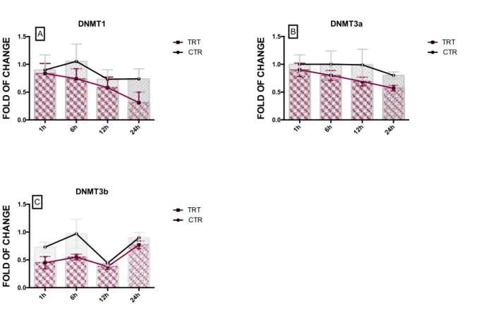
M checkpoint). At the G1/S checkpoint, cell cycle arrest induced by DNA damage is p53-dependent, Usually, the level of p53 is low but DNA damage can induce upregulation[54]. Moreover, the cell cycle mechanism is finely regulated by an epigenetic control involved in regulation of the genes mentioned upon, by activation of chromatin remodelling complexes SWI/SNF (SWItch/Sucrose Non-Fermentable) [55], histones modification as methylation by methylases(HMTs ) and demethylases(HDMTs)[56], acetylation histone acetyltransferases (HATs) and histone deacetylases (HDACs), phosforilation by histone kinases [57][58], control promoter areas (CpG island) by specific DNA metyl transferases (DNMTs)[59]. These last one, the DNMTs play a predominant role during the cell cycle control.
The DNA methyltransferases (DNMTs) are particular proteins that are involved in the establishment and maintainment of methylation in the fifth carbon of cytosine residues in DNA CG dinucleotides (CpG islands) patterns at specific regions in the genome for the regulation (contribute) of gene expression. Easily, we could consider them as “writers of epigenome”. DNMT1 is implicated in maintenance of methylated status, while DNMT3a and DNMT3b are recruited for de novo methylation [60]. Recent observations, showed the relevant role of DNMTs during the behaviour of cells. In fact, as observed by Robertson and colleagues, the mRNA levels of all three DNMTs change during the cell cycle, with DNMT1 and DNMT3b decreasing in G1 as well DNMT3a. During G0 phase, conversely the cells expressed high mRNA level of DNMT3a, DNMT3b and DNMT1[61].
The regulation of cell cycle, with its fine and elegant machine represent perhaps, one of the most interesting challenge to understand cell biology and its applications and solutions.
Cell polarity
Adult somatic stem cells have a central role during the homeostasis in the tissues presenting a high cellular turnover like the skin, intestine, and the hematopoietic system. It is thought that polarity is particularly important with respect to fate decisions on stem cell division (symmetric or asymmetric) as well as for the maintenance of stem cell adhesion and quiescence (interaction with the niche). Embryonic and adult stem cells can use their polarity to generate cell diversity by asymmetric cell division, whereas differentiated cells use their polarity to execute specific functions, like epithelial cells that have apical and basolateral cortical domains to guarantee the barrier maintenance, or the fibroblasts that have actin-rich leading edge during cell migration, or the neurons with their axonal and dendritic portion are able to convey the signal[62].
Finding a concise and clear definition about polarity is not easy, however it represent a main characteristic of the behaviour of a cell. In fact, a cell can be defined polarized when the organelles, proteins, RNAs, inside are distributed and maintained in a asymmetrical organization[63]. The polarization can occur in response to extracellular stimuli that induce a redistribution of cellular components to comply a functional need during adhesion, migration, or cell division. During cell migration, the symmetric arrangement is broken, which may be accomplished by (1) repositioning the centrosome, (2) by adding an asymmetric microtubule nucleation site, (3) by reorganizing the microtubules network, or (4) by altering microtubules dynamics [64].The orientation of the polar axis in a cell can be determined by the shape of the cell, the direction of cell protrusions, the orientation of
microtubule and actin networks; for example during migration the protruding front, a retracting rear, and the cell polarity axis is oriented in a define direction[ 23].
Many recent studies have demonstrated that plasma membrane domains with specialized lipid commonly enriched in cholesterol are distributed asymmetrically in polarized cells. For example, during the migration in lymphocytes it was observed that the membrane-anchored cell surface receptors Inter-cellular adhesion molecule (ICAM) with lipid rafts, together with their respective signal transduction pathway regulate extravasation and crawling.
One obvious hypothesis currently supported by experimental evidence is that polarity establishment during mitosis regulates the mode/outcome (symmetric vs. asymmetric) of stem cell divisions and it is hypothesized that during the mitosis in stem cells, only one daughter cell remains in contacts with the niche, maintaining the ability to self-renew. In a review, Florian explained how Dpp and Hh, the bone-morphogenic protein (BMP)2/4 homo- released from the niche/hub cells are involved in regulating the mode of division and polarity of the cells[63]. It was also recently demonstrated that a planar cell polarity pathway is activated by Wnt receptor Fzd7 and that its candidate ligand Wnt7, controls the homeostatic level of satellite stem cells hence regulating the regenerative potential of muscle [65]. Even the cytokine release is correlated with cell division, by activation of receptors that activate a multitude of intracellular signalling cascades [66].
Recent data indicate a correlation between altered stem cell polarity and stem cell aging. The first pioneering studies were done on animal models, in aged drosophilae, shown a disoriented
centrosomes and thus altered polarity relative to their niche cells. This was correlated with a reduced self-renewal activity caused by aging[67], even in another model.
In yeast the same behaviour was linked to high activity of Cdc42[68]. In the human stem cells, the altered polarity is correlated with aging and with an alteration of cell mobilization, homing, engrafting, and lineage choice[69].
Methodology for stem cell differentiation
Concept of plasticity
Since year, scientists have postulated that some cells in the body appear to participate in the formation of new cells to replace injured, aged, or infected cells in tissues and organs. The cell population which constitutes an organ can be divided into three groups: (static) somatic cells, differentiated, produced during development, which have also lost the minimum proliferative capability, in a slow decay process during adult life; (Transit) cells that produce precursors of differentiated cells, that have a relatively short period of existence in the organ, and their life usually is determined by a suicide at the end of maturation process (apoptosis); (Stem) a type of cells present in a predefined microenvironment, with extensive proliferative capability and plasticity to differentiate.
The current knowledge about the factors that control the biology of a stem cell, or the self-renewal, the maintenance of the undifferentiated state and the ability to take symmetric or asymmetric divisions, are still rather limited. The molecular pathways of stem cells are controlled genetically and epigenetically, so depend on mechanisms able to turn on or off specific genes. The shift in the balance between stem cell and differentiation is influenced by
intrinsic and extrinsic factors that can induce stem cell differentiation or maintenance. Watt proposed that stem cells contribute to the maintenance of homoeostasis, with an average of 50% for the retention of progeny and 50% for the differentiation.
Since years researches in the regenerative field are trying to control in vitro the plasticity and differentiation of stem cells. The numbers of papers produced since the fist discovery of stem cells and their possible management and control in vitro in exponentially growing. It is known, that there are several ways to interact with the commitment and differentiation of the cells throw the different linages (mesoderm, ectoderm, endoderm), below will discuss the principals.
Chemical induction Mesodermal linages
Differentiation of MSCs into adipocytes is induced by proper media supplementations, which activate transcription factors (genes) responsible for adipogenesis. or adipogenesis, MSCs were cultured in growth medium supplemented with dexamethasone, indomethacine, insulin and isobutyl methyl xanthine for 3 weeks, cells are then analysed by accumulation of lipid droplets and expression of adipocytes-specific genes as peroxisome proliferator- activated receptor γ (PPARγ ), adipocyte protein 2(ap2) and lipoprotein lipase (LPL) genes. Adipogenic differentiation is characterized by two phases, during the first one, cells are committed toward pre-adipocytes showing a fibroblast-like morphology thus being difficult to be distinguished from the MSCs, during the second phase pre-adipocyte become mature adipocytes with lipid droplets while changing their morphology.
The most common methodology to differentiate MSCs into osteocytes is based on culturing the cells with ascorbic acid, β-glyceralphosphate and dexamethasone for 3 weeks in growth conditioned media; in a recent work[70] our group described that MSCs isolated from adult dental pulp hDPSCs and exposed to a mixture of hyaluronic, butyric, and retinoic acids (HA + BU + RA) in combination with melatonin exhibited the transcription of genes involved in osteogenesis, as VEGF A, which orchestrate osteogenesis trough by modulating both ZBTB16 and NR4A3 gene expression. The osteogenic differentiation of MSCs in vitro continue with an increase in the expression of osteogenic genes as runt-related transcription factor 2 (Runx2), osteonectin, bone morphogenic protein 2 (BMP2), mineral aggregation, exhibiting an increase in alkaline phosphatase activity at the end of differentiation.
Many investigators have reported that a correlation exists between adipogenesis and osteogenesis [71][72], in fact PPARγ, a key transcription factor implicated in adipogenesis, lipid metabolism, and glucose homeostasis, has shown to promote osteogenesis through enhanced osteoblast formation, by the pathways of Hedgehog, NEL-like protein 1 (NELL-1) [72] and β catenin- dependent Wnt[73][74], the latter being a Wnt glycolipoproteins directly affecting cell proliferation, cell polarity and cell fate determination during embryonic development, tissue homeostasis, and during differentiation toward the in cardiogenic lineages.
According to the standard protocol for chondrogenesis, it is possible induce using insulin transferrin selenium, linoleic acid, selenious acid, pyruvate, ascorbate 2-phosphate, dexamethasone and transforming growth factor-β III (TGF-βIII) in culture medium. The commitment toward the chondrogenic phenotype resulted in the formation of
pre-chondrocytes which expressed type I and type II collagens, then the pre-pre-chondrocytes differentiate into mature chondrocytes, expressing the typical transcription factors like Sox9, L-Sox5 and Sox6[75]andTGF-β1,the latter interacting with Wnt/β- catenin pathway inhibiting osteoblast differentiation while promoting chondro-differentiation.
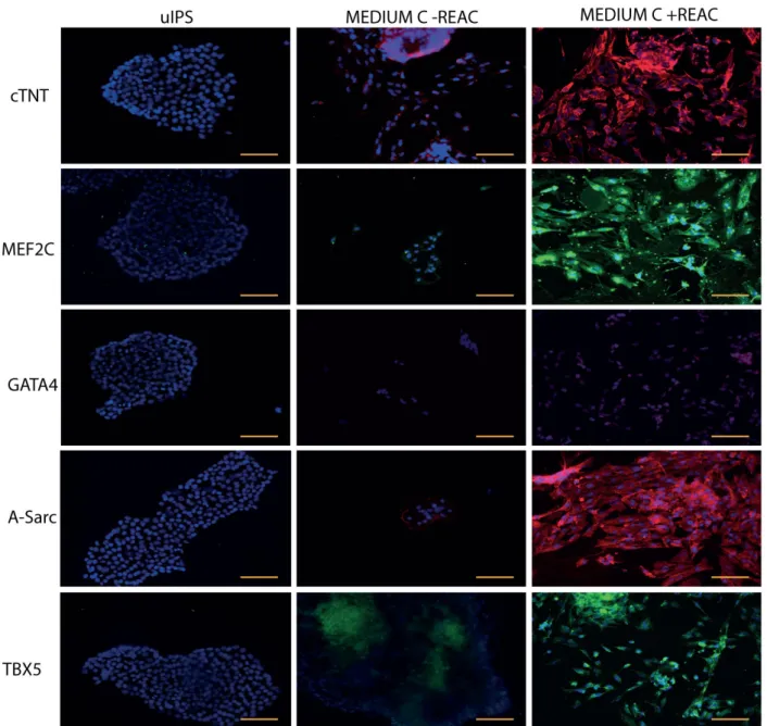
MSCs, embryonic and iPS can even differentiate into other mesodermal lineages, the pioneer work by Wakatani on bone marrow derived MSCs from mouse showed the effect of 5-azacytidine cardiogenic commitment [76], moreover recent works show how it is possible to commit stem cells from amniotic fluid (hAFSCs) toward the cardiovascular phenotype in the presence of a mixture of hyaluronic (HA), butyric (BU), and retinoic (RA) acids[77] in human mesenchymal stem from placenta (FMhMSCs) and mouse embryonic stem (ES) cells[78] by an activation on Smad1/4 pathway. However, the most common protocol for cardiac commitment is based on the addition of specific growth factors important for cardiovascular development like fibroblast growth factor 2 (FGF2), transforming growth factor β (TGFβ) Activin A and BMP4, vascular endothelial growth factor (VEGF), and the Wnt inhibitor DKK-1[79].
Ectodermal linages
Despite the mesodermal origin, hMSCs displayed the capability to trans-differentiate into cells from ectodermal lineages. In fact it was demonstrated that trans-differentiation into neuronal cells can occur upon stem cells are exposed to a neural induction media supplemented with cocktails of growth factors, as the nerve growth factor (NGF, able to induce BMSC transdifferentiation into cholinergic phenotype[80], or the sonic hedgehog
can induce neuronal plasticity and dopamine release [81]. Many other studies have shown that factors like insulin, retinoic acid, bFGF, EGF, valproic acid, BME and hydrocortisone support adipose derived stem cells neuronal differentiation [82][83], as well as glial cell line-derived neurotrophic growth factors (GNDF), brain-line-derived neurotrophic factors (BDNF), retinoic acid, 5-azacytidine, isobutyl- methylxanthine (IBMX) and indomethacin are able to enhance the MSCs differentiation into mature neuronal cells[84]. The dental derived MSCs even called dental pulp stem cells (DPSCs) which originate from neural crest, successfully differentiate into mature neuronal cells [85].
Even the embryonic stem cells as well the IPs are able to commit toward the neuronal linage, because both use the same transcriptional network to generate neuro- epithelia and functionally appropriate neuronal types over the same developmental time course in response to the same set of morphogens treatment as SHH and Wnts[86], or their agonists/antagonists [87].
Endoderm Linages
The differentiation of endodermal linages, is already a challenge for researches, however a lot of protocols and studies comes out during the last years. The discovery that even the MSCs have the capability to trans-differentiate into hepatocytes and pancreatic cells upon induction with conditioned media supplemented with EGF, bFGF and nicotinamide followed by stimulation with dexamethasone insulin, transferrin, selenium [88]; In other studies, it was shown that valproic acid, which is histone deacetylase inhibitor, up-regulated the expression of hepatic marker through activation of protein kinaseB(AKT) and extracellular
signal-Physical energies induction
Recently, alongside the conventional methodologies largely studied by researchers using chemical inductors mentioned upon, new strategies were applied to commit specific cellular phenotypes. Since years, the scientific community, tried some how to reproduce the physiological behaviour of organ, tissue and organs, by physical devices, able to generate specific energies.
In 1974, the group direct by Nucitelli studied the endogenous ionic current and the electric field in multicellular animal tissues, aiming at proving that electric field was able to influence the physiological functions in living organism[90]. Many following studies supported these observations and many protocols were published. Magnetic fields, commonly showed to affect cell proliferation and growth factor expression [91]–[97]. Extremely low frequency (ELF)-PEMF applied to MSCs were able to commit the adipogenic, osteogenic, neural, and glial linage together with conditioned media [98], Moreover pulsed electromagnetic fields (PEMFs) were proved to have a regulatory role in bone marrow derived stem cells (BMSCs) and in adipose derived stem cells (ASCs) differentiation toward the osteogenic phenotype [99]. As well, PEMF of different intensities as 1, 2, and 5mT with a modulation frequency of 750 Hz, a carrier frequency 75Hz and a duty ratio of 0.8, 3 h/day for 4 weeks were able to optimize the process of in vitro endochondral ossification Other study showed how the 50 Hz, 1mT ELF-MFs 5-day exposure implemented NGF-induced PC12 cells neuronal differentiation [97]. [97]. Ventura’s group demonstrated that the extremely low frequency (ELF) pulsed magnetic fields (PMF) affected opioid peptide gene expression and opioid dependent signalling pathways in adult ventricular myocytes [100]. Other devices able to
modulate the behaviour of the cells were projected, as the REAC (Radio electric asymmetric conveyor) which produce extremely low-intensity electromagnetic energy circuit, conveyed directely to the tissue or the cell culture, with a frequency of 2.4 GHz and power of only 2mW. It was observed that REAC elicited human embryonic mouse cells differentiation toward , cardiac, neuronal, and skeletal muscle lineages[101]. Even on Lipogems-derived hASCs the radioelectric asymmetric conveyer (REAC) remarkably enhanced the transcription of prodynorphin, GATA-4, Nkx-2.5, VEGF, HGF, vWF, neurogenin-1, and myoD, indicating the commitment toward cardiac, vascular, neuronal, and skeletal muscle lineages respectively[102]. The light therapy, light-emitting diode (LED) at 620 nm and 2 J/cm2 can modulate the fate of human umbilical cord mesenchymal stem cells (hUMSCs) cultured in osteogenic differentiation medium, in term of proliferation and increase of yield of osteogenic differentiation[103], Other studies showed that the modulation of genes involved in osteogengesis in vitro, depended on the LED length frequency used(680-nm, 760-nm and 830-nm)[104]. On human skin-derived fibroblast LED induced an increase in cell behaviour as, proliferation, viability, migration speed and reactive oxygen-species (ROS) generation[105].
The 532 nm green light LLLT performed with KTP laser at 4 J/cm2 influenced the osteoblastic differentiation of mesenchymal stromal cells [106].
However, the basic physics behind al of these instrument is almost the same: a modulation of an electromagnetic signal or a radiofrequency signal that can interact with the biological behaviour of the cells.
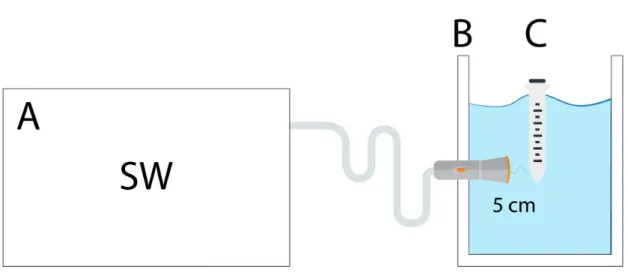
On the other hand, the Extra Corporeal Shock Wave (ECSW or SW) technology, is based on another principle, the ultra sound and cavitation. ECSW have been clinically introduced for lithotripsy since the 1980s. At the beginning, the aim was to brock up the urinary stones by high-energy pulses[107], then several findings demonstrated that changing the modulation of energetic intensity, different effect on tissues can be observed. In particular , SW could affect wound healing with an increase of angiogenesis[108], resolution of tendinitis[109][110] and bone repair increase[111]. SW modulate the differentiation of hASCs toward myofibroblasts[112]
Problems with stem cells
Ethical problems
Scientists plan to differentiate pluripotent cells into specialized cells that could be used for transplantation. However, human stem cell (hSC) research also raises sharp ethical and political controversies because it involves the destruction of human embryos, in fact totipotent stem cell lines can be derived from the inner cell mass of the 5- to 7-d-old blastocyst[113][114]. On the other hand the new discovery by Yamanaka [26] of induced pluripotent stem cells (IPS) has paved the way to new hopes for regenerative medicine and clinical application, because are ethically unproblematic and acceptable to use in humans. Even though IPS cells still exhibit some problems that the researcher are trying to solve, like the possibly tumour formation, immunological reactions, unexpected behaviour of the cells, and unknown long-term health effects[115], based on retroviral integration into the host genome that can create spontaneous and directed differentiations in the expression pattern [116] that cannot really be controlled by researchers.
On the other hand, the MSCs are easier to isolate from many tissues[117], are able to differentiate into many cell types thus representing a starting point of many novel therapies, especially in tissue engineering.
Technical problems
Currently, MSC clinical applications requires the use of high cellular doses (up to several million cells/ patient body weight) together with efficient expansion protocols to generate a large number of cells based on traditional culture techniques[118], meaning culturing MSCs into plastic tissue flasks, a limited process in terms of cell productivity involving at least 2 to 3 cell passages to achieve a clinically relevant cell numbers in an acceptable period of time. On the other hand the effects of extended ex-vivo MSC cells obtained by consecutive cell passaging during long-term cultivation may lead to a senescent state of the cultured cells ultimately compromising clinical safety and efficacy[117]. It is known that after 7 passages BM-MSCs, the proliferative and clonogenic potential is seriously affected, as well as their proteome profile, like cell cycle regulation and apoptosis. [119].
Nevertheless, in practice, cell transplantation does have a number of limitations, adult stem cells or iPSCs, are expensive and need an intensive work, moreover they require specialized facilities for cell collection, expansion, quality control and transplantation as mentioned ATMP standards parameter[120].
Senescence in cells in vivo and in vitro
researchers observed a limitation of the normal cells unlimitedly proliferation in vitro, showing that human fibroblasts post-isolation stages in culture, at the beginning exhibit a strong proliferation rate, gradually decreasing during passages, and progressively losing the ability to divide[121][122]. That phenomena, at a later time, was called “senescence” by Haflick, who demonstrated that the mechanism was correlated to stressors forces causing the cellular senescence. These stressors factors include dysfunctional telomeres, genotoxic stresses/DNA damage, perturbations to chromatin organization, and strong mitogenic signals. Based on this observation, the scientific community, at that time, proposed two important hypotheses. The first one assumed that the senescence mechanism was a way of the body and the cell to prevent aberrant proliferation like cancers, so was proposed senescence as a beneficial event in order to protect unregulated cell proliferation. The second hypothesis, stated senescence as a phenomenon related to the progressive decline in tissue regeneration and repair, with age, together with then loss of regenerative capability of cells in vivo. Nevertheless, the understanding of the senescence processes grew and these two hypotheses combined together, brought new insights to the fields of cancer, ageing and regeneration. In the field of regenerative medicine, the senescence is considered a problem, because for stem cell-based therapies a substantial number of cells are needed, requiring extensive ex vivo cell expansion with many related problems.
The primary cells do not grow indefinitely , but only for a limited number of cell division [122]. On the other hand cell therapy protocols usually require hundreds of million cells per treatment, with the need to be expanded in vitro several passages before implantation (http://www.clinicaltrials.gov).
Cell senescence is strictly dependent on another important factor, the patient. It is known, in fact, that the age, together with genetic patterns strongly the quality of the obtained cells, and the of the lifespan in vitro[123][124]. SCs senescence could affect the clinical therapeutic potential, immunomodulatory activity, differentiation potential, and cell migration ability[125]. It was found that senescent cells exhibit striking changes in the expression of genes, as cell cycle activators [126][127] including the cyclin-dependent kinase inhibitors (CDKIs) p21 (also termed CDKN1a, p21Cip1, Waf1 or SDI1) and p16 (also termed CDKN2a or p16INK4a),. These CDKIs are components directly regulated by the p53 and retinoblastoma (pRB) proteins respectively, involved in growth arrest control during senescence. Senescent cells repress genes encoding proteins that stimulate or facilitate cell-cycle progression, as c-FOS, cyclin A, cyclin B and PCNA[128] by modulating, the transcription factor E2F, that is which is in turn inactivated by pRB. In some senescent cells, E2F target genes are silenced by a pRB-dependent reorganization of chromatin.
The senescence can be induced by many stimuli: • Telomere-dependent senescence
Telomeres are the last part of a linear chromosome and are characterized by the repetition of 5-TTAGGG-3 (in vertebrates) which are associated proteins that have the main role to protect them from degradation or fusion by DNA-repair processes (during replicative event)[129]. The precise telomeric leght sequence is not known and structurally present in the end a large circular structure like a t-loop[130]. During replicative event in S phase of the cell cycle polymerases loose 50–200 base pairs of telomeric DNA [131] the length of telomere decrease, that is one of the reason why
cells do not proliferate indefinitely. Another event inducing senescence, is the double-strand breaks (DSBs) in telomere; in fact, after a telomere break, the cell can arrest cell-cycle progression and try to repair the damage, otherwise if it is not senescence [132][133].
However, physiologically the end-replication problem can be avoided in cells a particular enzyme, the telomerase. This enzyme contains a catalytic protein component (telomerase reverse transcriptase; TERT) and a template RNA component, that adds telomeric DNA repeats directly to the chromosome ends. The activity of telomerase is strictly correlated with the senescence in cells.
• DNA-damage induced senescence
Severe DNA damage that occurs anywhere in the genome, causes in many type of cells senescence, it was observed that DNA and telomerase damages increase cell senescence through the activation of p53, that in turn up regulate p21 thus causing G1 phase arrest [134][135].
• Senescence caused by chromatin perturbation
The chromatin state determines the extent of genes which are active (chromatin) or silent (heterochromatin), and depends mainly on epigenetic control by histone modifications as acetylation, methylation and remodelling of chromatin associated complexes. Recent observation, demonstrated that chemically-induced inhibition of histone deacetylase (HDAi), which promotes euchromatin formation, induces cell cycle arrest and senescence in fibroblast[136] because HDAi induces p21 and p16 expression and in turn p53 up regulation.
• Oncogene-induced senescence
During cancer transformation, some genes show mutations, thus increasing the seriousness of cellular deregulation. These mutated genes, typical of cancerous cells are known as oncogenes. Usually, cells respond to cancer transformation by activating senescence or apoptosis pathways. For examples oncogenes molecularly activate cell senescence. This phenomenon was first observed when an oncogenic form of RAS, a cytoplasmic transducer of mitogenic signals, was expressed in normal human fibroblasts together with an up regulation of p53 and p16(INK4a)[137]. These finding clearly highlight a defence mechanism established by the cells, in the attempt to counteract abnormal growth c stimulation and cancer formation[138].
• Stress and other inducers of senescence
There are a lot of observations that stressor event as oxidative condition in the cells or chronic inflammatory states induced by cytokines, such as interferon-β may induce a senescent state together with cell growth arrest. Chronic stimulation by transforming growth factor-β in epithelial cell induces senescence by promoting p16–pRB-dependent target genes [139].
In conclusion, cell senescence still represent a problem in the field of regenerative medicine because influences the therapeutic potential of human stem cell that can be used for transplantation by influencing many cellular features as migratory ability, differentiation, immunomodulation ability, cell expansion and cell quality[140].
Most cell-based therapies are currently experimental, with a few exceptions such as haematopoietic stem cell (HSC) transplantation which is already a well-established treatment for blood related disorders[141], or the transplantation of cultured sheets of autologous epidermal or corneal cells to repair burn injuries[142], the used transplantation of ex vivo– expanded autologous chondrocytes to repair cartilage defects[143]in clinical practical, or experimental trials like transplantation of embryonic stem cells for treatment of spinal cord injuries[144] a pilot trial conduct in Australia called Geron Phase 1 or the application of pluripotent stem cells for treatment of blindness on a Japanese woman by Masayo Takahashi group.
Conventionally, cell therapies can be classified by the therapeutic indication and the aim to address (neurological, cardiovascular etc.) or by where are from like the same individual (autologous) or derived from a donor (allogeneic) or most commonly by the cell types, often using the EU regulatory classification, the EU regulatory classification of cell-based therapies discriminates between minimally manipulated cells for homologous use (transplants or transfusions) and those regulated as medicines which are required to demonstrate quality, safety and efficacy standards to obtain a marketing authorization before becoming commercially available (referred to as Advanced Therapy Medicinal Products; ATMPs) which are subdivided into somatic cells, gene therapy and tissue engineered products [145]. However, the majority of cell-based therapies till now are an early stage of development (clinical trial Phases I and II focused on demonstration of safety and early indication of efficacy) with relatively few reaching the later stages of clinical trial and marketing authorization [146].
However, since tissue engineering and regenerative medicine emerged as an industry about two decades ago, a number of therapies have received Food and Drug Administration (FDA) clearance or approval and are commercially available[147]. Carticel, the first FDA-approved biologic product in the orthopaedic field, uses autologous chondrocytes for the treatment of focal articular cartilage defects autologous chondrocytes are harvested from articular cartilage, expanded ex vivo, and implanted at the site of injury, resulting in recovery comparable with that observed using micro fracture and mosaicplasty techniques [148]. Other examples include laViv, which involves the injection of autologous fibroblasts to improve the appearance of nasolabial fold wrinkles, or Epicel that are autologous keratinocytes used for severe burn wounds. Tissue engineering includes materials that are often an important component of current regenerative medicine strategies because the material can mimic the native extra cellular matrix (ECM) of tissues and direct cell behavior, contribute to the structure and function of new tissue, and locally present growth factors[149]. Decellularized donor tissues are also used to promote wound healing (Dermapure, a variety of proprietary bone allografts)[150] or as tissue substitutes the CryoLife and Toronto’s heart valve substitutes and cardiac patches[151]. These products provide benefit in terms of healing and regeneration but are unable to fully resolve injuries or diseases[152].
2.Use of materials able to increase the repair processes and cell growth and migration Tissues generally consist of cells and extracellular matrix (ECM). The main role of biomaterials is mimicking the ECM, giving both structural and functional support. During the last few years, ECM has been shown to play a key role in many different functions, such as gene expression, survival, death, proliferation, migration, differentiation. Therefore, all of
them should be reproduced by biomaterials enriched with bioactive factors, such as growth factors and cytokines.
The biomaterials, called “scaffold” in regenerative medicine, can be either natural or synthetic with different advantages and disadvantages. Synthetic materials can be identically reproduced on a large scale with specific properties of microstructure, and degradation rate. However, they still have some problems of biocompatibility with cells. That problem should be solved using natural biomaterial from living organism, able to integrate themselves with cells, creating decellularized tissue matrices[153], but unfortunately even acellular tissue matrices are not so easy to obtain large quantities according to good manufacturing practice and ATMP standards parameter [120] and in some cases they present cellular components which may induce an immune response [153].
The ideal biomaterial should be biocompatible and biodegradable at the same rate as regeneration well porosity, which allows the exchange of nutrients and wastes, not toxic, not inflammative and may help regeneration in term of time and quality.
3.3D printing cells
The new era of regenerative medicine is trying to solve the problems concerning the use of cells and the biomaterials directly printing all together. The biofabrication technique[154], for example, is based on a photo-induced solidification process, which uses soft biocompatible hydrogels containing living cells and forms one layer of solid structure at a time[154], but in a continuous fashion, by shining light on a selected area of a solution containing photo-sensitive biopolymers and cells Organovo is a medical start-up aiming at
delivering bioprinted organs, like liver, for surgical therapy and transplantation. Cytofuse is a Japanese company that without biopolymer directly prints in 3D articular cartilage and subchondral, starting from 3D adipose tissue-derived mesenchymal stem cells[155].
Principle aim and challenges in regenerative medicine
Stem cells have the capability to differentiate into a wide range of adult cells, the discovery and isolation of them paved the way to new hopes in the regenerative field. But most of the applications of stem cells directly on patient are still under experimental trial phase, except for some procedures actually used in clinical practice (bone marrow transplantation in haematology). Even tissue engineering, one of branch of the regenerative medicine based on the regeneration of novel tissues from cells with the aid of biomaterials and growth factors still have some problems. The regenerated tissues usable by the patients are still very limited, as skin, bone, cartilage, capillary and periodontal tissues.
One of the reason which strongly prevents the use of stem cells to regenerate organs and tissues deal with the need of a large number of stem cells as MSCs for clinical applications. Most of the time the isolation of tissue could be dangerous and painful for the patient. Then, a proper set up of in vitro MSCs expansion and subsequent cryopreservation and banking are necessary to establish safety and efficacy in transplanted patients. And is well known that expansion of primary cells is strongly influenced by senescence problems, which represent also one of the challenges in the application of tissue engineering. The artificial tissue engineered still have some limitations correlated to the dimensions of the construct, that can not be used for the recovery of serious defects. The other problem is correlated with the
architecture and tridimensional structure, actually the only usable engineered tissues are vases or cave structure like the trachea [156], or tissues not physiologically scattered because the viability of cells seeded on a scaffold gradually decrease with the thickness. On the other hand, the use of growth factors alone or associated with the 3D construct are still not considered completely safe, since it is unknown the the influence that they could have on the environment of the donors. All of these items need to be addressed before cells or engineered constructs can be used routinely in the clinical setting.
The challenges in regenerative medicine are still countless, (1) the safely, for instance the use of stem cell that can differentiate in all types of mature cells include cancer cell, (2) the use of a method that can commit all the stem cell toward a specific phenotype with the 100% yield of final differentiation, (3) the use of a population of cells that can be fast and easily isolated and with high quantity, and safe for the patient. Also for tissue engineering some clues are still open, (4) find the best scaffold, (5) the best bioreactor (6) the best solution for seeding different population in order to have a relevant mature material implantable on patients.
However, since years other technologies were tested on patients in order to modulate some physiological behaviour involved in the homeostasis of tissue and activation of niche.