Università degli Studi di Sassari
University of Sassari
Dipartimento di Scienze Biomediche
Department of Biomedicals Sciences
XXIX Cycle (Curriculum: Microbiology and Immunology)
Coordinator: Prof. Leonardo A. Sechi
Corso di Dottorato in Scienze della Vita e Biotecnologie
PhD Course in Life Sciences and Biotechnologies
CHARACTERIZATION AND PHYLOGENETIC
ANALYSIS OF USUTU VIRUS ISOLATES IN
SARDINIA
PhD Student: Roberto Bechere
Tutor: Prof. Alberto Alberti
Co -Tutor: Dott.ssa Giantonella Puggioni
Roberto Bechere
“CHARACTERIZATION AND PHYLOGENETIC ANALYSIS OF USUTU VIRUS ISOLATES IN SARDINIA” PhD Thesis in Life Sciences and Biotechnologies Curriculum Microbiology and Immunology
University of Sassari
I
NDEX
Pag.
Index 2
Chapter 1
Usutu virus
1.1 Classification of Usutu virus (USUV) 4
1.2 Molecular features of USUV 7
1.3 Cellular tropism and pathogenesis of USUV 9
1.4 Enzootic cycle, reservoir animals and “bridge-vectors” 12
1.5 Epidemiology of USUV 14
Chapter 2
Aim of the Project 18
Chapter 3
Materials and Methods
3.1 Entomological surveillance 19
3.2 Birds surveillance 20
3.3 Phylogenetic analysis 22
Roberto Bechere
“CHARACTERIZATION AND PHYLOGENETIC ANALYSIS OF USUTU VIRUS ISOLATES IN SARDINIA” PhD Thesis in Life Sciences and Biotechnologies Curriculum Microbiology and Immunology
University of Sassari
Chapter 4
Results
4.1 Surveillance plan on birds and mosquitoes 34
4.2 Phylogenetic analysis 37
4.3 Molecular characterization 41
Chapter 5
Discussion 53
Roberto Bechere
“CHARACTERIZATION AND PHYLOGENETIC ANALYSIS OF USUTU VIRUS ISOLATES IN SARDINIA” PhD Thesis in Life Sciences and Biotechnologies Curriculum Microbiology and Immunology
University of Sassari
Chapter 1
1.1 Classification of Usutu virus (USUV)
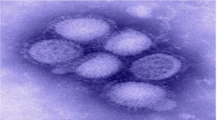
Usutu virus (USUV) (fig. 1.1) is an African mosquito-borne flavivirus, belonging to the
Japanese encephalitis virus (JEV) serocomplex.
USUV was originally isolated from mosquitoes (C. Neavei) collected in Ndumu, Natal,
South Africa in 1959 and named after the river homonymous (Usutu river) where it was
found. It is one of the most neglected Old World encephalic flaviviruses.
USUV is a zoonosis and, like other Flavivirus, is able to propagate both in Arthropods and
in Vertebrates, including Humans.
Fig. 1.1 Usutu virus (from Steinmetz HW et al. 2010)
USUV is classified in the group of Arbovirus, family Flaviviridae, genus Flavivirus and the
type specie is the Yellow fever virus (YFV).
The genus Flavivirus, of the Flaviviridae family, is composed of more than 70 viruses.
Among them we can find the Japanese encephalitis virus (JEV), West Nile virus (WNV),
Roberto Bechere
“CHARACTERIZATION AND PHYLOGENETIC ANALYSIS OF USUTU VIRUS ISOLATES IN SARDINIA” PhD Thesis in Life Sciences and Biotechnologies Curriculum Microbiology and Immunology
University of Sassari
Murray Valley encephalitis virus (MVEV), Dengue virus (DENV) and Yellow fever virus
(YFV) which are all extremely dangerous to human health. (Karabatsos N. 1985; Kuno et al.
1998; Poidinger et al. 1996).
In addition, USUV is closely related to several human and animal pathogenic members of
this family including WNV, MVEV and JEV (Calisher CH et al. 2003).
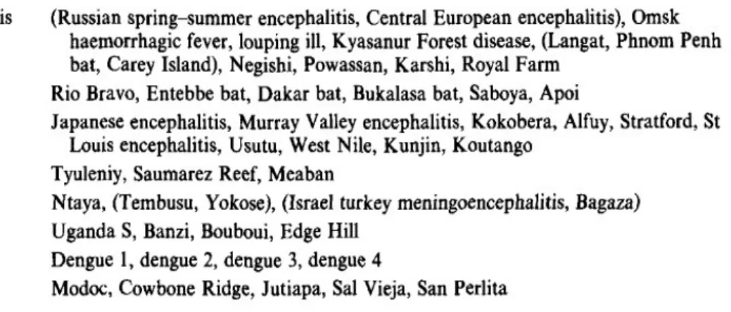
We can classify the members of the genus Flavivirus in two different ways:
- Based on cross-neutralization studies, they have been divided into 8 antigenic
complex (table 1.1) (Calisher CH et al. 1989);
Table 1.1 Antigenic serocomplex of Flavivirus (from Calisher CH et al. 1989)
Most of these viruses are serologically classified into 8 antigenic serocomplex, however 17
others viruses are not sufficiently related to each other to include them in any of these
complexes.
Roberto Bechere
“CHARACTERIZATION AND PHYLOGENETIC ANALYSIS OF USUTU VIRUS ISOLATES IN SARDINIA” PhD Thesis in Life Sciences and Biotechnologies Curriculum Microbiology and Immunology
University of Sassari
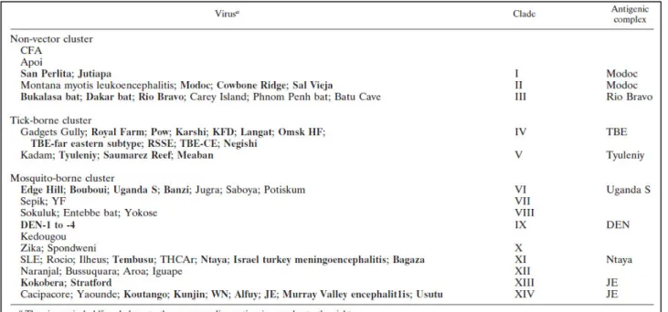
- Based on phylogenetic analysis carried out on partial nucleotide sequences of the
NS5 coding and 3’ noncoding regions (Kuno et al. 1998).
Table 1.2 Assignment of Flaviviruses to clusters and clades (from Kuno et al. 1998)
If we analyse the genetic features of the Flavivirus genus, it is segregated into 14 clades
belonging to 3 different clusters. Each cluster is then divided in different clades, as shown in
the table (1.2) above.
USUV is classified in the JEV group of the mosquito-borne cluster, together with
Cacipacore virus (CPCV), Koutango virus (KOUV), JEV, MVEV, Alfuy virus (ALFV), St
Louis encephalitis virus (SLEV), WNV, Kunjin virus (KUNV), and Yaounde virus (YAOV)
(Heinz et al. 2000).
Roberto Bechere
“CHARACTERIZATION AND PHYLOGENETIC ANALYSIS OF USUTU VIRUS ISOLATES IN SARDINIA” PhD Thesis in Life Sciences and Biotechnologies Curriculum Microbiology and Immunology
University of Sassari
1.2 Molecular features of USUV
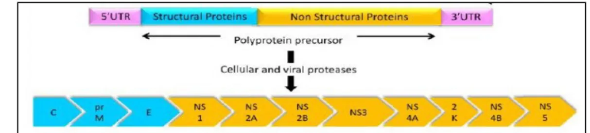
Similar to other flaviviruses, USUV is a spherical, small-enveloped virus with a diameter
40-60 nm. The virion comprises three structural proteins, the capsid (C), the membrane (M),
and the envelope (E) proteins and an 11 kb single-stranded, positive sense RNA genome
(Rice et al. 1986). The genomic RNA (fig 1.2) contains a 5’ cap structure, but lacks a
polyadenylated tail. It acts as mRNA for translation of a single open reading frame (ORF)
encoding the viral proteins as a large polyprotein that is co- and post- translational processed
by cellular and viralproteases into at least 10 separate products. The N-terminal region
encodes the structural proteins C, pre-M, and E, followed by the nonstructural proteins NS1
(soluble complement-fixing antigen), NS2A and NS2B, NS3 (serine protease/RNA
helicase), NS4A, 2K, NS4B, and NS5 (RNA dependent RNA polymerase/methyltransferase)
(Rice et al. 1985).
Fig. 1.2 USUV: its gene structure and the proteins encoded by its genome.
The polyprotein precursor is cleaved by cellular and viral proteases to yield three structural
proteins C (97-474 nt), prM (475-975 nt), and E 976-2475 nt) and eight non-structural
proteins NS1 (2476-3531 nt), NS2A (3532-4212 nt), NS2B (4213-4605 nt), NS3
(4604-6462 nt), NS4A (6463-6840 nt), 2K (6841-6909 nt), NS4B (6910-7683 nt), and NS5
(7684-10398 nt) (from Ashraf et al. 2015).
At the 5’-terminal there is a UTR which is long about 100 nt and, at the 3’-terminal, is
usually long about 670 nt.
Roberto Bechere
“CHARACTERIZATION AND PHYLOGENETIC ANALYSIS OF USUTU VIRUS ISOLATES IN SARDINIA” PhD Thesis in Life Sciences and Biotechnologies Curriculum Microbiology and Immunology
University of Sassari
It is noticeable how the UTRs play a crucial role in the Flavivirus, not only for the initiation
and for the regulation of the translation but also for the replication and the virion assembly.
In particular the 3’-UTRs of various flaviviruses demonstrate extensive heterogeneity in size
and sequence of their proximal parts where long deletions, insertions, sequence repeats and
even poly (A) stretches have been observed (Mandl et al. 1993; Wallner et al. 1995; Wang
et al. 1996; Gritsun et al. 1997). In contrast, the distal part of the 3
’-UTR (330–400
nucleotides in length) exhibits relatively high sequence identity and was previously defined
as the functional
‘core’ element of the flaviviral 3’-UTR (Wallner et al. 1995).
The functions of the 3
’-UTR can be mediated through signal sequences and elements of
RNA secondary structure. Indeed, a number of conserved sequence motifs have been
identified in the 3
’-UTR of flaviviruses (Hahn et al. 1987; Mandl et al. 1993), although
there is no direct evidence for their function.
On the other hand, a region of about 100 nt at the 3
’-terminal of all flaviviruses is known to
form a
“long stable hairpin structure” (3’-LSH) which shows conformational conservation
despite differences in sequence. This would suggest that the 3’- LSH is under selective
constraint and, therefore, of functional importance (Grange et al. 1985; Brinton et al. 1986;
Hahn et al. 1987; Mohan and Padmanabhan, 1991; Mandl et al. 1993; Shi et al. 1996). The
3
’-LSH can exhibit a specific interaction with host cellular proteins (Blackwell and Brinton,
1995, 1997) and viral polymerase (Chen et al. 1997), which points to the direct involvement
of this structure in the initiation of viral minus strand RNA synthesis (Proutsky et al. 1999).
In 2004, with the use of phylogenetic analyses to examine the whole genome sequence of
USUV, it was established the inclusion of one African (SouthAfrica-1959) and one
European (Vienna-2001) lineage of USUV (fig.1.3) (Bakonyi et al. 2004). These two strains
Roberto Bechere
“CHARACTERIZATION AND PHYLOGENETIC ANALYSIS OF USUTU VIRUS ISOLATES IN SARDINIA” PhD Thesis in Life Sciences and Biotechnologies Curriculum Microbiology and Immunology
University of Sassari
are 97% and 99% identical at nucleotide and amino acid level, respectively (Bakonyi et al.
2004).
A research was published in 2016 which established that USUV can be classified into six
distinct lineages and that the most recent common ancestor of the recent European
epizootics emerged in Africa at least 500 years ago (Engel et al. 2016).
Currently, the virulence of the specific USUV lineages is unknown, and further studies are
necessary to determine the biological characteristics of each lineage.
When comparing USUV to other JEV serocomplex viruses, the closest relative that can be
found is MVEV, which exhibits 73% and 82% identity at the nucleotide and amino acid
levels, respectively. JEV and WNV exhibit 71% and 68% identity with USUV at the
nucleotide level and 81% and 75% at amino acid level, respectively (Bakonyi et al. 2004).
1.3 Cellular Tropism and Pathogenesis of USUV
USUV can infect cells of various tissues types derived from humans and a wide variety of
animal species (Bakonyi et al. 2005).
Fig.1.3 Phylogenetic tree illustrating the genetic
relationship between flaviviruses based on their
complete genome sequences. (from Bakonyi et al.
2004)
Roberto Bechere
“CHARACTERIZATION AND PHYLOGENETIC ANALYSIS OF USUTU VIRUS ISOLATES IN SARDINIA” PhD Thesis in Life Sciences and Biotechnologies Curriculum Microbiology and Immunology
University of Sassari
USUV infection on birds is often characterizes by encephalitis, neuronal necrosis,
hepatosplenomegaly, myocardial degeneration and necrosis of the liver and spleen (Chvala
et al. 2004; Bakonyi et al. 2004; Calzolari et al. 2012; Weissenböck et al. 2003). However
the demyelination of infected neurons was found to be a unique feature of USUV infection
(Weissenböck et al. 2004).
Upon infection, human can develop a variety of signs such as encephalitis, meningitis and
fever
.USUV is not considered a significant human pathogen but it is rarely pathogenic in
immune-compromised patients (Cavrini et al. 2009; Pecorari et al. 2009). These organ
lesions can be regarded as a cause of death, because they were generally associated with
sites of viral replication. However, not all of the mentioned lesions are always existing
together.
USUV does not have a specific cell, tissue or organ tropism but, on the contrary, it is present
in a large variety of cells (neurons, different epithelial cells and mesenchymal cells). The
cause of death in birds is most likely due to a multi-organ failure, and brain lesions seem to
play a central role (Chvala et al. 2002).
After the uptake into the cell by endocytosis, the viral particles are transported to the
endosome. Here, the acidic environment enables the release of the capside into the
cytoplasm. After replication of the genome and synthesis of the viral proteins, the viral
particles are assembled and the maturation takes place in the endoplasmic reticulum and are
released by exocytosis.
Roberto Bechere
“CHARACTERIZATION AND PHYLOGENETIC ANALYSIS OF USUTU VIRUS ISOLATES IN SARDINIA” PhD Thesis in Life Sciences and Biotechnologies Curriculum Microbiology and Immunology
University of Sassari
In relation to genus Flavivirus, autophagy is associated with multiple aspects of replication
and pathogenic of some members of this genus, including DENV (Khakpoor et al. 2009);
JEV (Li et al. 2012) and USUV (Blàzquez AB et al. 2013).
Some viruses, including USUV, can take advantage of the autophagic process by
incorporating the components of this cellular pathway in their own replication (Blàzquez AB
et al. 2013; Miller S and Krijnse-Locker J 2008).
“Autophagy” literally means “self-digestion” as it is a cellular process by which cytoplasmic
components are isolated into double-membrane vesicles and degraded to maintain cellular
homeostasis. In addition to this,
“Autophagy” constitutes an evolutionarily ancient process
of survival during different forms of cellular stress, including infection with viruses
(Orvedhal A and Levine B 2008; Mizushima et al. 2008).
Upon infection, USUV induced the Xbp-1 pathway activation of the unfolded protein
response, revealed by the splicing of Xbp-1 mRNA. It also increased cytoplasmic
aggregation of microtubule-associated with the lipidated form of
“protein 1 light chain 3”
(LC3) (Weissenböck et al. 2004), which are considered as marker of autophagosome
formation during viral infections that facilitates replication and immune evasion (Yu et al.
2006).
Roberto Bechere
“CHARACTERIZATION AND PHYLOGENETIC ANALYSIS OF USUTU VIRUS ISOLATES IN SARDINIA” PhD Thesis in Life Sciences and Biotechnologies Curriculum Microbiology and Immunology
University of Sassari
1.4 Enzootic cycle, r
eservoir animals and “bridge-vectors”
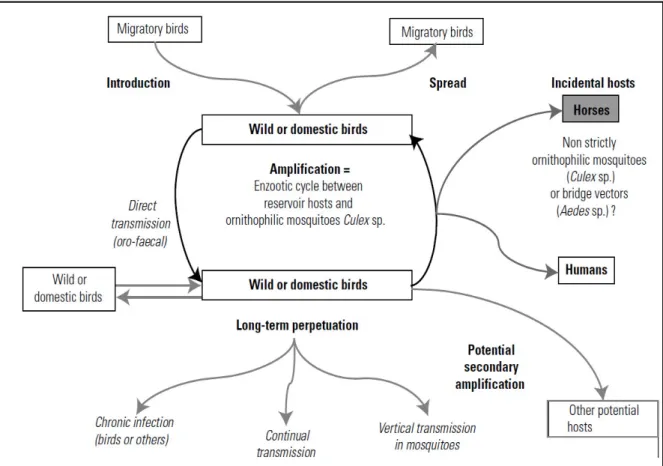
The natural transmission cycle (fig. 1.4) occurs between ornithophilic mosquitoes (vector)
and birds that serve as reservoir or amplifying host.
Fig. 1.4 Usutu virus transmission cycle (from Pradier et al 2012)
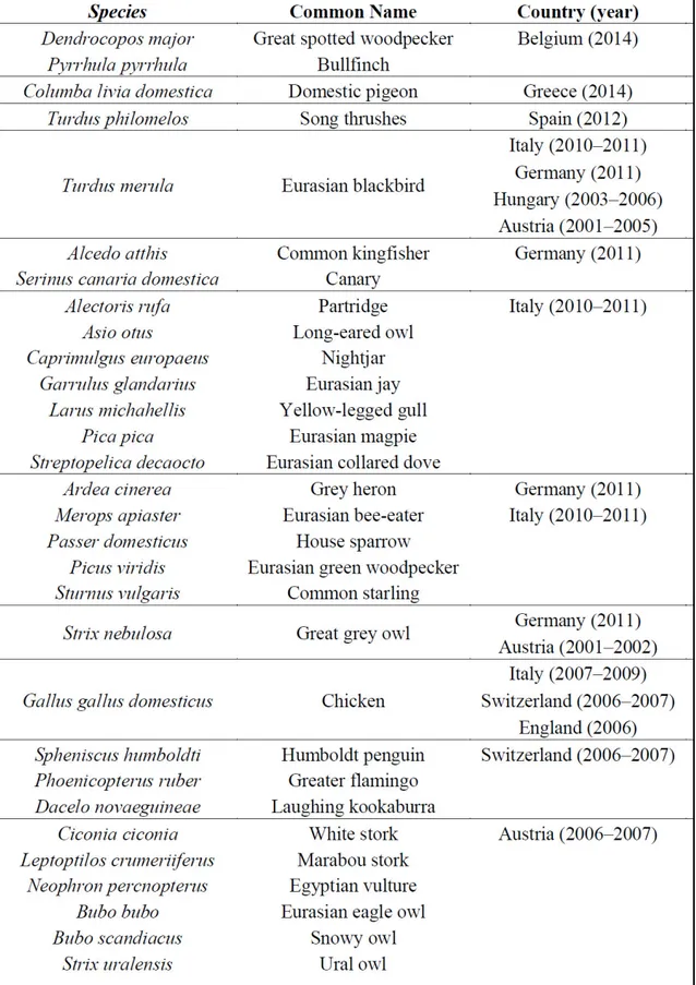
Many mosquitoes and avian species (table 1.3) are involved in perpetuating the USUV life
cycle (Vazquez A et al. 2011; Steinmetz HW et al 2011).
Roberto Bechere
“CHARACTERIZATION AND PHYLOGENETIC ANALYSIS OF USUTU VIRUS ISOLATES IN SARDINIA” PhD Thesis in Life Sciences and Biotechnologies Curriculum Microbiology and Immunology
University of Sassari
Roberto Bechere
“CHARACTERIZATION AND PHYLOGENETIC ANALYSIS OF USUTU VIRUS ISOLATES IN SARDINIA” PhD Thesis in Life Sciences and Biotechnologies Curriculum Microbiology and Immunology
University of Sassari
Mosquitoes are not strictly ornithophilic, in particular the genus Culex
, are called
“bridge-vector” because facilitate viral transmission to incidental host like humans, horses and
rodents. Humans and horses are considered dead-end hosts due to their inability to develop a
sufficient viremia to infect mosquitoes (Kramer et al. 2008; Weaver et al. 2010).
In 2015 in Germany USUV was also isolated from bats for the first time (Cadar et al. 2014)
proving that it is able to emerge in other hosts. This observation raised questions regarding
the USUV host range and the ability to adapt to new hosts.
1.5 Epidemiology of USUV
Following its discovery in South Africa in 1959, USUV was found in other African
countries such as CAR (Central Africa Republic) (1969; 1981); Senegal (1974; 1993; 2007)
(Adam F and Digoutte J-P 2014; Cornet M et al 1979; Nikolay B et al 2011; Chevalier et al
2009) and Tunisia (2014) (Ben Hassine et al 2014). USUV was typically isolated from
mosquitoes, except for the strain “CAR 1981” which was isolated from a man who showed
fever and rush (Institut Pasteur de Dakar 1984). The virus then moved from Africa into
Europe, presumably carried by migratory birds and their related mosquitoes.
The first emergence of USUV in Europe was reported in Austria in 2001 and it derived from
an episode of mass mortality in Eurasian blackbirds (Turdus merula) (Weissenböck et al.
2002).
Interestingly, a similar death of blackbirds was also reported in Italy in 1996 (Mani et al.
1998) but the causes, in this instance, remained unknown. Only in 2013, the partial
nucleotide sequence of that unknown virus was compared with the Austrian strain
“Vienna
Roberto Bechere
“CHARACTERIZATION AND PHYLOGENETIC ANALYSIS OF USUTU VIRUS ISOLATES IN SARDINIA” PhD Thesis in Life Sciences and Biotechnologies Curriculum Microbiology and Immunology
University of Sassari
2001
”, revealing a complete identity sequence (Weissenböck et al. 2013). On the base of
this data, the emergence of USUV in Europe happened much earlier than previously
thought.
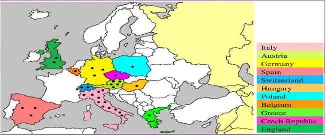
In the following years, USUV was found in several other European countries (fig. 1.5) with
virus isolation from mosquitoes, birds and bats. These countries included Hungary
(2003-2006) (Bakonyi et al. 2007); Switzerland ((2003-2006) (Steinmetz HW et al 2011); Spain
(2006-2009) (Vàzquez et al. 2011; Busquets et al. 2008); Italy (2006) (Calzolari et al. 2009;
Manarolla et al. 2010; Savini et al. 2011; Tamba et al. 2011); Germany (2013) (Cadar et al.
2014) and Belgium (2014) (Garigliany et al. 2014)
USUV was detected in Sardinia for the first time in 2013 from mosquito
’s pool (C. pipiens).
Moreover, USUV infection has also been proved serologically in birds in England (2001-
2004) (Buckley et al. 2006), Czech Republic (2005) (Hubàlek et al. 2008), Spain (2003
–
2006) (Figuerola et al. 2007), Poland (2006) (Hubàlek et al. 2008), Switzerland (2006)
(Steinmetz HW et al. 2007), Germany (2007) (Linke et al. 2007), Italy (2007) (Lelli et al.
2008), and Greece (2014) (Chaintoutis et al. 2014). The recurrence of the virus in Italy
(2010
–2011) (Calzolari et al. 2012; Calzolari et al. 2013), Germany (2011) (Becker et al.
2012), Spain (2012) (Höfle et al. 2013), and Czech Republic (2011
–2012) (Hubàlek et al.
2014) suggests persistence of the transmission cycle in the affected areas, possibly through
overwintering mosquitoes (Vàzquez et al. 2011).
In the summer 2016, the virus was detected for the first time in the continent of South
America, specifically in Colombia (Paniz-Mondolfi AE et al. 2016).
Roberto Bechere
“CHARACTERIZATION AND PHYLOGENETIC ANALYSIS OF USUTU VIRUS ISOLATES IN SARDINIA” PhD Thesis in Life Sciences and Biotechnologies Curriculum Microbiology and Immunology
University of Sassari
Fig. 1.5 Geographic locations of USUV-related epidemiological studies on birds and
mosquitoes in Europe (from Ashraf et al. 2015).
Following its emergence in Europe, USUV has proven to be highly pathogenic for several
species of wild birds, especially Eurasian blackbirds (Turdus merula) (Bakonji et al. 2004).
In addition to avian species, USUV has also been detected in humans. The first reported case
of USUV infection was observed in Africa in two patients with fever, rash and jaundice
(Nikolay et al. 2011).
Subsequently, in Europe two USUV-positive cases of meningoencephalitis were reported in
immune-compromised patients in Italy (Cavrini et al. 2009; Pecorari et al. 2009; Gaibani et
al. 2010). The first case was developed following orthotropic liver transplantation with an
infected organ donor, and the second case was found in a cancer patient.
Recently, the first 5 cases of USUV specific-IgG positive sera in healthy blood donors have
been reported, confirming the theory that USUV actively circulates amongst asymptomatic
subjects in Europe (Gaibani et al. 2012; Allering et al. 2012).
Roberto Bechere
“CHARACTERIZATION AND PHYLOGENETIC ANALYSIS OF USUTU VIRUS ISOLATES IN SARDINIA” PhD Thesis in Life Sciences and Biotechnologies Curriculum Microbiology and Immunology
University of Sassari
Therefore, the introduction onto a new continent of a previously exotic flavivirus that is
closely related to other pathogenic flavivirus is a potential serious threat for a wide range of
avian and mammalian species, including human (Bakonyi et al. 2004).
Roberto Bechere
“CHARACTERIZATION AND PHYLOGENETIC ANALYSIS OF USUTU VIRUS ISOLATES IN SARDINIA” PhD Thesis in Life Sciences and Biotechnologies Curriculum Microbiology and Immunology
University of Sassari
Chapter 2
Aim of the Project
This research has been carried out by the Veterinary Medicine Department, Infectious
Diseases Division of the University of Sassari, and the Istituto Zooprofilattico Sperimentale
della Sardegna
“G. Pegreffi”, Animal Health Department, Virology and Entomology
Division.
The aim of the project is the study of USUV circulating in Sardinia through the screening of
surveillance samples.
On the base of positive samples, we have then proceeded to full genomic sequencing
through the technique
of “Primer-Walking” with the use of primers which have been
designed by us.
From the data acquired, we have then proceeded to molecular characterization and
phylogenetic analysis of strains that have been found, with the aim of investigating
mutations associated with increased virulence or associated with geography - or
host-adaptation.
Roberto Bechere
“CHARACTERIZATION AND PHYLOGENETIC ANALYSIS OF USUTU VIRUS ISOLATES IN SARDINIA” PhD Thesis in Life Sciences and Biotechnologies Curriculum Microbiology and Immunology
University of Sassari
Chapter 3
Materials and Methods
3.1 Entomological surveillance
The aim of entomological surveillance is to identify the mosquitoes involved in viral
transmission and their seasonal abundance in the framework of the entomological activities,
as defined in the Italian surveillance plan for WNV. In Sardinia we are also able to expand
this surveillance plan for USUV.
In recent years, there have been increasing global reports of diseases due to Arboviruses
(arthropod-borne viruses) (Gratz NG, 2006; Weaver SC and Reisen WK, 2010; Hubàlek Z,
2008). The detection of viruses in Invertebrate vectors and Vertebrate reservoir hosts is an
important part of public health surveillance systems that allows the analysis of the intensity
and seasonality of viral circulation in the environment. It provides data for the timely
planning of preventive measures such as blood donation screening (CDC Guidelines for
Surveillance, Prevention, & Control. 3
rdrevision, 2011).
Surveillance based on mosquitoes, wild and synanthropic birds sampling can provide early
detection of the virus circulation before the onset of the disease in humans and horses
(Calzolari et al. 2015).
Entomological surveillance in Sardinia was conducted using CDC light-traps operating for
24 hours in 35 geo-referenced sites every two weeks, from April until October 2015 or until
two consecutive catches were detected without finding mosquitoes.
Roberto Bechere
“CHARACTERIZATION AND PHYLOGENETIC ANALYSIS OF USUTU VIRUS ISOLATES IN SARDINIA” PhD Thesis in Life Sciences and Biotechnologies Curriculum Microbiology and Immunology
University of Sassari
Mosquitoes were identified to specie level by using morphological characteristics according
to four classification keys (Stojanovich CJ and Scott HG, 1997; Schaffner et al. 2001;
Severini et al. 2001; Becker et al. 2010).
Mosquitoes were pooled according to species, sex, place and date of sampling and on
whether they were engorged or not, with a maximum of 50 individuals per pool.
To avoid cross-contamination due to possible loss of mosquito parts, the pools were
obtained by handling specimens individually with tweezers.
The batches of mosquitoes were stored in 2 ml polypropylene cryotubes (Eppendorf) and
frozen at -80 °C, up to their biomolecular analysis.
One tungsten ball (Tungsten Carbide Beads, Qiagen) of 3 mm in diameter, was added to
each 2 ml tube. Different amount of PBS 1X were added to each tube according to the
number of stored mosquitoes (350 ul up to 10 specimens; 500 ul up to 30 specimens; 600 ul
up to 50 specimens).
The samples were then ground for 1 min and 30 sec (frequency 17 1/s) using the
homogenizer TissueLyserII (Qiagen) and centrifuged in a refrigerated centrifuge at 10,000g
for 20 minutes at 4 °C. Finally, samples were submitted to bio-molecular analysis through
specific Real Time RT-PCR for the detection of USUV (Cavrini et al. 2011).
3.2 Birds surveillance
A total of 525 birds were collected within the specific wildlife population control program in
2015 and were checked for the presence of USUV.
Roberto Bechere
“CHARACTERIZATION AND PHYLOGENETIC ANALYSIS OF USUTU VIRUS ISOLATES IN SARDINIA” PhD Thesis in Life Sciences and Biotechnologies Curriculum Microbiology and Immunology
University of Sassari
The active surveillance was carried out on synanthropic birds. Weekly samples of
approximately 20 - 40 birds that were caught or shot were collected. Monitoring mainly
focused on the European magpie (Pica pica); Eurasian jay (Garrulus glandarus) and in
particular on the Hooded crow (Corvus cornix).
The corvid is considered as a pest, due to its wide diffusion and eating habitat.
The passive surveillance was carried out on animals found dead in the field or deceased in
wildlife rehabilitation centers.
Birds were necropsied and for each specimen we took the brain and a pool consisting of
spleen, heart and kidney separately.
Homogenate was made to 10% in PBS (0.5 g sample in 5 ml PBS 1X). Samples were then
ground using the homogenizer Omni Bead Ruptor 24 (Omni International, Inc., USA) with
the following program: two steps to 45 sec with a pause for 15 sec; frequency 5,65 1/s.
Twenty (2.8 mm in diameter) Ceramic Beads Bulk (Omni International, Inc., USA) was
added to each 5 ml tube.
Viral RNA was extracted from 100 ul homogenate sample (pool of mosquitoes; organs)
using the commercial kit Biosprint 96 One-for-all Vet Kit (384) (Qiagen) according to the
manufacturer’s instructions.
After RNA extraction with the automatic nucleic acid extractor Biosprint 96 (Qiagen) we
submitted them to biomolecular analysis; samples from every bird were processed
individually. All the samples were assessed with a specific Real-time RT-PCR for the
detection of USUV RNA (Cavrini et al. 2011).
Roberto Bechere
“CHARACTERIZATION AND PHYLOGENETIC ANALYSIS OF USUTU VIRUS ISOLATES IN SARDINIA” PhD Thesis in Life Sciences and Biotechnologies Curriculum Microbiology and Immunology
University of Sassari
3.3 Phylogenetic analysis
For phylogenetic analysis, the most informative genes of USUV, and generally for
Flavivirus reported in bibliography, are the gene E, which encodes Envelope protein, and
the gene NS5, which encodes RNA dependent RNA polymerase.
Initially, a preliminary amplification of partial NS5 and E gene region was made before
sequencing the entire genome of our isolates of USUV.
The purpose of this step was not only related to phylogenetic analysis but also to obtain
information about the origin of our isolates (example European or Africans lineages), in
order to develop specific primers for the whole genome sequencing and to have a strong
confirmation of USUV positivity in our isolate based on data sequence.
Firstly, we tried to use the degenerated primers specific for Flavivirus designed by
Sanchez-Seco MP et al. 2005 and Kuno et al. 1998 but unexpectedly we did not have any
amplification on our samples.
After these negative results, we designed new primers to cover the partial genomic regions
of the genes E and NS5. Primers are developed on viral variant obtained from a positive
sample.
The pairs of primer designed by us were:
gene NS5:
Usuv8885 F TAAGAGGAAGGTCAACAGCA /
Usuv9597 R TTCCTGCCCAATCACTCCTT (712 bp)
Roberto Bechere
“CHARACTERIZATION AND PHYLOGENETIC ANALYSIS OF USUTU VIRUS ISOLATES IN SARDINIA” PhD Thesis in Life Sciences and Biotechnologies Curriculum Microbiology and Immunology
University of Sassari
gene E:
Usuv1124 F CACGAACCTGGCTGAAGTGA /
Usuv1789 R CTGGAAYAGCTCCTGCCAAK (665 bp)
Thermo cycler parameters (except T° annealing 60°C) and PCR mix reaction are the same to
the primers used for the full-length genome amplification.
We analised all the isolates found but surprisingly we obtained only the partial sequence of
NS5 and E genes of two isolates, 91418 (2013) and 62059/4 (2015).
After sequencing, they confirmed the USUV positivity and a very close relation with Italian
strains
“Bologna2009” and “Italia2009”.
Based on these results, however, we proceeded into analysing only the isolates 91418 (2013)
and 62059/4 (2015).
With data analysis, the gene NS5 has proved globally much more informative in comparison
to the gene E, and for this reason we decided to use the gene NS5 for the phylogenetic
analysis of USUV.
List USUV strains used for phylogenetic analysis:
1. Usutu virus strain UR-10-Tm polyprotein gene, complete cds
KX555624.1
2. Usutu virus strain OS1-10-Tm polyprotein gene, complete cds
KX555628.1
3. Usutu virus strain SE1-10-Tm polyprotein gene, complete cds
KX555626.1
4. Usutu virus strain Bologna 2009, complete genome
HM569263.1
Roberto Bechere
“CHARACTERIZATION AND PHYLOGENETIC ANALYSIS OF USUTU VIRUS ISOLATES IN SARDINIA” PhD Thesis in Life Sciences and Biotechnologies Curriculum Microbiology and Immunology
University of Sassari
6. Usutu virus strain Vienna 2001 from Austria, complete genome
AY453411.1
7. Usutu virus strain Meise H polyprotein gene, complete cds
JQ219843.1
8. Usutu virus isolate Budapest, complete genome
EF206350.1
9. Usutu virus strain V86, complete genome
KJ438781.1
10. Usutu virus strain V20, complete genome
KJ438769.1
11. Usutu virus strain V191, complete genome
KJ438779.1
12. Usutu virus isolate S.nebulosa-7890/Fra/2016, complete genome
KY128481.1
13. Usutu virus strain V248, complete genome
KJ438774.1
14. Usutu virus strain V321, complete genome
KJ438766.1
15. Usutu virus strain 3358, complete genome
KJ438721.1
16. Usutu virus gene for polyprotein, genomic RNA, isolate BH65/11-02-03
HE599647.1
17. Usutu virus strain VB-15-Tm polyprotein gene, complete cds
KX555629.1
18. Usutu virus strain V211, complete genome
KJ438764.1
19. Usutu virus strain V72, complete genome
KJ438761.1
20. Usutu virus strain V49, complete genome
KJ438756.1
21. Usutu virus strain V210, complete genome
KJ438763.1
22. Usutu virus strain V10, complete genome
KJ438760.1
23. Usutu virus strain V245, complete genome
KJ438747.1
24. Usutu virus strain V93, complete genome
KJ438746.1
25. Usutu virus strain 6424, complete genome
KJ438736.1
26. Usutu virus strain 1477, complete genome
KJ438705.1
27. Usutu virus isolate BAT2USUTU-BNI, complete genome
KJ859683.1
28. Usutu virus isolate BAT1USUTU-BNI, complete genome
KJ859682.1
29. Usutu virus isolate HB81P08, complete genome
KC754955.1
30. Usutu virus isolate ArD101291, complete genome
KC754956.1
31. Usutu virus isolate ArD192495, complete genome
KC754957.1
32. Usutu virus strain Bonn, complete genome
KM659877.1
33. Usutu virus isolate S.nebulosa-6004/NL/2016, complete genome
KY128482.1
34. Usutu virus isolate ArD19848, complete genome
KC754954.1
35. Usutu virus strain SAAR-1776 from South Africa, complete genome
AY453412.1
Roberto Bechere
“CHARACTERIZATION AND PHYLOGENETIC ANALYSIS OF USUTU VIRUS ISOLATES IN SARDINIA” PhD Thesis in Life Sciences and Biotechnologies Curriculum Microbiology and Immunology
University of Sassari
As outgroup we included the Murray Valley encephalitis virus (MVEV), by using the
reference strain “MVEV_NC000943.1”.
Pairwise/multiple sequence alignments and sequences similarities were calculated using the
ClustalX software (Larkin et al. 2007).
The evolutionary history of USUV was inferred using the Neighbor-Joining method (NJ)
(Saitou and Nei 1987) identified as the best-suited evolutionary model for our data. The
phylogenetic tree was obtained by applying Neighbor-Joining (NJ) algorithms to a matrix of
pairwise distances estimated using the maximum composite likelihood (MCL) method
(Tamura et al. 2004) and were calculated as units of the number of base substitutions per
site. A discrete Gamma distribution was used to model evolutionary rate differences among
sites (parameter = 0.45880082). The analysis involved 39 nucleotide sequences. Codon
positions included were 1st+2nd+3rd+Noncoding. Positions containing gaps and missing
data were disposed of.
A total of 1490 positions were included in the final dataset and evolutionary analyses were
conducted in MEGA6 (Tamura et al. 2013). Statistical support for internal branches (shown
next to the branches) of the trees was evaluated by bootstrapping with 100 iterations
(Felsenstein 1985). NJ (Saitou and Nei 1987) trees and consensus values were generated
using the same software.
Roberto Bechere
“CHARACTERIZATION AND PHYLOGENETIC ANALYSIS OF USUTU VIRUS ISOLATES IN SARDINIA” PhD Thesis in Life Sciences and Biotechnologies Curriculum Microbiology and Immunology
University of Sassari
3.4 Whole genome sequencing Protocol
Regarding the molecular characterization of USUV, it is very important to know the
complete genome sequence, because there are many hot points for the virulence spread in
the viral genome.
However, initially we had to solve a problem. In fact, in the current method of sequencing
the whole genome of USUV with
“Primer-Walking”, we had too many PCR reactions, and
this was not suitable especially with a small amount of RNA sample to work with, like in
our case.
To solve our problem
we developed a new protocol divided in 4 steps:
1) cDNA optimization
2) new primers design to cover the whole genome in a few PCR reactions
3) PCR products cloning
4) Sequencing
1. cDNA optimization
The first step we took was the cDNA optimization. At first
we made two different types of
cDNA using two different specific primers
for USUV: UsuvF1 and Usuv11066Rdeg. This
step was undertaken to ensure the retro-transcription of the entire viral genome.
Then, we tested several combinations of primer concentration and different amount of
sample by using a specific Real time RT-PCR for USUV (Cavrini et al. 2011). This was
Roberto Bechere
“CHARACTERIZATION AND PHYLOGENETIC ANALYSIS OF USUTU VIRUS ISOLATES IN SARDINIA” PhD Thesis in Life Sciences and Biotechnologies Curriculum Microbiology and Immunology
University of Sassari
done to try to limit the amount of RNA sample.
The best arrangement was 20 uM primer
concentration and 2,5 ul of RNA sample.
cDNA Protocol
We used the commercial K
it “SuperscriptII reverse transcriptase”, (Invitrogen).
It is divided in 3 steps:
1
stStep Program name: “65 Usutu”
Add:
- RNA 2.5 ul
- Primer 1 ul
- BSA 1X 0.5 ul
- dNTPs 10 mM 1 ul
- H
2O milliQ 4.5 ul
for 5’ at 65 °C ;
2
ndStep Program name:
“Mix Usutu”
Add:
- 5X first-strand buffer 4 ul
- DTT 0,1 M 2 ul
Roberto Bechere
“CHARACTERIZATION AND PHYLOGENETIC ANALYSIS OF USUTU VIRUS ISOLATES IN SARDINIA” PhD Thesis in Life Sciences and Biotechnologies Curriculum Microbiology and Immunology
University of Sassari
Mix gently pipetting up and down, then:
for 2’ at 42 °C ;
3
rdStep Program name:
“cDNA Usutu”
Add:
- 1 ul reverse transcriptase 200 U/ul
Mix gently pipetting up and down, then:
for 50
’ at 42 °C for 15’ at 70 °C ;
2. New primers design to cover the whole genome in a few PCR reactions
We designed new primers that overlapped between them to cover the whole genome in a
few PCR reactions. Primers are developed on viral variant obtained from a positive sample.
To design our primers we aligned several strains of USUV from African and European
origin using MUSCLE software (available at
http://www.ebi.ac.uk/Tools/msa/muscle/
). (See
Table S1 in the supplemental material).
List USUV strains used to design our primers:
Spain_MB119/06_2006 (KF573410)
Dakar_ArD192495 (KC754957)
Senegal_ArD101291_1993 (KC754956)
Germania_V45_2011 (KJ438770)
Roberto Bechere
“CHARACTERIZATION AND PHYLOGENETIC ANALYSIS OF USUTU VIRUS ISOLATES IN SARDINIA” PhD Thesis in Life Sciences and Biotechnologies Curriculum Microbiology and Immunology
University of Sassari
Germany_V382_2013 (KJ438758)
Vienna_2001 (AY453411)
Bologna_2009 (HM569263)
Italia_2009 (JF266698)
Dakar_ArD19848_1974 (KC754954)
South_Africa_SAAR-1776 (AY453412)
Primer design performed using the free software available on:
www.promix.cribi.unipd.it/cgi-bin/promix/melting
PCR reaction Protocol (50 ul)
We used the commercial Kit
“Platinum Taq DNA Polymerase” (Invitrogen)
PCR mix:
10X PCR Buffer, -Mg 5 ul
50 mM MgCl
21.5 ul
10 uM dNTP mix 1 ul
10 uM forward primer 1 ul
10 uM reverse primer 1 ul
Template cDNA 4 ul
Roberto Bechere
“CHARACTERIZATION AND PHYLOGENETIC ANALYSIS OF USUTU VIRUS ISOLATES IN SARDINIA” PhD Thesis in Life Sciences and Biotechnologies Curriculum Microbiology and Immunology
University of Sassari
Platinum Taq DNA Polymerase 2 U/ rxn 0.2 ul
H
2O milliQ, nuclease free 36.3 ul
Thermo cycler parameter:
Initial denaturation 2’ at 94°C 30sec at 94°C
30sec at (depending on primers T° melting) x 35 1’/ Kb (depending on the length of amplified)
Infinite hold at 4°C
T° annealing:
- PCR reactions: 2; 3; 5 (using the cDNA made with the primer UsuvF1) and 8 (using the
cDNA made with the primer Usuv11066Rdeg) is 60°C ;
- PCR reactions: 4 and 6 is 55 °C (using the cDNA made with the primer UsuvF1).
- PCR reaction 1 is 62 °C (using the cDNA made with the primer Usuv11066Rdeg).
- PCR reactions: 7; 9, 10, 11 and 12 is 58°C (using the cDNA made with the primer
Usuv11066Rdeg).
Pairs of primer used for the full-length amplification of USUV genome:
1) UsuvF1deg AGWYGTTBGYCTGYGTGAGC / Usuv327R TGCCGTGGTCTTGTTGATGC (327 bp)
Roberto Bechere
“CHARACTERIZATION AND PHYLOGENETIC ANALYSIS OF USUTU VIRUS ISOLATES IN SARDINIA” PhD Thesis in Life Sciences and Biotechnologies Curriculum Microbiology and Immunology
University of Sassari
3) Usuv1124F CACGAACCTGGCTGAAGTGA / Usuv2353R TGATCCAGGACATGCCACCG (1229 bp)
4) Usuv2295F GTACATCAGGTCTTTGGAGG / Usuv4693R TGTAACGGCTCATGATACGG (2398 bp)
5) Usuv4578F TGGCTCACTGTCAAGTACGC / Usuv5509R CAATGTAGCCTCGTGCTGCTAT (931 bp)
6) Usuv5443F ACCTCTTTGTGATGGATGAG / Usuv6935R GTTCTCTCTAACATTCCGTA (1492 bp)
7) Usuv6861F GCTGTGTTTCTGATCTGTGT / Usuv8956R TGGCACTGCTCCATTGGTTC (2095 bp)
8) Usuv8885F TAAGAGGAAGGTCAACAGCA / Usuv9994R ACAACAGGAGCCACATCTGA (1109 bp)
9) Usuv9643F* GAGAACGGAGAAGAAAGGGT / Usuv10823R* ACCAGTTCGCATCACCGTCT (1181bp) 10) UsuvADF* (10646) GAAAGCCCCTCAGAACCGTTTC / Usuv11066Rdeg AGATCCTGTGGTCTWGTYCC (420 bp) or 11) UsuvADF* (10646) GAAAGCCCCTCAGAACCGTTTC / Usuv11014R* AGATCCTGTGKTCTWSYYCMCCAYCAG (420 bp) or 12) Usuv10794F** CAAGCGAACAGACGGTGATG / Usuv11027R* GCGCTCTGTGCCTTGTGGTTGAT (233 bp)
Roberto Bechere
“CHARACTERIZATION AND PHYLOGENETIC ANALYSIS OF USUTU VIRUS ISOLATES IN SARDINIA” PhD Thesis in Life Sciences and Biotechnologies Curriculum Microbiology and Immunology
University of Sassari
Pairs of primer used for closing gap (because the amplicon was too long to be sequenced in
one-step by Sanger’s method) in reactions 4 and 7:
1) Usuv6901F GTGGCTGCCAATGAATACGGAA / Usuv7845R TGGGTGTCCTCCAGTTTTGT (944bp)
2) Usuv7769F AGAGGCCATCACTGAAGTCG / Usuv8908R CGTTGCTGTTGACCTTCCTC (1139bp) 3) Usuv3167F TTGGAGTGATGGCGTTGTTG / Usuv4600R TTGCGTACTTGACAGTGAGC (1433bp)
(for these pairs primer the T° annealing is 60°C)
(*) from (Cadar et al. 2014)
(**) from (Bakonyi et al. 2004)
Nucleotide position are according to the
“Vienna2001” reference strain USUV genome
sequence (AY453411) (Bakonyi et al. 2004). (See Table S2 in the supplemental material).
Therefore, with this protocol the whole genome of USUV could be sequenced from only 10
PCR reactions, which were far less in comparison to others protocols available in
bibliography (Cadar et al. 2014; Bakonyi et al. 2004).
Subsequently, the PCR products were all gel purified and cloned. Gel purification
electrophoresis was carried out in a 1% Tris acetate-EDTA-
agarose gel at 80 V for 60’.
The gel was stained with Gel Star
TMNucleic Acid Gel Stain, 10.000X concentrate in DMSO
(LONZA Rockland, ME USA).
Roberto Bechere
“CHARACTERIZATION AND PHYLOGENETIC ANALYSIS OF USUTU VIRUS ISOLATES IN SARDINIA” PhD Thesis in Life Sciences and Biotechnologies Curriculum Microbiology and Immunology
University of Sassari
Then, the band with the molecular size expected, was completely removed from the gel and
purified with the commercial Kit Zymoclean Gel DNA Recovery Kit (200 Preps) (ZYMO
RESEARCH, USA) according to the instructions of the manufacturer.
After gel purification, the PCR products were all cloned using the commercial
kit “TOPO
TA Cloning Kit for sequencing” (Invitrogen) that included “One Shot TOP10 Chemically
Competent Escherichia coli cells
” (Invitrogen) and the plasmid vector “pCR-4-TOPO”,
according to the instructions of the manufacturer.
Plasmid DNA was extracted from positive colonies with
“PureLink Quick Plasmid MiniPrep
Kit
” (Invitrogen) according to the instructions of the manufacturer.
To check if the correct insert was present, we made a specific enzymatic cleavage (EcoR1
enzyme) on the plasmid with the following protocol:
Mix (20 ul)
EcoR1enzyme 0.3 ul
10x Buffer 3 ul
100x BSA 0.3 ul
H
2O milliQ (depending on the sample concentration)
Plasmid DNA (depending on the sample concentration*)
*For each reaction needs average 600 ng of plasmid DNA;
Incubation was performed at 37°C for 2 hours. Lastly, we checked the band expected
through a gel electrophoresis carried out in a 1% Tris acetate-EDTA-agarose gel at 80 V for
60 min.
Roberto Bechere
“CHARACTERIZATION AND PHYLOGENETIC ANALYSIS OF USUTU VIRUS ISOLATES IN SARDINIA” PhD Thesis in Life Sciences and Biotechnologies Curriculum Microbiology and Immunology
University of Sassari
Chapter 4
Results
4.1 Surveillance plan on birds and mosquitoes
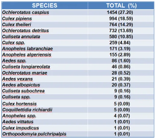
In relation to the surveillance plan, during the year 2015, 5,345 adult mosquitoes
representing 17 different species were collected (table 4.1). The most represented species
were O. caspius with 1,454 specimen(27.2%); C. pipiens 994 (18.6%); C. theileri 764
(14.3%) and O. detritus 732 (13.7%).
Table 4.1 Species of mosquitoes caught during 2015 (from Rossi et al. 2016).
Roberto Bechere
“CHARACTERIZATION AND PHYLOGENETIC ANALYSIS OF USUTU VIRUS ISOLATES IN SARDINIA” PhD Thesis in Life Sciences and Biotechnologies Curriculum Microbiology and Immunology
University of Sassari
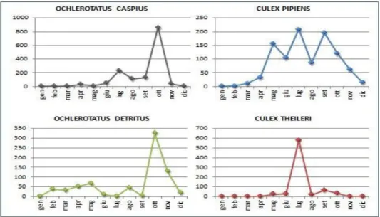
The seasonal abundance (table 4.2) peak of O. caspius and O. detritus were recorded in
October with a maximum of 859 and 324 individuals, respectively while C. pipiens and C.
theileri showed a peak in July with 208 and 577 adults, respectively.
Table 4.2 Seasonal trends spp. Culex and Ochlerotatus (from Rossi et al. 2016).
The Ochlerotatus taxon was considered as an Aedes sub-genus (Savage et al. 2004).
These results confirm the gradual adaptation of the mosquito belonging to Aedes genus to
our climate and environment which could be a very dangerous future health threat because
the Aedes mosquito is the main vector for Zika virus (ZIKV).
During the year 2015, we also observed the over-wintering of mosquitoes in Sardinia,
probably due to a drought condition.
Overall, 1,004 pools were sorted and tested for USUV and it was detected in 4 pools
consisting of not-engorged females of C. pipiens captured from August to October in
Siniscola (ID 54860/1), Oschiri (ID 59265), Alghero (ID 62059/4) and Olbia (ID 71253/2)
municipalities.
Roberto Bechere
“CHARACTERIZATION AND PHYLOGENETIC ANALYSIS OF USUTU VIRUS ISOLATES IN SARDINIA” PhD Thesis in Life Sciences and Biotechnologies Curriculum Microbiology and Immunology
University of Sassari
During 2015, 525 birds were collected within the specific wildlife population control
programs and USUV was detected in a pool (spleen, heart, and kidney) of owl (Athene
noctua) in Olbia (ID 81088) municipality.
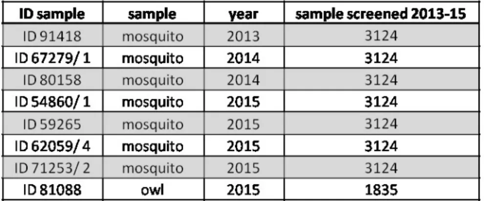
In total we found eight isolates of USUV in Sardinia (table 4.3): five in 2015 which have
been already described above, two isolates of USUV found in 2014 in Badesi (ID 67279/1)
and San Teodoro (ID 80158) municipalities and finally, the first isolate of USUV found in
Sardinia in 2013 in Sassari (ID 91418) municipality.
Table 4.3 Positive samples for USUV found in Sardinia from 2013 to 2015.
All isolates were detected and confirmed by specific Real Time RT-PCR for USUV (Cavrini
et al. 2011). However only the strains 91418 and 62059/4 were also confirmed with data
sequence.
Roberto Bechere
“CHARACTERIZATION AND PHYLOGENETIC ANALYSIS OF USUTU VIRUS ISOLATES IN SARDINIA” PhD Thesis in Life Sciences and Biotechnologies Curriculum Microbiology and Immunology
University of Sassari
4.2 Phylogenetic analysis
We analized all the isolates found (fig. 4.1) but surprisingly we obtained only the partial
sequence of NS5 and E genes of two isolates, 91418 (2013) and 62059/4 (2015) and only by
using the primers that were designed by us.
Fig. 4.1 Photo gel electrophoresis for phylogenetic analysis; the top row in the gel represents
the gene E while the row below represents the gene NS5. We used Marker8 (Roche).
Unfortunately, we did not achieve any result with the use of the primers developed by
Sanchez-Seco MP et al. 2005 and Kuno et al. 1998.
Following these results, we proceeded only with the analysis of the isolates 91418 and
62059/4.
In order to see if they were different, we aligned the sequence of the two isolates with
MUSCLE software (available at
http://www.ebi.ac.uk/Tools/msa/muscle/
) through
preliminary data sequence, and the percentage of identity in both genes was of 100%. We
gave to
this type of sequence the temporary name of “USUTUSARD2013-2015”.
Roberto Bechere
“CHARACTERIZATION AND PHYLOGENETIC ANALYSIS OF USUTU VIRUS ISOLATES IN SARDINIA” PhD Thesis in Life Sciences and Biotechnologies Curriculum Microbiology and Immunology
University of Sassari
Afterwards we compared these results with other sequences of USUV present in the
database. The phylogenetic tree was obtained by MEGA software (Tamura et al. 2013)
using the maximum parsimony in the analysis of taxa, simply because this is more suitable
for much preserved sequences such as USUV genome.
In preliminary analysis, the gene E (fig. 4.2) did not prove to be more informative globally
but it distinguished well between Italian strains.
Fig. 4.2 Provisional phylogenetic tree of gene E USUV.
Instead, the gene NS5 was much more informative globally, for this reason we decided to
use it for the phylogenetic analysis of USUV. In addition, in order to differentiate better
Roberto Bechere
“CHARACTERIZATION AND PHYLOGENETIC ANALYSIS OF USUTU VIRUS ISOLATES IN SARDINIA” PhD Thesis in Life Sciences and Biotechnologies Curriculum Microbiology and Immunology
University of Sassari
between the isolates 91418 and 62059/4 we decided to extend the sequence to investigate
within the NS5 gene from (8885-9597 nt) to (8885-10398 nt).
Percent Identity Matrix - created by Clustal2.1
1: 62059 100.00 99.46
2: 91418 99.46 100.00
In this way, we were able to discriminate between our strains and we designed them
respectively in strain “91418_Sassari2013” and strain “62059_Sardegna2015”.
Subsequently, we compared them with all USUV strains available in the GenBank from
African and European origin using MEGA6 software (Tamura et al. 2013) to be able to
obtain the phylogenetic tree of NS5 gene (fig. 4.3).
The phylogenetic tree shows that Sardinian strains are closely related to Italian strains, in
particular with strain “Bologna2009”. This was an extremely interesting discovery because
“Bologna2009” has been the first strain isolated from a human patient presenting with
neurological symptoms (Gaibani et al. 2009).
However, Sardinian isolates show some unique molecular properties in comparison to all
Italian strains, like the deletions in 3’-UTR which, until now have not been detected in Italy.
The phylogenetic analysis of the European strains demonstrated that they can be divided in 3
major groups defined by location of sampling: German group, Austrian group and Italian
group.
On the other hand, Spanish strains are linked with African strains as well as strains
“Holland2016” and “Bonn2014”. These strains probably belong to the third recent
Roberto Bechere
“CHARACTERIZATION AND PHYLOGENETIC ANALYSIS OF USUTU VIRUS ISOLATES IN SARDINIA” PhD Thesis in Life Sciences and Biotechnologies Curriculum Microbiology and Immunology
University of Sassari
independent introduction (Engel et al. 2016) in Western Europe (Spain) of USUV via
migratory birds.
Roberto Bechere
“CHARACTERIZATION AND PHYLOGENETIC ANALYSIS OF USUTU VIRUS ISOLATES IN SARDINIA” PhD Thesis in Life Sciences and Biotechnologies Curriculum Microbiology and Immunology
University of Sassari
4.3 Whole genome sequencing and molecular characterization
This protocol allows to sequence the whole genome of USUV with a smaller number of
PCR reactions, specifically in only 10 PCR reactions, much less in comparison to others
protocols available in bibliography (Cadar et al. 2014; Bakonyi et al. 2004).
The PCR reactions functioned well but there were a lot of unspecific band (fig. 4.4) because
pool of mosquitoes are a very complex matrix. In fact, there are different type of
“mosquito-only flavivirus
” that maybe can cross-react between them. (Calzolari et al. 2012; Grisenti et
al. 2015; Roiz et al. 2012; Vàzquez et al. 2012).
Fig. 4.4 Gel electrophoresis for all PCR reactions to amplify the entire genome of USUV.
Isolates 91418 and 62059/4 gave the same results except for the reaction 10.
We used Marker8 (Roche) on the left and Marker7 (Roche) on the right.
In relation to
the 3’-terminal region, we had many difficulties in obtaining the expected
amplification. For the isolate 91418 we obtained a band at 190 bp (but we expected a band
at 420 bp) with the PCR reaction ADF/ 11066Rdeg. For the isolate 62059 we obtained two
Roberto Bechere
“CHARACTERIZATION AND PHYLOGENETIC ANALYSIS OF USUTU VIRUS ISOLATES IN SARDINIA” PhD Thesis in Life Sciences and Biotechnologies Curriculum Microbiology and Immunology
University of Sassari
bands at 190 bp and 320 bp (but we expected a band at 420 bp) with the PCR reaction ADF/
11014R but after cloning and sequencing unfortunately they turned out to be unspecific
amplification.
The PCR products have been all gel purified and cloned successfully (fig.4.5).
Fig. 4.5 Gel electrophoresis to check if there were the correct inserts. Isolates 91418 and
62059/4 gave the same results except for the reaction 10. We used Marker8 and Marker7
(Roche).
Therefore, we tried
again to use the previous primer combinations (pairs primer already
described before) but the cDNA was made with "random hexamers" (Roche) rather than
with a specific primer for USUV (11066Rdeg).
In addition, we tried to use a reverse primer (11027R) in a slight previous of the theoretical
end of the viral genome.
Roberto Bechere
“CHARACTERIZATION AND PHYLOGENETIC ANALYSIS OF USUTU VIRUS ISOLATES IN SARDINIA” PhD Thesis in Life Sciences and Biotechnologies Curriculum Microbiology and Immunology
University of Sassari
We achieved amplification (fig.4.6) only with the pair primer 10746F/ 11027R (band
expected at 220 bp) and only in the isolate 62059/4 (B). Unfortunately, we found no valid
results for the isolate 91418 (A).
Fig.4.6 The following pairs primer were used: (1) ADF/ 11066Rdeg; (2) ADF/11014R; (3)
10746F/ 11027R; We used Marker8 (Roche). After sequencing confirmed to be USUV.
This particular difficulty to amplify the 3'-terminal of viral genome can be explained by the
fact that some USUV strains (in particular in the African lineages) have typically high
variable size (short or large deletion). Therefore we assume that our isolates presented a long
or short deletion.
BMR GENOMICS Company carried out the sequencing of plasmids and PCR products.
FinchTV
software
version
1.4.0
(GEOSPIZA,
Inc.)
(Available
at
Roberto Bechere
“CHARACTERIZATION AND PHYLOGENETIC ANALYSIS OF USUTU VIRUS ISOLATES IN SARDINIA” PhD Thesis in Life Sciences and Biotechnologies Curriculum Microbiology and Immunology
University of Sassari
The nucleotide sequences were aligned using MUSCLE software (available at
http://www.ebi.ac.uk/Tools/msa/muscle/
) and confirmed by BLAST software (available at
https://blast.ncbi.nlm.nih.gov/Blast
).
The 91418 USUV isolate complete genome sequence was designated “91418_Sassari2013”
while the 62059/4 USUV isolate complete genome sequence was designated
“62059_Sardegna2015”.
After obtaining the complete genome sequences, we started the molecular characterization
on our viral strains of USUV.
Isolate 91418_Sassari2013
The genome length for the isolate 91418_Sassari2013 is of 10823 nt and contains one large
open reading frame (ORF) comprising nucleotides (nt) 97 to 10401 that encodes a unique
polyprotein precursor of 3424 amino acid (aa).
This strain is characterized for having
a putative large deletion in the 3’-UTR.
Through a comparison of the entire genome nucleotide sequence between our isolates, the
Percent Identity Matrix created by Clustal2.1 is:
1: 91418_Sassari2013 100.00 99.43
2: 62059_Sardegna2015 99.43 100.00
After this, we used BLAST software (
https://blast.ncbi.nlm.nih.gov/Blast
) to estimate the
degree of identity among the strain 91418_Sassari2013 and all the strains of USUV
available in the GenBank at nucleotide level.
Roberto Bechere
“CHARACTERIZATION AND PHYLOGENETIC ANALYSIS OF USUTU VIRUS ISOLATES IN SARDINIA” PhD Thesis in Life Sciences and Biotechnologies Curriculum Microbiology and Immunology
University of Sassari
This analysis showed a Percent Identity Matrix of 99% with all USUV strains except
African strains: "ArD101291"
KC754956.1
; "HB81P08_CAR_1981"
KC754955.1
;
"ArD192495"
KC754957.1
; "ArD19848"
KC754954.1
and "SAAR-1776_South Africa"
AY453412.1
(98-97%) and the following European strains: "S.nebulosa-6004/NL/2016"
KY128482.1
(97%) and "Bonn"
KM659877.1
(97%).
This is closely related with the Italian strains of USUV, in particular with:
"UR-10-Tm_2015"
KX555624.1
; "Bologna2009"
HM569263.1
; "SE1-10-Tm_2010"
KX555626.1
.
Usually the accumulation of mutations in Arbovirus is given by the transmission and
infection modes as effect of alternation between the arthropod vector and avian or mammal
hosts (Holmes EC 2003).
Regarding the UTRs, which is extremely important in Flavivirus, we analyzed the complete
5’-UTR sequence of this strain that is 96 nt long. Its sequence was closely related with all
European strains of USUV and we have not found any unique mutation.
Comparing the complete 5’-UTR nucleotide sequence between our isolates, the Percent
Identity Matrix created by Clustal2.1:
1: 91418_Sassari2013 100.00 96.88
2: 62059_Sardegna2015 96.88 100.00
We have not found any particular mutation in this genomic region.
T
he 3’-UTR sequence in USUV is typically high and variable in size. In the isolate
91418_Sassari2013 is 425 nt long such as many African strains of USUV (fig. 4.7).
Roberto Bechere
“CHARACTERIZATION AND PHYLOGENETIC ANALYSIS OF USUTU VIRUS ISOLATES IN SARDINIA” PhD Thesis in Life Sciences and Biotechnologies Curriculum Microbiology and Immunology
University of Sassari