R E S E A R C H
Open Access
Oral mucosal lesions in teenagers: a
cross-sectional study
Francesca Amadori
1, Elena Bardellini
1,3*, Giulio Conti
2and Alessandra Majorana
1Abstract
Background: Adolescence is a period of transition to adulthood. Little is known about oral mucosal lesions (OMLs) in teenagers, in which the emergence of new habits, unfamiliar to children, could affect the type of lesions. The aim of this study was to evaluate the distribution of oral mucosal lesions (OMLs) in a wide sample of adolescents.
Methods: A retrospective cross-sectional study was carried out examining all medical records of adolescents (aged 13–18 years) treated at the Dental Clinic of the University of Brescia (Italy) in the period from 2008 to 2014. Cases with OMLs were selected. Data regarding age, gender, type of OML, bad habits, systemic chronic diseases were collected.
Results: A total of 6.374 medical records (mean age 15.2 +−1.7 years) were examined. We found 1544 cases (31.7%) of oral mucosal lesions; 36 different types of mucosal alterations were detected and the most frequent were: aphthous ulcers (18%), traumatic ulcerations (14.3%), herpes simplex virus (11%), geographic tongue (9.6%), candidiasis (5.5%), and morsicatio buccarum (4.7%). Papilloma virus lesions (1.7%), piercing-related lesions (4%), multiform erythema (0.13%), oral lichen planus (0.13%) and granular cell tumour (0.06%) were also diagnosed.
Conclusions: The prevalence of OMLs in adolescents are different from those in children and, in some conditions, it could increase with age.
Keywords: Epidemiology, Oral mucosal lesion, Adolescent Background
In literature, epidemiological studies of oral mucosal lesions (OMLs) are still poor, if compared with reports regarding dental caries or periodontal diseases [1]. This gap is even more apparent in case of children and adolescents, where studies focus above all on cancer pa-tients or on samples with specific chronic diseases [2, 3]. In addition, there is a tendency of using different experi-mental methods, non-standardized diagnostic criteria and small samples which leads to a controversial and underestimated prevalence of OMLs in adolescents [4, 5]. However, literature demonstrates that OMLs prevalence seems to change and increase with age along with the development of bad habits [1, 6].
The investigation of OMLs prevalence in specific population groups is mandatory in order to understand its extension and characteristics, but it is also important for the improvement of oral health promotion and pre-vention programs for specific age groups, as recom-mended by the World Health Organization [7].
In a previous report [2], we evaluated the prevalence of OMLs in a wide sample of children, including both those who were healthy and those who had a chronic disease, aged 0–12 years. This study focuses on a differ-ent age group, which is that of teenagers, in order to determine the prevalence of OMLs in adolescence.
Methods
Study population
This study was designed as a retrospective cross-sectional study. All the medical records of adolescents (aged 13– 18 years) who were treated at the Dental Clinic of the Uni-versity of Brescia (Italy) in the period from January 2008 to
* Correspondence:[email protected]
1Dental School, Pediatric Dentistry Department, University of Brescia, P.le Spedali
Civili, 1, 25123 Brescia, Italy
3Clinica Odontoiatrica, P.le Spedali Civili 1, 25123 Brescia, Italy
Full list of author information is available at the end of the article
© The Author(s). 2017 Open Access This article is distributed under the terms of the Creative Commons Attribution 4.0 International License (http://creativecommons.org/licenses/by/4.0/), which permits unrestricted use, distribution, and reproduction in any medium, provided you give appropriate credit to the original author(s) and the source, provide a link to the Creative Commons license, and indicate if changes were made. The Creative Commons Public Domain Dedication waiver (http://creativecommons.org/publicdomain/zero/1.0/) applies to the data made available in this article, unless otherwise stated.
December 2014 were reviewed in order to select teenagers with OMLs. The medical records of patients with lesions linked to dental caries, periodontal diseases and endodontic problems were excluded from the study.
Medical records forms
The medical records of the patients with OMLs were filled out by three clinicians who had undergone the same train-ing and therefore permitted the standardization of the procedures. The calibration to detect OMLs was the same as our team’s previous study [2]; each examiner repeated the calibration every 2 years (three times during the study period). Each patient’s examination was conducted on a dental chair, under artificial lighting and with a mirror. Oral mucosal lesions were classified following the WHO criteria [8]. When necessary, complementary laboratory tests were performed.
Data collection
The following data were recorded from each patient’s medical records: age at time of diagnosis, gender, smoker or non-smoker (if smoker, were they a current or former one), alcohol consumption, systemic chronic diseases, use of drugs and the clinical aspects of the OMLs (clas-sification, location, symptoms, medications). The clinical data of the adolescents were recorded on a specifically designed chart.
Data analysis
The data were inserted on a spread sheet. A 5% level of significance was used and the data was analysed using R® software for Mac. Descriptive analysis, bivariate analysis and Fisher’s test were used.
Ethical considerations
Parents or caregivers signed an informed consent before the beginning of the study. Helsinki Declaration guide-lines were followed for this analysis.
Results
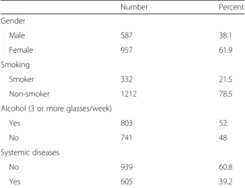
A total of 6.374 medical records belonging to adoles-cents (mean age 15.2 +−1.7 years) were examined (3387 boys and 2987 girls). Out of the total sample, 1544 (31.7%) teenagers showed OMLs. Sociodemographic data are shown in Table 1.
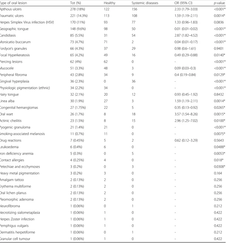
Out of the 1544 adolescents, 36 types of OMLs were detected and the most frequent were: aphthous ulcers (n = 278; 18%), traumatic ulcerations (n = 221, 14.3%), herpes simplex virus (n = 170, 11%), geographic tongue (n = 148, 9.6%), candidiasis (n = 85, 5.5%), and morsica-tio buccarum (n = 73, 4.7%). Papilloma virus lesions (1.7%), piercing-related lesions (4%), multiform erythema (0.13%), oral lichen planus (0.13%) and granular cell tumour (0.06%) were also diagnosed.
39.2% of the patients (n = 605) affected by OMLs also suffered from some form of systemic disease. The distri-bution of OMLs in healthy patients and in adolescents with systemic diseases is shown in Table 2.
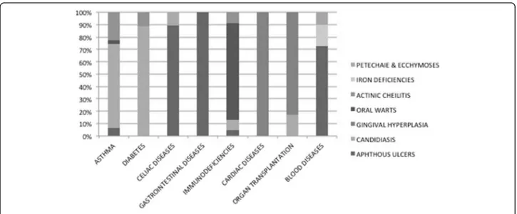
These systemic diseases included diabetes (10%), asthma (8%), heart disease (4%), organ transplant (5%), encephalopathies (9%), gastrointestinal diseases (16%), nephropathies (4%), syndromes (9%), blood diseases (21%) and primary and secondary immunodeficiencies (14%). In particular, of the gastrointestinal diseases, ce-liac disease was the most common. The adolescents with primary immunodeficiencies suffered from Di George syndrome, Hyper-IgE syndrome or IgA deficit T and B cell immunodeficiencies. Acquired immunodeficiencies were secondary to HIV infection. Teenagers with blood diseases had anemia, platelets deficit, leukaemia or lymphomas.
The association between OMLs and the main systemic diseases is displayed in Fig. 1.
Discussion
This retrospective study describes the distribution of OMLs among teenagers who were/are patients of the Dental Clinic of the University of Brescia over a 6-year period. Before discussing the findings of this study, it should be explained that the sample included adolescents admitted both as outpatients for dental emergencies and as inpatients for dental care or consultation from other departments of the Spedali Civili of Brescia (i.e. general pediatrics, pediatric hematology and oncology, auxology, etc.…). The sample is therefore heterogeneous, including both healthy and diseased adolescents. The percentage of healthy people included in this study (61.8%) is almost double compared to a previous study carried out in the same clinic, which focused on children from 0 to 12 years
Table 1 Demographic and behavioral features of the patients with OMLs (n = 1544) Number Percent Gender Male 587 38.1 Female 957 61.9 Smoking Smoker 332 21.5 Non-smoker 1212 78.5 Alcohol (3 or more glasses/week)
Yes 803 52
No 741 48
Systemic diseases
No 939 60.8
Yes 605 39.2
old (39.3%) [2]. The reason lies in the greater number of outpatient teenagers compared to inpatient teenagers i.e. diseased patients from internal departments of the hos-pital. Most requests of internal consultation from the dental clinic are for the 0–12 age range. With in-creasing age, health problems tend to stabilize, thus requiring less hospital check-ups and so the diseased adolescents begin to be treated by private dentists.
Comparison with other reports is not easy, due to the different socio-demographic characteristics of the sam-ples (e.g. schoolchildren or clinic attendees), dissimilar clinical diagnostic criteria and methods, i.e. examination settings and calibration among examiners [9]. These dif-ferences should be taken into consideration when com-paring the frequency and distribution of OMLs in these two age groups (ages 0–12 versus 13–18). Our study
Table 2 OMLs in healthy patients and in adolescents with systemic diseases
Type of oral lesion Tot (%) Healthy Systemic diseases OR (95% CI) p-value Apthous ulcers 278 (18%) 122 156 2.33 (1.79–3.03) <0.001* Traumatic ulcers 221 (14.3%) 113 108 1.59 (1.19–2.11) 0.0014* Herpes Simplex Virus infection (HSV) 170 (11%) 93 77 1.33 (0.96–1.83) 0.0836 Geographic tongue 148 (9.6%) 98 50 0.01 (0.01–0.02) <0.001* Candidiasis 85 (5.5%) 31 54 2.87 (1.82–4.52) <0.001* Morsicatio buccarum 73 (4.7%) 71 2 0.04 (0.01–0.17) <0.001* Fordyce’s granules 66 (4.3%) 37 29 0.98 (0.6–1.61) 0.9401 Focal Hyperkeratosis 65 (4.2%) 49 16 0.49 (0.29–0.88) 0.0140* Piercing lesions 62 (4%) 62 0 - <0.001* Mucocele 51 (3.3%) 48 3 0.09 (0.03–0.3) <0.001* Peripheral fibroma 43 (2.8%) 34 9 0.4 (0.19–0.84) 0.0129* Gingival hyperplasia 36 (2.3%) 0 36 - <0.001* Physiologic pigmentation (ethnic) 34 (2.2%) 34 0 - <0.001* Hairy tongue 32 (2.1%) 20 12 0.93 (0.45–1.92) 0.8432 Linea alba 30 (1.9%) 27 3 1.59 (1.19–2.11) 0.0014* Congenital hemangiomas 27 (1.75%) 22 5 0.35 (0.13–0.92) 0.0265* Oral wart 26 (1.7%) 8 18 3.57 (1.54–8.26) 0.0015* Actinic cheilitis 23 (1.5%) 8 15 2.96 (1.25–7.02) 0.0100* Pyogenic granuloma 21 (1.4%) 21 0 - <0.001* Smoking-associated melanosis 11 (0.7%) 11 0 - 0.0075* Drug reactions 7 (0.45%) 5 2 0.62 (0.12–3.29) 0.5643 Leukoedema 6 (0.4%) 6 0 - 0.0488* Iron deficiency anemia 5 (0.3%) 0 5 - 0.0053* Contact allergies 4 (0.25%) 4 0 - 0.018* Petechiae and ecchymoses 3 (0.2%) 0 3 - 0.0308* Heavy metal pigmentation 3 (0.2%) 3 0 - 0.164 Amalgam tattoo 2 (0.13%) 2 0 - 0.256 Erythema multiforme 2 (0.13%) 2 0 - 0.256 Oral lichen planus 2 (0.13%) 2 0 - 0.256 Pleomorphic adenoma 2 (0.13%) 2 0 - 0.256 Neurofibroma 1 (0.06%) 0 1 - 0.212 Necrotizing sialometaplasia 1 (0.06%) 1 0 - 0.422 Herpes Zoster infection 1 (0.06%) 1 0 - 0.422 Pemphigus vulgaris 1 (0.06%) 1 0 - 0.422 Dermatitis herpetiforme 1 (0.06%) 0 1 - 0.212 Granular cell tumour 1 (0.06%) 1 0 - 0.422
reports a prevalence of OMLs of more then 31% in teen-agers; this data is in accordance with literature: a report carried out in Turkey only on adolescents [5] found a slightly lower prevalence (26.2%) and our previous study [2] reported a prevalence of 28.9% in a younger sample (0–12 years). Instead, a study done in the USA [10] showed a prevalence of 10.26% of OMLs, but in a wider age range sample (2–17 years old children). The preva-lence rate of OMLs increases with age [1, 11] is most likely due to the deterioration of health status and asso-ciated to certain behaviors such as smoking and/or drinking. In our experience, we found the prevalence in the two age ranges of 0–12 years and 13–18 years to be quite similar, but it could be due to the fact that the per-centage of chronic diseases potentially associated to oral lesions was higher in the previous and younger study group. The association between OMLs and systemic dis-ease in the adolescent patients is reported in Table 2.
In literature, the most commonly reported OMLs are geographic tongue, herpetic stomatitis and oral ulcers [9]. These lesions were in fact the most frequent among the adolescents of this study (Tab. 2).
The most common type of OML observed in the teen-agers was recurrent aphthous stomatitis (RAS) (18%). The general prevalence of this lesion has a wide range varying from 0.5% to 39.2%, but it reduces to 10.8% in studies recording their findings on the same day the pa-tient was examined [9]. Since the ulcers may occur at in-tervals and usually heal within 2 weeks [12], its lifetime prevalence is frequently underestimated [13]. In accord-ance with literature [14], in this report RAS resulted sig-nificantly frequent (p < 0.005) in teenagers affected by systemic diseases, in particular those with gastrointes-tinal disturbances (i.e. coeliac disease) and anemia. RAS was more frequent in girls when compared to boys
(p < 0.005). This result is in contrast with the findings reported by Parlak et al. [5] who found no significant differences between the two sexes.
Traumatic lesions were the second most frequently detected (14.3%). The prevalence of lesions caused by trauma found in this study is difficult to compare, because not every study investigated this type of alteration [5] or they would include it in a wider group of lesions [1, 9]. Traumatic ulcers result from heterogeneous causes i.e. from physical, thermal or chemical injuries. Accidental biting during mastication or rough-edged/hot food may cause acute traumatic ulceration, as well as overzealous tooth brushing or iatrogenic damage caused by dental treatment. Such ulcers generally heal within a few days with no complications. However, chronic trauma caused by sharp edges on teeth, restorations and orthodontic ap-pliances may cause chronic ulcers. The majority of such injuries were unintentional. We found some cases of self-inflicted injuries, i.e. in 2 cases of epilepsy attack [15].
Chronic biting (nibbling) of the buccal mucosa often leads to loose threads such as keratin shreds, tissue tags or desquamative areas on the mucosal surface [16]. Such lesions have been referred to as “morsicatio buccarum” when it occurs on the buccal mucosa, “morsicatio labiorum” when on the labial mucosa and “morsicatio linguarum” when it is on the lateral borders of the tongue [17]. A significant percentage of adolescents in our group had this type of reactive OML, which was usually linked to a period of stress and intense study-ing. We found no differences regarding gender or state of health.
The infection of Herpes Simplex Virus (HSV) was found in 11% of the sample. Even if this result is slightly in contrast to literature [18], it highlights the fact that it is difficult to calculate the exact prevalence because of
the characteristic intermittence of the lesions and the different precipitating factors (sun exposure, antibiotic treatments etc.…). Another confounding factor is the methodology used to diagnosis this viral infection: through clinical observations, serological assay or using saliva samples [19, 20]. Despite immunosuppression be-ing one of the causes of HSV reactivation, no difference was recorded between healthy adolescents and those with chronic diseases (p = 0.08) in our sample. The same was for smoking, which in our sample did not seem to be a precipitating cause.
Geographic tongue, also called benign migratory gloss-itis, was found to be more frequent in adolescents when compared to previous reports in children [2, 5]. In our previous study on children under the age of 13 there seemed to be an association between geographic tongue and chronic diseases [2], while in the present sample there is a significant link with healthy adoles-cents. These finding seem to support the still unclear etiology of the pathology.
About 15.7% of teenagers (n = 243/1544) of the sam-ple were smokers. In Italy, as in many other parts of the world, tobacco use by young people remains a serious problem: by the age of 15 years, over 50% have already tried smoking and nearly 15% are daily smokers. Fur-thermore, the global youth tobacco survey’s (GYTS) data on susceptibility to start smoking within 1 year, among those who have not yet started, indicate that 35.4% of the males and 46.6% of the females fall into this suscep-tible category [21]. We found that smoking in teenagers was significantly associated with hairy tongue, smoking-associated melanosis, and focal hyperkeratosis (p < 0.05%). This confirms that smoking has an effect on oral mucosa which may lead to different OMLs, by altering the micro-bial flora, with proliferation of fungi and chromogenic bacteria and over growth of the lingual papillae, or by stimulating the melanocytic pigment extravasation or by promoting the thickening of the oral mucosa.
It is interesting to point out that OMLs normally not described in children and adolescents were found in this sample; in particular, piercing related lesions (4%), oral lichen planus (0.13%) and granular cell tumour (0.06%).
Piercing related lesions consisted of abrasions or thick-ening of the oral mucosa in areas corresponding to points of contact with jewelry, usually on the interior side of the lips or on the surface of the tongue. A small percentage of the lesions was due to trauma caused by piercings no lon-ger present. Literature focuses above all on gingival thick-ness and recessions [22] but, since there has been growing attention to piercing aesthetics in adolescents the last few years, we can speculate that further studies about OMLs will detect an increasing number of these lesions.
Granular cell tumour (GCT) often affects people aged 40–60, with a male female ratio of 1:2. It is rare in
childhood, and usually affects individuals ages 10–18 [23]. Although most of GCTs are benign, a very small percentage can be malignant and can also metastasize [24], therefore its early detection is important, especially at such a young age.
In this analysis, we detected only one adolescent (0.06%) with oral lichen planus (OLP); literature esti-mates that OLP affects 1–16% of patients younger than the age of 15, particularly in cases of chronic disease, stress and infections [25]. In this sample, the patient did not have a chronic disease; however, both in healthy and in diseased patients, a prompt diagnosis and treatment are mandatory to prevent complications.
Some types of OMLs (Fig. 1) resulted significantly more frequent in the adolescents affected by systemic diseases, as a direct consequence of the pathology or as an indirect effect of the pharmacological therapy to cure it. As previously mentioned, aphthous ulcers resulted directly related to some diseases, in particular to celiac disease, to other intestinal diseases and to blood diseases (e.g. anemia). This association seems to confirm that-even if the cause of aphthous ulcers remains unknown-several etiologic factors, like nutritional intolerance or iron deficiencies, seem to play an important triggering role [26]. Oral candidiasis depended both on the diseases (i.e. immunodeficiencies and diabetes) and both on the pharmacological treatment, as in the case of asthma (due to inhaled or topical corticosteroids) and of organ transplantation (due to immunosuppressive drugs). This finding reflects the complex relationship between the commensal state and the pathogenicity of this organism (based on local factors in some cases and systemic fac-tors in others), which can determine the transformation from a state of commensalism to pathogen [26]. Gingival hyperplasia, an overgrowth of gingival fibrous connective tissue, is the result of an unusual tissue response to chronic inflammation. This OML resulted predomin-antly linked to heart diseases and organ transplantation for the hyperplastic effect respectively of antihyperten-sive/antiarrhythmic drugs and of immunosuppressive drugs, often associated with local factors, such as bacter-ial plaque. Oral warts resulted more common among adolescents with immunodeficiencies. Some of oral squamous papillomas have been associated with the same human papilloma virus (HPV) subtypes that cause cutaneous warts. Other oral papillomas have been asso-ciated with different HPV subtypes [26]. Whether all oral papillomas are of viral aetiology is open to question [26]. However, our patients resulted to have viral oral warts, certainly favoured by their immunological status.
Actinic cheilitis resulted significantly more common among adolescents with systemic diseases, especially asthma, diabetes and immunodeficiencies. This is a hardly explainable finding as prolonged exposure to sun-light is the major etiologic factor of actinic cheilitis [26].
We can only speculate that the behaviour (direct expos-ition to sun or solar lamps) associated to a fair skin and a local/systemic immunosuppression could precipitate the development of actinic cheilitis.
Conclusion
In comparison to our previous study on the prevalence of OMLs in children, the prevalence in adolescents re-flects the transition to adulthood, given the increase of some mucosal conditions as well as the appearance of habits unfamiliar to children. Unlike adulthood, however, preventive interventions in this age group can be very effective.
The majority of lesions and their causes are largely avoidable and could be contained with initiatives pro-moting oral health, targeted specifically at teenagers. Therefore, public health interventions should aim at re-ducing various risk factors; from the elimination of local irritants and excellence in dental care, to campaigns de-signed to prevent compulsive habits and psychological dependencies like smoking and the consumption of alcohol.
Acknowledgements Not applicable.
Funding Not applicable.
Availability of data and materials
The datasets used and/or analysed during the current study are available from the corresponding author on reasonable request.
Authors’ contributions
FA conceived the study and drafted the manuscript. EB helped to draft the manuscript and performed the data analysis. GC helped with the data analysis. AM revised the manuscript for intellectual content. All authors read and approved the final manuscript.
Competing interests
The authors declare that they have no competing interest.
Consent for publication Not applicable.
Ethics approval and consent to participate
The research was performed in compliance with the Declaration of Helsinki and Good Clinical Practice. All patients and their caregivers were informed about the research and signed an IRB approved informed consent.
Publisher’s Note
Springer Nature remains neutral with regard to jurisdictional claims in published maps and institutional affiliations.
Author details
1Dental School, Pediatric Dentistry Department, University of Brescia, P.le Spedali
Civili, 1, 25123 Brescia, Italy.2University Vita-Salute San Raffaele, Milan, Italy.3Clinica
Odontoiatrica, P.le Spedali Civili 1, 25123 Brescia, Italy.
Received: 20 March 2017 Accepted: 18 May 2017
References
1. Gheno JN, Martins MAT, Munerato MC, Hugo FN, Sant’Ana Filo M, Weissheimer C, et al. Oral mucosal lesions and their association with sociodemographic, behavioral, and health status factors. Braz Oral Res. 2015;29:1–6.
2. Majorana A, Bardellini E, Flocchini P, Amadori F, Conti G, Campus G, et al. Oral mucosal lesions in children from 0 to 12 years old: ten years’ experience. Oral Surg Oral Med Oral Pathol Oral Endod. 2010;110:e13–8.
3. Rioboo Crespo MR, Planells del Pozo O, Rioboo Garcia R. Epidemiology of the most common oral diseases in children. Med Oral Patol Cir Bucal. 2005; 10:376–87.
4. Bessa CF, Santos PJ, Aguiar MC, et al. Prevalence of oral mucosal alterations in children from 0 to 12 years old. J Oral Pathol Med. 2004;33:7–22. 5. Parlak AH, Koybasi S, Yavuz T, Yesildal N, Anul H, Aydogan I, et al. Prevalence of
oral lesions in 13- to 16- years-old students in Duzce, Turkey. Oral Dis. 2006;12: 553–8.
6. Garcia-Pola Valejo MJ, Martinez Diaz-Canel AI, Garcia Martin JM, González GM. Risk factors for oral soft tissue lesions in an adult Spanish population. Community Dent Oral Epidemiol. 2002;30:277–85.
7. Petersen PE, Estupian-Day S, Ndiaye C. WHO’s action for continuous improvement in oral health. Bull World Health Organ. 2005;83:642.
8. Kramer IR, Pindborg JJ, Bezroukov V, Infirri JS. Guide to epidemiology and diagnosis of oral mucosal diseases and conditions. World Health Organization. Community Dent Oral Epidemiol. 1980;8:1–26.
9. Furlanetto DLC, Crighton A, Topping GVA. Differences in methodologies of measuring the prevalence of oral mucosal lesions in children and adolescents. Int J Ped Dent. 2006;16:31–9.
10. Shulman JD. Prevalence of oral mucosal lesions in children and youths in the USA. Int J Paediatr Dent. 2005;15:89–97.
11. Castellanos JL, Diaz-Guzman L. Lesions of the oral mucosa: an epidemiological study of 23785 Mexican patients. Oral Surg Oral Med Oral Pathol Oral Endod. 2008;105:79–85.
12. Scully C, Porter S. Oral mucosal disease: recurrent aphtous stomatitis. Br J Maxillofac Surg. 2008;46:198–206.
13. Shulman JD. An exploration of point, annual, and lifetime prevalence in characterizing recurrent aphthous stomatitis in USA children and youths. J Oral Pathol Med. 2004;33:558–66.
14. Volta U, Caio G, Stanghellini V, De Giorgio R. The changing clinical profile of celiac disease: a 15-years experience (1998–2012) in an Italian referral center. BMC Gastroenterol. 2014;14:194.
15. Anura A. Traumatic oral mucosal lesions: a mini review and clinical update. Oral Health Dent Manag. 2014;13(2):254–9.
16. Woo SB, Lin D. Morsicatio mucosae oris-a chronic oral frictional keratosis, not a leukoplakia. J Oral Maxillofac Surg. 2009;67:140–2.
17. Neville BW, Damm DD, Allen CM, Bouquot JE. Oral and maxillofacial pathology. Philadelphia: Saunders; 2009.
18. Sawair FA, Jassim ZA, Malkawi ZA, Jamani KD. Epidemiologic aspects of recurrent herpes labialis among Jordanian University students. Saudi Med J. 2010;31:808–13.
19. Shulman JD. Recurrent herpes labialis in US children and youth. Community Dent Oral Epidemiol. 2004;32:402–9.
20. Gilbert S, Corey L, Cunningham A, et al. An update on short-course intermittent and prevention therapies for herpes labialis. Herpesviridae. 2007;14(Suppl 1):13A–8A.
21. Charrier L, Berchialla P, Galeone D, Spizzichino L, Boraccino A, Lemma P, et al. Smoking habits among Italian adolescents: what has changed in the last decade? Biomed Res Int. 2014;2014:287139. doi:10.1155/2014/287139. 22. Maroso FB, Gaio EJ, Rösing Cassiano K, Fernandes MI. Correlation between
gingival thickness and gingival recession in humans. Acta Odontol Latinoam. 2015;28:162–6.
23. Brannon RB, Anand PM. Oral granular cell tumors: an analysis of 10 new pediatric and adolescent case and a review of the literature. J Clin Pediatr Dent. 2004;29:69–74.
24. Krishnamurthy A, George R, Majhi U. Malignant granular cell tumor of the tongue: a clinico-pathological challenge. Indian J Surg Oncol. 2014;5:71–4. 25. Luis-Montoya P, Domínguez-Soto L. Vega- Memije E. Lichen Planus in 24
children with review of the literature. Pediatr Dermatol. 2005;22:295–8. 26. Regezi J, Sciubba J, Jordan R. Oral Pathology. 7thed. United States: