1
EPUB
ACTA oTorhinolAryngologiCA iTAliCA:EPUB February 09, 2012
Case report
Palliative combined treatment for unresectable
cutaneous basosquamous cell carcinoma
of the head and neck
Trattamento palliativo combinato di un carcinoma basosquamoso cutaneo
non resecabile del distretto testa-collo
A. DegAnello, g. gitti, B. StruijS1, F. PAiAr2, o. gAllo
Clinic of otolaryngology/Head and neck Surgery, Department of Surgical Sciences, university of Florence, italy;
1 Department of otolaryngology Head and neck Surgery, Vu Medical Center, Amsterdam, the netherlands; 2 Department of radiotherapy, university of Florence, italy
SUmmAry
A case is presented of a patient with a skin basosquamous cell carcinoma of the frontal region infiltrating the cerebral tissue and with a widespread unresectable regional metastatic ulceration of the left parotid region. The patient underwent combined palliative treatment: sur-gical coverage of the ulceration by means of a pectoralis mayor flap transposition and radiotherapy. After 18 months of follow-up, no signs of tumour progression were noted, the patient is currently free from pain, no increase in trismus was seen, and a slight gain in weight was recorded. Unresectable cancer is mainly treated by concurrent chemoradiation; radiotherapy, however, is contraindicated in deep neoplastic ulcerations with exposure of large vessels. The data reported suggest that surgical coverage of an unresectable neoplastic ulcer is feasible, and combined with early administration of radiation permits a palliative approach in an otherwise untreatable condition.
KEy wordS: Basosquamous cell carcinoma • Palliative surgery • Palliative radiotherapy • Head and neck cancer
riASSUnTo
Presentiamo il caso di un paziente con carcinoma basosquamoso della regione frontale arrivato alla nostra osservazione avendo già sviluppato un’estesa infiltrazione del tessuto cerebrale ed un’ampia ulcera metastatica laterocervicale sinistra non resecabile. Per poter offrire un trattamento palliativo al paziente abbiamo eseguito una copertura dell’ulcera neoplastica attraverso la trasposizione di un lembo miocutaneo di gran pettorale a cui ha fatto seguito radioterapia palliativa. Dopo 18 mesi di follow-up non sono stati riscontrati segni di progressione di malattia a livello loco-regionale, il paziente non ha dolore, il trisma non è aumentato e si è registrato un guadagno ponde-rale. Le forme tumorali non resecabili vengono trattate con protocolli chemio-radioterapici; questi tuttavia sono controindicati in caso di ulcere profonde con esposizione dei grandi vasi. Il nostro caso clinico suggerisce che è tecnicamente possibile coprire efficacemente ulcere neoplastiche non resecabili ai fini di poter eseguire ulteriori trattamenti palliativi che devono necessariamente essere tempestivi.
PArolE ChiAvE: Carcinoma squamocellulare • Chirurgia palliativa • Radioterapia palliativa • Tumore cutaneo • Cancro del distretto testa-collo
Acta Otorhinolaryngol Ital:EPUB February 09, 2012
Introduction
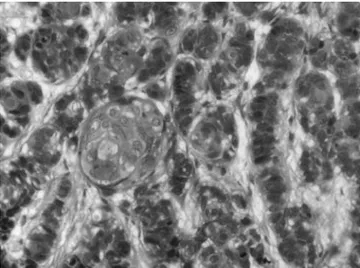
Basosquamous cell carcinoma (BSCC) is a rare malig-nancy with features of both basal cell carcinoma (BCC) and squamous cell carcinoma (SCC) (Fig. 1). The recent world health organization (who) classification of head and neck tumours 1 defines BSCC as an aggressive
sub-type of SCC. The incidence of BSCC among BCC has been estimated to be 0.4-12% 2 and its higher metastatic
propensity compared to SCC, is clearly highlighted in the literature 3 4. rare cases of BSCC with either
leptomenin-geal carcinomatosis 5 or infiltrating cerebral tissue 6 have
been reported. disease onset usually occurs between the sixth and seventh decade, with the skin of the head and neck being the sites most involved. A more aggressive be-haviour has been recorded in patients with recurrence, and male sex has been statistically correlated with likelihood of recurrence 7.
Palliative treatment in head and neck cancer aims to im-prove the patient’s quality of life. in some cases, palliative treatment can prevent life-threatening complications (such as rupture of vessels) thus prolonging patient survival. herewith the case is presented of a patient with BSCC of the supraorbital skin, infiltrating the cerebral tissue and
A. deganello et al.
2
EPUB
with widespread regional metastatic ulceration of the leftparotid region. The patient underwent palliative treatment with surgical coverage and radiotherapy.
Case report
A 54-year-old male patient consulted our first aid unit on account of moderate bleeding from the neck. Upon physi-cal examination, on the left side, a 3 × 2 cm non-bleeding ulceration of the frontal skin was found and a wide (ap-proximately 10 × 5 cm) ulcer involving the left parotid area from the zygomatic arch to the level of the hyoid bone, eroding the external ear canal, disrupting most of the pavilion and causing complete peripheral facial nerve paralysis (Fig. 2). The patient also presented a moderate trismus with 1.5 cm maximal mouth opening. Upon visual inspection, it was possible to clearly perceive the carotid pulse at the jugulo-digastric level.
laboratory tests revealed a haemoglobin value of 12 mg/ dl with normal electroytes.
discussing the medical history, the patient reported hav-ing undergone surgery abroad (Albania) for BCC of the frontal skin. Unfortunately, earlier detailed charts were not available, and relatives could not provide exhaustive information about previous surgical treatment. it was cer-tain, however, that the patient had never received either radiotherapy or chemotherapy and that, abroad, no fur-ther treatment options were advised besides pain control. From these findings, it was assessed that the disease ap-peared 2 years before as a left-sided frontal skin lesion for which the patient had undergone local excision. Six months later, the patient developed a left parotid-cervical mass that was treated with parotidectomy and lymph-node excision (no codified type of neck dissection was performed). Furthermore, 6 months after parotid-cervical surgery, a non-healing ulceration, which became progres-sively enlarged, appeared in the parotid region with con-comitant facial paralysis. moreover, a necrotic sore devel-oped from the operated frontal skin. Since then the patient experienced progressive fatigue with consistent limitation in his daily activities, increasing pain that required mor-phine patches, and weight loss (8 kg in the last 6 months). he never experienced either seizure or any other kind of cerebral symptom.
Contrast enhanced CT scan of the head and neck and thorax showed infiltration of the meningeal and cerebral tissue of the left frontal lobe, infiltration of the mastoid middle ear, complete encasement of the left internal and external carotid arteries with involvement of the pterygoid muscles and skull base (Fig. 3). no evidence of distant lung metastasis was observed. After appropriate counsel-ling and multidisciplinary evaluation, the patient under-went surgery. Extensive biopsies of the parotid ulcera-tion’s borders were submitted for frozen section analysis that revealed BSCC (tumour staging rT4n1m0 8). The
edges of the skin were resected until macroscopic healthy tissue was found. A generous undermining of the cervi-cal skin, reaching the superior border of the clavicle, was carried out in order to avoid further incisions in the neck. A pectoralis major flap with a skin paddle of 20 × 8 cm was transposed to cover the defect. The external auditory canal was obliterated.
Fig. 1. Photomicrograph of the biopsy specimen (haematoxylin/eosin stain-ing), showing basaloid cells and focal areas of squamous differentiation with keratinization.
Fig. 2. Pre-operative aspect of the left parotid region.
Fig. 3. Pre-operative CT scan. A: infiltration of the frontal bone and cerebral tissue. B: Involvement of the lateral pterygoid plate, tumour extension to the parapharyngeal space with encasement of the styloid process and internal carotid artery that lie close to the bottom of the ulcer.
Palliative combined treatment for unresectable cutaneous basosquamous cell carcinoma of the head and neck
3
EPUB
The post-operative course was uneventful, flap viability was optimal and no wound dehiscences occurred. The pa-tient was discharged on the 4th post-operative day having
been advised to undergo palliative radiotherapy that was started on the 15th day. External beam three-dimensional
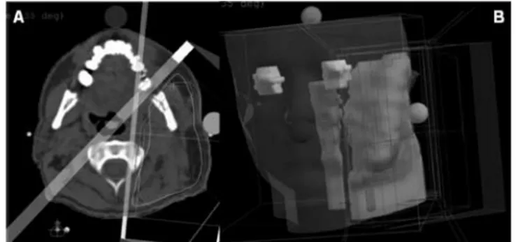
conformal radiotherapy was carried out using 6-mv pho-tons (linear accelerator) equipment. Planning CT scan was performed in treatment position with a customized head mask. The gross tumour volume (gTv), the clinical target volume (CTv), the planning target volume (PTv) and the organs at risk (spinal cord, lens, eyes, etc.) were delineated on each slice. radiotherapy was delivered to the cervico-parotid region with standard fractionation (2 gy per day, 5 days a week) using three coplanar converging wedged beams (Fig. 4) and was stopped, at a total dose of 44 gy, when a neck abscess developed at the operated neck side. The abscess was treated with surgical evacuation by means of simple pen rose drain introduction and antibiotic admin-istration. Since gTv, at this site, was strictly related to the spinal cord, it was not possible to achieve a higher total dose. The supraorbital frontal skin recurrence was treated with electron beam radiotherapy with standard fractiona-tion until a total dose of 44 gy was reached.
Adjuvant chemotherapy was planned and advised, after the end of radiation, but the patient refused.
After 18 months of follow-up, no signs of tumour progres-sion were noted, the patient is currently pain free, no in-crease in trismus was observed, and a slight gain in weight was recorded (4 kg since the end of radiation) (Fig. 5).
Discussion
in the literature, there are no reports giving guidelines for the management of unresectable neoplastic head and neck ulcerations with vessel exposure.
Unresectable cancer is mainly treated by radiotherapy with comitant chemotherapy, the setting of which is de-signed for a curative or a palliative intent based on the realistic chances of tumour control. Several host/tumour factors must be taken into consideration in treatment plan-ning: patient’s general conditions (performance status) and specific comorbidities that might prevent withstand-ing of the treatment, the possibility of deliverwithstand-ing curative doses of radiation without damaging vital structures, the locoregional volumetric extension of the disease, the pres-ence or abspres-ence of distant metastases.
in the present case, due to cerebral involvement, it was not possible to offer the patient a chemoradiation protocol with curative intent.
in the case presented, the need for surgical coverage arose from the evidence that radiotherapy is contraindicated in deep neoplastic ulcerations with exposure of great vessels, on account of the serious risk of blow-out with fatal haem-orrhage. regional and distant tissue transfer techniques have increased the possibility of covering vital organs with well vascularised tissue allowing otherwise impossible ra-diation delivery. nevertheless, we were unable to predict whether flap transposition would have efficiently covered the defect without dehiscence and without an immediate neoplastic colonization of the transposed tissue from the neoplastic recipient. This was our major concern, but we had to face the fact that no other options were available and that carotid rupture is such a catastrophic event that its expo-sure represents a surgical priority in itself 9. we felt that the
transposition of a pectoralis major flap was more appropri-ate than reconstruction with a microvascular free flap, since the quality of the donor vessels for microvascular anasto-mosis was questionable.
in head and neck cancer, it is recommended to start post-operative adjuvant radiation, within 4-6 weeks after sur-gery 10 to maximize loco-regional control. our prompt
onset of post-operative radiation within 2 weeks after sur-gery, despite the development of a neck abscess, might have been a crucial factor in successful palliation.
Even if, however, we obtained a pathologic assessment only of the parotid-neck ulceration, we believe that the frontal le-sion had to be considered the primary tumour, while the pa-rotid ulceration was the regional metastatic extension. Since the frontal ulceration appeared as a non-bleeding necrotic sore, we felt that it was not appropriate to take biopsies that
Fig. 5. Post-operative result 18 months after palliative treatment. Fig. 4. A: Planning CT scan, transversal dose distribution for three-dimen-sional conformal radiation therapy, three wedged fields. B: Organs at risk and treatment volumes, three-dimensional three fields isodose reconstruction.
A. deganello et al.
4
EPUB
could have worsened the local status and delayed thepossi-bility of radiation delivery. in fact, necrotic tissue frequent-ly hides cancer proliferation, therefore thus preventing the diagnosis. This slight likelihood of obtaining the correct diagnosis with a single bite biopsy leads the surgeon physi-cian to perform multiple biopsies that could jeopardize the clinical condition. Furthermore, confirmation histology of the primary lesion would not have changed our treatment strategy in this particular patient. in the sixth edition of the Tnm classification 8 (the only one available at the time
the patient was treated), a giant unresectable metastatic in-volvement of the neck and parotid was classified n1 like a single small lymph node metastasis. recently, in the sev-enth edition of the Tnm classification 11, a more accurate n
classification for non-melanoma skin cancer has been intro-duced, accordingly our case would now be classified n3.: n0 no lymph node metastasis; n1 single < 3 cm; n2 single ≥ 3 to 6 cm, multiple ≤ 6 cm; n3 > 6 cm. These changes certainly improve the hazard consistency (homogeneity within the group) and hazard discrimination (heterogeneity between the groups) of the classification. however, we feel that a further discrimination between resectable and unre-sectable regional neck disease might be helpful, consider-ing that treatment and prognosis of these conditions differ
consistently. Furthermore, parotid disease, facial nerve in-volvement and tumour size greater than ≥ 6 cm within the parotid, had less favourable prognosis in terms of survival in several studies 12-14.
Based on this concept, several years ago, a new staging system was introduced that separates parotid involve-ment from cervical lymph node involveinvolve-ment 10-12. Parotid
disease was more prognostic of poor survival than neck involvement; in particular, facial nerve involvement and tumour size ≥ 6 cm within the parotid 11 13. These studies
were conducted on cutaneous SCC but probably the same findings might be valid also for BSCC.
Conclusions
in cases of unresectable neoplastic head and neck ul-ceration, the combination of surgical coverage with post-operative radiation radiotherapy offers a valid treatment option to achieve a palliation in an otherwise untreatable situation. This report indicates that surgical coverage of vital organs with well vascularised tissue is feasible even in the case of neoplastic recipient ulceration; the authors believe that, in these conditions, early administration of palliative radiation radiotherapy is mandatory.
Address for correspondence: dr. Alberto deganello, Clinic of otolaryngology, head and neck Surgery, department of Surgical Sciences, University of Florence, v.le morgagni 85, 50134 Firenze, italy. Fax: +39 055 435649; E-mail: [email protected]
received: September 29, 2010 - Accepted: march 15, 2011
References
1 Barnes l, Eveson Jw, reichart P, et al. Pathology and
genet-ics of head and neck tumors. lyon: iArC Press; 2005.
2 Kirkham n. Tumors and cysts of the epidermis. in: Elder dE,
editor. Lever’s Histopathology of the Skin, 8th edn.
Philadel-phia, PA: lippincott william & wilkins; 1997. p. 728-9.
3 lopes de Faria J, nunes Ph. Basosquamous cell carcinoma
of the skin with metastases. histopathology 1988;12:85-94.
4 Bowman Ph, ratz Jl, Knoepp Tg, et al. Basosquamous
car-cinoma. dermatol Surg 2003;29:830-2.
5 Pena ym, Bason mm, grant-Kels Jm. Basosquamous cell
carcinoma with leptomeningeal carcinomatosis. Arch der-matol 1990;126:195-8.
6 Sendur n, Karaman g, dikicioglu E, et al. Cutaneous
baso-squamous carcinoma infiltrating cerebral tissue. J Eur Acad dermatol venereol 2004;18:334-6.
7 martin rC 2nd, Edwards mJ, Cawte Tg, et al. Basosquamous
carcinoma: analysis of prognostic factors influencing recur-rence. Cancer 2000;88:1365-9.
8 Sobin lh, wittekind Ch, editors. TNM classification of
ma-lignant tumors, 6th ed. international Union Against Cancer.
new york: wiley-liss; 2002.
9 deganello A, gallo o, de Cesare Jm, et al. Surgical
man-agement of surgery and radiation induced peristomal neck ulcerations. B-EnT 2008;4:169-74.
10 huang J, Barbera l, Brouwers m, et al. Does delay in
start-ing treatment affect the outcomes of radiotherapy? A system-atic review. J Clin oncol 2003;21:555-63.
11 Sobin lh, gospodarowicz m, wittekind Ch, editors. TNM
classification of malignant tumors. 7th ed. international
Un-ion Against Cancer. new york: wiley-liss; 2009.
12 Palme CE, o’Brien CJ, veness mJ, et al. Extent of parotid
disease influences outcome in patients with metastatic cu-taneous squamous cell carcinoma. Arch otolaryngol head neck Surg 2003;129:750-3.
13 Audet n, Palme CE, gullane PJ, et al. Cutaneous metastatic
squamous cell carcinoma to the parotid gland: analysis and outcome. head neck 2004;26:727-32.
14 Andruchow Jl, veness mJ, morgan gJ, et al. Implications
for clinical staging of metastatic cutaneous squamous carci-noma of the head and neck based on a multicenter study of treatment outcomes. Cancer 2006;106:1078-83.