Outcomes of using a sutureless bovine pericardial
patch graft for Ahmed glaucoma valve implantation
Luciano Quaranta
1, Ivano Riva
1, Irene C. Floriani
21 Ophthalmology Department, University of Brescia, Brescia - Italy
2 Laboratory of Clinical Research, Istituto di Ricerche Farmacologiche “Mario Negri”, Milan - Italy
Ophthalmology Department, University of Brescia, Brescia Ophthalmology Department, University of Brescia, Brescia Laboratory of Clinical Research, Istituto di Ricerche Farmacologiche “Mario Negri”, Milan - Italy
Purpose: To evaluate the long-term outcomes of a surgical technique using a sutureless bovine
pericardial patch graft for the implantation of an Ahmed glaucoma valve (AGV).
Methods: This was a pilot study on patients with primary open-angle glaucoma refractory to repeated
surgical filtering procedures. All patients underwent AGV implant technique using a sutureless bovine pericardial patch graft. The pericardial membrane was cut using an ordinary corneal trephine with a diameter of 9.0 or 10.0 mm. The anterior part of the tube was covered with the graft and kept in place with fibrin glue. Subsequently, the cap was stitched all around the tube and the dissected conjunctiva was laid over it. Intraocular pressure (IOP) and complications were evaluated 1 week and 1, 3, 6, 12, and 24 months after surgery.
Results: The procedure was used to treat 20 eyes of 20 consecutive patients (12 men and 8 women:
mean age [SD] 64.8 [7.8] years). Mean IOP was 28.1 mm Hg (SD 4.9) at baseline and decreased to 14.9 mm Hg (SD 1.5) 24 months after surgery (p<0.001). The overall mean number of topical medica-tions was 3.1 (SD 0.5) at baseline and decreased to 1.4 (SD 0.8) after 24 months (p<0.001). During follow-up, there was no conjunctival erosion, thinning of pericardial patch graft over the tube, or tube exposure; no signs of endophthalmitis were recorded.
Conclusions: The results suggest that the sutureless technique using a bovine pericardial graft patch
is a safe and rapid procedure for AGV implantation.
Keywords: Ahmed glaucoma valve, Complications, Pericardial patch graft, Refractory glaucoma,
Sutureless surgery
Accepted: February 5, 2013
INTRODUCTION
The practice of implanting a glaucoma drainage device (GDD) in patients with surgically refractory glaucoma is rapidly growing (1, 2). Glaucoma drainage devices lower intraocular pressure (IOP) by creating a space between the sclera and the conjunctiva within which aqueous humor may flow. The distal end of the tube can be inserted into the anterior chamber or vitreous cavity. However, if left uncovered, the extraocular part of the tube can erode the conjunctiva, and this represents a major risk factor for
en-dophthalmitis (3, 4). Various tissue patch grafts have been proposed for covering the distal part of the tube (human donor dura mater, fascia lata, human donor sclera, and pericardial tissue) (5-8). The patches of different materials and sizes are anchored to the bulbar sclera using 9-0 or 10-0 nylon sutures, or 7-0 polyglactin sutures. Zeppa et al (9) have recently proposed a sutureless human sclera donor patch technique for Ahmed glaucoma valve (AGV) implantation, but its use has been limited by costs, com-mercial availability, and the poor uniformity and quality of donor sclera. In this study, we present the outcomes of
a sutureless procedure for AGV implant using Tutopatch®
(Tutogen Medical GmbH, Neunkirken am Brand, Germany).
METHODS
Twenty eyes of 20 consecutive patients (12 men and 8 women; mean age [SD] 64.8 years [7.8]) with primary open-angle glaucoma (POAG) refractory to repeated sur-gical filtering procedures (previous surgeries, mean [SD] 2.7 [0.8]) who were receiving maximal tolerated medical therapy were treated (defined as 3-4 topical drugs in fixed or unfixed combinations). All of the operations were per-formed by an expert glaucoma surgeon (L.Q.) working at the Glaucoma Service of the University of Brescia.
Informed consent was obtained from all the studied sub-jects. A fornix-based conjunctival incision was made in the superotemporal quadrant, and a blunt dissection was performed. After identifying the 2 adjacent rectus mus-cles, the AGV (Model S2, New World Medical, Rancho Cucamonga, Louisiana, USA) was implanted between them approximately 10 mm posterior to the surgical lim-bus. The plate was then secured to the sclera using inter-rupted 10-0 nylon sutures (Alcon Surgical, Milan, Italy), and the tube inserted into the anterior chamber through a scleral fistula created by means of a 23-gauge needle. Tutopatch® is a collagen membrane that is extracted from
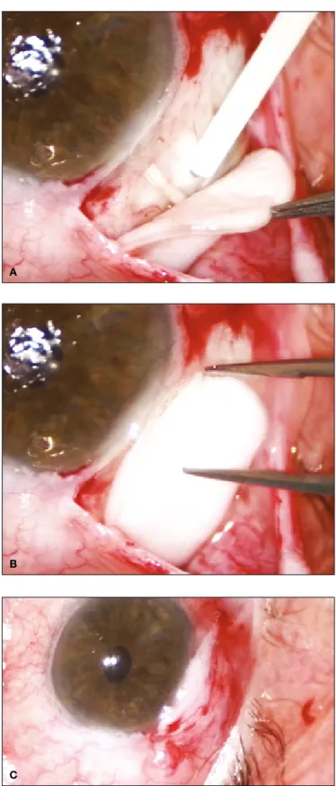
solvent-preserved bovine pericardium sterilized by means of gamma radiation. Before use, the collagen membrane was rehydrated with saline solution for 5-10 minutes, and the membrane was then cut using an ordinary corneal trephine with a diameter of 9.0 or 10.0 mm. The anterior edge of the patch graft was positioned just anterior to the tube insertion, and kept in place with fibrin glue (Tisseel fibrin glue, Baxter AG, Vienna, Austria) (Fig. 1A). Subsequently, the cap was stitched all around the tube and the dissected conjunctiva was laid over it (Fig. 1B); fibrin glue was then applied to the underside of the conjunctiva and smooth forceps were used to hold the edges in place until adherence was assured, tak-ing care to cover the graft totally (Fig. 1C). The eye was then examined for leaks as the anterior chamber was filled with balanced salt solution through a paracentesis.
The IOP and complications were evaluated 1 week and 1, 3, 6, 12, and 24 months after surgery. Success was de-fined as an IOP of ≤18 mm Hg, with (qualified success) or without medical hypotensive therapy (complete success) in all study visits during 2-year follow-up.
Fig. 1 - (A) The patch graft is positioned just anterior to the tube
insertion, and kept in place with fibrin glue. (B) The patch graft is
stitched all around the tube by using fibrin glue. (C) Final result: full
approximation of the conjunctiva to the limbus.
a
B
The comparison of the postoperative IOP values with the baseline was performed by means of the paired t test. The Bonferroni approach was adopted in order to make allow-ance for multiple tests; therefore, statistical significallow-ance level was set to 0.008, calculated as 0.05 divided by 6 comparisons.
The time to treatment failure was calculated as the time from the date of registration to the date in which the IOP was >18 mm Hg; patients with an IOP always within the normal values were censored at the end of observation in-terval. Time to treatment failure was described using the Kaplan-Meier method.
RESULTS
Sixteen patients (80%) were followed up for two years with an IOP ≤18 mmHg; the remaining four patients withdrew due to not sufficiently controlled IOP (Fig. 2). At the end of
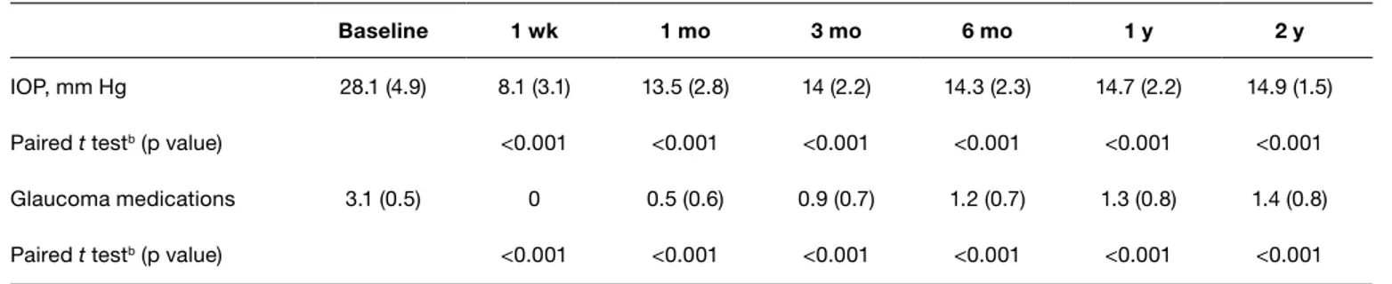
taBle i - INTRAOCULAR PRESSURE AND MEDICAL HYPOTENSIVE THERAPYa
Baseline 1 wk 1 mo 3 mo 6 mo 1 y 2 y
IOP, mm Hg 28.1 (4.9) 8.1 (3.1) 13.5 (2.8) 14 (2.2) 14.3 (2.3) 14.7 (2.2) 14.9 (1.5)
Paired t testb (p value) <0.001 <0.001 <0.001 <0.001 <0.001 <0.001
Glaucoma medications 3.1 (0.5) 0 0.5 (0.6) 0.9 (0.7) 1.2 (0.7) 1.3 (0.8) 1.4 (0.8)
Paired t testb (p value) <0.001 <0.001 <0.001 <0.001 <0.001 <0.001
IOP = intraocular pressure. aResults are reported as mean (SD). bComparison versus baseline.
Fig. 2 - Ahmed glaucoma valve implantation: time to treatment
failure. taBle ii - POSTOPERATIVE COMPLICATIONS AFTER
AHMED GLAUCOMA VALVE IMPLANTATION
Complicationsa no. (%) of eyes
Increased intraocular pressureb 4 (20)
Hypotonyc 2 (10)
Shallow or flat anterior chamber 3 (15)
Anterior chamber bleeding 2 (10)
Choroidal effusion 4 (20)
aSome patients had more than one complication.
bIntraocular pressure >21 mm Hg during maximal medical therapy. cIntraocular pressure <6 mm Hg.
Fig. 3 - Patch graft 2 years after Ahmed glaucoma valve implantation.
the 2-year follow-up, complete and qualified success was obtained in 3 and 13 eyes respectively. In the 16 patients followed up for 2 years, the IOP significantly decreased from the first postoperative visit to the end of the follow-up
patch.
The reported sutureless surgical technique is similar to that described by Kahook and Noecker (15). However, differ-ently from our technique, they had applied a 6 × 6-mm bo-vine pericardial patch graft at the level of tube entry site, sealed with fibrin glue. There is a substantial advantage to using a round patch graft of 9.0 to 10.0 mm in diameter. As a matter of fact, it allows to cover the majority of the length of the tube, and it offers greater safety in avoiding conjunctival erosion and tube exposure.
Limitations of the present study include limited follow-up, small sample size, and lack of inclusion of cases more at risk for conjunctival erosion (uveitic glaucoma, congenital and juvenile glaucoma).
In conclusion, this sutureless technique using a bovine pericardial graft patch seems to be a safe and rapid pro-cedure for AGV implantation. There is a need for longer follow-up in order to determine whether there are any long-term complications.
Financial support: None.
Conflict of interest statement: The authors report no proprietary interest.
Address for correspondence: Prof. Luciano Quaranta
USVD “Centro per lo studio del Glaucoma” Piazzale Spedali Civili 1
25123 Brescia Italy
after 24 months (mean IOP preoperatively 28.1 mm Hg [SD 4.9], mean IOP 24 months postoperatively 14.9 mm Hg [SD 1.5]; p<0.001). In all the follow-up visits, the number of top-ical hypotensive medications was significantly decreased when compared with the baseline (p<0.001) (Tab. I). Table II shows the postoperative complications. Through-out the period of follow-up, there was no conjunctival ero-sion, thinning of the patch graft over the anterior part of the tube, or tube exposure; no signs of endophthalmitis were recorded (Fig. 3).
DISCUSSION
The advantages of using fibrin glue in ocular surgery are well-known and include a shorter operative time and less postoperative pain (10-13). Furthermore, the absence of sutures is also of some advantage postoperatively, be-cause they can promote inflammation and provide a nidus for infection or neovascularization.
As the components of fibrin glue often come from pooled human plasma, there is a theoretical risk of human disease transmission. However, to our knowledge, there have not been any documented cases of disease transmission de-spite the extensive use of these products in various surgi-cal fields (14), and so the risk appears to be minimal. The total sterility of the Tutopatch® material is guaranteed by
gamma radiation and chemical treatments. The relatively high cost of fibrin glue is counterbalanced by the low cost of Tutopatch®, when compared with human sclera donor
REFERENCES
1. Gedde SJ, Schiffman JC, Feuer WJ, Herndon LW, Brandt
JD, Budenz DL; Tube versus Trabeculectomy Study Group. Treatment outcomes in the Tube Versus Trabeculectomy (TVT) study after five years of follow-up. Am J Ophthalmol 2012;153:789-803.
2. Gedde SJ, Singh K, Schiffman JC, Feuer WJ; Tube
versus Trabeculectomy Study Group. The Tube Versus Trabeculectomy Study: interpretation of results and appli-cation to clinical practice. Curr Opin Ophthalmol 2012;23: 118-26.
3. Krebs DB, Liebmann JM, Ritch R, Speaker M. Late
infec-tious endophthalmitis from exposed glaucoma setons. Arch Ophthalmol 1992;110:174-5.
4. Al-Torbak AA, Al-Shahwan S, Al-Jadaan I,
Al-Homma-di A, Edward DP. Endophthalmitis associated with the Ahmed glaucoma valve implant. Br J Ophthalmol 2005;89: 454-8.
5. Brandt JD. Patch grafts of dehydrated cadaveric dura ma-ter for tube-shunt glaucoma surgery. Arch Ophthalmol 1993;111:1436-9.
6. Tanji TM, Lundy DC, Minckler DS, Heuer DK, Varma R. Fas-cia lata patch graft in glaucoma tube surgery. Ophthalmol-ogy 1996;103:1309-12.
7. Freedman J. Scleral patch grafts with Molteno setons. Oph-thalmic Surg 1987;18:532-4.
8. Raviv T, Greenfield DS, Liebmann JM, Sidoti PA, Ishikawa H, Ritch R. Pericardial patch grafts in glaucoma implant sur-gery. J Glaucoma 1998;7:27-32.
9. Zeppa L, Romano MR, Capasso L, Tortori A, Majorana MA, Costagliola C. Sutureless human sclera donor patch graft for Ahmed glaucoma valve. Eur J Ophthalmol 2010;20: 546-51.
10. Srinivasan S, Dollin M, McAllum P, Berger Y, Rootman DS, Slomovic AR. Fibrin glue versus sutures for attaching the conjunctival autograft in pterygium surgery: a prospective ob-server masked clinical trial. Br J Ophthalmol 2009;93: 215-8. 11. Ozdamar Y, Mutevelli S, Han U, et al. A comparative study of
tissue glue and Vicryl suture for closing limbal-conjunctival autografts and histologic evaluation after pterygium exci-sion. Cornea 2008;27:552-8.
12. Uy HS, Reyes JM, Flores JD, Lim-Bon-Siong R. Compari-son of fibrin glue and sutures for attaching conjunctival au-tografts after pterygium excision. Ophthalmology 2005;112: 667-71.
13. Pfister RR, Sommers CI. Fibrin sealant in corneal stem cell transplantation. Cornea 2005;24:593-8.
14. Horowitz B, Busch M. Estimating the pathogen safety of manufactured human plasma products: application to fibrin sealants and to thrombin. Transfusion 2008;48: 1739-53.
15. Kahook MY, Noecker RJ. Fibrin glue-assisted glaucoma drainage device surgery. Br J Ophthalmol 2006;90:1486-9.