258
CASE REPORT
Tracheotomy-related posterior tracheal wall
rupture, trans-tracheal repair
Rottura della parete posteriore della trachea da tracheotomia.
Riparazione trans-tracheale
A. DEGANELLO, M.C. SOFRA1, F. FACCIOLO2, G. SPRIANO
Department of Otolaryngology Head and Neck Surgery, Azienda Ospedaliero-Universitaria “Careggi”, Firenze;
1 Department of Anesthesiology; 2 Department of Thoracic Surgery, National Cancer Institute “Regina Elena”, Rome, Italy
SUMMARY
Laceration of the membranous part of the tracheo-bronchial tree is a rare complication that can occur after single lumen intuba-tion, double-lumen intubaintuba-tion, percutaneous and surgical tracheotomy. The case of a 76-year-old male is presented in whom a posterior tracheal wall laceration, related to tracheotomy, was diagnosed and immediately treated at the end of a head and neck operation. A 6 cm long laceration started 1.5 cm below the tracheotomy level and ended 2 cm above the carina. The tear was closed from distal to proximal area via the tracheotomy opening with PDS 4/0 interrupted sutures using a thoracoscopic needle-holder. This original surgical technique is described in detail. In tracheotomy related tears, the fact that an opening in the trachea already exists and that the lesion rarely extends beyond the carina, should guide the surgeon to make every effort to repair the laceration through this already existing access.
KEY WORDS: Trachea • Tracheotomy • Complication • Tracheal rupture • Surgical treatment
RIASSUNTO
La rottura della pars membranacea tracheo-bronchiale è un’evenienza rara che si può riscontrare in seguito ad intubazioni con tubi a singolo o doppio lume ed in seguito a tracheotomie percutanee o chirurgiche. Viene presentato il caso di un paziente di 76 anni trattato chirurgicamente per un carcinoma del cavo orale. Al termine dell’intervento una lacerazione della pars membrana-cea tracheale, dovuta alla tracheotomia, è stata prontamente diagnosticata e trattata. La rottura tracheale era situata in posizione mediana, si estendeva per 6 cm, iniziava 1,5 cm sotto il livello dell’apertura tracheotomica e terminava 2 cm al di sopra della carena. La riparazione transtracheale da distale a prossimale è stata eseguita con fi lo PDS 4/0 a punti staccati utilizzando un pas-safi li toracoscopico. L’originale tecnica chirurgica viene descritta dettagliatamente. Nel trattamento delle lesioni iatrogene della pars membranacea dovute alla tracheotomia, il fatto che esista già un accesso diretto alla trachea e che la lesione non si estenda ai bronchi, deve spingere il chirurgo a fare ogni sforzo per riparare la breccia attraverso l’apertura tracheotomica.
PAROLE CHIAVE: Trachea • Tracheotomia • Complicazioni • Rottura della trachea • Trattamento chirurgico
Introduction
Laceration of the membranous part of the tracheobronchial tree is a rare complication that can occur after single lumen intubation, double-lumen intubation, percutaneous and sur-gical tracheotomy. The exact incidence of this complication cannot be reasonably estimated since the large number of intubations and tracheotomies performed daily, on a world-wide basis, is unknown. When diagnosis is made, surgical repair should be performed immediately in order to minimize the risk of life-threatening events such as respiratory insuf-fi ciency, mediastinal emphysema, mediastinitis, compressive pneumothorax, that may lead to cardio-pumonary failure.
Case report
The case of a 76-year-old male is presented in whom a pos-terior tracheal wall laceration, related to tracheotomy, was
diagnosed and immediately treated at the end of a head and neck operation.
The patient represented a high operative risk with reduced life expectancy (ASA IV), the medical record was posi-tive for chronic obstrucposi-tive airway disease, diabetes mel-litus, renal insuffi ciency, obesity, anaemia and ischaemic cardiopathy for which the patient had undergone triple aorto-coronary bypass and aortic valve substitution 4 years before the head and neck operation. The patient pre-sented with a cT4aN2b retromolar trigone squamous cell carcinoma on the left hand side and because of the severe co-morbidity the patient was initially treated with elective external beam radiotherapy. Surgical indication was given due to failure of the local radiotherapy: tumour restage rT4aNO.
Informed consent was obtained after appropriate counsel-ling regarding the technical aspects of surgery and the high general anaesthesia-related risks.
259 Pre-medication was carried out with midazolam 0.02 mg/kg
and ondansetron 00.1 mg/kg.
General anaesthesia was induced by fentanyl 1 mcg/kg and propofol 2 mg/kg, both given intravenously, and maintained with 2% sevofl urane and oxygen/air mixture. Regional sur-face anaesthesia of the base of the tongue, posterior tracheal wall of the pharynx and vocal cords was obtained using 2% lidocaine and muscle relaxation using cis-atracurium be-sylate 0.2 mg/kg.
Oro-tracheal intubation was performed in a single attempt, placing a single lumen n. 8 tube. Oxygen saturation value of 99%, End Tidal CO2 value of 33% and airway pressure value of 18 mbar demonstrated the lack of any problem in the intubation.
Through a horizontal cervical incision (3 cm in length) pass-ing 1.5 cm above the jugulum, the anterior tracheal wall was easily exposed, without any bleeding, just below the thyroid isthmus, the trachea was opened between the second and third tracheal ring, an inferior based tracheal fl ap accord-ing to the Björk technique was created and sutured to the inferior skin edge of the cervical incision. After withdraw-ing the tube, a cuffed cannula n. 8 was introduced smoothly using the obturator, without any diffi culty and no bleeding. Ventilation through the cannula was impossible, a suction tube was easily passed through the cannula but still there was no CO2 return, so the cannula was removed to check if any damage had occurred in the trachea. At visual inspec-tion, through the tracheotomy opening, everything looked normal without bleeding, so at this moment, there was no explanation as to why ventilation resulted impossible, how-ever a cuffed cannula n. 10 was placed (always using the obturator), again without any diffi culty and no bleeding. The patient was then effectively ventilated through the can-nula, with normal airway resistances, during the entire oper-ation which took 4 hours. Surgery included modifi ed radical neck dissection type II with en bloc segmental mandibular resection, and pectoralis major fl ap reconstruction. Whilst the patient was waking up, the Anaesthesiologist claimed diffi culty in passing the suction tube through the cannula but no problems with the patient’s ventilation were expe-rienced, albeit immediate bronchoscopy was performed, at the operatoring table.
Bronchoscopy was carried out fi rst through the cannula then directly through the stoma after removing the cannula and a 6 cm long, full thickness, longitudinal tear was revealed in the posterior wall of the trachea, at a median position. The laceration started 1.5 cm below the tracheotomy level and ended 1.5 cm above the carina. Chest X-ray fi ndings were normal except for the presence of a minimal pneumome-diastinum at the right hand side, and clinically no subcu-taneous emphysema developed. Oesophagoscopy showed no injuries.
Surgical technique
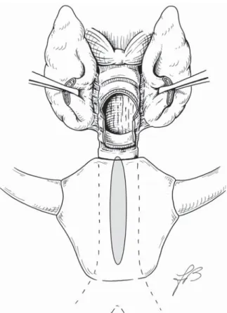
A single lumen tube was introduced endoscopically through the tracheotomy opening with the tip of the tube touching the carena. The Björk fl ap was left in place, the tracheotomy skin incision was enlarged 2 cm both sides, the sterno-hy-oid and sterno-thyrsterno-hy-oid muscles were divided, the thyrsterno-hy-oid isthmus was bound and divided to expose the anterior
tra-cheal wall. At the tracheotomy opening, a complete separa-tion between the second and third tracheal ring was created maintaining continuity of the trachea only via the posterior wall to gain exposure to the laceration (Fig. 1).
The tube was removed, the patient was sedated with a tar-get-controlled infusion of propofol (target 1.5 mcg/ml) and kept spontaneously breathing with extra oxygen provided (FiO2 = 50%) with a tube n. 4 maintained in correspondence to the tracheotomy. Local anaesthesia with 10% spray lido-caine, at the tracheotomy opening, was given to minimize coughing.
A Langenback retractor was introduced through the tracheal opening to retract the anterior tracheal wall, this manoeuvre allowed the surgical exposure to be increased and, at the same time, the edges of the tear were mechanically brought closer together.
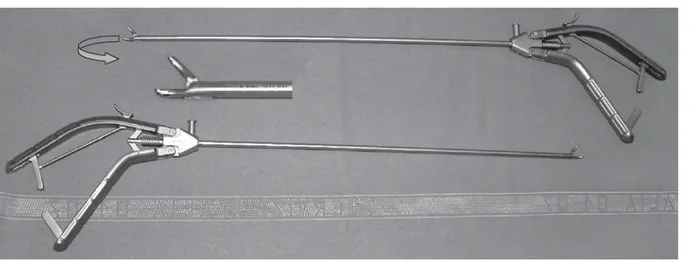
The tear was then closed from distal to proximal area via the tracheotomy opening with PDS 4/0 interrupted sutures (8 stitches) using a thoracoscopic needle-holder (Fig. 2). The needle was passed through the external layer of the anterior wall of the oesophagus, then through the edges of the tear, so that each knot (with the exception of the most proximal one) remained outside the lumen and the anterior wall of the oesophagus sealed the suture (Fig. 3).
Once the needle had been passed, each surgical knot was ligated outside the tracheotomy, then the extremities of the wire were kept outside the tracheal lumen and the knot was pushed towards the laceration by the needle holder. To fi rmly tie the knot, one side of the wire was grabbed by the needle holder, very close to the knot, while the other end
Fig. 1. Thyroid isthmus is divided and a complete separation
between second and third tracheal ring is created maintaining con-tinuity of trachea only via posterior wall.
260
was simply held by the surgeon’s hand, outside the trache-otomy. The needle holder also helped by pressing upon the oesophagus and, therefore, the knot resulted in an extra-tra-cheal position. The last surgical knot (the most proximal) was eventually located in the intra-tracheal area and the needle was passed under the ligature of the second most proximal knot.
A very soft sylastic cannula without cuff and without in-ner-cannula was positioned to avoid further damage to the trachea, and the patient awoke, breathing spontaneously, and was then monitored, in the intensive care unit, for 4 days.
Results
The patient recieved wide spectrum antibiotics for 3 weeks, a control chest X-ray and computed tomography of the thorax showed no pneumomediastinum. Endoscopic visu-alization through the tracheotomy, after temporary removal of the cannula (that was then cleaned and sterilized), was performed daily for 2 weeks, showing no dehiscence in the suture. On post-operative day 5, the extremely soft cannula was replaced by a conventional micro-fenestrated cannula n. 6 without a cuff but with an inner-cannula to allow easier management of toilette of the cannula, on day 18, the pa-tient was able to keep the cannula closed for 48 hours, so the patient was decannulated on day 20.
Discussion
Posterior tracheal wall laceration is an uncommon, although well recognized, potentially fatal injury. Even if the mem-branous part of the trachea is very friable and susceptible to tearing, especially in elderly patients and in short wom-en 1, iatrogenic laceration is a relatively infrequent event.
Chronic obstructive airway disease has been suggested as a possible risk factor since enlargement of the trachea by coughing results in an increase in the surface of the mem-branous portion which could explain the risk of damage in this already vulnerable area 2. Over infl ation of the cuff and
direct trauma, caused by diffi cult intubation or in emergency procedures, are recognized as aetiologic factors, but even in “atraumatic” airway entry procedures, tracheal lacerations may occur 1-3.
Diagnosis is usually made by tracheo-bronchoscopy that follows the onset of symptoms and allows the entity and location of the rupture to be established.
Clinical symptoms occur either immediately, during me-chanical ventilation (pneumomediastinum, compressive pneumothorax, extensive subcutaneous emphysema), or arise in the post-operative period (respiratory distress, me-diastinitis and tracheal stenosis).
In our case (to our knowledge, the only one in the litera-ture), fortunately, the diagnosis was made before any major symptom occurred, the patient was in such poor general conditions that, probably, development of related symp-toms could have been fatal.
Fig. 2. Thoracoscopic needle holder allows needle to easily pass into the narrow tracheal lumen.
Fig. 3. Needle is passed through external layer of anterior wall of
261 Several specifi c risk factors could have played a role, in this
patient: the fi brosis related to the previous cardiothoracic surgery and the previous radiotherapy, together with the chronic obstructive airway disease that stretches the posterior tracheal wall. The presence of the extensive fi brosis might have prevented the development of severe pneumothorax by sealing the mediastinum. After this experience, we do recommend endoscopic control, if any problem occurs during airway entry in patients at risk.
Most publications report a single or a few case reports in which the laceration occurred mainly after intubation or percutaneous tracheotomy 4.
In our patient, the mechanism of the rupture remains un-clear. It is possible that the oro-tracheal tube caused a direct trauma in the posterior wall without the tube going into the false passage, this is actually possible, but unlikely; maybe the cuff infl ation of the tube did provoke the tear, but usual-ly traumatic ruptures caused by overinfl ation of the balloon are localized in the corner between the posterior wall and trachea rings. In this case, the laceration was in a median position.
Even if the cannulas were always introduced using the obtu-rator and without any diffi culty, the most probable explana-tion is a direct trauma to the posterior tracheal wall during introduction of the cannula n. 8. Probably, due to the fragil-ity of the posterior wall, the obturator of the cannula perfo-rated the pars membranacea thus creating a false passage directly between the posterior wall of the trachea and the anterior wall of the oesophagus. Infl ation of the cuff might have increased the length of the rupture, the suction tube was easily passed in the mediastinum but ventilation was impossible. The cuff of the second cannula, larger in size (n. 10), probably fi lled the rupture thus allowing effective ventilation during the entire operation.
In 1978, Jacobs et al. 5 fi rst reported two similar cases of
posterior tracheal wall laceration as a complication of tra-cheotomy, both without entrance in the oesophagus, which were repaired directly through the same tracheotomy. Both patients were female and previously irradiated. In this re-port, both patients developed pneumothorax which oriented the diagnosis, the ruptures, 3 cm and 5 cm long, started at tracheotomy level so that they were rather proximal and could easily be recognized and managed through the tra-cheotomy opening.
In our patient, the fact that laceration started 2 cm below the tracheotomy level prevented direct visualization of the tear, diagnosis was then made only after tracheo-bronchoscopy and the repair required an extended tracheotomy access to gain distal suffi cient exposure.
Non-surgical treatment can be considered in small (length < 2 cm) uncomplicated tears, in stable patients, since, un-der these conditions, healing can be achieved with minimal risks and discomfort for the patient 6. Surgery is,
neverthe-less, the treatment of choice for the large majority of pa-tients and should be performed promptly to avoid feared complications such as descendent mediastinitis 7.
During the eighties and early nineties, posterior wall lac-erations were treated mainly with a right thoracotomy, and, occasionally, through a lateral cervical approach. In 1995, Angelillo-Mackinlay 8 described the cervical
trans-tracheal approach, in a posterior trans-tracheal wall laceration, after oro-tracheal intubation, in which a midline incision is made in the anterior wall of the trachea to expose the pos-terior wall.
In our opinion, if the injury occurs while performing a tra-cheotomy, the surgical repair through the same tracheotomy should be the gold standard on account of its limited inva-siveness and lack of major morbidity. The tear caused by tracheotomy always lies proximal to the carina, the com-plete separation of the trachea rings, at the tracheotomy opening provides a suffi cient surgical exposure to perform a surgical repair up to the carina. The use of the external part of the anterior oesophageal wall as a re-enforcement patch in the repair can be very helpful in sealing the laceration, a naso-gastric feeding tube is required, for at least a week, to minimize peristalsis. The surgeon must be aware of the well-recognized pressure confl ict that can arise between the naso-gastric feeding tube and the cuff of the cannula, this is a major risk factor for developing tracheo-oesophageal fi stulas. Thus, after the repair of the laceration, the best op-tion is to place a non-cuffed cannula, as soft as possible, if a cuffed cannula is needed, for ventilation purposes, then the pressure in the cuff has to be maintained as low as pos-sible.
If the aetiology is not related to tracheotomy, the rupture should not extend to the bronchi and if the patient does not require tracheotomy after surgical repair, a trans-tracheal closure, with a longitudinal opening of the anterior tracheal wall is an excellent option.
Conclusions
Immediate surgical repair remains the standard treatment of posterior tracheal wall lacerations. The surgical approach has always been codifi ed upon the extension and location of the tear, in general a thoracotomy is required to man-age ruptures extending in the bronchial tree, otherwise a left cervical approach or a trans-tracheal approach are indicat-ed. In tracheotomy related tears, the fact that an opening in the trachea already exists and that the lesion rarely extends beyond the carina, should guide the surgeon to make every effort to repair the laceration through this existing access. Our extended tracheotomy technique together with the use of thoracoscopic instruments has been shown to be very helpful in the immediate successful treatment of this fear-some complication
References
1 Marty-Ané CH, Picard E, Jonquet O, Mary H. Membranous tracheal rupture after endotracheal intubation. Ann Thorac
Surg 1995;60:1367-71.
2 Hasan A, Low DE, Ganado AL, Norton R, Watson DC. Tra-cheal rupture with disposable polyvinylchloride double-lumen endotracheal tubes. J Cardiovasc Anaesth 1992;6:208-11.
3 Sternfeld D, Wright S. Tracheal rupture and the creation of
a false passage after emergency intubation. Ann Emerg Med
2003;42:88-92.
4 Walz MK, Schmidt U. Tracheal lesion caused by percutane-ous dilatational tracheostomy – a clinico-pathological study.
Intensive Care Med 1999;25:102-5.
262
Posterior tracheal laceration: a rare complication of trache-ostomy. Laryngoscope 1978;88:1942-6.
6 Ross HM, Grant FJ, Wilson RS, Burt ME. Nonoperative management of tracheal laceration during endotracheal intu-bation. Ann Thorac Surg 1997;63:240-2.
7 Mussi A, Ambrogi MC, Menconi G, Ribechini A, Angeletti
CA. Surgical approaches to membranous tracheal wall
lac-erations. J Thorac Cardiovasc Surg 2000;120:115-8. 8 Angelillo-Mackinlay T. Transcervical repair of distal
membra-nous tracheal laceration. Ann Thorac Surg 1995;59:531-2.
Received January 10, 2007 - Accepted May 24, 2007
Address for correspondence: Dr A. Deganello, Clinica Otorinolarin-goiatrica, Azienda Ospedaliero-Universitaria “Careggi”, viale Mor-gagni 85, 50100 Firenze, Italy. Fax +39 055 435649.