Index
Summay
...I
Summary [Italian]
... IIIIntroduction
...1
Hormonal Risk And Protective Factors for Breast Cancer
...1
Endocrine disruptors in Breast Cancer
...5
Endocrine Therapy: Estrogens and Antiestrogens
...9
Tamoxifen Mode of Action in Breast Cancer
...10
Tamoxifen Use in Advanced Breast Cancer
...11
Estrogen Receptordependent and Estrogen Receptorindependent
pathways for tamoxifeninduced apoptosis
...13
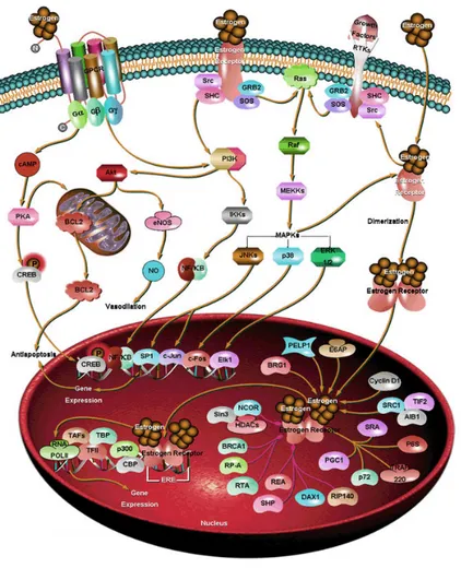
Hormone induced activation of cellsurface receptors:
signaling from plasma membrane to nucleus
...16
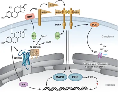
The membrane receptor GPR30
...23
Activation Protein 1 (AP1)
...29
Results
...36
Discussion
...47
References
...54
Publications
...69
Summary
Estrogens are pleiotropic hormones that regulate the growth and differentiation of
many tissues. By acting as mitogens they also promote the development of breast
and ovarian tumors. The biological effects of estrogens are classically mediated by
the estrogen receptor (ER)s α and β which function as hormoneinducible
transcription factors binding to the estrogenresponsive element (ERE) located
within the promoter region of target genes. Many studies have identified
membraneassociated estrogen signals which may alter gene expression
independently of the nuclear ERs. Previously, we suggested that evaluating the
levels of G proteincoupled receptor 30 (GPR30) in combination with those of a set
of GPR30 target genes might be more informative to assess the outcome in certain
types of cancer. We recently assessed that GPR30 triggers proliferative stimuli of
several natural compounds (phytoestrogens) as well as synthetic compounds
(xenoestrogens) in a variety of estrogensensitive cancer cells. Moreover, we
demonstrated that 4hydroxytamoxifen (OHT) like estrogens is able to induce cell
proliferation through GPR30 by induction of cFos and other target genes. On the
other hand, we have also examined, in specific cellular contexts, the molecular
mechanisms of OHTinduced apoptosis. In this regard, our studies were focused on
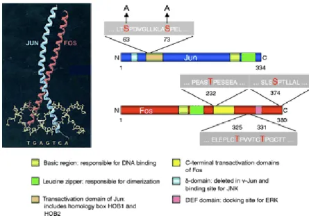
the activation of cJun, a major component of the AP1 transcription factor, which
represents a paradigm for the transcriptional response to stress. Transactivation of
cJun is regulated by JunNterminal kinases (JNKs) through phosphorylation at
serine 63 and 73 (S63/S73), as well as at threonine 91 and 93 (T91/T93). We show
that following a short exposure to the DNAdamaging compound etoposide, cJun
phosphorylation is restricted to S63/S73. In contrast, JNKdependent
Summary
II
(T95). Hence, our study suggests that cJun may sense the strength of genotoxic
stress, in apoptosis cell death, through DNAdamage dependent phosphorylation of
T95, which in turn augments cJun transactivation by JNKs. Finally, we analysed
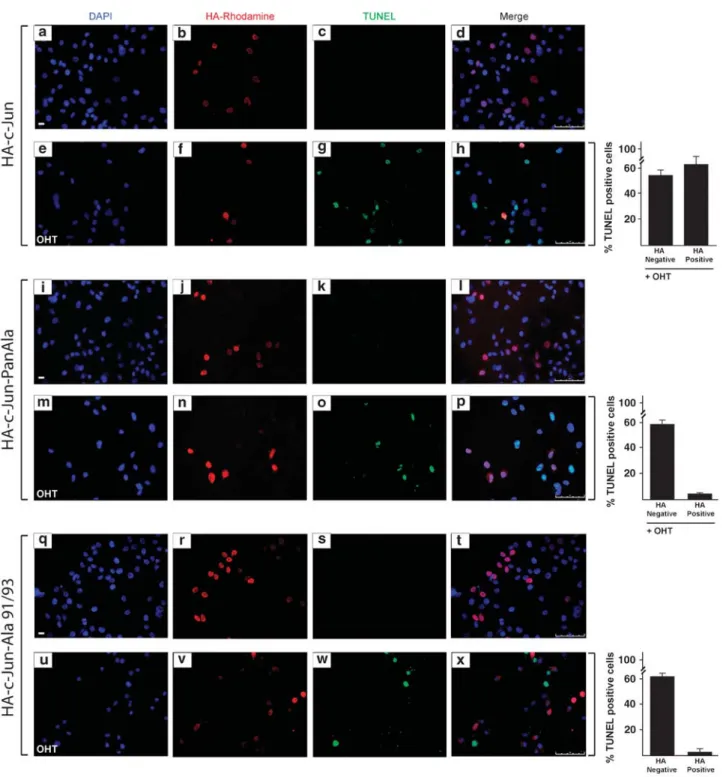
whether cJun, the major nuclear target of JNK, has a role in OHTinduced apoptosis
of SkBr3 breast cancer cells. We show that before DNA fragmentation and caspase
3/7 activation, cytotoxic concentrations of OHT induce JNKdependent
phosphorylation of cJun at JNK sites earlier shown to regulate cJunmediated
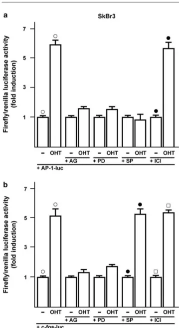
apoptosis. In addition, OHT induced ERKdependent expression of cFos and
transactivation of an AP1responsive promoter. In particular, the ectopic
expression of dominantnegative constructs blocking either AP1 activity or cJun N
terminal phosphorylation prevented DNA fragmentation after OHT treatment.
Furthermore, both cFos expression and cJun Nterminal phosphorylation preceded
OHTdependent activation of caspase 37 in different types of tamoxifensensitive
cancer cells, but not in OHTresistant LNCaP prostate cancer cells. Taken together,
our results indicate that the cJun/cFos AP1 complex has a proapoptotic role in
OHTtreated cancer cells and suggest that pharmacological boosts of cJun
activation may be useful in a combination therapy setting to sensitize cancer cells
to tamoxifenmediated cell death.
Our data contribute to better understand the molecular mechanisms involved in
biological effects elicited in different cancer cell types by estrogens and
antiestrogens, providing new insights for the comprehension of apoptotic
mechanisms induced by antiestrogenic therapy in cancer.
Summary [Italian]
Gli estrogeni sono ormoni pleitropici che regolano la crescita e la differenziazione di
molti tessuti. Agendo da mitogeni sono inoltre in grado di promuovere lo sviluppo di
tumori estrogenosensibili come il tumore mammario ed ovarico. Gli effetti biologici
degli estrogeni sono mediati dal Recettore Estrogenico (ER) α e β, che agendo da
fattori di trascrizione, legano le sequenze responsive agli estrogeni (ERE) presenti
nelle regioni promoter dei geni target. Numerosi studi hanno identificato segnali
estrogenici associati alla membrana che modificherebbero l’espressione genica
indipendentemente da ERs nucleari. Negli ultimi anni, attraverso lo studio dei livelli
di espressione di un recettore accoppiato a proteine G (GPR30) e dei geni target da
esso regolati, il nostro gruppo di lavoro ha potuto fornire ulteriori importanti
informazioni riguardo alcuni tipi di tumore.
Recentemente, abbiamo dimostrato che GPR30 media gli effetti proliferativi di
alcuni composti naturali (fitoestrogeni), così come di composti sintetici
(xenoestrogeni) in diverse linee cellulari tumorali sensibili all’azione degli
estrogeni. Successivamente abbiamo dimostrato che il 4hydroxytamoxifen (OHT),
al pari degli estrogeni, è capace di indurre proliferazione cellulare attraverso
GPR30. Abbiamo inoltre valutato, in specifici contesti cellulari, i meccanismi
molecolari dell’apoptosi indotta da OHT. A tal proposito, abbiamo focalizzato i
nostri studi sull’attivazione di cJun, il principale componente del fattore di
trascrizione AP1. La transattivazione di cJun, fondamentale nella risposta
trascrizionale allo stress cellulare, è regolata dalla fosforilazione da parte di una
classe di chinasi denominate JunNterminal kinases (JNKs) delle serine 63 e 73
(S63/73), così come delle treonine 91 e 93 (T91/T93) situate nella regione N
Summary [Italian]
IV
ristretta alle S63/73. Al contrario, la fosforilazione di T91/T93 richiede esposizioni
prolungate al trattamento ed è abolita dall’esposizione alla caffeina o dalla
sostituzione del sito di fosforilazione adiacente, treonina 95 (T95), con un’alanina.
Pertanto, i nostri studi indicano che cJun potrebbe comportarsi da sensore di
intensità dello stress genotossico capace di innescare la morte cellulare per
apoptosi, attraverso la fosforilazione di T95 in seguito a danno al DNA. Infine,
abbiamo valutato il ruolo di cJun, principale target delle JNK, nell’apoptosi indotta
da OHT in cellule di tumore mammario SkBr3. Abbiamo dimostrato che prima della
frammentazione del DNA e dell’attivazione delle caspasi 3/7, concentrazioni
citotossiche di OHT sono in grado di indurre la fosforilazione di cJun a livello dei
siti target delle JNK, coinvolte, come dimostrato in precedenza, nella regolazione
dell’apoptosi mediata da cJun. Inoltre, OHT è in grado di aumentare l’espressione
di cFos e la transattivazione di AP1 in maniera dipendente dalle ERK. In
particolare, l’espressione ectopica di costrutti in grado di bloccare sia l’attività di
AP1 che la fosforilazione di specifici siti a livello Nterminale di cJun, è stata in
grado di prevenire il danno al DNA dopo trattamento con OHT. Inoltre, l’aumento
dei livelli proteici di cFos e della fosforilazione di cJun indotti da OHT, precedono
l’attivazione delle caspasi 37 in diversi tipi di cellule tumorali sensibili al
trattamento con OHT, ma non nella linea cellulare di tumore prostatico LNCaP,
resistente a OHT. I nostri risultati dimostrano che il complesso AP1, formato da c
Jun e cFos, svolge un ruolo proapoptotico nelle cellule tumorali trattate con OHT e
individuano l’attivazione di cJun come bersaglio farmacologico da utilizzare in una
terapia combinata che sensibilizzi le cellule tumorali alla morte cellulare indotta
dal tamoxifene.
Summary [Italian]
I nostri dati contribuiscono ad una migliore conoscenza dei meccanismi molecolari
coinvolti negli effetti biologici esercitati dagli estrogeni e dagli antiestrogeni in
diverse tipologie di cellule tumorali suggerendo, nuovi campi di studio per la
comprensione del meccanismo apoptotico indotto dagli antiestrogeni nella terapia
antitumorale attualmente in uso.
INTRODUCTION
Introduction
Hormonal Risk And Protective Factors for Breast Cancer
Worldwide, breast cancer is by far the most common cancer amongst women,
with an incidence rate more than twice that of colorectal cancer and cervical
cancer and about three times that of lung cancer. However breast cancer
mortality worldwide is only 25% greater than that of lung cancer in women
(World Health Organization International Agency for Research on Cancer, 2003).
In 2004, breast cancer caused 519,000 deaths worldwide (7% of cancer deaths;
almost 1% of all deaths) (World Health Organization, 2006).
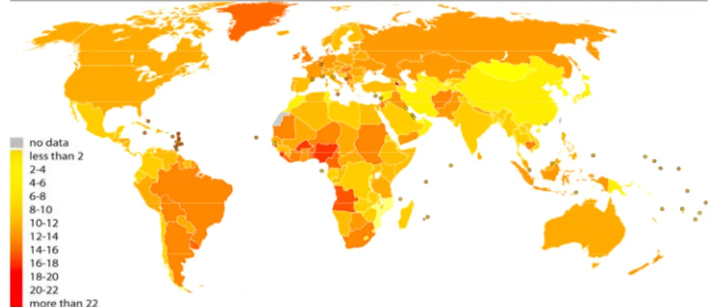
The number of cases worldwide has significantly increased since the 1970s, a
phenomenon partly blamed on modern lifestyles in the Western world
(Laurance, 2006) (Fig. 1).
Fig. 1.
Agestandardised death rates from Breast cancer by country (per 100,000 habitants)
Breast cancer is the most common cancer in women and is estimated to have
accounted for 182,460 new cancer diagnoses and 40,480 deaths in 2008 (Jemal
et al., 2008). The incidence is highest in highly industrialized countries like North
America, Northern Europe, and Australia, where age‐adjusted rates are 75‐92
Introduction
2
2001). Ovarian cancer is the fourth leading cause of tumor death in Western
countries representing the most fatal gynecologic malignancy with the overall 5‐
year survival rate about 10% to 20% (Boete et al., 1993) and is also estimated to
have accounted for 21,650 new cases and 15,520 deaths in 2008 (Jemal et al.,
2008) (Fig. 2).
Fig. 2.
Ten Leading Cancer Types for the Estimated New Cancer Cases and Deaths, by Sex, United
States, 2008.(Jemal et al., 2008).
There has been an evident decline in breast cancer mortality since 1997, most
likely the result of therapy with tamoxifen and perhaps other forms of
chemotherapy (McKeanCowdin et al., 2000). Existing evidence regarding the
hormonal etiology of breast cancer supports the hypothesis that estrogen is the
primary stimulant for breast cell proliferation (Henderson and Feigelson, 2000).
Introduction
Hormonally related risk factors for female breast cancer equate with a greater
cumulative lifetime exposure to estrogen and include early age at menarche, late
age at menopause, null parity or late age at first full‐term pregnancy, and obesity
(Table 1). Protective factors include higher parity, long lactation, and bilateral
ovariectomy (Davis et al., 1997; Kreiger et al., 1999; Parazzini et al., 1997).
Reproductive risk factors are associated with exposure to estradiol,
progesterone, and other hormones; and reproductive hormones are also
believed to underlie increased risk associated with alcohol consumption, lack of
physical activity, higher body mass index and weight gain after menopause, and
low premenopausal body mass index (Bernstein et al., 2002). Much attention has
been focused on dietary differences, particularly fat consumption, to explain
both the international pattern of breast cancer occurrence and changes in rates
of breast cancer following migration to high‐risk, usually Western nations from
low‐risk countries (Armstrong and Doll, 1975). Diet seems very likely to affect
breast cancer risk, as it does in animals, but epidemiologic studies have failed to
identify specific dietary constituents that increase or decrease risk. Effects of fat
and fruits and vegetables have been extensively studied, so far providing no
consistent evidence of dietary risk factors (Gandini et al., 2000; Holmes et al.,
1999; Hunter and Willett., 1996; Michels., 2002; SmithWarner et al., 2001; Willett,
1999). High soy intake in Asia has been proposed as a factor in reduced breast
cancer rates there, although epidemiologic studies so far provide limited
evidence of a protective effect (Adlercreutz, 2002; HilakiviClarke et al., 2001;
Trock et al., 2000). Pharmaceutical hormones similarly affect risk. Both
Introduction
4
hormonal exposures experienced by women and therefore are of concern as
potential contributors to breast cancer risk. Other more common allelic
variations in estrogen metabolism genes (e.g. CYP17, CYP19, HSD17B1) in breast
cancer risk are also under study (Fig. 3; Table 1‐2) (Henderson and Feigelson,
2000). Sequence variants in CYP17 and HSD17B1 have been reported to
individually, and in an additive manner; increase the risk of advanced breast
cancer (Feigelson et al., 2001). Genes in the growth factor pathway, as well as
genes encoding proteins in intracellular pathways, steroid receptor
transactivation, and DNA repair, are additional important sources of such
candidate genes.
Fig. 3.
Schematic presentation of estrogen metabolism in the ovaries and breast epithelium and
four candidate genes that may play a role in breast cancer etiology. The genes of interest are the
cytochrome P450c17a (CYP17) gene, the aromatase cytochrome P450 (CYP19) gene, the 17
β
hydroxysteroid dehydrogenase 1 (HSD17B1) gene, and the estrogen receptor (ER) gene.
Introduction
Endocrine disruptors in Breast Cancer
A class of hormonally active chemicals, referred to as endocrine disruptors, may
affect breast cancer primarily at the phase of tumor promotion. They may also
affect mammary gland development and responsiveness to other carcinogens. It
has been suggested that exposure to endocrine disruptors, including chemicals
that mimic estrogens or xenoestrogens, might play an important role in breast
cancer risk (Davis et al., 1993). More than 500 chemicals have been found to be
weakly estrogenic in various assays, including many chemicals in common use,
such as constituents of detergents, pesticides, and plastics (Jobling et al., 1998;
Introduction
6
vitro to proliferate (Korach and McLachlan, 1995; Shelby et al., 1996; Soto et al.,
1995). Short‐term in vivo assays, such as increase in uterine weight in rodents,
are also used to demonstrate estrogenic activity (O'Connor et al., 1996).
Furthermore, the effects of these compounds have been frequently observed in
wildlife in a more natural context. Sexual disruption of wild fish has been
reported in rivers receiving wastewater effluent from industries containing
mixtures of endogenous and pharmaceutical estrogens and industrial chemical
endocrine disruptors (Jobling et al., 1998). The identification of estrogenic
compounds in the environment has raised significant issues regarding the
relevance of the potential adverse health effects (Rudel, 1997). Some researchers
maintain that the potency of many of these endocrine‐disrupting pollutants is
typically much lower than the potency of endogenous estrogens, and so their
effects will likely be insignificant (Safe, 1995). Others are concerned about the
exposure to endocrine‐disrupting chemicals levels of endogenous hormones are
very low, such as in utero or during prepubertal, or postmenopausal time
periods. Furthermore, it must be taken into consideration that additive effects of
low‐level estrogenic pollutants can act together even when each individual
component of the mixture is present below a threshold for effect (Silva et al.,
2002). Finally, the in vivo estrogenic effects of a range of compounds
demonstrate that estrogenic compounds can exhibit different mechanisms and
effects (Gould et al., 1998; Rudel, 1997). This diversity is attributed, at least in
part, to the fact that the shape of the ER ligand (either E2 or an endocrine
disruptor) affects the binding of the ER‐ligand complex to DNA sequences and
subsequent gene expression. Current research into selective estrogen response
modifiers (SERMs, like tamoxifen) for menopause and breast cancer prevention
Introduction
is a result of this phenomenon (Emmen and Korach, 2001). Endocrine disruptors
can also act indirectly, for example, by up‐ or down‐regulating the enzymes that
metabolize endogenous estrogens or by affecting synthesis of endogenous
hormones (NRC, 1999). Although research in this area focuses on measuring
circulating serum or urinary levels of endogenous hormones, it is important to
note that human breast tissue, both normal and tumour tissue can metabolize
hormones and create its own local hormonal environment independent of
circulating levels (Adams, 1991; Adams et al., 1992). Thus, effects of chemicals on
the local hormone environment in the breast may be more relevant than effects
on circulating hormone levels. Estrogens can enhance the development of breast
cancer by stimulating cell proliferation rate and thereby increasing the number
of errors occurring during DNA replication (epigenetic effects), as well as by
causing DNA damage via their genotoxic metabolites produced during oxidation
reactions (genotoxic effects) (Gadducci et al., 2005).
Synthetic estrogenic compounds, called xenoestrogens, environmental estrogens
or disruptors, include a variety of pesticides, polychlorinated biphenyls and
plasticizers and are almost ubiquitous in our society (Starek, 2003; Jacobs and
Lewis, 2002). Atrazine, belongs to the 2‐chloro‐s‐triazine family of herbicides and
is the most common pesticide contaminant of groundwater and surface water
(Fenelon and Moore, 1998; Kolpin et al., 1998; Miller et al., 2000) (Tab. 3).
Atrazine is able to interfere with androgen‐ and estrogen‐mediated processes
(Cooper et al., 1999, 2000, 2007; Cummings et al., 2000; Friedmann, 2002;
Narotsky et al., 2001; Stoker et al., 2000). This action occurs without direct
Introduction
8
(BabicGojmerac et al., 1989; Kniewald et al., 1995) as well as stimulates estrogen
production (Heneweer et al., 2004; Keller and McClellanGreen 2004; Sanderson et
al., 2002). Epidemiologic studies, also have related long‐term exposure to
triazine herbicides with increased risk of ovarian cancer in female farm workers
in Italy (Donna et al., 1989) and breast cancer in the general population of
Kentucky in the United States (Kettles et al., 1997).
Introduction
Endocrine Therapy: Estrogens and Antiestrogens
It was first shown over 100 years ago (Powles, 2002) that approximately one‐
third of premenopausal women with advanced breast cancer will respond to
oophorectomy or removal of the ovaries. Since advances in the understanding of
reproductive endocrinology and steroid biochemistry permitted the
development of specific strategies to restrict the availability of estrogen, the
hormone was widely believed to be responsible for the development of breast
carcinoma (Henderson et al., 1988). As early as 1936, researchers predicted that a
therapeutic agent might be found that could block the stimulatory effects of
estrogen in breast tissue (Lacassagne, 1936). The first non‐steroidal
antiestrogens synthesized produced toxic side effects, but only tamoxifen (ICI
46474, Nolvadex), a synthetic triphenylethylene agent demonstrated efficacy
and a low incidence of side effects (Fig. 4). Furthermore aromatase inhibitors,
such as letrozole, which inhibit the synthesis of estrogens (Powles, 2002), have
been successfully developed for the treament of advanced breast cancer.
Fig. 4.
Structure and pharmaceutical/commercial form of tamoxifen.
Introduction
10
Tamoxifen Mode of Action in Breast Cancer
Tamoxifen is a competitive inhibitor of estradiol binding to the ER and can
reversibly prevent estrogen‐stimulated growth in vitro. Similarly, tamoxifen will
prevent estrogen‐stimulated growth of ER‐positive breast cancer cells
transplanted into immune deficient (athymic) mice (Nilsen et al., 2000).
Estrogens are believed to modulate cell growth by causing an increase in
stimulatory growth factors (e.g. TGF‐α) and a decrease in inhibitory growth
factors (e.g. TGF‐β) (Dickson and Lippman, 1987). These growth factors are
thought to influence the cell cycle by interaction with their respective membrane
receptors. The regulatory mechanism functions as an autocrine loop, however
also paracrine (cell‐cell) influences of growth factors (e.g. IGF‐1) can play a role
in modulating the replication of epithelial cells. Antiestrogens interfere with the
stimulatory effects of estrogen by blocking the ER (Fig. 5), causing the cell to be
held at the G1 phase of the replicative cycle (Osborne, 1983).
Figure 5.
Antiestrogenic activity of tamoxifen.
Introduction
Initially, experimental studies indicated that tamoxifen acted as an anti‐
estrogenic competititor by preferentially binding to the ER and denying access to
estrogen. Since then it has become apparent that tamoxifen can initiate both
estrogenic and antiestrogenic events (Powles, 2002) by binding to the ER.
Tamoxifen binds to the LBD pocket in the same way as other ligands, such as
estradiol to form transcriptional complexes to activate or switch off genome
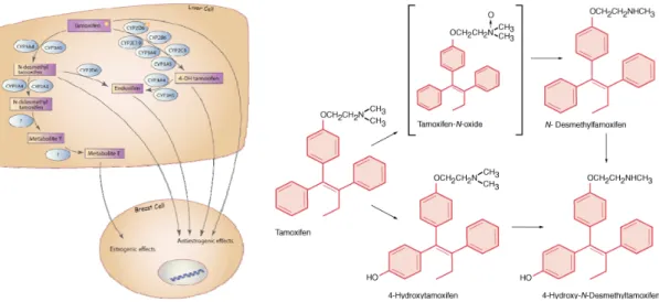
transcription. Tamoxifen is rapidly absorbed and is extensively metabolized (Fig.
6) to N‐desmethyltamoxifen and 4‐hydroxytamoxifen, the latter has a high
affinity for the ER (Jordan et al., 1977) and may play a significant role in the
antitumor actions of tamoxifen (Etienne et al., 1989).