Alma Mater Studiorum – Università di Bologna
DOTTORATO DI RICERCA IN
Progetto 2: Biologia Cellulare, Molecolare e Industriale
Ciclo XXV
Settore concorsuale di afferenza: 05/E2Settore scientifico disciplinare: BIO/11
TITOLO TESI
Functional and Genetic characterization of new genomic
islands from an E. coli strain associated with neonatal
meningitis.
Presentata da: Vanja Alberto Mariani Corea
Coordinatore Dottorato
Relatori
Chiar.mo Prof.
Chiar.mo Prof.
Vincenzo Scarlato
Vincenzo Scarlato
Dott.
Roberto Rosini
Dott.ssa
Michèle A. Barocchi
Esame finale anno 2013
ABSTRACT ... 5
1
INTRODUCTION: ... 7
1.1 ESCHERICHIA COLI ...7
1.1.1E. COLI CLASSIFICATION METHODS ... 8
1.2 E. COLI DIVERSITY: WHEN BOUNDARIES ARE NOT SO CLEAR. ... 10
1.3 PATHOGENESIS OF EXPEC ... 12
1.4 E. COLI AND GENETIC ISLANDS: EVOLUTION AT A FAST PACE. ... 15
1.4.1GENOMIC ISLANDS AND VIRULENCE FACTORS IN EXPEC. ... 18
1.4.2DICTYOSTELIUM DISCOIDEUM: A BACTERIAL HUNTER. ... 20
2
MATHERIALS AND METHODS. ... 22
2.1 BACTERIAL GROWTH. ... 22
2.1.1BACTERIAL STRAINS. ... 22
2.1.2ISOLATION OF CHROMOSOMAL DNA. ... 22
2.1.3SINGLE AND MULTIPLE GEI-DELETION MUTANTS . ... 23
2.1.3.1 Preparation of electro-competent cells. ... 23
2.1.3.2 Transformation of bacterial cells by electroporation. ... 23
2.1.3.3 Single and multiple Genomic island deletion by Red recombinase-mediated mutagenesis. ... 23
2.2 DICTYOSTELIUM DISCOIDEUM GROWTH AND GRAZING ASSAY. ... 24
2.2.1WORKING CULTURE. ... 24
2.2.2AMOEBA SPORE GENERATION. ... 25
2.2.3BACTERIAL GROWTH CURVES. ... 25
2.2.4DICTYOSTELIUM D. GRAZING ASSAY. ... 25
2.3 BIOINFORMATIC ANALYSIS AND PRIMER DESIGN. ... 26
2.3.1PRIMER DESIGN... 27
2.3.2STATISTICAL ANALYSIS. ... 28
2.4 GEI DISTRIBUTION AND EXCISION STUDIES. ... 28
2.4.1PCRAMPLIFICATION AND SEQUENCING. ... 32
2.4.2NUCLEASE RESISTANCE OF CIRCULAR INTERMEDIATES. ... 32
2.4.3RELATIVE REAL-TIME PCR. ... 33
2.5 PRIMER LIST. ... 34
2.6 MEDIA AND BUFFERS. ... 40
2.6.1MEDIA. ... 40 2.6.1.1 LB ... 40 2.6.1.2 SM ... 40 2.6.1.3 HL5 ... 40 2.6.1.4 SoC ... 40 2.6.2BUFFERS. ... 41 2.6.2.1 50x Soerensen buffer ... 41 2.6.2.2 Arabinose 20%... 41
2.6.2.3 50x TBE buffer ... 41 2.6.2.4 SM Buffer (Phage extraction) ... 41
3
RESULTS ... 42
3.1.1EXPEC ISOLATES HAVE A GREATER NUMBER OF GEIS THAN INPEC AND NON- PATHOGENIC STRAINS. ... 42 3.2 DISTRIBUTION OF IHE3034 ISLANDS AMONG A PANEL OF DIVERSE E. COLI ISOLATES. ... 43
3.3 MOLECULAR AND GENETIC CHARACTERIZATION OF IHE3034 GENOMIC ISLANDS. ... 48 3.3.1DELETION OF GENOMIC ISLAND (GEI)INTEGRASES PREVENTS THE FORMATION OF
CIRCULAR INTERMEDIATES. ... 49 3.3.2GEI11,13 AND 17 CIRCULAR INTERMEDIATES ARE RESISTANT TO DNASE TREATMENT. ... 50 3.3.3GROWTH CONDITIONS ALTER THE EXCISION RATE OF IHE3034 GENOMIC ISLANDS. ... 51
3.4 IHE3034 GENOMIC ISLANDS 13, 17 AND 19 ARE LINKED TO SURVIVAL IN THE
DYCTIOSTELIUM DISCOIDEUM GRAZING ASSAY. ... 55 3.4.1GEI13,17,19 DELETION AFFECT IHE3034 ABILITY TO RESIST TO THE DYCTIOSTELIUM
DISCOIDEUM GRAZING ASSAY. ... 55
4
DISCUSSION... 58
5
BIBLIOGRAPHY ... 65
Abstract
Enterobacteriaceae genomes evolve through mutations, rearrangements and
horizontal gene transfer (HGT). The latter evolutionary pathway works through the acquisition DNA (GEI) modules of foreign origin that enhances fitness of the host to a given environment. The genome of E. coli IHE3034, a strain isolated from a case of neonatal meningitis, has recently been sequenced and its subsequent sequence analysis has predicted 18 possible GEIs, of which: 8 have not been previously described, 5 fully meet the pathogenic island definition and at least 10 that seem to be of prophagic origin.
In order to study the GEI distribution of our reference strain, we screened for the presence 18 GEIs a panel of 132 strains, representative of E. coli diversity. Also, using an inverse nested PCR approach we identified 9 GEI that can form an extrachromosomal circular intermediate (CI) and their respective attachment sites (att). Further, we set up a qPCR approach that allowed us to determine the excision rates of 5 genomic islands in different growth conditions. Four islands, specific for strains appertaining to the sequence type complex 95 (STC95), have been deleted in order to assess their function in a Dictyostelium discoideum grazing assays.
Overall, the distribution data presented here indicate that 16 IHE3034 GEIs are more associated to the STC95 strains. Also the functional and genetic characterization has uncovered that GEI 13, 17 and 19 are involved in the resistance to phagocitation by
Dictyostelium d thus suggesting a possible role in the adaptation of the pathogen
1 Introduction:
1.1 Escherichia coli
Escherichia coli is a gram-negative bacteria belonging to the gamma-proteobacteria
class of microorganisms. The vast majority of Escherichia coli strains live within the healthy human organism without causing disease; the colonization generally begins a few hours after birth setting up a mutual benefit relationship. E. coli are generally non-pathogenic bacteria but in an immune-compromised host they find a way to breach the gastrointestinal barriers it
may happen that strains that were harmless in the digestive tracts start to cause diseases. E. coli has been identified as a versatile bacteria with a an ability to reorganize its genetic material in order to adapt to the environmental conditions in which it grows[42].
Pathogenic E. coli can be divided into two major sub-groups depending on the location where they cause disease. The Intestinal Pathogenic E. coli (InPEC) cause bowel diseases such as diarrhea, bloody stools and comprises pathotypes such as enterotoxigenic (ETEC), enteropathogenic (EPEC), enterohemorrhagic (EHEC),
enteroinvasive (EIEC), diffusely adherent (DAEC) all causing infections to the human intestinal tract. The second group of strains are the Extraintestinal Pathogenic E. coli (ExPEC) includes both human and animal pathogen causing urinary tract infections (UPEC) while others cause neonatal meningitides (NMEC) [5, 14].
ExPEC strains represent a major cause of morbidity, as it is responsible for 85-95% of uncomplicated cystitis cases and for over 90% of the episodes of uncomplicated pyelonephritis in premenopausal women. It has been estimated that 40-50% of
Fig. 1: Sites of pathogenic Escherichia coli colonization. (Croxen et. al 2010)
Pathogenic Escherichia coli colonize various sites in the human body. EPEC, ETEC, DAEC colonize the small bowel and cause diarrhoea, whereas EHEC, EIECcause disease in the large bowel; EAEC can colonize both the small and large bowels.
UPECenters the urinary tract and travels to the bladder to cause cystitis. Septicaemia can occur with both UPEC and NMEC, whereas the latter can cross the blood–brain barrier into the central nervous system, causing meningitis 3.
women will experience at least one case of UTI due to E. coli during the lifetime, with one fourth of these cases becoming a recurrent infection within 6 months of initial infection. Extra-intestinal strains are also responsible for episodes of catheter-associated UTIs (25-35%). NMEC together with Streptococcus agalactiae (GBS) are the leading causes of neonatal meningitis, accounting for an estimated 20 to 40% of the cases, with a fatality rate ranging from 25 to 40% and with neurological sequelae affecting 33 to 50% of survivors. These strains account for 17% of the cases of severe sepsis, with a mortality rate of approximately 30%. There are also strains that can be associated with intra-abdominal infections and nosocomial pneumonia and that occasionally participate in other extraintestinal infections, such as osteomyelitis, cellulitis and wound infections[25, 26, 65].
These kinds of diseases have never captured the public attention because they do not cause dramatic epidemics like those that cause food borne illness, thus underestimating the health and economic impact that they have. The high plasticity and rate of mutation of the E. coli genome is one variable that leads to the high number of diseases and the increasing antimicrobial resistance of the ExPEC strains. These characteristics translate into a large burden on the healthcare systems increasing the already heavy strain due to the actual economical environment; it is thus clear that a better understanding of E. coli is necessary in order to reduce the impact on healthcare[40].
1.1.1 E. coli classification methods
In order to classify E. coli many different typing methods have been developed; the most used ones are the multi-locus sequence typing (MLST) and the Phylogenetic groups.
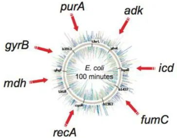
MLST is a generic typing method for the molecular characterization of bacterial isolates that is robust and easily accessible. It has been employed principally, but not solely, to type bacterial pathogens and its strength relies on the fact that it is based explicitly on the same population genetics on which was based the multilocus enzyme electrophoresis (MLEE). MLST has the additional aims of providing a unified bacterial isolate characterization approach that generates data that can also be used for evolutionary and population studies of a wide range of bacteria regardless of their diversity, population structure, or evolution[47]. This methodology is based on the sequencing PCR of seven highly conserved housekeeping genes (purA, adk, icd, fumC,
recA, mdh, gyrB) as it can be seen
in Fig. 2. The sequences are then concatenated and aligned through a program such as eBurst; each unique combination of alleles was assigned a sequence type (ST). Related STs were assigned to so-called ST complexes (STC), using the principles of the eBurst algorithm: each ST complex includes at least three STs that differ from their nearest neighbour by no more than two of the seven
loci while ST complexes differ from each other by three or more loci. STs not matching the criteria for inclusion were referred to by their ST designation[81]. E. coli demonstrated to be a clonal bacteria and as Wirth and colleagues have pointed out the strains appertaining to a given ST or STC tend to have similar virulence factors and thus phenotypes. This is particularly true for very conserved STCs like: STC10 were almost all the non-pathogenic phylogenetic group A strains group or STC95 strains that are all of the phyl. group B2 and carry the K1 capsule gene cluster.
This typing method is highly used by clinical microbiologists and epidemiologists as it is very robust and reproducible. The limit of this typing technique is that, in order to create an homogeneous classification of the bacterial populations, it requires a common agreement on the alleles (and their order) to be used [47].
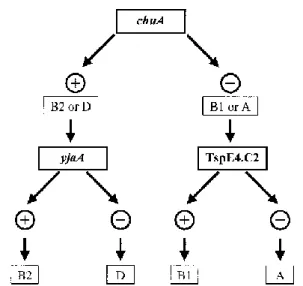
Originally phylogenetical grouping was performed using techniques like MLEE and ribotyping that are complex, time-consuming and also require a collection of typed strains. Thirteen years ago (2000) Clermont et. al. proposed a rapid and simple method for typing Escherichia coli that uses a triplex polymerase chain reaction to amplify three targets genes. The markers were: chuA, a gene required for heme transport in enterohemorrhagic O157:H7; yjaA, a gene of unknown function identified in a E. coli K-12 strain and an anonymous DNA fragment designated TSPE4.C2.
Fig. 2: MLST genes and genomic position (Wirth et.
al 2010)
Genomic disposition of all the 7 alleles used for the MLST analysis.
The results of these three amplifications made it possible to establish a dichotomous decision tree (Fig. 3) that could attribute to any typed strain a phylogenetical group out of the four possible (A, B1, B2, D)[12]. This new typing method used by Clermont allows a faster and easier discrimination of the strains appurtenance to a phylogenetical group with an accuracy ranging from 80-85%[30]. The previous methods, due to not-common pattern of bands, assigned some strains to smaller sister groups (ABD, AxB1) that had a
typing profile which was intermediate to between A and B1.
1.2 E. coli diversity: when boundaries are not so clear.
The high rate of mutation and plasticity of E. coli genome is the peculiarity that allows this bacteria to survive and thrive in different enviroments ranging from waste water to human/animal body. Bio-informatic analysis of the ever-growing collection of E.
coli genomes allowed to understand that bacterial genomes comprise stable regions
that form the “core” genome and variable regions that form the flexible gene pool. [3] Also genomic comparisons revealed that non-pathogenic E. coli genomes size varies from the 4,6Mb of the non-pathogenic strains to the 5.7Mb of the pathogenic and Asymptomatic Bacteriuria (ABU) strains. ExPEC virulence factors exhibit distinct patterns of phylogenetic distribution. This provides evidence of both, vertical and horizontal transmission of the corresponding virulence-associated genes as well as of host-specific associations and strong associations among different virulence-associated genes[18]. The constant typing effort allowed the identification of phylogenetic groups into which the major E. coli pathotypes cluster together (Fig. 4).
Fig. 3: Phylogenetic group decision tree (Clermont et al.)
Dichotomous decision tree to determine the phylogenetic group of an E. coli strain by using the results of PCR amplification of the chuA and yjaA genes and DNA fragment TSPE4.C2.
The majority of the non-pathogenic strains cluster together in the group A, ABD, AxB1; while the intestinal pathogenic strains tend to cluster in the AxB1, B1 and D groups. It is important to understand that the groups AxB1 and ABD are sister groups to the B1 group. Intestinal pathogenic E. coli strains derive from phylogenetic groups A, B1 or D or from ungrouped lineages and are seldom found in the fecal flora of healthy individuals as the mere acquisition of these bacteria by the naïve host is sufficient for disease to ensue. Each intestinal pathotype possesses a characteristic combination of virulence and fitness factors that allow the colonization of specific niches and results in a unique diarrheal syndrome.
The B2 cluster is where almost all the ExPEC strains group while the remaining strains belong to cluster D. Extraintestinal strains have acquired various virulence genes that allow them to induce infections outside the digestive system in both normal and compromised hosts. ExPEC are incapable of causing gastrointestinal disease, but they can asymptomatically colonize the human intestinal tract and become the predominant strain in approximately 20% of normal individuals[40, 71].
Fig. 4: Schematic representation of the E. coli pathogenic organization.
E. coli can be divided in pathogenic and non-pathogenic strains. Pathogenic strains can be further
divided in Intestinal pathogenic or in Extra Intestinal pathogenic strains depending on where they cause a disease. In the image are represented to which phylogenetic groups the different pathogroups are associated.
ExPEC strains carry a broad range of virulence factors, distinct from those found in InPECs, that allow them to colonize host mucosal surfaces, avoid or subvert local and systemic host defense mechanisms, scavenge essential nutrients such as iron, injure or invade the host, and stimulate a noxious inflammatory response[40]. Due to extra-intestinal E. coli ability to survive either in or out of the gastroextra-intestinal tract the definition of non-pathogenic strains has been hard to define. As colonizing sites outside the gut are unlikely to provide any selective advantage in terms of transmissibility, it is clear that any so-called ‘‘extra-intestinal virulence factors’’ are likely to have evolved to enhance survival in the gut and/or transmission between hosts, and therefore will be shared with at least some commensal strains. So this ability to fluctuate between mutualism, commensalism, opportunistic pathogenesis or even specialized pathogenesis make Escherichia coli the perfect candidate to study the boundaries between pathogenicity and commensalism[18, 74].
1.3 Pathogenesis of ExPEC
Among ExPEC strains, uropathogenic E. coli and neonatal meningitis E. coli are characterized by different molecular mechanisms of pathogenicity.
Urinary tract infection usually begins with the colonization of the bowel with a uropathogenic strain in addition to the commensal flora these strains, by virtue of its virulence factors, are able to colonize the periurethral area and to ascend the urethra to the bladder. Between 4 and 24 hours after infection, the new environmental conditions in the bladder select for the expression of type 1 fimbriae that allow the adhesion to the uroepithelium[42]. This attachment is mediated by fimbrial adhesin H (FimH), which is located at the tip of type 1 pili. FimH binds to mannose moieties of the receptors uroplakin Ia and IIIa that coat terminally differentiated superficial facet cells in the bladder, stimulating also unknown signaling pathways that induce invasion and apoptosis (Figure 5). Bacteria internalization is also mediated by FimH binding to 3 and 1 integrins that are clustered with actin at the sites of invasion, as well as by microtubule destabilization.
These interactions trigger local actin rearrangement by stimulating kinases and Rho-family GTPases, which results in the envelopment and internalization of the attached bacteria.
Once internalized, UPEC can rapidly replicate and form biofilm-like complexes called intracellular bacterial communities (IBCs), which act as transient, protective environments. UPEC can also leave the IBCs through a fluxing mechanism and enter again the lumen of the bladder. Filamentous UPEC has also been observed fluxing out of an infected cell, looping and invading surrounding superficial cells in response to innate immune responses. During infection, the influx of polymorphonuclear leukocytes (PMNs) causes tissue damage, while apoptosis and exfoliation of bladder cells can be induced by UPEC attachment and invasion, as well as by sublytic concentrations of the pore-forming toxin HlyA. This breach of the superficial facet cells temporarily exposes the underlying transitional cells to UPEC invasion and
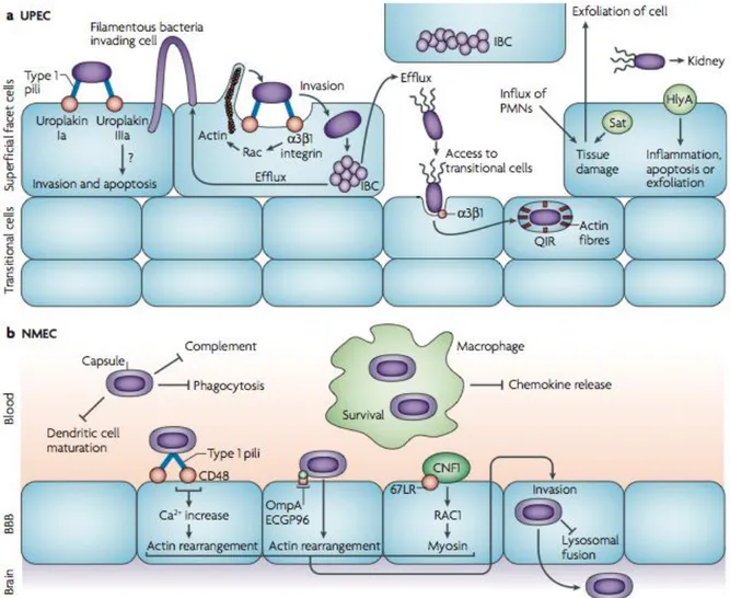
Fig. 5: Pathogenic mechanisms of ExPEC (Croxen and Finlay, 2010). The different stages of
extraintestinal E. coli infections are shown. (A) UPEC attaches to the uroepithelium through type 1 pili, which bind the receptors uroplakin Ia and IIIa. Sublytic concentrations of the pore-forming toxin HlyA can inhibit the activation of Akt proteins and lead to host cell apoptosis and exfoliation. Exfoliation of the uroepithelium exposes the underlying transitional cells to further UPEC invasion. (B) NMEC is protected from the host immune response by its K1 capsule and outer-membrane protein A (OmpA). Invasion of macrophages may provide a replicative niche for high bacteremia, allowing the generation of sufficient bacteria to cross the blood-brain barrier (BBB) into the central nervous system.
dissemination. Invading bacteria are trafficked in endocytic vesicles enmeshed with actin fibers, where replication is restricted. Disruption of host actin allows rapid replication, which can lead to IBC formation in the cytosol or fluxing out to the cell. This quiescent state may act as a reservoir that is protected from host immunity and may, therefore, permit long-term persistence in the bladder, as well as recurrent infections[14]. In strains causing cystitis, type 1 fimbriae are continuously expressed and the infection is confined to the bladder. In strains that are able to cause pyelonephritis, the invertible element that controls type 1 fimbriae expression turns to the “off” position and type 1 pili are less well expressed. This releases the UPEC strain from bladder epithelial cell receptors and allows the microorganism to ascend through the ureters to the kidneys, where it can attach by P fimbriae to digalactoside receptors that are expressed on the kidney epithelium. At this stage, hemolysin could damage the renal epithelium inducing an acute inflammatory response with the recruitment of PMNs to the infection site. Hemolysin has also been shown to cause calcium oscillations in renal epithelial cells, resulting in increased production of interleukin-6 (IL-6) and -8 (IL-8). Secretion of the vacuolating cytotoxin Sat damages glomeruli and is cytopathic for the surrounding epithelium. In some cases, bacteria can cross the tubular epithelial cell barrier and penetrate the endothelium to enter the bloodstream, leading to bacteremia[42]. The pathogenesis of NMEC strains is a complex mechanism, as the bacteria must enter the bloodstream through the intestine and ultimately cross the blood-brain barrier (BBB) into the central nervous system, which leads to meningeal inflammation and pleocytosis, that means presence of a higher number of cells than normal, in the cerebrospinal fluid (Fig. 5). Bacteria can be acquired perinatally from the mother and, after the initial colonization of the gut, they can translocate to the bloodstream by transcytosis through enterocytes. The progression of disease is dependent on high bacteremia (>103colony forming units per ml of blood), therefore survival in the blood is crucial. NMEC is protected from the host immune responses by its K1 antiphagocytic capsule, made up of a homopolymer of polysialic acid, and by outer membrane protein A (OmpA), which confers serum resistance through manipulation of the classical complement pathway. NMEC has also been shown to interact with immune cells: invasion of macrophages and monocytes prevents apoptosis and chemokine release, providing a niche for replication before dissemination back into the blood. Bacterial attachment to the BBB
is mediated by FimH binding to CD48 and by OmpA binding to its receptor, ECGP96. Invasion of brain microvascular endothelial cells involves CNF-1 binding to the 67 kDa laminin receptor (67LR), which leads to myosin rearrangement, as well as OmpA and FimH binding to their receptors, which results in actin rearrangement. The K1 capsule, which is found in approximately 80% of NMEC isolates, also has a role in invasion by preventing lysosomal fusion and thus allowing delivery of live bacteria across the BBB. Collectively, these mechanisms allow NMEC to penetrate the BBB and gain access to the central nervous system, where they cause edema, inflammation and neuronal damage[14].
1.4 E. coli and genetic islands: evolution at a fast pace.
The ability to adapt and thrive across a huge diversity of hosts both human and animal make microbial pathogens a considerable threat all around the world [3]. The versatility that pathogens show is caused, at a molecular level, by the ability of the bacteria to adapt and evolve to
evade detection.
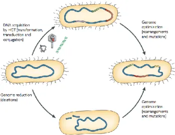
Bacterial genome evolution is a continuous process that can be analysed from two points of view: a long-term ‘macroevolution’, which leads to the development of new species or subspecies over millions of years, and short-term ‘microevolution’, which spans shorter time frames (days or weeks) and leads to the alteration of genes and traits[84]. Bacterial evolution takes place following three main mechanisms of large-scale genome alteration: DNA deletions, rearrangements a point mutations, gene duplication and gene acquisition through
Fig. 6: Mechanisms that contribute to bacterial genome evolution(Ahmed et. al. 2004)
Genome plasticity results from DNA acquisition by horizontal gene transfer (HGT; for example, through the uptake of plasmids, phages and naked DNA) and genome reduction by DNA deletions, rearrangements and point mutations. The concerted action of DNA acquisition and gene loss results in a genome-optimization process that frequently occurs in response to certain growth conditions, including host infection or colonization.
horizontal gene transfer (HGT). Upon selection, such modifications to the genome, create subgroups of strains able to resist environmental stress and possibly cause a diseases using a common set of virulence/fitness factors (pathotypes) (Fig. 6).
As previously noted pathogenic genomes are bigger than non-pathogenic ones, bioinformatic analysis has shown and that these areas of difference are generally very variable, thus dividing the bacterial genome in very conserved “core” areas and variable areas that are more susceptible to rearrangements[22]. Such areas can be hotspots for insertions and stabilization pieces of DNA carried by phages, transposons and larger mobile chromosomal elements such as genomic islands (GEIs).
GEIs are very long non replicative mobile elements, ranging from 10Kbp to 120Kbp, that have features taken by other mobile elements (ICEs, prophages, plasmids,…) allowing them to integrate and excise from the genome. Given the great amount of genes carried by such mobile elements, the acquisition of a GEI, is generally considered to be a big evolutionary event that may cause a marked variation in the microorganism phenotype[31]. After such an event the genomic islands become integrant part of the bacteria and are subsequently subject to mutation to prevent further transmission and integration depending on the usefulness of the island itself[32]. Of course, the line that separates these conditions can be very subtle, according to the niche and to the right combination of factors.
Genomic islands take different names based on the kind of fitness advantage they furnish with the genes encoded on them: help the microorganism to live in the environment (ecological islands) or to persist as saprophyte (saprophytic islands), to colonize the host and provide benefit (symbiosis islands) or to cause disease (PAIs)[31, 60]. Most notably the recent German E. coli outbreak was caused by a mildly pathogenic InPEC strain integrating the Shiga toxin-encoding genomic island (stx island) in its own genome thus creating a new microorganism more fit to survive against the immune system.
Among the most characterized islands such as: the shiga phage or the high pathogenicity island (HPI). The stx island is a GEI of prophagic origin that carries the shiga toxin genes. This island has been throughoutly studied as it carries genes that significantly enhance the pathogenicity of the host and that are highly over expressed when DNA interfering or oxidative agents are added to the media[46]. The HPI island
or Yersinia island is devided in two portions: a conserved “core” portion and a variable AT-rich. The stable encodes a functional cluster of genes coding for biosynthesis, transport and regulation of the siderophore yersiniabactin, the recombinase gene and siderophore (intHPI); while the AT-rich carries genes carries
the excisionase (XisHPI) and Hex two genes foundamental for the island mobilization.
It is of interest that this island is able to successfully colonize Enterobacteriacea such as E. coli, but in the majority of this strains (Yersinia excluded) this AT-rich zone is truncated and missing the attR site and thus is immobilized[4].
Genomic islands are identified by bioinformatic means as this genetic elements have very distinct features such as: the presence of an integrase gene, a GC content lower than the surrounding core DNA, the presence of a tRNA (facultative), the presence of direct repeat sites (att sites) at each side of the area. The integrase gene and the att sites play a fundamental role in the island mobilisation as they are the molecular machinery that allows GEIs to mobilize themselves. There are GEIs though missing some of this features that have been stabilized by the selective pressure; all these islands are not able to mobilize themselves anymore and generally carry virulence/fitness factors. To excide from the genome and release the plasmid-like structures, called circular intermediates (CI), in the cytoplasm the integrase protein brings the att sites close to each other, thus allowing for a site-specific recombination event to happen[9, 37, 50]. This ability to excise from the genome and create discrete CIs is thought to be an adaptation of the one used by bacteriophages to integrate and excide from the genome[50]. Continuous non-perfect integration and mobilization events may also have been the cause for the creation of this stretches of DNA that do still carry some prophagic elements, but should not able to create a fully working prophage. This assumption may be considered true to the point that genomic islands can be divided in two groups depending on their gene content and integrase gene (GEI-encoded, Phage-encoded)[55]. Also If we take into account that, for the prophages, the passage from a lysogen to a lytic cycle is considered to be their way to survive to stressful conditions[56, 78] we can also understand that the variations of genomic island excision rates in bacteria may be affected by external stress conditions (temperature, minimal medium, iron depletion, oxidative stress).
E. coli has been selected as the representative the pathogen genomic fluidity due to its
GEIs that have been fully described. Plasmids, phages and PAIs all play a crucial part in the evolution of different E. coli pathotypes[18, 42].
One main feature of the different intestinal E. coli pathotypes is the presence of pathotype-specific plasmids that often encode toxins. The characteristic protein toxins of enterotoxigenic, enteroaggregative, enteroinvasive, enterohaemorrhagic and enteropathogenic E. coli (and also extraintestinal pathotypes) are plasmid-encoded.
Also it is important to understand that as whole GEIs have a mosaic-like, modular structure and, although many of them superficially resemble each other (presence of certain virulence determinants), a great variability exists with regard to GEI composition, structural organization and chromosomal localization among strains even if they are of the same patho- or sero- type.
1.4.1 Genomic islands and virulence factors in ExPEC.
As previously stated genomic islands are the main effectors of the HGT due to their ability to transfer themselves from a donor to a host; their importance is also due to the high amount of open reading frames (ORFs), many of which of unknown origin, that encode for fitness or virulence factors.
IHE3034 is a neonatal meningitis strain appertaining to the phylogenetical group B2 and to the clonal complex (STC) 95 that it has been sequenced in 2010. The bioinformatic analysis carried out by Moriel et. al. uncovered 19 possible genomic islands present in IHE3034 (Tab. 1) [53].
Table 1
GEI Virulence / Fitness Factors Kbp Related islands 1 Putative type VI secretion system 30 PAI IIAPECO1
2 Prophage DNA 57 F-CFT073-smptB
3 Prophage DNA 22 Moriel DG et al.
4 Prophage DNA 33 Moriel DG et al.
5 S-perfimbriae, IroN, putative TonB-dependant receptor, Antigen 43
61 PAI III536, PAI-CFT073-serX, PAI
INissle1917
6 sitABCD 47 PAI-CFT073-icdA
7 Prophage DNA 46 F-CFT073-potB
8 Yersiniabactin and cdtABC 78 PAI-CFT073-asnT
9 Colibactin gene cluster 54 PAI VI536, GI-CFT073-asnW
10 Putative TonB-dependent receptors and ibrAB 44 PAI VI536, GI-CFT073-cobU, PAI
IVAPECO1
11 Prophage DNA 37 Moriel DG et al.
13 Enterohemolysin 1 39 Moriel DG et al.
14 Prophage DNA 43 Moriel DG et al.
15 Putative type VI secretion system 36 PAI-CFT073-metV, PAI-536-metV
16 T2SS and K1 capsule 28 PAI V536, PAI IAPECO1
17 Prophage DNA 16 Moriel DG et al.
18 IbeA and IbeT 20 GimA
19 Prophage DNA 46 Moriel DG et al.
Among virulence factors carried by ExPEC GEIs, a fundamental role is played by adhesins (GEI 5), which allow the strict interaction of the pathogen with the host, facilitating the colonization and invasion processes and avoiding clearance by the host immune defences. Also the presence of group K1 (GEI 16) capsule confers additional selective advantages to ExPEC strains. Indeed, their molecular mimicry to host tissue components helps the bacteria to evade the immune response, providing protection against phagocytic engulfment and complement-mediated bactericidal activity[24, 79]. GEI 16 though is not a genomic island but a hotspot of integration; bioinformatic analysis has shown that it is a highly variable region and that the tRNA present in the middle of it is a typical insertion point for mobile elements. Other proteins are also associated with the virulence of ExPEC strains. For example IbeA and IbeT (GEI 18) that are involved in the invasion of brain microvascular endothelial cells [38, 85]. Antigen 43 (Ag43 – GEI 5) is associated with a strong aggregation phenotype and with biofilm formation, promoting long-term persistence in the bladder, although its relevance and contribution in the pathogenesis are far from clear[76].
Growth of ExPEC strains in iron-limited conditions, such as urine, requires successful mechanisms for the scavenging of iron, which rely on siderophores and iron-complex receptors [80]. Several iron and siderophore receptors, which are highly expressed during infection of the urinary tract, have already been described in E. coli, for example the salmochelin siderophore receptor IroN[33] and the ferric and manganese receptor sitABCD[83].
Eight out of nineteen islands of IHE3034 islands are or prophagic origin and have been identified for the first time by Moriel et. al.; many of the ORFs on these GEIs are of unknown function. These islands altogether account for the 0,5% of the genome of IHE3034 and the percentage of ORFs of known function ranges from 50% to 75%. Understanding the mechanisms behind GEI mobilization and functions is a key point
to develop preventive and therapeutic approaches that could aim to selectively induce PAI deletion and reduce the incidence of E. coli diseases.
1.4.2 Dictyostelium discoideum: a bacterial hunter.
Bacteria like E. coli are mainly environmental microorganism; they live in the soil were they are constantly threatened by bacteria-eating predators such as protozoa and nematodes. These evolutionary pressures may affect bacterial populations in multiple ways, like creating defense strategies that allow them to survive and to establish new replicative niches. For example, to protect themselves from predators, produce biofilms thus preventing engulfment and phagocytosis, or use molecular machinery to avoid lysosomal killing[34]. As a soil amoeba and a phagocyte
Dictyostelium discoideum can be a natural host of opportunistic bacteria that may
have developed strategies to invade, survive and replicate intracellularly inside the amoeba itself[10].
D. discoideum is a fascinating member of the amoebozoa, its natural habitat is
deciduous forest soil and decaying leaves, where the amoebae feed on bacteria, yeast and grow as separate, independent, single cells. The organism offers unique advantages for studying fundamental cellular processes with powerful molecular genetic, biochemical, and cell biological tools[23].
Phagocytosis is a very complex, evolutionarily conserved mechanism that is used by higher eukaryotes to clear dead cells and cell debris and to counter the constant threat posed by pathogens. For this purpose they harbour specialized cells such as macrophages, neutrophils or dendritic cells that have the ability to rapidly and efficiently internalize a variety of organisms and particles and degrade them. For lower eukaryotes like D. discoideum phagocytosis is a means to internalize bacteria that are used as food source. The ingested microorganism is trapped in a phagosome and, via the phago-lysosomal pathway, is ultimately delivered to a lysosome where it is degraded by a cocktail of hydrolytic enzymes[11, 13].
Bacterial pathogenicity was certainly largely developed to resist predatory bacteriovorous microorganisms in the environment, and this accounts for the fact that a large number of bacterial virulence traits can be studied using Dictyostelium as a host.
The increasing number genome sequences and the genetic tractability of E. coli generate many opportunities for the study of host-pathogen interactions. The use of
Dictyostelium cells as a screening system for bacterial virulence combining the use of E. coli mutant cells will allow to identify determinants of susceptibility and resistance
2 Matherials and Methods.
2.1 Bacterial growth.
2.1.1 Bacterial strains.
IHE3034 (O18:K1:H7), ST95, is a neonatal meningitis-associated strain isolated in Finland in 1976[1]. Its genomic sequence has been revealed in 2012[53].
The 132 E. coli strains extra intestinal, intestinal pathogenic and non-pathogenic that have been used in this analysis are described in table 3. The eleven ST131 strains have been kindly provided by Marina Cerquetti from the Istituto Superiore di Sanità (Rome). 77 strains mixed ExPEC, InPEC and non-pathogenic have been kindly provided by Lothar H. Wieler from the Freie Universität Berlin. 35 strains ExPEC, InPEC and non-pathogenic have been kindly provided by Ulrich Dobrindt from the Universitätklinikum of Münster. The collection is composed of strains belonging to the A, AxB1, ABD, B1, B2 phylogenetic group[39]. Throughout the manuscript, GEI deletion mutants (partial or whole) of E. coli strain IHE3034 are named by the symbol “G” followed by the numbers of the deleted GEI and if needed, the letter of the deleted portion. The numbers indicating the GEI are expressed in the Arabic form instead of the Roman one (classically used to number GEIs in the literature) to ease readability. Bacteria were routinely grown in LB broth at 37°C except when otherwise stated. Ampicillin (Amp 100g/ml), Kanamycin (Kan 25g/ml), Trimethoprim (Trim 100g/ml), Mitomycin C (Mit. C 0,5g/ml) or Chloramphenicol (Clm 8 g/ml) were added to the media when necessary.
2.1.2 Isolation of chromosomal DNA.
Genomic DNA was prepared by culturing bacteria overnight at 37 °C, with antibiotics added when needed, and left overnight in an orbital shaker. The extraction took place the following day using either the GenElute Bacterial Genomic DNA Kit (Sigma) according to the manufacturer’s instructions or preparing a raw genomic extract. Final DNA concentration, of the genomic kit preparation sample, was assessed by optical density determination at 260 nm.
Raw genomic extract preparations were prepared adding 200l of ON culture (c.a. 4,6*107 CFU) in a clean Eppendorf. The culture was centrifuged and the supernatant
for 10 minutes. The raw extraction was then centrifuged for 5 minutes at 1100 g in a table top centrifuge (Eppendorf) and the supernatant was transferred in a new tube. 2.1.3 Single and multiple GEI-deletion mutants .
2.1.3.1 Preparation of electro-competent cells.
For electro-competent IHE3034 cell preparation, 2ml LB were inoculated starting from the glycerol stocks and set to grow overnight at 37°C shaking at 180rpm. If the red recombinase (p434, pKOBEG) or the flipase plasmid (pCP20) were present the culture was grown at 30°C. The next day 25ml of fresh LB were inoculated to a final OD/ml of 0,1 and left to grow, with Arabinose to a final concentration 0,2%, up to 0,6-0,8OD/ml. When the OD/ml of 0,6-0,8 is reached the culture was poured into a Falcon tube and the cells precipitated for 30min at 3650g at 4°C in a Heraeus MULTIFUGE 3 S-R centrifuge with a 75006445 rotor. The pellet was then washed 3 times with 25ml of cold sterile water (4°C) and one time with 25ml of cold a 10% glycerol solution. The pellet was resuspended in 500l of 10% glycerol solution and divided in 60l aliquots in Eppendorf tubes and stored at -80°C.
2.1.3.2 Transformation of bacterial cells by electroporation.
For electroporation 60l of electro-competent cells were thawed on ice and mixed with 1-12l of plasmid or purified PCR constructs to the final concentrations of up to 100ng plasmid, 1g PCR product. Cells were transferred in a 1-mm-wide GenePulser electroporation cuvette and then electroporated using program Ec1 of the Biorad GenePulser Xcel (1,8kV). Transformations with a time constant no lower than 4 were recovered in 250l of SoC media and set to grow 1-2hrs at 37°C (30°C for temperature-sensitive plasmids) in a shaking thermal block before being plated on selective LB-Agar plates. The following day colonies were PCR screened after being streaked in a fresh plate.
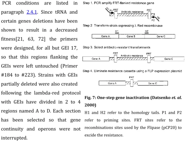
2.1.3.3 Single and multiple Genomic island deletion by Red recombinase-mediated mutagenesis.
4, 13, 17 and 19: each single GEI deletion mutant was generated using the
-Red recombinase gene inactivation method [16]. Flipase recognition target (FRT)-flanked kanamycin or chloramphenicol cassettes were generated by PCR using as a template pKD4 or pKD3. Primers carried tails of 60 to 71 bases homologous with both ends of the GEI to be deleted (Fig. 7).
PCR conditions are listed in paragraph 2.4.1. Since tRNA and certain genes deletions have been shown to result in a decreased fitness[21, 63, 72] the primers were designed, for all but GEI 17, so that this regions flanking the GEIs were left untouched (Primer #184 to #223). Strains with GEIs partially deleted were also created following the lambda-red protocol with GEIs have divided in 2 to 4 regions named A to D. Each section has been selected so that gene continuity and operons were not interrupted.
Multiple Knock-out strain: t 4 mutant was used to successively remove GEIs,
13, 17, and 19 using the Red recombinase method as for single GEI deletion mutants. After the deletion of two GEIs, the antibiotic resistance cassette was removed using Flp before proceeding to the next GEI deletion [16].
2.2 Dictyostelium discoideum growth and grazing assay.
Amoeba spore aliquots where thawed and allowed to grow for three days in 10ml of HL5 medium in 25cm2 cell-culture flasks in order to generate pre-cultures[27, 70].
All the amoeba cultures were grown at room temperature (21-25°C) in a thermally controlled laboratory.
2.2.1 Working culture.
After all the spores have germinated 5 ml of pre-culture were inoculated in 15ml of fresh HL5 medium to generate a new Working culture a 75cm2 cell-culture flask. The
new culture was then left to grow for three to four days in a temperature controlled room at 21-25°C. [27, 70].
Fig. 7: One-step gene inactivation (Datsenko et. al. 2000)
H1 and H2 refer to the homology tails. P1 and P2 refer to priming sites. FRT sites refer to the recombinations sites used by the Flipase (pCP20) to excide the resistance.
2.2.2 Amoeba spore generation.
A shaking culture of 25ml HL5 was inoculated in a conical cylinder using 1,5-2ml of matured pre-culture. After 3 to 4 days the cells are counted using the Neubauer improved counting chamber using the following formula:
⁄
If needed cell were concentrated in Soerensen Buffer 1x[28] to a final concentration no less than 1x10⁷cell/ml. In each Soerensen-Agar Plate were then plated 400l of
amoeba resuspension and gently distributed by rotating the plate with circular motions.
The plates were put in a closed-lid box with wet towel papers in the bottom and left to incubate for 3-4 days at room temperature. The Dyctyostelium d. yellowish spore heads were harvested by rinsing the plates with 2ml of Soerensen buffer 1X until the majority of the spore heads were in solution. Each stock cryotube was filled with 1ml of spore suspension and stored at -80°C[28].
2.2.3 Bacterial growth curves.
The Dyctiostelium discoideum grazing assay is heavily influenced by the bacterial fitness. In order to confirm that the assay is not biased by such a problem, both IHE3034 wild type and all the mutant strains have been tested in a growth curve assay. This analysis has been carried out to assess if the deletions that have been made reduce the fitness of the bacteria.
For each strain, a 0,01 OD/ml LB or SM inoculum was prepared in a 96 well plate with a final volume of 200l. The plate was then sealed tight with parafilm and placed in a pre-warmed Tecan Infinite 200 with a Heating and Shaking module. The instrument temperature has been pre-set to 37°C shaking at 180rpm; the OD measurements (600nm) were taken every 10 min for 24hrs. The data gathered were then plotted in an OD – Time graph with the X axis (OD) expressed in logarithmic scale.
2.2.4 Dictyostelium d. grazing assay.
The IHE3034 wild type and the deletion mutant E. coli strains were inoculated from the glycerol stock and let to grow for 3-4 hrs in LB media at 37°C shaking at 180rpm. 300l of bacterial growth (c.a. 4,5x10⁸CFU) were then evenly plated on fresh
1 hr. The amoeba population in the working culture was then quantified and four working dilutions were prepared in HL5 medium: 106cell/ml, 2x10⁵cell/ml,
2x10⁴cell/ml and 2x10³cell/ml. The bacterial SM plates were divided in four areas in
which different amount of amoeba cells (5000, 1000, 100 and 10) were plated. In order to reduce the experimental variability two 5l drops were placed on each plate; each working dilution was vortexed after each use to reduce the error caused by the fast sedimentation rate of the amoeba cells. The plates were then left at room temperature for a variable period between 3 and 7 days and the plaque formation assessed daily; each plate was photographed with an acquiring time of 230ms using the Protocol 2 (Synbiosis) cell counter. The plating assay was repeated 3 times for each bacterial strain under study and for the two control strains. As a negative control, immune to the amoeba grazing, the highly virulent UPEC strain 536 was used and due to its susceptibility to Dictyostelium, the non-pathogenic E. coli DH5 has been selected as positive control.
2.3 Bioinformatic analysis and primer design.
All of the E. coli genomes sequences used for the analysis were gathered from the NCBI website (www.ncbi.gov) and have been imported in the Geneious 5.6 software[49] (for the list of the genomes refer to Tab. 2). Full genome alignments have been carried out using the plug-in MAUVE program implemented on Geneious using the default settings[15]. The primer designs have been carried out using the Primer 3 plugin for Geneious [67]. Real time data analysis and images generation has been don using the REST2009 program[57, 62]
Table 2
Name Pathotype Group Sequence Length (Bp)
1 ABU 83972 ABU Non-pathogenic 5131397
2 LF82 AIEC InPEC 4773108
3 O83:H1 str. NRG 857C AIEC InPEC 4747819
4 UM146 AIEC InPEC 4993013
5 APEC O1 APEC ExPEC 5082025
6 SMS-3-5 AREC InPEC 5068389
7 042 EAEC InPEC 5241977
8 55989 EAEC InPEC 5154862
9 O26:H11 str. 11368 DNA EHEC InPEC 5697240
10 O103:H2 str. 12009 EHEC InPEC 5449314
12 O157:H7 str. EC4115 EHEC InPEC 5572075
13 O157:H7 str. Sakai EHEC InPEC 5498450
14 O157:H7 str. TW14359 EHEC InPEC 5528136
15 O111:H- str. 11128 EHEC InPEC 5371077
16 O55:H7 str. CB9615 EPEC InPEC 5386352
17 O127:H6 str. E2348/69 EPEC InPEC 4965553
18 E24377A ETEC InPEC 4979619
19 H10407 ETEC InPEC 5153435
20 UMNK88 ETEC InPEC 5186416
21 ATCC 8739 Laboratory strain Non-pathogenic 4746218
22 BL21(DE3) Laboratory strain Non-pathogenic 4570938
23 B str. REL606 Laboratory strain Non-pathogenic 4629812 24 DH1 (ME8569) Laboratory strain Non-pathogenic 4621430
25 BW2952 Laboratory strain Non-pathogenic 4578159
26 DH10B (K-12) Laboratory strain Non-pathogenic 4686137 27 MG1655 (K-12) Laboratory strain Non-pathogenic 4639675 28 W3110 (K-12) Laboratory strain Non-pathogenic 4646332
29 KO11 Laboratory strain Non-pathogenic 4920168
30 MDS42 (K-12) Laboratory strain Non-pathogenic 3976195
31 W Laboratory strain Non-pathogenic 4900968
32 IHE3034 NMEC ExPEC 5108383
33 O7:K1 str. CE10 NMEC ExPEC 5313531
34 S88 NMEC ExPEC 5032268
35 HS Non-pathogenic Non-pathogenic 4643538
36 SE11 Non-pathogenic Non-pathogenic 4887515
37 SE15 DNA Non-pathogenic Non-pathogenic 4717338
38 IAI1 Non-pathogenic Non-pathogenic 4700560
39 ED1a Non-pathogenic Non-pathogenic 5209548
40 536 UPEC ExPEC 4938920
41 CFT073 UPEC ExPEC 5231428
42 IAI39 UPEC ExPEC 5132068
43 NA114 UPEC ExPEC 4971461
44 str. 'clone D i2' UPEC ExPEC 5038386
45 str. 'clone D i14' UPEC ExPEC 5038386
46 UMN026 UPEC ExPEC 5202090
47 UTI89 UPEC ExPEC 5065741
2.3.1 Primer design.
The distribution studies were carried out using 3 sets of primers; the first set was a single pair of primers while the second and third sets were combined together in a Multiplex PCR design. Primers (#72 to #183) have been designed on three open reading frames (ORF) spanning along the whole island. Each ORF was selected only if it was present, in the BLAST analysis, in IHE3034 or in pathogenic strains and absent
in non-pathogenic strains.
Primers for the identification of the circular intermediates (CI) and for the exclusion PCR were designed to be functional only if the CI was formed and thus the island was not integrated in the genome (Primer #1 to #71).
Primers for the generation of knock out strains were designed to have a fixed portion that could anneal on pKD3/4[16] and had a tail spanning between 60 and 71bp that was completely homologous to the flanking regions that were to be knocked-out (#184-#223).
Primers designed for the screening of the knock-out strains were a forward annealing at the 5’ region of the KO region and a reverse annealing either on the inserted resistance gene or on the 3’ of the insertion region. The external oligonucleotides were also used to test if the islands had lost their resistances after pCP20 transformation (#241-#262).
Primers for the Relative Real Time PCR were designed to be no more than 20bp and each pair had to have a Tm of 60°C, equal GC content when possible, a Ta difference within 0,5°C and a final amplicon of no more than 350bp. Before being used in the real-time experiment a test PCR was run and the product of these primers were analysed on an agarose gel for aspecific bands. All the products were sequenced to check for the specificity of the reaction (#224-#240).
2.3.2 Statistical analysis.
Data were analyzed by Fisher’s exact tests to evaluate associations. Results with p values lower than 0.5 indicate a low significance, 0,05 indicate good significance while a p value lower than 0,01 indicates a strong significance. The statistical significance of expression ratios of the real time data has been calculated using the integrated randomization and bootstrapping methods in the REST2009 program[77].
2.4 GEI distribution and excision studies.
Using the PCR approach a panel of 132 E. coli isolates (listed in table 2) were examined for the presence of 3 genes carried on the genomic islands of IHE3034[53]. The GEIs have also been analyzed for their ability to form circular intermediates, their dependence on the int gene to excide and for CI resistance to DNase.
Table 3
Strain ST STC Pathotype Group Phyl. Group
1 042 n.a. n.a. EAEC InPEC n.a.
2 764 14 14 UPEC ExPEC B2
3 3970 155 155 ETEC InPEC B1
4 40956 n.a. n.a. EAEC InPEC n.a.
5 A5/10 40 40 STEC InPEC B1
6 05-07839-2 38 n.a. EAEC InPEC D
7 06-04456-2 131 n.a. EAEC InPEC B2
8 08-24489 10 n.a. EAEC InPEC A
9 303/89 29 29 EHEC InPEC B1
10 312/00 n.a. n.a. EPEC InPEC n.a.
11 350 C1A 10 n.a. ETEC InPEC n.a.
12 37-4 n.a. n.a. EPEC InPEC n.a.
13 413/89-1 113 29 STEC InPEC B1
14 537/89 298 306 STEC InPEC nt
15 540/00 n.a. n.a. EPEC InPEC n.a.
16 5477/94 n.a. n.a. EAEC InPEC n.a.
17 7476A 58 n.a. ETEC InPEC n.a.
18 76-5 n.a. n.a. EIEC InPEC n.a.
19 B10363 95 95 NMEC ExPEC B2
20 B13155 390 95 NMEC ExPEC B2
21 B616 390 95 NMEC ExPEC B2
22 E 34420 A 1312 n.a. ETEC InPEC n.a.
23 E1392-75 2353 n.a. ETEC InPEC n.a.
24 E22 20 20 EPEC InPEC B1
25 E2348/69 n.a. n.a. EPEC InPEC n.a.
26 E457 95 95 Commensal Non-pathogenic B2
27 Ecor19 48 10 Commensal Non-pathogenic A
28 Ecor20 48 10 Commensal Non-pathogenic A
29 Ecor34 58 155 Commensal Non-pathogenic AxB1
30 Ecor35 59 59 Commensal Non-pathogenic ABD
31 Ecor64 14 14 UPEC ExPEC B2
32 EDL1284 n.a. n.a. EIEC InPEC n.a.
33 F630 23 23 APEC ExPEC B1
34 F645 62 n.a. SEPEC ExPEC ABD
35 F911 12 12 SEPEC ExPEC B2
36 H10407 n.a. n.a. ETEC InPEC n.a.
37 IHE3034 95 95 NMEC ExPEC B2
38 IHE3036 390 95 NMEC ExPEC B2
39 IHE3080 390 95 NMEC ExPEC B2
40 IHIT0578 29 29 EHEC InPEC B1
41 IHIT0608 28 28 EHEC InPEC ABD
42 IHIT2087 21 29 STEC InPEC B1
43 IMT10651 10 10 ExPEC ExPEC A
45 IMT10740 1159 n.a. Commensal Non-pathogenic B2
46 IMT14782 69 69 Commensal Non-pathogenic D
47 IMT14967 73 73 UPEC ExPEC B2
48 IMT14973 12 12 UPEC ExPEC B2
49 IMT14993 127 127 UPEC ExPEC B2
50 IMT15000 95 95 UPEC ExPEC B2
51 IMT15006 88 23 UPEC ExPEC B2
52 IMT15007 141 n.a. UPEC ExPEC B2
53 IMT15009 80 568 UPEC ExPEC B2
54 IMT15010 12 12 UPEC ExPEC B2
55 IMT15014 117 117 UPEC ExPEC ABD
56 IMT15019 127 127 UPEC ExPEC B2
57 IMT15020 88 23 UPEC ExPEC B1
58 IMT15146 95 95 Commensal Non-pathogenic B2 59 IMT15150 131 131 Commensal Non-pathogenic B2
60 IMT15991 32 32 STEC InPEC ABD
61 IMT16101 73 73 Commensal Non-pathogenic B2
62 IMT17424 10 10 UPEC ExPEC A
63 IMT1930 88 23 APEC ExPEC B1
64 IMT1932 23 23 APEC ExPEC B1
65 IMT1939 155 155 APEC ExPEC B1
66 IMT2111 38 38 APEC ExPEC D
67 IMT2113 101 101 APEC ExPEC B1
68 IMT2120 356 23 APEC ExPEC B1
69 IMT2121 357 n.a. APEC ExPEC n.a.
70 IMT2283 23 23 APEC ExPEC B1
71 IMT2312 10 10 APEC ExPEC A
72 IMT2358 915 117 APEC ExPEC ABD
73 IMT2470 95 95 APEC ExPEC B2
74 IMT2487 69 69 APEC ExPEC D
75 IMT2490 117 117 APEC ExPEC ABD
76 IMT5112 127 127 APEC ExPEC B2
77 IMT5124 369 23 APEC ExPEC B1
78 IMT5155 140 95 APEC ExPEC B2
79 IMT5214 95 95 APEC ExPEC B2
80 IMT5215 93 168 APEC ExPEC A
81 IMT8103 10 10 UPEC ExPEC A
82 IMT8897 141 n.a. APEC ExPEC B2
83 IMT9087 131 131 UPEC ExPEC B2
84 IMT9096 73 73 UPEC ExPEC B2
85 IMT9213 88 23 SEPEC ExPEC B1
86 IMT9258 73 73 UPEC ExPEC B2
87 IMT9286 80 568 UPEC ExPEC B2
88 IMT9650 372 372 UPEC ExPEC B2
89 IMT9713 372 372 APEC ExPEC B2
91 IN16/R 131 131 SEPEC ExPEC B2
92 IN22/R 131 131 SEPEC ExPEC B2
93 IN30/R 131 131 SEPEC ExPEC B2
94 IN31/R 131 131 SEPEC ExPEC B2
95 IN33/R 131 131 SEPEC ExPEC B2
96 IN36/R 131 131 SEPEC ExPEC B2
97 IN40/R 131 131 SEPEC ExPEC B2
98 IN6/R 131 131 SEPEC ExPEC B2
99 MG1655 10 10 Lab Strain Non-pathogenic A
100 Ref. Str. O164 270 n.a. EIEC InPEC n.a.
101 RL318/96 17 20 EPEC InPEC B1
102 RS168 59 59 NMEC ExPEC ABD
103 RS179 62 n.a. NMEC ExPEC ABD
104 RS226 95 95 Feacal Non-pathogenic B2
105 RW1374 17 20 EHEC InPEC B1
106 RW2297 113 29 STEC InPEC B1
107 St5119 141 n.a. SEPEC ExPEC B2
108 TB156A 335 n.a. EPEC InPEC n.a.
109 U3454 95 95 UPEC ExPEC B2
110 U4252 48 10 UPEC ExPEC A
111 U5070 69 69 UPEC ExPEC D
112 UEL31 101 101 APEC ExPEC B1
113 Uli 2038 59 59 Human Fec. Non-pathogenic B2 114 Uli 2039 405 405 Human Fec. Non-pathogenic A 115 Uli 2040 93 168 Human Fec. Non-pathogenic A 116 Uli 2041 10 10 Human Fec. Non-pathogenic A 117 Uli 2042 1497 n.a. Human Fec. Non-pathogenic A 118 Uli 2043 59 59 Human Fec. Non-pathogenic B2 119 Uli 2044 95 95 Human Fec. Non-pathogenic B2 120 Uli 2045 73 73 Human Fec. Non-pathogenic A 121 Uli 2046 1298 469 Human Fec. Non-pathogenic A 122 Uli 2047 1298 469 Human Fec. Non-pathogenic A 123 Uli 2048 59 59 Human Fec. Non-pathogenic D 124 Uli 2049 636 n.a. Human Fec. Non-pathogenic A 125 Uli 2050 59 59 Human Fec. Non-pathogenic D 126 Uli 2051 350 350 Human Fec. Non-pathogenic B2 127 Uli 2052 567 n.a. Human Fec. Non-pathogenic B2 128 Uli 2053 93 168 Human Fec. Non-pathogenic A
129 UR14/R 131 131 UPEC ExPEC B2
130 UR3/R 131 131 UPEC ExPEC B2
131 UR40/R 131 131 UPEC ExPEC B2
2.4.1 PCR Amplification and Sequencing.
For screening and distribution purposes 100ng of chromosomal DNA were used as the template for the amplification of the target genes. The amplification of the knockout inserts was carried out in two PCR steps: a 50l reaction with 4ng of plasmid (pKD3 or pKD4) and a second PCR in 100l using 1l of the previous reaction. The amplification enzymes used were either the Phusion® DNA Polymerase (Finnzymes) for sequencing, KO amplification or the GoTaq® Green Master Mix (PROMEGA) for the screening and distribution studies. The amplification of the GEI portions to create the complementation vectors was carried out using the PfuUltra II Fusion HS (Agilent). Primers were designed in conserved DNA region and the sequences are reported in Table #. The sequencing and KO PCRs were run for 30 cycles of denaturation at 98 °C for 10 s, annealing at 57-60°C for 20 s, and elongation was carried out depending on the length of the amplicon at 72 °C considering the speed of the Taq enzyme to be around 17bp/sec. The distribution PCRs were carried out in 20l for 30 cycles, denaturation at 94 °C for 30 s, annealing at 57-60 °C for 30 s, and elongation at 72 °C for 1 min 10 s. The complementation vector’s inserts has been amplified using the following cycles: 92°C for 2 min, 10 cycles with Tm of 92°C for 10 s
Ta of 57°C for 30 s and an elongation at 68°C of between 14 min / 34 min and 24 s, 20
cycles Tm=92°C for 10 s - Ta=57°C for 30 s and elongation at 68°C between 14 min /
34 min and 24 s with 10 s added after the end of each cycle.
PCR products used for the knockout generation and the complementation were treated for 2Hrs with DpnI at 37°C to erase any leftover of template plasmid/genomic DNA. All the products were purified with Wizard® SV Gel and PCR Clean-Up System protocol (PROMEGA) and sent for sequencing at the in-house facility. Sequences were assembled with Geneious 5.6 (Biomatters), aligned and analyzed using its clustalW plugin.
2.4.2 Nuclease resistance of circular intermediates.
To examine whether the circular DNA intermediates were nuclease resistant, we extracted DNA from culture supernatants. To this aim E. coli IHE3034 strain was grown until late exponential phase in a 250ml conical flask with 50ml of LB. Next, cells were precipitated at 4000g for 5 minutes at 4°C in a Heraeus MULTIFUGE 3 S-R centrifuge with a 75006445 rotor. The supernatant was then transferred in a 50ml
syringe with a 0,45m PES filter attached to it. After filtration the supernatant was split in two 25ml aliquots and one was treated with DNAse I (Roche) and RNAse A (PROMEGA) to the final concentration of 25g/ml for one hour at 37°C while the other Falcon was left in ice. Following enzymes inactivation for 10 minutes at 75°C the prophage particle extraction protocol was started. NaCl and Polyetilene Glycol 8000 were added to the final concentration of 1M and 10%vol respectively to both the samples The supernatants were then gently mixed and left overnight at 4°C to precipitate. Samples were centrifuged at 11000g for 30 minutes at 4°C, the supernatant gently poured away and the tubes left to dry upside down on a sheet of paper. The transparent pellet was gently re-suspended in 400l of SM Buffer and, after the addition an equal volume of Chloroform, lightly vortexed for 30 seconds. The solution is then left to separate in two phases for 5-10 minutes and the superior acqueos portion is recovered and tested by PCR or frozen a -20°C.
2.4.3 Relative Real-Time PCR.
The frequency of detection of GEI excision was assessed by relative quantification using real-time PCR on IHE3034 raw DNA extractions. The PCR mix was composed of 2l of raw DNA extract (see chapter 2.1.2.2), 2l of each primer (final conc. 0,3M), 12.5l of FastStart Universal SYBR green master mix (ROX)(Roche), and PCR-grade water to a final volume of 25l in a LightCyclerII 480 real-time PCR system (Roche). The reaction was initiated by enzyme activation and DNA denaturation at 95°C for 10 min and 40-45 cycles at 95°C for 10 s, annealing at 60°C for 8 s, and extension at 72°C for 14 s. The specificity of the reaction was assessed by melting curve analysis using the LightCycler 4.5 software V1.5.5 and by running the qPCR results in 1,2% agarose gels. The melting curve has been carried out by heating the PCR amplicons to 95°C for 1 s, then by cooling them down at 65°C for 15 s and heated again slowly with a ramping temperature of 0.1°C/s to 99°C under continuous fluorescence monitoring. Each real-time experiment included in the plate a negative control without DNA. The Cq values were extrapolated from the software and the fold-change of the frequency
of excision was estimated using the Cq method. All the conditions have been tested
in three independent experiments and each sample had three technical replicates. The primers used for the real time analysis were from #224 to #240.
2.5 Primer List.
Table 4
# Thesis NAME Sequence (5'->3') Bp Tm
1 3034P01ciF GTTATGGTCTTTTGTTTGATGTTATTG 27 57°C 2 3034P01ciF GCTTTCATCTTTGTTTTTGTCTTTATT 27 57°C 3 3034P02ciF TTTCCGTCATACCTTTCTCTTTCAG 25 57°C 4 3034P02ciF GTATCAACTCAGACAAAGGCAAAGC 25 57°C 5 3034P03ciF CGGACTGATTTACCTTTTCTCAATATG 27 57°C 6 3034P03ciF ATCGTTCAGTATGGTTGTAAATGTGTG 27 57°C 7 3034P04ciF GTAAAACTGAAACGCAAAAAGAAAGA 26 57°C 8 3034P04ciF GTAAATCGGCATCTGGCAATAATG 24 57°C 9 3034P05ciF AAACGATGATGTCAGATATCACAATCTC 28 57°C 10 3034P05ciF TGTAAGTATCACCATTAATAACAGTGCG 28 57°C 11 3034P06ciF TTTTTATTGTTTTATCTTGTTGACTTTG 28 57°C 12 3034P06ciF AAACCCAAAACTCCAAAGGATAATC 25 57°C 13 3034P07ciF TAAAACCCAGTTCCAACACCAATATC 26 57°C 14 3034P07ciF GGAAAAGGATGGTTACTTTTTACAG 25 57°C 15 3034P08ciF ATATCATCGTTTTCAGGTTCTTTTTAC 27 57°C 16 3034P08ciF GAAGAGGAGCAAGAAGATGAAAACAG 26 57°C 17 3034P09ciF TTTTAGCGGAGAACAACAACAGATAG 26 57°C 18 3034P09ciF TATCTTGTTTCGTGTTGTATCCCATCT 27 57°C 19 3034P10ciF AGTAAATCTTAACCACCGATAAGGAG 26 57°C 20 3034P10ciF GTCAACCACCAAAAGAAAATACAATAC 27 57°C 21 3034P11ciF ACTAAACCAAAAGGATAACAAAATGAAA 28 57°C 22 3034P11ciF TCAAAAGATAGCTGAAGGATTGAAAC 26 57°C 23 3034P12ciF CGCTATAAAGGTGAATATCGACAATG 26 57°C 24 3034P12ciF CGAGATACTGAGCATGGTTGTAAATAC 27 57°C 25 3034P13ciF GCATCACCAGCAGATTTAAGAAAATG 26 57°C 26 3034P13ciF AAACATGGAGATTAAACAATTCCAGC 26 57°C 27 3034P14ciF TAAGGACTACACCAACAAAAACAGGAA 27 57°C 28 3034P14ciF CAATAACAACCTTCACTTTTCCTTCC 26 57°C 29 3034P15ciF CAAACAAACCAAGACTAACAATGAAATC 28 57°C 30 3034P15ciF GTACAGACATCAGCATTTCCTTTTCAG 27 57°C 31 3034P17ciF TGAACATACTGCGATAGTTATCAACCTC 28 57°C 32 3034P17ciF AACCGATATGAGGGAATATATAAAGCTC 28 57°C 33 3034P18ciF CTTATTGTTCTGTGCTTTACCTTTTTG 27 57°C 34 3034P18ciF GTGTATTGTTCTGTTGCTCAGGCTTT 26 57°C 35 3034P19ciF CTATGCTCTGATACCTCCAAAATGTA 26 57°C 36 3034P19ciF ATGACCTAGCATTATTTCTGCAATATG 27 57°C 37 3034P01esF CTACGAAATAGATAACAGTAAACGA 25 57°C 38 3034P01esR CTAGCACAAGATGACGTAGTGAAC 24 57°C 39 3034P02esF AACGTTCCTCTTGCGGTAAAGACAC 25 57°C 40 3034P03esF TGGAATAATGAGCGAAAATATCTTC 25 57°C 41 3034P03esR AAGACAATATTGAAATGCAAGGTA 24 57°C 42 3034P04esF ATCGAGTTTGTATTCTTCACCCATTG 26 57°C 43 3034P04esF AAATCTTCAACGGTAACTTCTTTA 24 57°C 44 3034P04esR GTTGTGTATGGTAAGAAAACTGGTAA 26 57°C 45 3034P04esR ACTTCTAACGTTGTGTATGGTAAG 24 57°C 46 3034P05esF GAATAAAGTTAGTGAAAACACAAAAC 26 57°C 47 3034P05esR GACTAAAGCATAATCAGCAGAGTC 24 57°C 48 3034P06esF AAAAATCCACACAGGTTTATGGTCAG 26 57°C 49 3034P06esF ATTGAAGATGTAGAAAATAATAAACC 26 57°C