UNIVERSITA’ DEGLI STUDI DI FOGGIA
DIPARTIMENTO DI SCIENZE MEDICHE E CHIRURGICHE
Dottorato di ricerca in
Medicina sperimentale e rigenerativa
XXX Ciclo
Monocyte-derived miRNA and Extracellular Vesicles in
patients with Multiple Sclerosis: evaluation as biomarkers
MED/26
Tutor: Prof. Carlo Avolio
Supervisor: Prof. Nazzareno Capitanio
PhDstudent: Antonella Amoruso
ANNO ACCADEMICO 2016-2017
CONTENTS
Abbreviations………...2
Abstract………...4
Riassunto………..6
Introduction………8
Multiple Sclerosis ……….8
Extracellular vesicles ………..29
microRNA………....37
Aims………44
Materials and methods……….45
Results………51
Discussion………...59
ABBREVIATIONS
APCs = Antigen-Presenting Cells A-SMase = Acid Sphingomyelinase BBB = Blood-Brain Barrier
BCSFB = Blood-Cerebrospinal Fluid Barrier CIS = Clinically Isolated Syndrome
CNS = Central Nervous System
CPE = Epithelium Of The Choroid Plexus CSF = Cerebrospinal Fluid
DCs = Dendritic Cells
DIS = Dissemination In Space DIT = Dissemination In Time DMF = Dimethylfumarate
EAE = Experimental Autoimmune Encephalomyelitis EDSS = Expanded Disability Status Scale
EVs = Extracellular Vesicles EXOs = Exosomes GA = Glatiramer Acetate HCs= Healthy Controls HDs = Healthy Donors IFN = Interferon Igs = Immunoglobulins mAB = Monoclonal Antibody MBP = Myelin Basic Protein
MHC = Major Human Histocompatibility System miRNAs = microRNA
MoAs = Mechanism Of Actions
MOG = Myelin Oligodendrocyte Glycoprotein MS = Multiple Sclerosis
MVs = Microvesicles NK = Natural Killer NTZ = Natalizumab
OCBs = Oligoclonal Bands
PLP = Proteolipid Protein
PML = Progressive Multifocal Leukoencephalopathy PP-MS = Primary Progressive Multiple Sclerosis PRMS = Progressive Relapsing Multiple Sclerosis RA = Rheumatoid Arthritis
RIS = Radiologically Isolated Syndrome ROS = Reactive Oxygen Species
RR-MS = Relapsing Remitting Multiple Sclerosis Sema3A = Semaphorin 3A
S1PR = Sphingosin-1 Phosphate Receptors
SP-MS = Secondary Progressive Multiple Sclerosis TF = Teriflunomide
ABSTRACT
Multiple Sclerosis (MS) is a chronic autoimmune inflammatory demyelinating disease of the central nervous system (CNS). Recently, growing attention has been given to extracellular vesicles (EVs), microvesicles (MVs) and exosomes (EXOs), as important mediators of intercellular communication, in both physiological and pathological conditions such as MS. In immune cells, especially of the myeloid lineage, MV shedding is induced by the stimulation of the ATP receptor P2X7 through activation of acid sphingomyelinase (A-SMase) and mediates release of the inflammatory cytokine IL-1 . Accumulating evidence indicates that MVs may contain and transfer between cells small non-coding RNAs (miRNAs), which are deregulated in the immune and CNS of MS patients and are emerging as diseases biomarkers.
In this study project we firstly evaluated how Fingolimod, a second line treatment for MS, may affect MVs production by the monocytes of the affected patients, as well as P2X7R, IL-1 expression and A-SMase activity.
Thirty-seven MS patients were enrolled, nineteen of which started assuming Fingolimod. Purified monocytes from PBMCs were isolated from venous blood samples after 12 months of treatment. Eighteen healthy donors (HDs) were also recruited and similarly investigated. NBD C6-Sphingomyelin-labelled MVs were quantified by fluorimetry. A-SMase activity was
determined using Amplex Red sphingomyelinase assay. P2X7R, IL-1 , A-SMase expression in monocytes were quantified by qRT-PCR.
We found that basal production of MVs was higher in monocytes from untreated MS patients than Fingolimod treated patients or HDs. Upon BzATP stimulation, MVs production significantly increased in HDs and in patients treated with Fingolimod but not in untreated MS patients. Fingolimod was able to decrease such production compared to MS patients. Treatment, instead, increases P2X7R expression in Fingolimod treated patients compared to HDs in both conditions (KRH and BzATP stimulation). However, the drug reduced IL-1 expression and A-SMase activity in BzATP-stimulated monocytes from MS patients. This evidence reveals that
treatment with Fingolimod reduces MVs production in MS patients by inhibiting A-SMase activity and suggests that monocyte MVs can be considered as a possible diseasebiomarker. As a second aim, we evaluated EV-miRNA cargo and the relative expression of the same miRNAs in parental cells of MS patients and HDs. For this purpose, 35 MS patients (21 RRMS and 14 PPMS) were enrolled and 16 HDs were similarly investigated. A set of specific miRNAs, important in the immune system and CNS as well as in the crosstalk between monocytes/macrophages and oligodendrocytes or neurons, was evaluated. In preliminary results we found detectable levels of a set of miRNAs, known to be altered in MS, in monocyte-derived EVs from both MS and HDs, i.e.miR-146a, miR-181a, miR-223, miR-23a, miR-30c, and miR125a. Differential expression analysis of these miRNAs in monocytes from HDs and MS patients was then evaluated. Taken together, our results suggest that the study of cellular miRNAs provides interesting information about their role in inflammatory response. Moreover their possible involvement in the synaptic function, in MS as well as in other neuroinflammatory disorders (OND), may certainly deserve consideration for future investigation.
The challenge facing future research will be the optimization and standardization of methods to isolate and characterize EVs content in order to consider them as a possible diagnostic biomarker.
RIASSUNTO
La sclerosi multipla (SM) è una patologia infiammatoria autoimmune cronica, demielinizzante e invalidante, del sistema nervoso centrale (SNC). Recentemente, un crescente interesse è stato rivolto alle vescicole extracellulari (EVs), in particolare microvescicole (MVs) ed esosomi (EXOs), risultati essere importanti mediatori della comunicazione tra cellule, in condizioni sia fisiologiche che patologiche.
Diverse evidenze indicano che il rilascio delle MVs aumenta nelle cellule immunitarie, in particolare quelle della linea mieloide, dopo stimolazione con APT del recettore purinergico P2X7 (un recettore di segnale dell’ATP), attraverso l'attivazione della sfingomielinasi acida (A-SMase), che media anche il rilascio della citochina infiammatoria IL-1 . Il contenuto delle MVs comprende acidi nucleici, proteine cellulari e lipidi che possono essere trasferiti alle cellule target. In particolare, le MVssono in grado di trasferire piccole sequenze di RNA non codificanti (miRNA), che sono deregolati nel sistema immunitario e nel SNC di pazienti affetti da SM e che stanno emergendo come nuovi biomarcatori di malattia.
Il principale obiettivo di questo studio è indagare l’effetto del Fingolimod, un trattamento di seconda linea per la SM, sulla produzione delle MVs e dei pathways cellulari coinvolti in monociti di pazienti affetti, utilizzando un approccio sperimentale che sfrutta la quantificazione spettrofotometrica e la valutazione dell’espressione genica mediante Real-time PCR.
Trentasette pazienti con SM sono stati arruolati, diciannove dei quali in trattamento con il Fingolimod e valutati dopo 12 mesi di trattamento. Diciotto donatori sani (HDs) sono stati inoltre reclutati e analizzati nello stesso modo. Le MVs, marcate con NBD C6-Spingomielina, sono state quantificate mediante metodo spettrofotometrico. L'attività di A-SMase è stata determinata utilizzando il dosaggio fluorimetrico del reagente Amplex Red, mentre l'espressione del recettore P2X7, IL-1 e A-SMase nei monociti è stata quantificata mediante qRT-PCR.
Abbiamo riscontrato che la produzione basale di MVs è più alta nei monociti di pazienti con SM non trattati rispetto ai pazienti trattati con Fingolimod o HDs. La stimolazione con il
Benzoil-ATP, un analogo sintetico dell’Benzoil-ATP, incrementa significativamente la produzione di MVsnegli HDs e nei pazienti trattati con Fingolimod, ma non nei pazienti con SM. Abbiamo osservato che il trattamentoè in grado di ridurre la produzione delle MVs nei monociti rispetto ai pazienti SM non trattati, sia in condizioni di stimolo che di non stimolo. L'espressione del recettore P2X7 aumenta, invece, nei pazienti trattati con Fingolimod rispetto ai HDs in entrambe le condizioni. Tuttavia, il farmaco riduce l'espressione di IL-1 e l'attività di A-SMase nei monociti stimolati con BzATP in pazienti con SM. Complessivamente, i nostri risultati suggeriscono che le MVs aumentano in corso di malattia e che il trattamento con Fingolimod è in grado di ridurre la produzione delle stesse inibendo l'attività di A-SMase. I nostri dati suggeriscono, inoltre, che le MVs monocitarie possono essere considerate come un possibile biomarker di malattia.
In secondo luogo, abbiamo valutato l’espressione di alcuni microRNA nelle EVs e la relativa espressione degli stessi miRNA nei monociti di pazienti affetti da SM e HDs. A tale scopo, sono stati arruolati 35 pazienti affetti da SM (21 RRMS e 14 PPMS) e 16 HDs, analizzati in maniera analoga. Sono stati valutati dei miRNA specifici, noti per essere importanti nella regolazione del sistema immunitario e del SNC, così come nel crosstalk tra monociti/macrofagi e oligodendrociti o neuroni. Risultati preliminari rivelano livelli detectabili di un set di miRNAs, per esempio il miR-146a, miR-181a, miR-223, miR-23a, miR- 30c e il miR125a noti per essere alterati nella SM, in EVs isolate da monociti di pazienti e HDs. Successivamente abbiamo valutato l’espressione differenziale di questi miRNAs in monociti da pazienti e HDs. Complessivamente, i nostri dati suggeriscono che lo studio dei miRNAs cellulari può fornire informazioni sul loro ruolo nella risposta infiammatoria. Inoltre, il loro possibile coinvolgimento nelle funzioni sinaptiche, nella SM e in altre malattie neuroinfiammatorie, è sicuramente meritevole di ulteriori considerazioni per studi futuri.
L’ottimizzazione e la standardizzazione dei protocolli per isolare e caratterizzare il contenuto di EVs rappresenta la sfida che la ricerca futura dovrà affrontare al fine di identificare nuovi biomarcatori.
INTRODUCTION
MULTIPLE SCLEROSISMultiple Sclerosis (MS) is considered a chronic, inflammatory, demyelinating disease of the central nervous system (CNS) that affects young adults between 20 and 40 years of age, and women three times more often than men [Sospedra and Martin, 2005; Noseworthy, 2000]. Affecting over 2,5 million people worldwide, it is the most common cause of neurologic disability among young adults. Typical clinical signs include temporary loss of vision, sensory and motor problems, and neurocognitive dysfunction. There are two major forms of MS. Relapsing-remitting (RR)-MS is the most frequent (85-90%). This form of MS is characterized by episodes of neurologic dysfunction that usually recover. Most RR-MS patients (60%) later develop secondary progressive (SP)-MS, during which disease worsening may still occur with neurodegeneration and progressive intensification of disability. In 10-15% of patients a progressive course is seen from the beginning, which is referred to as primary progressive (PP)-MS [Sospedra and Martin, 2005]. It is not clear at present which factors are responsible for the different clinical presentations and disease courses [Compston and Coles, 2008].
Based on studies on experimental autoimmune encephalomyelitis (EAE), MS is considered a CD4+T-cell mediated autoimmune disease with a complex genetic background. In both EAE and MS, activation CD4+ autoreactive T cells and their differentiation into Th1 and Th17 cell subsets are pivotal events in the first steps of MS and these cells are probably also important players in the long-term development of the disease. The site of activation of these autoreactive CD4+T cells and their differentiation to specific Th subsets is still unknown in MS.
More recently it was also shown that in addition to T cell activation, other components of the immune system, such as B cells, antibodies, complement, CD8+ T cells, monocyte/macrophages and factors produced by innate immune cells are also involved in MS [Hirotani, 2010; Frohman,
Perturbations in immunomodulatory networks that include Th2 cells, regulatory CD4+ T cells, NK cells, and others may in part be responsible for the relapsing-remitting or chronic progressive course of the disease. However, in some forms of MS, as in many diseases, the monocytes are thought to be the primary cell type responsible for cellular pathology and tissue damage, while T lymphocytes are thought to orchestrate the process by secreting cytokines and other factors to promote leukocyte homing and activation in the lesion [Noseworthy, 2000; Davidson and
Diamond, 2001; Raine, 1994; Lucchinetti, 2000].
Etiology of Multiple Sclerosis
Etiologically, MS is a complex disease in which both genetic and environmental factors play a role. The most important genetic risk factor in MS is the major human histocompatibility system (MHC), a region on chromosome 6, responsible for the HLA-DR and -DQ genes [Sospedra and Martin, 2005]. This area produces human leukocyte antigens that present to T cells and may trigger an immune response. Less information is available about genetic risk conferred by HLA class I alleles [Sospedra and Martin, 2005]. Among the evidence are genetic studies demonstrating a protection by the A*02:01 allele and risk conferred by HLA-A*03:01. Other susceptibility loci such as IL2RA, IL7RA, CD58, EVIS, CLEC16A, TYK2, CD226, TNFRSF1A, IRF8, or CD6, which have all been involved in T-cell activation and proliferation, cytokine pathways, costimulation, and signal transduction, might be associated with dysregulation of the adaptive immune system in MS [Sospedra and Martin, 2016].
Additional non-genetic factors that are associated with MS have been identified, including latitude, vitamin D level, Epstein-Barr virus, and smoking [Files, 2015].
Immune cells and mechanisms involved in the pathogenesis of Multiple Sclerosis
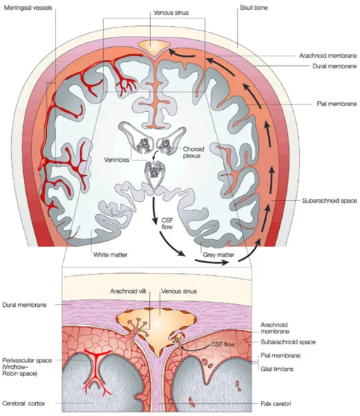
Anatomically the CNS is protected by the cerebrospinal fluid (CSF), produced by the secretory epithelium of the choroid plexus (CPE), and the meninges that surround the brain and
the spinal cord. The meninges comprised the innermost pial membrane that is intimately in contact with the parenchyma, the intermediate non-vascularized arachnoid membrane, and the outermost vascularized dural membrane, that is attached to the skull (Fig.1) [Louveau, 2015]. The meninges are an essential immunological site that make possible CNS immune surveillance [Raper, 2016].
Experiments in the mid 20th century gave rise to the concept of the CNS as immune-privileged
site, to which access of circulating immune cells is firmly controlled by an endothelial blood-brain barrier (BBB) and the epithelial blood-cerebrospinal fluid barrier (BCSFB) within the choroid plexus [Engelhardt and Ransohoff, 2012]. This property allows to protect post-mitotic neural cells from potential immune response-mediated injury and death. Immune privilege is based on multiple factors, including: 1) as mentioned above, the presence of the BBB; 2) a relative lack of classical lymphatic drainage of the parenchyma; 3) a poorness of professional antigen-presenting cells (APCs), such as dendritic cells (DCs); 4) low expression levels of MHC molecules; and 5) many anti-inflammatory soluble modulators [Harris, 2014].
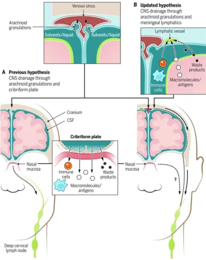
Under physiological conditions, there is a minimal entry of immune cells, mainly memory T cells, into the CNS through the choroid plexus for the purpose of immune surveillance. The CSF drains into the deep cervical lymph nodes via the cribriform plate into the lymphatic system of the nasal mucosa and then to the deep cervical lymph nodes [Louveau, 2015] (Fig.2A). Recent findings by Louveau and Aspelund have revealed the presence of a conventional and functional lymphatic system, which is placedin the dura matter and allows fluid, macromolecules and immune cells to drain from the CNS into the deep cervical lymph nodes (Fig.2B). The drainage in the deep cervical lymph nodes was dependent on the presence of meningeal lymphatic vessels in which immune cells (T cells, B cells, DCs) were present under physiological conditions, suggesting that the meningeal lymphatics play a role in the trafficking of immune cells out of the CNS in the stationary state [Louveau, 2015]. These data have dramatically altered classical viewpoint of immune privilege of CNS by revealing that the interactions between the CNS and
the immune system occur and are not limited to pathology, but also extend to homeostatic functions (Fig.2). Therefore, the CNS is still considered immunologically privileged site but our knowledge of the complex neuroimmune interactions occurring suggest that it is a mostly controlled system with an unique immunological environment.
Figure 1. CNS anatomy and the CSF circulation. The CNS is protected by the meninges (arachnoid, dural, and
pial membrane). The CSF is produced by the secretory epithelium of the choroid plexus that is placed in the ventricular system of the brain. CSF circulates from the ventricles to the subarachnoid space and is reabsorbed to the systemic circulation through the arachnoid villi that extend into the venous sinuses of the cerebral hemispheres [Louveau, 2015].
Figure 2. CNS drainage: New concepts for old.(A) Previous hypothesis suggested CSF drainage into deep
cervical lymph nodes via the cribriform plate. (B) New concept of CSF drainage into the deep cervical lymph nodes via meningeal lymphatics [Kipnis, 2016].
The triggering inflammatory event occurring in MS is not yet clear, thus generating contradictory hypotheses on the etiopathogenesis of the disease.
The most approved hypothesis for an inflammatory autoimmune pathogenesis of MS is the activation of CNS antigen-specific CD4+T cells in the periphery. This hypothesis origins mainly from animal models, in which disease can be induced by immunization with CNS-derived proteins and is largely driven by CNS-specific CD4+T cells [Sospedra and Martin, 2016]. This phenomenon occurs when peptide from pathogens share sequence or structural similarities with self-antigen. In this case an immune event outside the CNS starts the disease process in MS, in which autoreactive CD4+T cells are activated in the periphery by cross-reactivity of peptides derived from a foreign antigen, for example, during a viral infection. This concept, called
immunization of rabbits with a peptide from hepatitis B virus polymerase with similarity with myelin basic protein (MBP) can induce EAE, and that T cells with cross reactivity against the two peptides are implicated [Sospedra and Martin, 2016]. Cross reactivity of these T cells with non-self antigen can lead to activation, migration across BBB, CNS infiltration, and, if they recognize antigens expressed in the brain, tissue damage.
In any case, molecular mimicry alone may not be able to induce disease; in fact other evidences indicate that a focal inflammatory reaction at CNS level and triggered by a still unknown stimulus to a local injury could occur in MS patients [Martino, 2000]. In a typical MS brain, it is possible to observe lymphocytes and monocytes infiltrates within demyelinating areas, but also in normal white and grey matter as well as in meningeal spaces. The recruitment of leukocytes within the CNS induces the activation of the resident immune cells, such as astrocytes and microglia, determining a CNS-specific immune reaction [Martino, 2000].
Martino et al. proposed a theory, called the dual signal hypothesis, offering an integrated and more comprehensive view of the MS pathogenic process, in which two phenomena are merged [Martino, 2000]. They suggest that local CNS inflammatory process along with a concomitant, but possibly unrelated, systemic inflammatory event may trigger a CNS-specific autoimmune reaction cascade setting the stage for the MS pathogenesis.
To sum up, in this scenario, peripheral activated T cells cross the BBB and enter the CSF. They have access to the subarachnoid space where they can be restimulated by local myeloid APCs, particularly meningeal and perivascular macrophages. The re-activation of naive and effector T cells in the CNS induces cytokine and chemokine production that amplify the inflammatory response with activation of resident immune cells, upregulation of MHC class II molecules, the recruitment of monocytes, CD8+T cells, B cells from peripheral blood; and the formation of the inflammatory lesion [Sospedra and Martin, 2005]. Immune cells infiltrating the CNS can lead to neurodegeneration and tissue damage by secretion of different neurotoxic products, such as reactive oxygen species (ROS) [Sospedra and Martin, 2016]. During early stages of MS, new
lesions form frequently, in which the above mentioned adaptive immune mechanisms are the most important drivers. In contrast, during later phases of the disease, inflammation decreases, but the susceptibility of the target tissue for neurodegeneration increases [Sospedra and Martin,
2016]. All these events are summarizedin Fig.3.
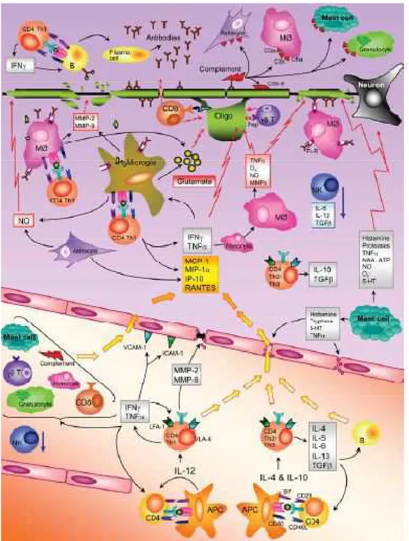
Figure 3. Immune mechanisms in the MS disease process. The figure summarizes the most important events in
Regarding the functional characteristics of autoimmune T cells in MS, T helper 1 cells, phenotype characterized by the production of interferon- (IFN- ) were thought originally to be the main pathogenic T cells in EAE and MS. It was postulated that during EAE, in the peripheral lymphoid organs, T precursor reactive T cells are induced to differentiate into myelin-reactive Th1cells when an antigen that crossreacts with a myelin antigen is presented to a T cell by an APC. Th1 cells that react with myelin antigens, such as myelin proteolipid protein (PLP), MBP and myelin oligodendrocyte glycoprotein (MOG), cross the BBB where the myelin antigen are represented to T cell by APCs in the brain (microglia cells), and an inflammatory cascade is triggered with the release of inflammatory mediators that cause damage to the myelin sheath and ultimately the underlying axon. This conclusion was based partly on the observation that IFN- was detected in MS lesions and in the CNS of EAE mice [Traugott and Lebon, 1988a; Traugott
and Lebon, 1988b].The concept of Th1 driven organ-specific autoimmunity was challenged
when Th17 cells central role in several EAE models was discovered. These cells produce IL-17 that was found highly expressed in tissue sites of many autoimmune diseases including MS lesions [Lock, 2002].
Although most evidence supports an important role of CNS antigen-specific CD4+T cells in sustaining MS, recent data suggest that also CNS antigen-specific CD8+T cells are involved in MS. These cells are activated in periphery by professional APCs presenting CNS-derived or cross-reactive peptides as previously described for CD4+T cells. CD8+T cells are detected in MS lesions and often clonally expanded, suggesting that these cells are involved in CNS tissue damage. Furthermore, because axons and neurons express MHC class I molecules, CD8+ T cells can recognize their cognate antigen on these cells and directly damage them [Sospedra and Martin, 2016]
Figure 4
subsets, producing different cytokines and mediating distinct effector functions
participate in the formation of increased levels of i
suggests
To date, the strongest evidence for B cells involvement in MS pathology arise from studies evaluating the effect and the efficacy of anti
ocrelizumab, and ofatumumab [ oligoclonal bands (OCBs) antibodies
structures, and a B cell
B cells, once inflammation has started, contribute to MS dise
Figure 4. Different T helper cell subsets.
subsets, producing different cytokines and mediating distinct effector functions B cells
participate in the formation of increased levels of i
suggests a role for B cells and antibodies in the pathology of MS [
To date, the strongest evidence for B cells involvement in MS pathology arise from studies evaluating the effect and the efficacy of anti
ocrelizumab, and ofatumumab [ oligoclonal bands (OCBs) antibodies in MS plaques and in structures, and a B cell
, once inflammation has started, contribute to MS dise
. Different T helper cell subsets.
subsets, producing different cytokines and mediating distinct effector functions B cells are APCs,
participate in the formation of
increased levels of immunoglobulins (Igs) in the CSF of MS patients is the earliest evidence a role for B cells and antibodies in the pathology of MS [
To date, the strongest evidence for B cells involvement in MS pathology arise from studies evaluating the effect and the efficacy of anti
ocrelizumab, and ofatumumab [ oligoclonal bands (OCBs) in the CSF,
in MS plaques and in structures, and a B cell-fostering milieu
, once inflammation has started,
contribute to MS disease pathogenesis with costimulation of autoreactive T cells, elevated
. Different T helper cell subsets. Upon activation, Th0 precursor cell differentiates into effector T cell
subsets, producing different cytokines and mediating distinct effector functions , implicated in
participate in the formation of ectopic lymphoid tissues (
mmunoglobulins (Igs) in the CSF of MS patients is the earliest evidence a role for B cells and antibodies in the pathology of MS [
To date, the strongest evidence for B cells involvement in MS pathology arise from studies evaluating the effect and the efficacy of anti
ocrelizumab, and ofatumumab [von Büdingen, 2015
in the CSF, detection of B cells, plasma cells in MS plaques and in the areas of demyelination,
fostering milieu [Sospedra and Martin, 2005 , once inflammation has started,
ase pathogenesis with costimulation of autoreactive T cells, elevated Upon activation, Th0 precursor cell differentiates into effector T cell subsets, producing different cytokines and mediating distinct effector functions
implicated in cytokine ectopic lymphoid tissues (
mmunoglobulins (Igs) in the CSF of MS patients is the earliest evidence a role for B cells and antibodies in the pathology of MS [
To date, the strongest evidence for B cells involvement in MS pathology arise from studies evaluating the effect and the efficacy of anti-CD20 B cell depleting therapy s
von Büdingen, 2015
detection of B cells, plasma cells areas of demyelination,
Sospedra and Martin, 2005
, once inflammation has started, can cross BBB and
ase pathogenesis with costimulation of autoreactive T cells, elevated Upon activation, Th0 precursor cell differentiates into effector T cell subsets, producing different cytokines and mediating distinct effector functions
cytokines and antibodies ectopic lymphoid tissues (Fig
mmunoglobulins (Igs) in the CSF of MS patients is the earliest evidence a role for B cells and antibodies in the pathology of MS [
To date, the strongest evidence for B cells involvement in MS pathology arise from studies CD20 B cell depleting therapy s
von Büdingen, 2015]. Others evidences are
detection of B cells, plasma cells
areas of demyelination, the presence of B cell follicle
Sospedra and Martin, 2005
can cross BBB and
ase pathogenesis with costimulation of autoreactive T cells, elevated Upon activation, Th0 precursor cell differentiates into effector T cell subsets, producing different cytokines and mediating distinct effector functions (image from Wikipedia)
and antibodies
Fig.5) [von Büdingen, 2015 mmunoglobulins (Igs) in the CSF of MS patients is the earliest evidence a role for B cells and antibodies in the pathology of MS [Sospedra and Martin, 2005 To date, the strongest evidence for B cells involvement in MS pathology arise from studies
CD20 B cell depleting therapy s Others evidences are detection of B cells, plasma cells
the presence of B cell follicle
Sospedra and Martin, 2005, Pröbstel, 2015
can cross BBB and enter the CNS.
ase pathogenesis with costimulation of autoreactive T cells, elevated Upon activation, Th0 precursor cell differentiates into effector T cell
(image from Wikipedia)
production, and they
von Büdingen, 2015
mmunoglobulins (Igs) in the CSF of MS patients is the earliest evidence
Sospedra and Martin, 2005
To date, the strongest evidence for B cells involvement in MS pathology arise from studies CD20 B cell depleting therapy such as rituximab, Others evidences are the presence of detection of B cells, plasma cells and myelin
the presence of B cell follicle
, Pröbstel, 2015].
enter the CNS. In loco, ase pathogenesis with costimulation of autoreactive T cells, elevated
Upon activation, Th0 precursor cell differentiates into effector T cell (image from Wikipedia).
production, and they
von Büdingen, 2015]. The
mmunoglobulins (Igs) in the CSF of MS patients is the earliest evidence that
Sospedra and Martin, 2005].
To date, the strongest evidence for B cells involvement in MS pathology arise from studies uch as rituximab, the presence of and myelin-specific the presence of B cell follicle-like
In loco, they ase pathogenesis with costimulation of autoreactive T cells, elevated Upon activation, Th0 precursor cell differentiates into effector T cell
production, and they The that ]. To date, the strongest evidence for B cells involvement in MS pathology arise from studies uch as rituximab, the presence of specific like
they ase pathogenesis with costimulation of autoreactive T cells, elevated
production ofthe Igs in the CSF, myelin-specific antibodies secretion [Sospedra and Martin,
2005,]. It is necessary to control B cell tolerance to reduce autoimmunity risk that can randomly
develop, nevertheless, peripheral B cell tolerance mechanisms seems to be impaired [von
Büdingen, 2015].
Figure5. B cell functions. Antigen presentation, antibody production, establishment of ectopic lymphoid follicles at
sites of inflammation and cytokine production are the most important functions relevant to MS pathogenesis [von
Büdingen, 2015].
As a matter of fact, myelin-reactive memory B cells can be found in the peripheral blood of MS patients where they may act as APCs. These cells, expressing high levels of CD20, could be depleted following treatment with anti-CD20 targeting monoclonal antibodies reducing MS severity [von Büdingen, 2015].
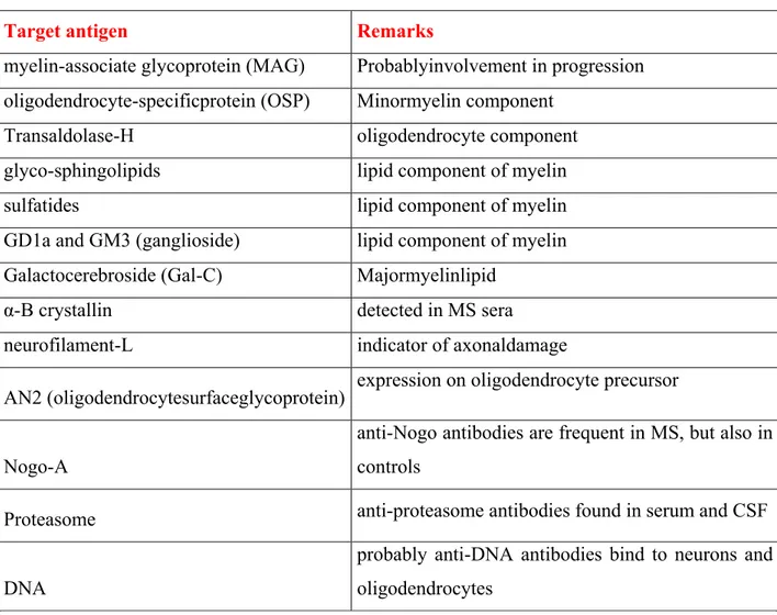
To date, it is difficult to identify the antigen specificity of CSF antibodies in MS, because OCBs could target both myelin proteins and foreign agents. However, known pathogenic nature of autoantibodies, the research for autoantigens has focused on myelin and other CNS components [Sospedra and Martin, 2005]. MOG appeared as a very promising candidate B cell autoantigen in MS. Numerous other potential targets for autoantibodies in MS have been described. In the
table below (Table 1), are summarized antibody specificities against CNS components other than MOG and MBP, adapted from Sospedra et al. [Sospedra and Martin, 2005].
Target antigen Remarks
myelin-associate glycoprotein (MAG) Probablyinvolvement in progression oligodendrocyte-specificprotein (OSP) Minormyelin component
Transaldolase-H oligodendrocyte component
glyco-sphingolipids lipid component of myelin
sulfatides lipid component of myelin
GD1a and GM3 (ganglioside) lipid component of myelin Galactocerebroside (Gal-C) Majormyelinlipid
α-B crystallin detected in MS sera
neurofilament-L indicator of axonaldamage
AN2 (oligodendrocytesurfaceglycoprotein) expression on oligodendrocyte precursor
Nogo-A
anti-Nogo antibodies are frequent in MS, but also in controls
Proteasome anti-proteasome antibodies found in serum and CSF
DNA
probably anti-DNA antibodies bind to neurons and oligodendrocytes
Table 1. Antibody specifities against CNS components. Adapted from [Sospedra and Martin, 2005].
To date, the most prominent are those directed against the potassium channel KIR4.1 and antibodies against neurofascin and contactin-2 [Sospedra and Martin, 2016].
In parallel with the activation of damaging inflammatory events, in MS patients there is wide evidence that regulatory mechanisms of inflammation are activated in order to restrain tissue damage and initiate regeneration. These mechanisms represented an intrinsic capability of the immune system to limit its overactivation. Regulatory T-cell (Tregs) populations have been described as the main cell population in controlling pro-inflammatory events. Tregs are generally defined as being CD4+ and CD25high and characterized by their transcription factor, Foxp3.
Their function is to inhibit effector T cells both by direct cell–cell contact and by secreting inhibitory cytokines, such as TGF-ß, IL-10, and IL-35 [Naegele and Martin, 2014]. However, in MS, a reduced number and activity of CD4+CD25hi Tregs are observed [Sospedra and Martin,
2016]. Apart from Tregs, other immune cells may also be involved in neuroprotection and lesion
resolution via relative production of Th2/Th3 cytokines, such as IL-10 and TGF- , and secretion of growth factors [Sospedra and Martin, 2016]. However, it should not be forgotten that resident inhibitory mechanisms of the immunoprivilegied CNS also contribute to the repression of inflammatory events. As a matter of fact, oligodendrocyte precursors that are still present in the adult CNS are also activated, and surviving oligodendrocytes begin to regenerate myelin sheaths, although they are structurally abnormal [Sospedra and Martin, 2005]. Despite this, a large number of lesions exhibit no remyelination.
Based on studies of MS pathology, possible explanations for differences in remyelination potential include disease heterogeneity or differences in disease chronicity. Lucchinetti et al have identified four pathologic MS subtypes on the basis of the relative contribution of different immune cells, antibody and complement deposition, myelin loss, and oligodendrocyte death [Lucchinetti, 2000]. The following pathologic subtypes are described: Pattern I. In this pattern, inflammation is dominated by T cells and macrophages, and the demyelinating process is induced mainly by TNF-α, IFN- , and ROS.
Pattern II. This pattern is very similar to pattern I, but in this case antibody and complement deposition predominate, and both MOG- and MBP-specific antibodies are involved.
Pattern III. Lesions present inflammatory infiltrates, composed mainly by T cells, macrophages and activated microglia. Igs and complement deposition are absent. Plaques are characterized by loss of oligodendrocytes without remyelination process.
Pattern IV. This pattern occurs primarily in PP-MS and show similarities to the classical pattern I and II lesions. Demyelination was associated with oligodendrocytes death [Lucchinetti, 2000].
Clinical course of Multiple Sclerosis
In 1996, the US National Multiple Sclerosis Society (NMSS) Advisory Committee on Clinical Trials in MS defined the clinical subtypes of MS [Lublin and Reingold, 1996]. The characterizations were rapidly adopted into practice and served to better communicate a patient’s clinical course and to define clinical trial populations. At the time, no biomarkers or magnetic resonance imaging (MRI) signals were available to distinguish between the different MS clinical courses.
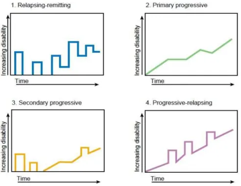
The Committee provided standardized definitions for 4 MS clinical courses: RR, SP, PP, and progressive relapsing (PR) (Fig.6). RRMSis the most common clinical form. Approximately 85% of patients present this form of pathology [Sospedra and Martin, 2005], which is characterized by acute episodes of disease (called relapses) followed by partial or complete recovery. Periods between disease relapses are characterized by lack of disease progression [Lublin and Reingold, 1996]. RRMS onset typically occurs in early adulthood, and, within around two decades, approximately half will go on to develop SPMS. SPMS is defined as a clinical form with initial RR course, followed by progression, with or without relapses [Lublin
and Reingold, 1996]. PPMS affects only 10–15% of the MS population [Sospedra and Martin, 2005] and is associated with a rapid disease progression from onset without remission or with
occasional plateaus and temporary minor improvements [Lublin and Reingold, 1996]. PRMS shows progression at the beginning of the disease with clear acute relapses with or without full recovery; and periods between relapses are characterized by sustaining progression.
Figura 6. Clinical course of MS.
In 2013, the International Advisory Committee on Clinical Trials of MS has introduced a revision with new insights and clarifications in order to better define MS disease phenotypes, in the light of the more recently identified clinical aspects of the disease.
First, it was introduced Clinically Isolated Syndrome (CIS) as a distinct MS phenotype. CIS was recognized as the first clinical presentation of a disease that shows characteristics of inflammatory demyelination that could be MS, but has yet to fulfill criteria of dissemination in time. Second, a more complicated phenotype was descripted as Radiologically Isolated Syndrome (RIS), in which inflammatory demyelination areas are observed in the absence of clinical signs or symptoms. RIS is not considered to be part of MS subtypes as patients lack clinical evidence of demyelinating disease (a current criterion for MS diagnosis) and MRI findings alone are insufficient to establish a diagnosis of MS [Lublin, 2014].
Actually, MS patients may be described as having 1) relapsing MS that is active or not active, with or without worsening; or 2) primary or secondary progressive disease that is active or not active, with or without progression. [Lublin, 2014] (Fig.7).
Disease activity can be assessed at least annually by clinical and brain imaging criteria, whereas disease course can be determined by clinical evidence of progression.
Clinical evaluation is usually made referring to the Expanded Disability Status Scale (EDSS), which is a method of quantifying disability in MS and monitoring changes in the level of disability over time. The EDSS rating scale ranges in half-point increments from 0 (normal neurologic examination) to 10 (death owing to MS).
As regarding diagnosis, the original diagnostic criteria for MS were previously based on clinical features of demyelination alone. Over time, several criteria have been developed and replaced. McDonald criteria for MS are usually used for MS diagnosis. Diagnostic criteria based on analysis of dissemination of lesions in space (DIS) and time (DIT) with other clinical and paraclinical evaluations in order to exclude alternative diagnosis. The McDonald criteria have been resulted very specific in earlier diagnosis of MS, allowing an earlier treatment [Polman,
Multiple Sclerosis Therapies
Treatment of MS is difficult, because the disease process itself is very complex. Much has changed in the management over the last 20 years, and it will likely continue to evolve in the near future. Currently the therapies are not able to determine a regression or an arrest of the disease, but they can modify the course of the disease in the long term evolution, reducing the accumulation of disability.While relapse treatment has not changed, there are now 10 drugs that have been approved for the various form of MS from the first clinical manifestation (CIS) over RRMS to SPMS, and, for the first time also for PPMS. The success of these several and varied treatments with different mechanism of actions (MoAs) has not only positive for people living with MS, but also enhanced our knowledge about immune mechanisms of MS.
The drugs are generally divided into two groups: first and second-line drugs. The first line drugs represent the initial therapeutic approach for MS patients, where the clinical benefits overcome the possible side effects. If patients do not respond to first-line treatment or in the case of a very active disease with a high risk of progression to disability, second-line therapy is applied.
Interferon-β. Interferon- (IFN- ) is a first line therapy for which we have clinical experience over two decades. There are a number of potential MoAs by which IFN- may inhibit several aspects of the pathophysiology of MS. They include stabilization of the BBB by blocking matrix metalloproteases, reduced activation of T lymphocytes via the downregulation of HLA class II molecules and antigen presentation on glial cells and B cells, and Th1-Th2 shift. Whether IFN- antiviral activity is relevant for the treatment of MS is not yet clear, but it was observed that therapy does not compromise immune system with respect to protection against viral, bacterial, or fungal infections. Major side effects include flu-like symptoms, as well as depression, local skin reactions, allergic reactions, and liver abnormalities [Martin, 2016; Cross and Naismith,
2014].
Glatiramer Acetate. Glatiramer acetate (GA), another first line drugs, is composed of a synthetic four amino acid copolymer developed in order to simulate MBP. GA was first
approved in 1996 after a complex clinical development. GA needs to be injected subcutaneously, inhibits relapse rates by approximately 30%, and is very well tolerated [Johnson, 1995]. Among immunomodulatory effects of GA, it affects antigen presentation, shifts the Th1 response in Th2 response, induces regulatory CD8+T cells, and induces the production of neurotrophic factors. Which of these GA immunomodulatory effects are most important in reducing the disease activity in MS is difficult to assess, but it is very well tolerated without any impairment of protective immune responses [Martin, 2016].
Teriflunomide. Teriflunomide (TF) is the active metabolite of leflunomide, which has been approved in the USA since 1998 to treat rheumatoid arthritis (RA). TF was approved in 2012 as first-line oral drug for treating RRMS. TF reduces the relapse rates similar to IFN- and GA treatments. The main MoA is the inhibition of dihydro-orotate-synthetase, a key enzyme in the
de novo pyrimidine synthesis. As a result TF blocks high levels of activated T lymphocytes
proliferation. Side effects include abnormal liver function tests, hair loss, gastrointestinal issues, and immunosuppression [Martin, 2016].
Dimethylfumarate. DMF (also known as BG-12) and other derivatives of fumaric acid ester compounds have been used initially to treat psoriasis prior to discover its efficacy in MS [Gold,
2012]. DMF was approved as a first-line oral treatment for RR-MS in 2013. DMF is an oral
prodrug that is converted into monomethylfumarate and fumarate inside cells. DMF has been shown to induce Th1-Th2 shift by modulating DC function [Martin, 2016]; to enhance Nrf2 activity, a transcription factor that controls the expression of a wide range of antioxidant proteins [di Nuzzo, 2014]. Further activities include the induction of regulatory B-cell subsets [Martin,
2016] and inhibition of NFkB-induced genes. Its efficacy in MS is moderate with some
important side effects such as gastrointestinal problems, flush; in some patients profound and long-lasting lymphopenia was observed. So far, no specific immunocompromise with respect to viral or bacterial infections has been reported, but a few progressive multifocal leukoencephalopathy (PML) cases have been observed [Martin, 2016].
Natalizumab. Natalizumab (NTZ) was the first monoclonal antibody (mAb) approved for the treatment of RR-MS. NTZ targets the α-4 chain of α4 1 integrin (very-late-antigen-4, VLA-4 ) that also binds to α4 7 integrin. VLA-4 receptoris expressed on all leucocytes except neutrophils [Lutterotti and Martin, 2008]. NTZ is an antagonist of VLA-4 that blocks it from binding to vascular cell adhesion molecule (VCAM), which is upregulated on the endothelium during inflammation. The rationale behind blocking of VLA-4 is to prevent inflammatory cell migration from the vasculature and within the CNS. Furthermore, inhibition of VLA-4/VCAM-1 interactions by NTZ also induces the release of certain B cell maturation precursors from bone marrow and lymphoid organs into the peripheral blood [Martin, 2016]. NTZ is efficacious in blocking relapse rates to a much higher extent than IFN- and GA [Miller, 2003]. NTZ is very well tolerated except for one adverse event: PML, a progressive demyelinating disease of the CNS that is caused by the infection of oligodendrocytes with the polyomavirus (JCV). Therefore, the patient must be monitored for the JC virus before starting therapy and can only be used with a restricted indication in the case of very active or breakthrough relapsing MS.
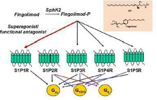
The Sphingosin-1 Phosphate Receptor Agonist Fingolimod. Fingolimod (FTY720), a derivative of the fungal antibiotic myriocin, is a functional antagonist of four of the five sphingosin-1 phosphate receptors (S1PRs), which are expressed by many cells including immune cells [Martin, 2016]. FTY720 was the first oral drug approved for the treatment of RRMS in 2011 as second line treatment. Fingolimod is an analog of sphingosine, which is a component of the lipid bilayer of the plasma membrane, and its phosphorylated metabolite S1P. Both sphingosine and fingolimodare phosphorylated inside the cells into their active forms, which bind to specific S1P receptors [Mehling, 2011]. In humans, 5 subtypes of S1PRs have been described. FTY720 binds to S1PRs and leads to their internalization and degradation, except for S1P2R (Fig.8). The main action of Fingolimod is to control the trafficking of lymphocytes between lymphoid organs and blood, in order to block the egress of lymphocytes from lymph nodes [di Nuzzo, 2014]. The result is a relative lymphopenia and change in composition of
peripheral lymphocytes, predominantly in naive and memory T cells [Mehling, 2011]. This effect of FTY720 is considered the main MoA in MS; however, effectson immune cells and CNS including the BBB have been observed [Martin, 2016].
Figure 8. Structure of Fingolimod and its mechanism of action. Fingolimod phosphate activates S1P1R, S1P3R,
S1P4R, and S1P5R, but not S1P2R [di Nuzzo, 2014].
Anti-CD52 (Alemtuzumab). Alemtuzumab is a humanized IgG1 mAb approved for RRMS treatment in 2014. It targets CD52, a cell-surface glycoprotein abundantly expressed on T and B cells, natural killer (NK) cells, monocytes and some subsets of DCs. Alemtuzumab lyses cells expressing CD52 by antibody-dependent cellular cytolysis, thereby rapidly producing a profound leucopenia [Lutterotti and Martin, 2008]. The physiological function of the CD52 molecule is still not known, but it has an elevated efficacy in MS with only few therapy cycles needed. Alemtuzumab significantly reduces clinical and MRI disease activity. However, some patients require additional treatment courses, and surprisingly, alemtuzumab is associated with a considerable risk of secondary autoimmune diseases [Martin, 2016].
Anti-CD20 (Rituximab, Ocrelizumab, Ofatumumab). The clinical success of B-cell targeting therapies with anti-CD20 antibodies have provided experimental evidence for an involvement of
B cells in MS pathogenesis. Rituximab, ocrelizumab, and ofatumumab are different anti-CD20 depleting agents.
Rituximab, a chimeric mouse-human anti-CD20 mAb, was first approached with the idea that B cells and their autoantibodies contribute to MS pathogenesis [Hauser, 2008]. Rituximab had been developed and approved for treating B-cell malignancies, such as non-Hodgkin’s lymphoma, and RA. CD20 is exclusively expressed on all B-cell in different stages of maturation; however, plasma cells and stem cells do not express CD20 on their surface. Thus, Rituximab selectively depletes CD20-expressing B cells, preserving the capacity for B-cell reconstitution and antibody production and secretion, via complement-mediated lysis [Martin,
2016, Lutterotti and Martin, 2008]. The most prominent adverse effects were related to the
infusion.
Ocrelizumab is a fully humanized mAb that deplets CD20+ B cells via cytolysis mechanism in a non-complement manner. Ocrelizumab appears to reduce relapse and disability in MS but quality of evidence is moderate [Filippini, 2017]. Ocrelizumabis generally well tolerated, with the most common side events associated to the infusion reactions and infections. Sometimes, in ocrelizumab MS trials the risk of malignancies was observed [Gelfand, 2017].
Ofatumumab is a human mAb that also targets CD20 molecule binding to a different epitope than rituximab (and ocrelizumab) resulting in a pronounced complement-mediated cytotoxicity
in vitro. It is currently approved for the treatment of chronic lymphatic leukemia [Bittner, 2017].
Anti-CD25 (Daclizumab). Daclizumab is a humanized mAb against CD25, the alpha chain of the IL-2 receptor (IL2Rα). It was originally approved for the prevention of allograft rejection of kidney in order to block the expansion of alloreactive T cells [Vincenti, 1998]. As a matter of fact, the IL2Rα is poorly expressed on resting T cells but is upregulated on activated or abnormal T cells [Lutterotti and Martin, 2008]. Therefore, the rationale for the use of anti-CD25 therapy in autoimmune disease is to inhibit autoreactive T cells proliferation. Despite the knowledge that daclizumab is a mAb with a singular target, its MoA in MS is not fully elucidated. Effectiveness
in MS appears to be related to the ability of daclizumab in expansion of a subset of NK cells that express the NK cell marker CD56 at high levels (CD56brightNK cells). CD56brightNK cells have
immunoregulatory effects in addition to their antiviral and antitumor properties [Lutterotti and
Martin, 2008]. The expansion of CD56brightNK cells strongly correlates with the reduction of
inflammatory lesions in the brain in multiple phases II and III studies [Martin, 2016,]. Several other interesting MoAS have been described for daclizumab, including inhibition of T cell activation via blocking CD25 on DCs [Wuest, 2011].
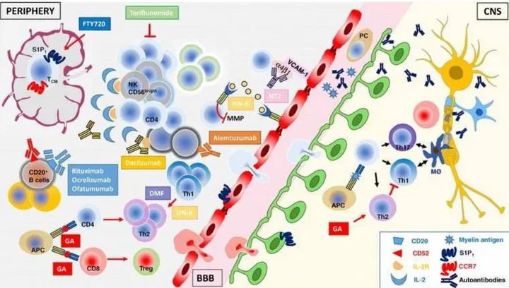
Fig.9 Summarizes the putative cellular and molecular therapeutic targets used in the MS treatment [Martin, 2016].
Figure 9. Schematic representation of the putative cellular and molecular therapeutic targets used in the MS treatment. On the left, cellular and molecular targets in the periphery; in the center, the endothelial BBB; on the
right, inflamed CNS parenchyma with a neuron (yellow) and its myelinated axon and the oligodendrocyte (blue) is shown [Martin, 2016].
EXTRACELLULAR VESICLES
Cell-to-cell communication is an essential hallmark of multicellular organisms required to assure proper coordination among several cell types. Communication can be mediated through
direct cell-cell contact or transfer of secreted molecules. Recent studies have suggested that cells may also communicate by the release of membrane vesicles [Camussi, 2010]. For a long time, microvesicles (MVs) were considered to be an in vitro artefact or inert cellular debris until De Broe et al. [De Broe, 1977] suggested that MVs released from human cells result from a specific process. Because of their small size, upon release, EVs can move from the site of discharge by diffusion and be retrieved in several biological fluids, for example, CSF, blood, urine .
Two classes of membrane vesicles have been described on the basis of size, content and mechanism of formation [Morel, 2011; Bartneva, 2013]: exosomes (EXOs) and, shed vesicles (MVs).
EXOs are a population of small membrane vesicles (30-120 nm in diameter) released by an endocytic pathway (Fig. 10) [Camussi, 2010]. EXOs formed by a series of processes involving the endosomal sorting complex required for transport (ESCRT) and multivesicular bodies (MVBs) [Février and Raposo, 2004; Schneider and Simons, 2013]. The first step is the invagination of the plasma membrane forming a vacuole. Once that process is completed, the ESCRT facilitates the development of the vacuoles into early endosomes. The early endosomes can fuse with endocytic vesicles, where they accumulate and mature into MVBs. These MVBs can either fuse with the lysosome if the content is destined for degradation or they can fuse with the plasma membrane releasing EXOs into the extracellular space [van der Pol, 2010; Frühbeis,
2012; Abels and Breakefield, 2016]. EXOs are enriched in several proteins and RNAs, such as
heat shock proteins, integrins and tetraspanins (CD63, CD9, CD81, CD82) [Frühbeis, 2012,
Théry, 2002; Chivet, 2012] . They are also characterized by the presence of high levels of
cholesterol, sphingolipids, ceramide and glycerophospolipids in their membrane [Simons and
Figure 10
MVs, also
vesicles that are more heterogeneous in size (100nm [Cocucci, 2009
extracellular activation
MVs appear to be formed though the outward budding and fission of the plasma membrane following a cytoskeletal reorganization
composition,
nucleic acids (mRNA, microRNA, DNA) the differential
the state (e.g. resting, stimulated) of [Bernimoulin, 2009
levels of phosphatidylserine (PS) on their surface A specialized type of MV
which release ion channel and microglia [
1979; Visentin and Levi, 1997
10. Generation of EXOs and MVs (
, also known as
vesicles that are more heterogeneous in size (100nm
Cocucci, 2009]. They bud directly from the plasma membrane and are released into the
extracellular space activation (Fig. 10
appear to be formed though the outward budding and fission of the plasma membrane following a cytoskeletal reorganization
composition, MVs contain a variety of cell surface receptors, intracellular signalling proteins and nucleic acids (mRNA, microRNA, DNA)
the differential composition
the state (e.g. resting, stimulated) of
Bernimoulin, 2009
levels of phosphatidylserine (PS) on their surface A specialized type of MV
release MVs from the cell surface when exposed to ATP. P2X7 receptor is ion channel, highly expressed in immune cells,
icroglia [Caragnano, 2012; Steinberg and Silverstein, 1987; Cockcroft and Gomperts,
1979; Visentin and Levi, 1997
Generation of EXOs and MVs (
known as shed vesicles or ectosomes vesicles that are more heterogeneous in size (100nm
. They bud directly from the plasma membrane and are released into the in physiological conditions, but their numb
10). Several mechanisms are responsible for the shedding of MVs. In general, appear to be formed though the outward budding and fission of the plasma membrane following a cytoskeletal reorganization
MVs contain a variety of cell surface receptors, intracellular signalling proteins and nucleic acids (mRNA, microRNA, DNA)
composition of proteins of various donor cells the state (e.g. resting, stimulated) of
Bernimoulin, 2009]. However, shed vesicles are generall
levels of phosphatidylserine (PS) on their surface A specialized type of MV
MVs from the cell surface when exposed to ATP. P2X7 receptor is y expressed in immune cells,
Caragnano, 2012; Steinberg and Silverstein, 1987; Cockcroft and Gomperts, 1979; Visentin and Levi, 1997
Generation of EXOs and MVs (adapted from
shed vesicles or ectosomes vesicles that are more heterogeneous in size (100nm
. They bud directly from the plasma membrane and are released into the in physiological conditions, but their numb
Several mechanisms are responsible for the shedding of MVs. In general, appear to be formed though the outward budding and fission of the plasma membrane following a cytoskeletal reorganization and plasma remodeling [
MVs contain a variety of cell surface receptors, intracellular signalling proteins and nucleic acids (mRNA, microRNA, DNA)
of proteins of various donor cells the state (e.g. resting, stimulated) of the parental
. However, shed vesicles are generall levels of phosphatidylserine (PS) on their surface
A specialized type of MVs release exists for cells that express the ATP receptor P2X7, MVs from the cell surface when exposed to ATP. P2X7 receptor is
y expressed in immune cells,
Caragnano, 2012; Steinberg and Silverstein, 1987; Cockcroft and Gomperts, 1979; Visentin and Levi, 1997]. It can act as a selective
adapted from Cocucci
shed vesicles or ectosomes [ vesicles that are more heterogeneous in size (100nm
. They bud directly from the plasma membrane and are released into the in physiological conditions, but their numb
Several mechanisms are responsible for the shedding of MVs. In general, appear to be formed though the outward budding and fission of the plasma membrane
and plasma remodeling [
MVs contain a variety of cell surface receptors, intracellular signalling proteins and nucleic acids (mRNA, microRNA, DNA) derived from the cell of origin.
of proteins of various donor cells the parental cells and on the . However, shed vesicles are generall
levels of phosphatidylserine (PS) on their surface.
release exists for cells that express the ATP receptor P2X7, MVs from the cell surface when exposed to ATP. P2X7 receptor is
y expressed in immune cells, particular
Caragnano, 2012; Steinberg and Silverstein, 1987; Cockcroft and Gomperts,
]. It can act as a selective
Cocucci et al., 2009
[Sadallah, 2011
vesicles that are more heterogeneous in size (100nm-1μm) and shape as compared to EXOs . They bud directly from the plasma membrane and are released into the
in physiological conditions, but their number is often increased upon cellular Several mechanisms are responsible for the shedding of MVs. In general, appear to be formed though the outward budding and fission of the plasma membrane
and plasma remodeling [
MVs contain a variety of cell surface receptors, intracellular signalling proteins and derived from the cell of origin.
of proteins of various donor cells, but their content depend also on cells and on the
. However, shed vesicles are generally characterized by the
release exists for cells that express the ATP receptor P2X7, MVs from the cell surface when exposed to ATP. P2X7 receptor is
particularly monocytes/
Caragnano, 2012; Steinberg and Silverstein, 1987; Cockcroft and Gomperts,
]. It can act as a selective ion channel or as a nonselective pore
2009) [Sadallah, 2011
Sadallah, 2011], are a
m) and shape as compared to EXOs . They bud directly from the plasma membrane and are released into the
er is often increased upon cellular Several mechanisms are responsible for the shedding of MVs. In general, appear to be formed though the outward budding and fission of the plasma membrane
and plasma remodeling [Schindler, 2014
MVs contain a variety of cell surface receptors, intracellular signalling proteins and derived from the cell of origin. Therefore,
, but their content depend also on cells and on the stimulus employed for
y characterized by the
release exists for cells that express the ATP receptor P2X7, MVs from the cell surface when exposed to ATP. P2X7 receptor is
monocytes/macrophages,
Caragnano, 2012; Steinberg and Silverstein, 1987; Cockcroft and Gomperts,
ion channel or as a nonselective pore
Sadallah, 2011].
, are a population of m) and shape as compared to EXOs . They bud directly from the plasma membrane and are released into the
er is often increased upon cellular Several mechanisms are responsible for the shedding of MVs. In general, appear to be formed though the outward budding and fission of the plasma membrane
Schindler, 2014]. In terms of
MVs contain a variety of cell surface receptors, intracellular signalling proteins and Therefore, MVs , but their content depend also on
employed for
y characterized by the presence of high
release exists for cells that express the ATP receptor P2X7, MVs from the cell surface when exposed to ATP. P2X7 receptor is an ATP
macrophages,
Caragnano, 2012; Steinberg and Silverstein, 1987; Cockcroft and Gomperts,
ion channel or as a nonselective pore population of small m) and shape as compared to EXOs . They bud directly from the plasma membrane and are released into the
er is often increased upon cellular Several mechanisms are responsible for the shedding of MVs. In general, appear to be formed though the outward budding and fission of the plasma membrane
. In terms of MVs contain a variety of cell surface receptors, intracellular signalling proteins and MVs reflect , but their content depend also on
employed for shedding presence of high
release exists for cells that express the ATP receptor P2X7, an ATP-gated macrophages, mastcells
Caragnano, 2012; Steinberg and Silverstein, 1987; Cockcroft and Gomperts,
ion channel or as a nonselective pore small m) and shape as compared to EXOs . They bud directly from the plasma membrane and are released into the er is often increased upon cellular Several mechanisms are responsible for the shedding of MVs. In general, appear to be formed though the outward budding and fission of the plasma membrane . In terms of MVs contain a variety of cell surface receptors, intracellular signalling proteins and reflect , but their content depend also on shedding presence of high
release exists for cells that express the ATP receptor P2X7, gated cells
Caragnano, 2012; Steinberg and Silverstein, 1987; Cockcroft and Gomperts,
depending on the ATP concentration. Activation of P2X7 receptor can induce efficient inflammosome assembly and rapid secretion of the inflammatory cytokines IL-18 and IL-1 [Qu, 2007]. P2X7 receptor differs from other members of the P2X family in its relatively low affinity for ATP and the presence of a long cytoplasmic C-terminus that contains several protein–protein interaction motifs. The activation of P2X7R induces several intra-signalling events, with the involvement of protein kinases and other effector enzymes [Duan and Neary,
2006]. In particular, it was known that P2X7-dependent blebbing is preceded by loss of plasma
membrane asymmetry and exposure of PS at the outer leaflet of the plasma membrane, commonly accepted as a marker of a cell undergoing apoptosis. Moreover, many studies have shown that blebbing induced byP2X7 receptor requires P38 MAP kinase and ROCK activation, with following disassembly of the cytoskeletal elements, similarly to apoptotic blebbing [Turola,
2012]. However, it has been showed by Surprenant and colleagues that P2X7-dependent
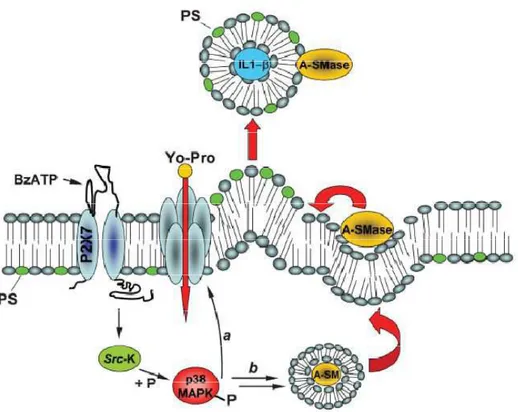
blebbing occurs within few minutes after receptor activation and may be a reversible event [MacKenzie, 2001]. Therefore, ATP stimulation time determines cell fate. In the model proposed by Surprenant, upon P2X7R-mediated macrophage activation, IL-1 is packaged into small plasma membrane blebs that are released into the extracellular space as MVs [MacKenzie, 2001]. These results contributed to the first evidence that P2X7R may act as a key player in IL-1 release and may represent a general mechanism for protein secretion from P2X7-expressing myeloid cells. Bianco et al. [Bianco, 2009] found that MV shedding from microglia is controlled by acid sphigomyelinase (A-SMase), a key enzyme that controls hydrolysis of sphigomyelin to ceramide. Following P2X7 receptor activation, p38 MAP kinase is phosphorilated; this in turn induces the translocation of A-SMase to plasma membrane outer leaflet, generating ceramide, and thereby inducing budding of MVs (Fig.11) [Bianco, 2009].
Figure 11. Model for P2X7 receptor-induced signaling pathway involved in MV shedding proposed by Bianco et al. [Bianco, 2009]. Stimulation of P2X7 receptor with BzATP, a synthetic agonist of ATP, leads to Src-k
phosphorylation that in turn activates p38 MAP kinase. This process induces A-SMase translocation from luminal lysosomal compartment to plasma membrane outer leaflet, altering membrane fluidity with IL-1 -MVs release. Based on the key role of A-SMase in MVs formation, it is possible to inhibit P2X7-induced release using pharmacological or genetic tools for the inactivation of the enzyme. Bianco et colleagues have showed that both approaches strongly abolished MV shedding and of IL-1 release from reactive glial cells [Bianco, 2009].
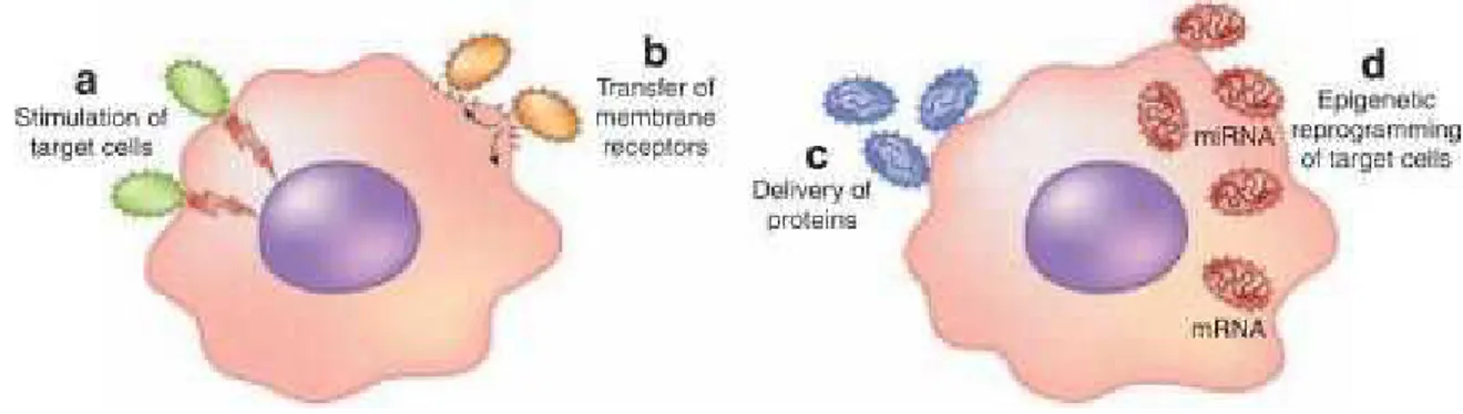
EVs from different cell types reflect composition and activation status of parental cells, thus interacting with target cells through specific receptors and leading to several responses in the target cells. EVs may act in multiple ways (Fig.12), for example, as a “signaling complex” by direct stimulation of target cells, a transfer of membrane receptors, a delivery system of proteins or genetic materials [Camussi, 2010].
Figure 12. EVs biological activities. Schematic representation of activities induced by EVs in the target cells
[Camussi, 2010].
It is now recognized that EVs are an integral part of the intercellular communication and the role of EVs has been reported inseveral physiological and pathological process such as cell proliferation, coagulation, vascular function, apoptosis, inflammation and tumor progression.The most well characterized membrane vesicles are those released from blood cells, i.e. platelets, leukocytes, erythrocytes, and endothelial cells. However, accumulating evidences demonstrate that MVs and EXOs can also be released by brain cells and play an important function in the CNS both in pathologic or physiological conditions. A number of studies have demonstrated the involvement of EVs in different scenarios, such as in neuronal development, synaptic activity; and nerve regeneration [Schindler, 2014]. It has been observed that EVs are released by neural cells, oligodendrocytes, neurons, microglia, astrocytes in the brain, and Schwann cells in the peripheral nervous system [Sáenz-Cuesta, 2014b].
Several studies demonstrate that EVs play an active role during pathogenesis of MS and EAE. Today researchers attention has been focused on the study of EVs as potential biomarkers and therapeutic targets that could have a possible involvement in MS pathogenesis. Increased numbers of MVs have been observed in the blood and in the CSF of MS patients as compared to healthy controls (HCs) and a role for EVs has been proposed in inflammatory progression and lesion repair [Sáenz-Cuesta, 2014b]. Endothelial cells, in addition to platelets, leukocytes, myeloid cells, and astrocytes, shed EVs containing metalloproteases and caspase 1 that promote BBB disruption and lymphocytes and myeloid cell transmigration into CNS [Carandini, 2015].
![Figure 7. The 1996 vs 2013 MS phenotype descriprions for relapsing and progressive disease [Lublin, 2014]](https://thumb-eu.123doks.com/thumbv2/123dokorg/5408571.58501/25.892.127.768.91.1093/figure-ms-phenotype-descriprions-relapsing-progressive-disease-lublin.webp)