UNIVERSITÀ DEGLI STUDI DI VERONA
DEPARTMENT OF MEDICINE SECTION OF GENERAL PATHOLOGY PhD SCHOOL OF LIFE AND HEALTH SCIENCES
DOCTORAL PROGRAM IN
INFLAMMATION, IMMUNITY AND CANCER
CYCLE XXX/2014
MOLECULAR MECHANISMS REGULATING
CYTOKINE PRODUCTION BY HUMAN
NEUTROPHILS
S.S.D. MED/04
Coordinator: Prof. GABRIELA CONSTANTIN
Tutors: Prof. MARCO A. CASSATELLA
Dr. NICOLA TAMASSIA
PhD Student: FABIO ARRUDA E SILVA
3
Sommario
I neutrofili, i leucociti più abbondanti nel sangue umano, sono noti per svolgere funzioni effettrici cruciali per la risposta immunitaria innata e adattativa contro le infezioni. Inoltre, i neutrofili sono in grado di rispondere a diverse componenti di origine microbica inducendo la sintesi e la secrezione di svariate citochine. In questo contesto, l'obiettivo principale di questo studio è stato quello di verificare la capacità dei neutrofili umani di esprimere e produrre citochine della famiglia di IL-17, tra cui IL-17A, IL-1B, IL-17F e IL-17AF, dato che attualmente la letteratura è in disaccordo su questo argomento. Attraverso metodiche quali RT-qPCR, immunoistochimica (IHC), immunoblotting, misurazione di proteine mediante ELISA, immunoprecipitazione della cromatina (ChIP) e ChIP-seq, abbiamo valutato la regolazione epigenetica e trascrizionale, così come la produzione di citochine utilizzando popolazioni di neutrofili isolati ad un elevato grado di purezza (> 99,7%). In accordo con alcuni dati precedentemente pubblicati sia dal nostro che da altri gruppi, abbiamo osservato che i neutrofili incubati con stimoli di diversa natura non esprimono ne producono, a livello di mRNA e proteina, nessuna citochina della famiglia di IL-17, comprese IL-17A, IL-17F, IL-17B o IL-17AF. Risultati analoghi sono stati ottenuti anche utilizzando neutrofili isolati da pazienti con psoriasi attiva. Inoltre, contrariamente a quanto osservato in studi pubblicati recentemente, anche in neutrofili incubati con concentrazioni molto elevate di IL-6 e IL-23, in combinazione con ife o conidi inattivati ottenuti da Aspergillus fumigatus, non è stata misurata nessuna espressione di mRNA o sintesi di proteina per IL-17A o IL-17F. In accordo con questi dati, sul locus genomico di 17A/F, in neutrofili stimolati o meno con 6 più IL-23, non è stata rilevata alcuna presenza di H3K27Ac e H3K4me1, due modificazioni istoniche post-traduzionali che contrassegnano elementi regolatori genomici attivi o "poised". Tale risultato è quindi coerente con l'incapacità dei neutrofili umani di esprimere l'mRNA di IL-17A o IL-17F. Un altro dato importante emerso in questo studio è stato la conferma che in immunoistochimica, anticorpi anti-IL-17A e IL-17B utilizzati in svariati lavori, danno un segnale positivo in citocentrifugati di neutrofili stimolati o meno con IL-6 più IL-23. In immunoblotting però gli stessi anticorpi non riconoscono proteine intracellulari del peso molecolare corretto per IL-17A e IL-17B ma altre proteine con peso molecolare molto più alto. Si può concludere quindi che il
4 segnale positivo dato da questi anticorpi in IHC è frutto di una reazione aspecifica ed è indipendente dalla presenza di IL-17A o IL-17B. In conclusione, i risultati esposti in questo studio non solo confermano e ampliano nostre osservazioni precedenti a riguardo dell’incapacità dei neutrofili umani di produrre IL-17A, IL-17B e IL-17F, ma forniscono anche una spiegazione del motivo per cui in altre pubblicazioni queste citochine sono rilevate nei neutrofili.
Abstract
Neutrophils are known to perform a series of effector functions that are crucial for the innate and adaptive responses towards infections. Furthermore, neutrophils respond to various stimuli, including microbial components, by synthetizing and secreting a variety of cytokines. In this context, the main objective of this study was to re-evaluate the capacity of human neutrophils to express and produce cytokines of the IL-17 family, including IL-17A, IL-17B, IL-17F and IL-17AF since the current literature on this topic is discordant. By performing RT-qPCR, immunohistochemistry (IHC), immunoblotting, protein measurement via commercial ELISA, chromatin immunoprecipitation (ChIP) and ChIP-seq, we evaluated transcriptional and epigenetic regulation, as well as production of the latter cytokines by highly pure (> 99.7 %) populations of human neutrophils. In agreement with some published data, we found that neutrophils do not express/produce 17A, 17F, 17AF or IL-17B mRNA/protein upon incubation with a variety of agonists. Similar findings were observed by analyzing neutrophils obtained from active psoriatic patients. No IL-17A and IL-17F mRNA expression/production was found even when human neutrophils from healthy donors were incubated with IL-6 plus IL-23 at very elevated concentrations in combination with inactivated hyphae or conidia from Aspergillus
fumigatus, unlike shown in multiple studies. Moreover, consistent with the inability of
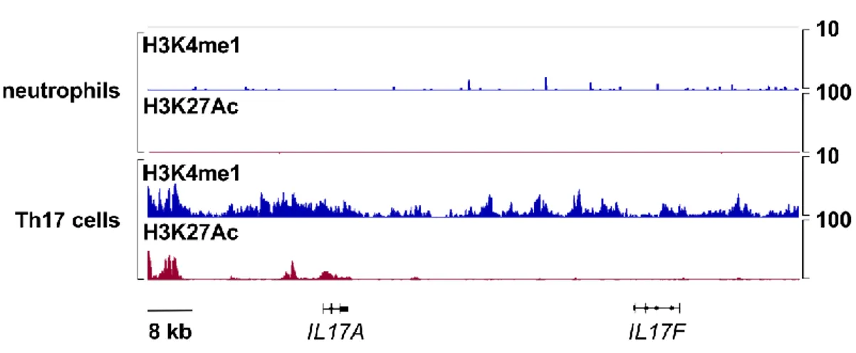
human neutrophils to express IL-17A and IL-17F mRNA, no deposition of H3K27Ac and H3K4me1, which are histone marks of, respectively, active and poised genomic regulatory elements, was detected at the IL-17A/F genomic locus in resting or IL-6 plus IL-23-stimulated neutrophils. In addition, although we found that anti-IL-17A and anti-IL-17B commercial antibodies positively stained cytospin preparations of resting and activated neutrophils by IHC, these antibodies do not recognize any intracellular protein having the correct MW of either IL-17A or IL-17B in corresponding lysates of the same neutrophil preparations by immunoblotting. Since the same antibodies were found to strongly stain other intracellular proteins of neutrophils, we conclude that their ability to positively stain neutrophils derives from IL-17A- or IL-17B-independent unspecific binding. In conclusion, our data not only confirm and further support our previous original findings on the inability of human
6 neutrophils to express/produce IL-17A, IL-17B and IL-17F mRNAs/proteins, but also attempt to explain why other published studies continue to report the opposite.
7
Table of contents
1. INTRODUCTION ... 15
POLYMORPHONUCLEAR NEUTROPHILS ... 15
Effector functions of neutrophils ... 16
Pattern recognition receptors by neutrophils ... 17
Cytokine expression by neutrophils ... 19
1.1.4 Chemokine expression by neutrophils ... 25
1.2 INTERLEUKIN 17... 26
IL-17 and receptors ... 26
IL-17 and Th17 cells ... 28
Neutrophils and IL-17 ... 29
2 MATERIALS AND METHODS ... 35
Cell purification and culture ... 35
Flow Cytometry ... 36
Respiratory burst activity ... 37
Immunohistochemistry ... 37
Enzyme-linked immunosorbent assay ... 38
Reverse transcription quantitative real-time PCR (RT-qPCR) ... 38
Immunoblotting experiments ... 39
Chromatin Immunoprecipitation (ChIP) assays ... 40
ChIP-seq ... 41
Gene Expression Data Set of normal hematopoietic stem and progenitor cells 42 Statistical analysis ... 42
Study approval ... 43
3 RESULTS ... 45
Human neutrophils incubated with various agonists in vitro do not express IL-17 members, either at mRNA or at protein levels ... 45
Human neutrophils incubated with IL-6 plus IL-23, in the presence or the absence of inactivated A.fumigatus hyphae or conidia, do not express IL-17RC. ... 51
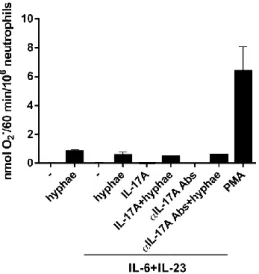
8 O2- production by neutrophils stimulated with inactivated A. fumigatus hyphae
after preincubation with IL-6 plus IL-23 is not modified by either exogenous
IL17A or IL-17A inhibitors ... 52
Chromatin organization at the IL-17A and IL-17F promoters and enhancers in human neutrophils ... 53
Human neutrophils from patients with psoriasis do not express/produce IL-17A and/or IL-17F ... 58
Commercial anti-IL-17A antibodies stain cytospins of resting and activated neutrophils but not their corresponding lysates ... 58
Human neutrophils do not express/produce IL-17B ... 62
4 DISCUSSION AND CONCLUSION ... 65
5 REFERENCES... 71
9
ABBREVATIONS
ANCA anti-neutrophil cytoplasm antibody-relatedAPC allophycocyanin
BATF basic leucine zipper ATF-like transcription factor
BLIMP PR domain zin finger protein 1
C/EBP CCAAT/enhancer binding protein
CCL chemokine CC motif ligand
CCR CC chemokine receptor
ChIP Chromatin immunoprecipitation
ChIP-Seq ChIP followed by high troughput sequencing
CLEC C-type lectin domain family
CMP common myeloid progenitor
COPD chronic obstructive pulmonary disease
CpG 5’- cytosineguanine-3’ dinucleotides
CXCL chemokine CXC motif ligand
CXCR CXC-chemokine receptor
DNA Deoxyribonucleic acid
ECAM endothelial cell adhesion molecule
EDTA ethylenediamine tetraacetic acid
ELISA enzyme-linked immunosorbent assay
FITC fluorescein isothiocyanate
fMLF Formyl-Methionyl-Leucyl Phenylalanine
FPKM Fragments per kilobase of transcript per million mapped reads
FPR formyl peptide receptor
G-CSF granulocyte colony stimulating factor
GADPH glyceraldehyde phosphate dehydrogenase
GM-CSF Granulocyte-macrophage colony-stimulating factor
HLA-DR major histocompatibility complex class II
HOMER Hypergeometric Optimization of Motif EnRichment
HT high-throughput
ICAM Intercellular Adhesion Molecule
IF immunofluorescence
IHC immunohistochemistry
iNKT invariant natural killer T cells
10
IL-17R Interleukin-17 receptor
ILC innate lymphoid cell
IFN Interferon
IGV Integrative Genome Viewer
IP Immunoprecipitation
IRF interferon regulatory factor
ISG interferon stimulated gene
JAM junctional adhesion molecule
kbp kilo base pairs
LPS lipopolysaccharide
LTA lipoteichoic acid
MAL MyD88-adaptor-like
MAPK mitogen-activated protein kinase
MDA5 Melanoma Differentiation-Associated protein 5
MDL myeloid DAP12-associating lectin
MMP matrix metalloproteinase
MNE mean normalized expression
MPO myeloperoxidase
mRNA messenger RNA
MTB Mycobacterium tuberculosis
mTOR mammalian target of rapamycin
MW molecular weight
MyD88 myeloid differentiation factor 88
NADPH nicotinamide adenine dinucleotide phosphate
NE neutrophil elastase
NETs neutrophil extracellular traps
NKT natural killer T cells
NLR nucleotide-binding oligomerization domain (NOD)-like receptor
NLRC NLR with a CARD domain
NLRP NLR with a pyrin domain
nt nucleotide
PAMPs pathogen-associated molecular patterns
PE phycoerythrin
PECAM platelet endothelial cell adhesion molecule
PerCP peridinin chlorophyll protein
11
PMA phorbol myristate acetate
PMN polymorphonuclear neutrophils
PRR pattern recognition receptors
PSA psoriatic arthritis
PSGL-1 P-selectin glycoprotein ligand 1
RA rheumatoid arthritis
RIG-I Retinoic acid-inducible gene I
RLH retinoic acid-inducible gene (RIG)-like helicase
RNAi RNA interference
ROI reactive oxygen intermediates
RORγt RAR-related orphan receptor gamma
ROS reactive oxygen species
RT-qPCR reverse transcription quantitative PCR
SAA serum amyloid A
SLE systemic lupus erythematosus
SP1 specific protein 1
STAT signal transducer and activator of transcription
TF Transcription factor
Th T helper
TLR Toll-like receptor
TNF tumor necrosis factor
TRAM TRIF-related adaptor molecule
TRIF TIR-domain-containing adaptor protein inducing interferon β protein
TSS transcription start site
TREM triggering receptor expressed on myeloid cells
13
LIST OF FIGURES AND TABLES
1
Figure 1 | Pattern recognition receptors expressed by neutrophils ... 18 2
Figure 2 | Cytokines that human neutrophils can potentially express and produce. . 20 3
Figure 3 | Examples of epigenetic mechanisms controlling gene expression in human 4
neutrophils. ... 21 5
Figure 4 | R848 induces a reorganization of the chromatin at the IL-6 locus of human 6
neutrophils. ... 25 7
Figure 5 | Members and related receptors of the IL-17 family. ... 28 8
Figure 6 | IL-17A, IL-17F, CXCL8 and IL-1ra mRNA expression levels in neutrophils 9
activated by a variety of stimuli. ... 46 10
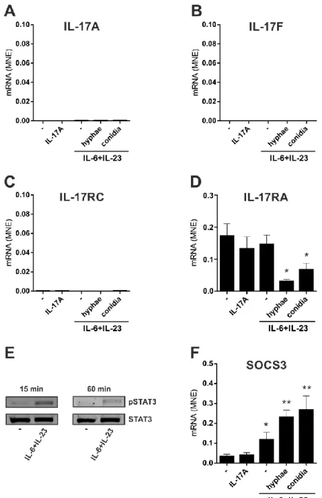
Figure 7| No induction of IL-17A, IL-17F and IL-17RC mRNA expression in 11
neutrophils incubated with IL-6 plus IL-23 in combination with inactivated A.fumigatus 12
hyphae or conidia ... 49 13
Figure 8 | Lack of IL-17A and IL-17F production by human neutrophils activated by 14
IL-6 plus IL-23 in a combination with inactivated A.fumigatus hyphae or conidia ... 50 15
Figure 9| No induction of intracellular IL-17A expression in CD66b+ neutrophils 50 16
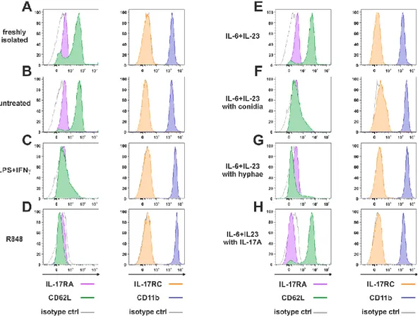
Figure 10 | IL-17RA, IL-17RC, CD62L and CD11b surface expression in neutrophils 17
activated under various experimental conditions. ... 52 18
Figure 11 | Superoxide anion production by neutrophils stimulated by inactivated 19
hyphae from A.fumigatus in combination with IL-6 plus IL-23, IL-17A or secukinumab. 20
... 53 21
Figure 12 | ChIP-Seq profiles of H3K4me1 and H3K27Ac at the IL17A and IL17F 22
loci in human neutrophils and Th17 cells. ... 55 23
Figure 13 | H3K4me1 or H3K27Ac levels at the IL-17A, IL-17F and SOCS3 genomic 24
loci of Th17 cells and resting/ IL-6 plus IL-23-activated neutrophils. ... 56 25
14 Figure 14 | IL-17A, CXCL8, TNF and SOCS3 mRNA expression, as well as IL-17R 26
surface expression, in neutrophils from patients with psoriasis ... 57 27
Figure 15 | Staining human neutrophils by anti-IL-17A (AF-317-NA) polyclonal 28
antibodies... 60 29
Figure 16 | IL-17A, IL-17B, IL-17F, IL-10, IL-17RC, IL-17RA azurocidin, neutrophil 30
elastase and MPO mRNA expression levels at different stages of neutrophil 31
maturation. ... 61 32
Figure 17 | Staining human neutrophils by anti-IL-17B (AF1248) antibodies... 63 33
34
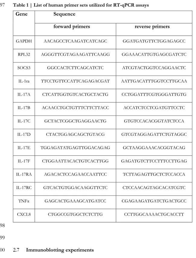
Table 1 | List of human primer sets utilized for RT-qPCR assays ... 39 35
Table 2 |List of human primer sets utilized for qPCR of ChIP assays ... 42 36
Table 3 | Lack of IL-17A production by neutrophils activated under various 37
experimental conditions. ... 47 38
39
15
1. INTRODUCTION
41 42 POLYMORPHONUCLEAR NEUTROPHILS 43Among leukocytes, the polymorphonuclear neutrophils are the most 44
numerous cells present in human bloodstream. These cells are crucial players in innate 45
immune response. Nonetheless, during the last years, research has demonstrated that 46
neutrophils also act as important regulators of adaptive immunity. Neutrophils are 47
indispensable for defence against pathogens, since they are the first cells to reach the 48
inflammatory sites where they rapidly exert their effector functions. In an injury or 49
microbial infection site, neutrophils are able to effectively counter the cause of the 50
injury by, for example, releasing enzymes, synthetizing reactive oxygen species and 51
producing inflammatory mediators. Neutrophils display a wide range of actions and 52
are believed to participate in protection against intracellular pathogens such as viruses 53
and mycobacteria, to interact with the adaptive immune system and to be involved in 54
the resolution of inflammation. However, neutrophils have a dark side in that if they 55
are improperly activated they lead to tissue damage, thus contributing to the 56
development of autoimmune diseases such as psoriasis, systemic lupus erythematosus 57
(SLE), rheumatoid arthritis (RA) and anti-neutrophil cytoplasmic antibody-related 58
(ANCA) vasculitis, or exaggerated inflammatory reactions such as inflammatory bowel 59
diseases [1]. 60
Prior to reaching the blood circulation, neutrophils pass through a 61
differentiation process in the bone marrow called myelopoiesis, arising from the 62
pluripotent stem cells in a generation rate of 1 to 2 × 10¹¹ mature neutrophils/day [2].
63
Once these cells are released into the bloodstream, they do not stay there for long, due 64
to their very short lifespan, that usually ranges between 6-20 hours under healthy 65
conditions. However, neutrophil life-span is increased when they migrate into the 66
infection site and enter in contact with cytokines such as tumour necrosis factor alpha 67
(TNF), type I and II interferons (IFN), granulocyte colony-stimulating factor (G-68
CSF) and granulocyte-macrophage stimulating factor (GM-CSF) [3, 4]. Furthermore, 69
bacterial products such as LPS and viral ssRNA have also been shown to prolong 70
neutrophil survival [3, 5]. In the absence of infection or inflammation, neutrophils 71
16 undergo through the apoptosis process and are in turn, removed by macrophages. 72
Apoptosis is an important homeostatic mechanism, as its alteration in neutrophils 73
would have serious consequences for the inflammatory response and resolution of 74
inflammation. Furthermore, the regulation of neutrophil turnover represents a critical 75
checkpoint because neutrophils constitute the majority of leukocytes in humans, 76
predominate at the infection sites and contain cytotoxic molecules that can damage 77
host tissues. 78
79 80
Effector functions of neutrophils 81
While circulating in the vasculature, neutrophils can rapidly migrate into the 82
infection sites in response to chemokines such as CXCL8, which is the most potent 83
neutrophil chemoattractant. CXCL8 is produced in response to pro-inflammatory 84
molecules by macrophages, epithelial cells, mast cells, endothelial cells, keratinocytes, 85
fibroblasts and neutrophils themselves [6, 7]. Moreover, products from bacteria such 86
as fMLF and peptidoglycan can also contribute to the recruitment of neutrophils [6]. 87
The process of neutrophil migration starts with a slow roll on the surface of the 88
endothelial cells mediated by constitutive expression of adhesion molecules of the 89
selectin family, which establish a low-affinity binding between L-selectin expressed by 90
neutrophils to P- and E-selectins present on the plasma membrane of activated 91
endothelial cells[6]. A high-affinity interaction can be then established, in the presence 92
of chemotactic factors, between 2-integrins present on the neutrophils surface and 93
endothelial cell intracellular adhesion molecule (ICAM)-1 and ICAM-2 on the 94
endothelial cells. Neutrophils start to crawl on the vasculature surface and ultimately 95
transmigrate through the endothelium into tissues without damaging these structures, 96
following a gradient that is believed to be set up by chemotactic factors [8, 9]. 97
Transmigration is facilitated by several surface molecules, including integrins and 98
CAMs (ICAM1, ICAM2 and vascular cell adhesion protein 1 (VCAM1)), junctional 99
proteins such as platelet/endothelial cell adhesion molecule 1 (PECAM1; also known 100
as CD31), CD99, junctional adhesion molecules (JAMs) and epithelial cell adhesion 101
molecule (ECAM) [8]. Once in the interstitial tissue, neutrophils follow chemotactic 102
gradients in order to move in an oriented manner, locate and kill microorganisms using 103
17 their antimicrobial arsenal, including reactive oxygen species (ROS) and release of 104
peptides, proteins and enzymes contained in granules and phagocytosis. Phagocytosis 105
is the process whereby neutrophils bind and ingest invading microorganisms. 106
Neutrophils recognize microbes via their pattern recognition receptors (PRRs) that 107
bind specific structures called pathogen-associated molecular patterns (PAMPs) such 108
as lipopolysaccharide (LPS), peptidoglycan (PGN), lipoteichoic acid (LTA) and 109
flagellin. These PAMPs generally trigger signal transduction pathways leading to the 110
expression of adhesion molecules, promotion of phagocytosis, release of cytokines, 111
chemokines, ROS and degranulation. Moreover, neutrophil phagocytosis can be 112
efficiently enhanced by antibodies and complement products that opsonize microbes. 113
Phagocytosis leads to the formation of a phagosome that, in turn, fuses with 114
azurophilic granules (peroxidase-positive granules) creating a vacuole lumen with 115
antimicrobial molecules including -defensins, cathepsins, proteinase-3, elastase, 116
azurodicin and lysozyme. 117
118 119
Pattern recognition receptors by neutrophils 120
One of the most important discovery regarding the innate immunity has been 121
the identification of pattern recognition receptors, whose importance was firstly 122
highlighted by Charles Janeway in 1989 [10]. Today, PRRs include members of Toll-123
like receptor (TLR) family, the C-type lectin receptors dectin 1 (also known as 124
CLEC7A), and CLEC2 (also known as CLEC1B), as well as cytoplasmic RNA sensors 125
(RIG-I and MDA5), cytoplasmic DNA sensors and cytoplasmic NACHT-leucine-rich 126
repeat receptors (NLRs), some of them being components of inflammasomes [11, 12]. 127
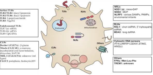
Among them, neutrophils express a broad range of PRRs, as illustrated in Figure 1. 128
18 130
Adapted from Thomas CJ and Schroder K. 2013. Trends Immunol. 34:317 [13]. 131
Based on the data from Tamassia N and Cassatella MA. 2013. Current Opinion in Pharmacology. 132
13:547 [12]. 133
Figure 1 | Pattern recognition receptors expressed by neutrophils
134
Abbreviations: CLEC, C-type lectin domain family; CLR, C-type lectin receptor; DAMP,
135
danger-associated molecular pattern; meso-DAP, meso diaminopimelic acid; FPR, formyl
136
peptide receptor; MDL, myeloid DAPs12-associating lectin; NLR, nucleotide-binding
137
oligomerization domain (NOD)-like receptor; NLRC, NLR with a CARD domain; NLRP,
138
NLR with a pyrin domain; PAMP, pathogen-associated molecular pattern; RLH, retinoic
acid-139
inducible gene (RIG)-like helicase; SAP, spliceosome-associated protein; TLR, Toll- like
140
receptor; TREM, triggering receptor expressed on myeloid cells. (a) Described in mouse but
141
not human neutrophils; (b) Described in human but not mouse neutrophils; (c) Described in
142
mouse neutrophils, no data available in humans.
143
144
TLRs, which to date comprises 10 members in humans (TLR1-10), were the 145
first PRRs to be discovered and are the most studied PRRs in neutrophils. Almost all 146
TLRs are expressed and functional in human neutrophils, with the exception of TLR3 147
and TLR7. TLR4, which recognizes LPS, has special features in its activation pathway. 148
In fact, while immune cells such as monocytes, macrophages and dendritic cells 149
activate two signalling pathways downstream of TLR4, respectively coordinated by 150
signal adaptors myeloid differentiation factor 88 (MyD88)/MyD88 adaptor-like (Mal) 151
and TIR domain-containing adaptor protein inducing interferon β (TRIF)/TRIF-152
related adaptor molecule (TRAM), neutrophils mobilizes only the MyD88-dependent 153
pathway [14]. Such a cell-specific activation by LPS influences the expression profile, 154
strength and kinetics of cytokine production by neutrophils. 155
19 Cytokine expression by neutrophils
157
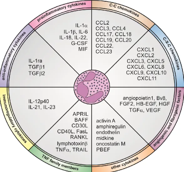
Neutrophils have been shown to express and produce a large number of pro- 158
and anti-inflammatory cytokines, chemokines, colony-stimulating and angiogenic 159
factors, either constitutively or following appropriate stimulation [11] (Figure 2). The 160
production of cytokines by neutrophils is controlled by regulatory mechanisms 161
occurring at the level of mRNA transcription [15], stability or translation, as well as at 162
the level of protein secretion. Moreover, cytokines are eventually stored in intracellular 163
pools following synthesis, ready to be promptly secreted when neutrophils are 164
stimulated with secretagogue-like molecules [16]. 165
It is important to mention two important issues for those people who want to 166
study neutrophil-derived cytokines: 1) neutrophils possess 10/20 times less RNA than 167
other leukocytes and consequently; 2) with few exceptions, they usually produce much 168
lesser cytokine amounts than monocytes/macrophages or lymphocytes on a per cell 169
basis. Hence, highly pure populations of neutrophils should be used for the evaluation 170
of their cytokine gene expression/production profiles. Accordingly, in a study 171
conducted in our lab, different published procedures to isolate neutrophils directly 172
from human blood were found to generate percentages of neutrophil purity very 173
diverse, in some cases causing artefacts [17]. By the way, the fact that a single 174
neutrophil, per se, produce little amounts of cytokine does not mean that neutrophil-175
derived cytokines are not important, as neutrophils usually outnumber mononuclear 176
leukocytes in infection/inflammatory sites by one to two orders of magnitude [15, 18, 177
19]. Neutrophil-derived cytokines can be measured in cell-free supernatants or cell 178
lysates by using a variety of methods, including enzyme-linked immunoadsorbent 179
assays (ELISA), immunoprecipitation, immunohistochemistry, intracellular staining by 180
flow cytometry or confocal microscopy. The latter two techniques should be not only 181
carefully interpreted, but also used only to support other cytokine detection methods, 182
since elevated neutrophil autofluorescence, antibody cross-reactivity or aspecific 183
binding of antibody to Fcγ receptors (especially CD16), could generate artefacts. 184
20 186
Adapted from:Tamassia, N., et al. (2018). "Cytokine production by human neutrophils: Revisiting the
187
"dark side of the moon”. Eur J Clin Invest, 2018 [20].
188 189
Figure 2 | Cytokines that human neutrophils can potentially express and produce.
190
Expression and/or production of the listed cytokines have been validated in human
191
neutrophils by gene expression techniques, immunohistochemistry, enzyme-linked
192
immunosorbent assays (ELISAs) or biological assays.
193 194 195
Interestingly, activation of the same PRR in autologous neutrophils and 196
monocytes may trigger distinct gene as well as cytokine expression programs [14, 21, 197
22], reflecting cell type-specific mechanisms of transcriptional regulation. For instance, 198
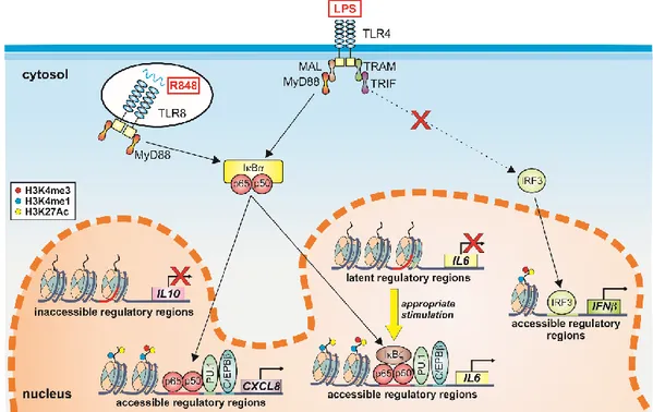
upon stimulation with LPS, IFN, an antiviral and immunomodulatory cytokine [23], 199
is induced in human monocytes, but not in neutrophils. As explained on page 18, the 200
activation of TLR4 by LPS in neutrophils fails to trigger the TRIF-signalling pathway, 201
that is indeed crucial for the activation of the transcription factor interferon regulatory 202
factor 3 (IRF3) (Figure 3). On the contrary, the activation of TLR4 by LPS in 203
21 monocytes leads to the triggering of both the MyD88-signalling pathway (and, in turn, 204
NF-B) and the TRIF-signalling pathway (mainly activating IRF3). Activation of both 205
NF-B and IRF3 is essential for the activation of IFN transcription [24, 25] which, 206
in an autocrine manner, stimulates the expression of a large set of interferon-stimulated 207
genes (ISG) displaying antiviral and immunomodulatory functions[23]. 208
209
210
Adapted from: Ostuni, R., et al. Epigenetic regulation of neutrophil development and function. Semin
211
Immunol 28(2): 83-93, 2016 [26].
212 213
Figure 3 | Examples of epigenetic mechanisms controlling gene expression in human
214
neutrophils.
215
The cartoon illustrates that the IL10 locus of neutrophils display inaccessible regulatory
216
regions, as evidenced by the absence of histone marks associated with active transcription.
217
Such a chromatin conformation prevents the binding of transcription factors (TFs) activated
218
by TLR ligands or other PAMPs, therefore preventing IL-10 mRNA transcription. By contrast,
219
the CXCL8 locus has an accessible conformation that is ensured by the constitutive binding
220
of both PU.1 and C/EBPβ. Upon neutrophil activation, NF-кB is recruited to its
221
corresponding binding sites and thus promotes transcription of CXCL8 mRNA. Figure also
222
shows that the IL6 locus of neutrophil is not accessible under basal state. However, upon
223
appropriate stimulation, pioneer TFs such as PU.1 and C/EBPβ initiate chromatin opening,
224
in turn favoring the binding of activated TFs and ultimately activating IL-6 mRNA
225
transcription. Finally, even if the IFNβ genomic locus is not in a closed conformation, no
226
IFNβ mRNA transcription occurs in TLR-stimulated neutrophils because of the inability to
227
activate IRF3 by TLR-dependent signals.
228 229 230
22 Beyond discrete transcription factor (TF) activation, chromatin organization 231
may represent another critical factor at the basis of the transcriptional differences 232
observed in neutrophils and monocytes. Indeed, pro-inflammatory TFs activated by 233
external stimuli such as TLR ligands largely act within a pre-established chromatin 234
landscape [27]. On the other hand, recent studies in neutrophils and monocytes have 235
shown that chromatin-dependent mechanisms control their expression of interleukin-236
10 (IL-10), a potent anti-inflammatory cytokine that prevents inflammatory and 237
autoimmune diseases[28]. Specifically, a comparative analysis at the IL10 genomic locus 238
of histone modifications associated with permissive chromatin, precisely H3K4me3 239
(localized at the transcription start site of active genes), H3K27Ac and H4Ac (both 240
associated with activated regulatory elements), and H3K4me1 (a histone mark mainly 241
associated with active and poised enhancers), revealed that these histone modifications 242
are detectable in autologous monocytes, but not in autologous neutrophils, already at 243
the steady-state. Furthermore, H3K4me3 and H3K27Ac marks were shown to further 244
increase in monocytes stimulated with TLR2 and TLR4 ligands (Pam3CYS4 and LPS 245
respectively) or serum amyloid A (SAA), while remaining undetectable in neutrophils 246
[21, 29]. In accordance with what observed for histone modifications, chromatin 247
immunoprecipitation (ChIP) of a number of transcription factors, previously proposed 248
to bind to and/or transactivate the IL-10 gene in various cells of human or mouse 249
origin [30], revealed no binding of C/EBPβ, c-FOS, SP1 and NF-Bp50 to the IL-10 250
promoter of neutrophils, either constitutively or upon activation with LPS. These data, 251
generated using highly purified human neutrophils isolated by antibody conjugated 252
magnetic beads, provided a mechanism explaining why human neutrophils are unable 253
to produce IL-10 [31-34]. Such an issue has been in fact controversial for many years, 254
as a number of studies were reporting a production of IL-10 by human neutrophils, 255
under resting or stimulatory conditions [35-39]. In our opinion, controversial reports 256
are explained by the scarce purity of neutrophil preparations used to evaluate the 257
production of IL-10, therefore avoiding the essential exclusion of contaminant cells, 258
such as lymphocytes or monocytes, which can strongly affect the final results [17, 21]. 259
Confirming this hypothesis, studies conducted by our laboratory with a preparation of 260
highly purified neutrophils (> 99.7 ± 0.2 %) have never detected mRNA 261
expression/production of IL-10 in response to plenty of inflammation-associated 262
stimuli, such as PRR agonists (LPS, R848, Pam3CSK4, poly(I:C), curdlan), SAA, 263
23 cytokines (IFNγ, TNFα, GM-CSF or G-CSF), chemoattractants (fMLF) or insoluble 264
immunocomplexes [21]. By contrast, autologous monocytes promptly produce 265
detectable amounts of IL-10 when stimulated with SAA, TLR ligands or curdlan [21]. 266
Although it cannot be excluded that specific stimulatory conditions may revert the 267
non-permissive state of the chromatin in their IL10 locus, human neutrophils are 268
unable to express IL-10 upon stimulation with TLR ligands or SAA. The bottom line 269
is that caution must be taken with studies that report IL-10 expression by human 270
neutrophils stimulated with TLR-ligands, especially when contamination with other 271
leukocytes is not excluded. The latter precaution is, anyway, mandatory when cytokine 272
production and/or gene expression by neutrophils need to be investigated [18, 40]. 273
Not only the expression of IL-10 in human neutrophils has been a target of 274
debate for a long time, but also of other cytokines such as IL-6 and IL-17. IL-6 is a 275
pleiotropic cytokine displaying both pro- and anti-inflammatory activities, playing a 276
crucial role in host defence against pathogens and acute stress [41]. However, 277
uncontrolled IL-6 regulation can contribute to the pathogenesis of inflammatory and 278
autoimmune diseases [41], as well as cancer [42]. Conflicting results have been 279
published on the ability of neutrophils to accumulate IL-6 mRNA, either constitutively 280
or after stimulation [43, 44]. In addition, several studies have reported a remarkable 281
production of IL-6 by neutrophils stimulated with LPS, GM-CSF or TNFα [43], while 282
other studies did not reproduce the same data [45, 46]. It is important to note that 283
different culture conditions, time points of analysis and methods to detect IL-6 284
production were used in these various studies. Still, these controversial publications 285
rarely met the employment of neutrophil preparations free of contaminant monocytes, 286
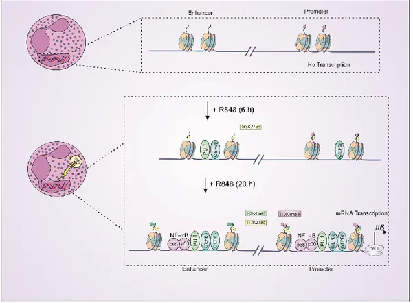
which are major producers of IL-6 [45]. As illustrated in Figure 4, the regulation of 287
expression/production of IL-6 by neutrophils was recently found to be regulated at 288
chromatin reorganization level. Specifically, data have shown the absence of 289
H3K4me3, H3K27Ac, H4Ac and H3K4me1 histone marks in highly purified resting 290
neutrophils [47]. However, incubation of neutrophils with either R848, a synthetic 291
mimic viral ssRNA recognized by TLR8, or very high concentrations (10 µg/ml) of 292
LPS increased the presence of these histone marks at the IL6 locus, in accordance with 293
the ability of TLR ligands to promote chromatin reorganization and de novo formation 294
of latent enhancers [48-50]. Interestingly, histone modifications were prominently 295
detected after an overnight incubation, while PU.1 and C/EBPβ were recruited at the 296
24
IL6 locus as early as 6 h after R848 stimulation, suggesting a “pioneer” activity of these
297
TF also in human neutrophils [51, 52]. On the other hand, in autologous monocytes 298
the IL6 locus was found to be in a “poised” conformation, which is a chromatin status 299
given by the presence of H3K4me1, PU.1 and C/EBPβ already under resting 300
conditions. Such a chromatin organization influences the kinetics of IL-6 transcription, 301
which in monocytes is much more accelerated than in neutrophils [47]. The different 302
kinetics of IL-6 gene expression observed between neutrophils and monocytes is 303
controlled also by other factors, including: 1) expression of IкBζ, a transcriptional 304
coactivator that is required for the IL-6 transcription [53], which in monocytes is more 305
rapidly induced by TLR stimulation than in neutrophils; 2) endogenous production of 306
IL-10, which occurs only in monocytes [21] and that turns off IL-6 transcription at 307
delayed time-points[54]; 3) induction of miR187 by endogenous IL-10, occurring only 308
in monocytes, which regulate IкBζ expression[55]; and 4) the presence of distinct cell-309
specific enhancers, located at -14 kb and at -49 and -64 kb from the IL-6 TSS in 310
neutrophils and monocytes, respectively [47]. 311
The importance of epigenetic in regulating cytokine gene expression in human 312
neutrophils is exemplified by the fact that R848 induces an increase of the chromatin 313
accessibility at their IL6 locus, and by doing so confers responsiveness to TNFα, which 314
by itself is not able to trigger IL-6 transcription in resting neutrophils [47]. When the 315
IL6 locus is accessible, as in TLR8-activated neutrophils, TNFα induces the 316
recruitment of C/EBPβ and histone acetylation, in turn promoting IL-6 transcription 317
[47]. Indeed, in the presence of adalimumab and etanercept [56], two TNFα inhibitors, 318
the mRNA expression and production of IL-6 by TLR8-activated neutrophils 319
dramatically decreases, indicating that endogenous TNFα plays a crucial role in the 320
amplification of IL-6 expression [47]. Furthermore, an important role of endogenous 321
TNFα in amplifying neutrophil-derived IL-6 was recently confirmed by studies using 322
TLR8-stimulated neutrophils in the presence of IFNα, hence mimicking a potential 323
situation occurring in systemic lupus erythematosus (SLE) patients [57]. These findings 324
indicate that TLR8 ligands, IFNα and TNFα, three players often coexisting in many 325
diseases of viral or autoimmune origin, promote a strong production of IL-6 by human 326
neutrophils, placing this cell type among potential targets for immunotherapeutic 327
interventions. 328
25 330
Adapted from: Zimmermann, M., et al., Chromatin remodelling and autocrine TNFalpha are
331
required for optimal interleukin-6 expression in activated human neutrophils. Nat Commun, 6: p. 6061,
332
2015 [47].
333
Figure 4 | R848 induces a reorganization of the chromatin at the IL-6 locus of human
334
neutrophils.
335
Scheme illustrating the occupancy of PU.1, NF-кB, C/EBPβ and histone modifications
336
H3K4me1, H3H4me3 and H3K27Ac, at the regulatory regions of the IL-6 locus of
337
neutrophils. IL-6 locus in neutrophils is inactive based on the absence of histone modifications
338
and TF binding. Upon neutrophils treatment with R848, recruitment of PU.1, C/EBPβ,
NF-339
кB and deposition of significant levels of histone marks (H3K4me1, H3H4me3 and
340
H3K27Ac) occur at various IL-6 locus positions, suggesting an induction of latent regulatory
341
sites (such as enhancers) leading to IL-6 gene transcription.
342 343
1.1.4 Chemokine expression by neutrophils 344
Among the cytokines produced by neutrophils, chemokines represent 345
interesting ones given their ability to recruit selective leukocytes into the site of 346
inflammation. In this manner, chemokines regulate leukocyte trafficking and immune 347
system responses, other than influencing B and T cell development and modulating 348
angiogenesis. As displayed in Figure 2, human neutrophils are a source of many 349
chemokines, including CXCL1, CXCL8, CXCL10, CCL2, CCL3, CCL4 [58] and, as 350
more recently described, CCL23 [59]. Interestingly, some of the chemokines released 351
by neutrophils potently amplify their own recruitment [60]. Furthermore, by releasing 352
26 chemokines, neutrophils orchestrate sequential recruitment and/or activation of 353
distinct leukocyte types into the inflamed tissue, such as monocytes, dendritic cells 354
(DCs), natural killer (NK) cells, and T-helper type 1 (Th1) and type 17 (Th17) cells [58, 355
61]. 356
Neutrophils express many chemokine receptors as well, including CXCR2, 357
CXCR4 and CCR1, and are the first cell types targeted by chemokines during 358
inflammation. CXCR4 expression, for example, progressively decreases during the 359
maturation of neutrophils in the bone marrow, allowing the release of mature 360
neutrophils into the bloodstream [62]. In this context, a decrease of CXCL12 (the 361
ligand of CXCR4) and CXCR4 expression by neutrophils can be promoted by G-CSF, 362
that in this manner further decreases the number of neutrophils that are retained in 363
the bone marrow. On the contrary, neutrophils increase their CXCR4 expression with 364
senescence, which favours their return to the bone marrow where they undergo 365
apoptosis [63]. As already explained on page 16, circulating neutrophils promptly 366
respond to activated endothelium by binding selectin and integrin ligands, thus rolling 367
and spreading along the endothelium to finally, transmigrate in response to 368
chemokines, including CXCL1, CXCL2 and CXCL8. Some of these chemokines are 369
produced by endothelial cells or other cell types at the infection site, including 370
neutrophils themselves. Once transmigrated, neutrophils follow chemoattractant 371
gradients created by chemokines/chemotactic factors to migrate to the inflammation 372
site and exert their various effector functions. 373
374
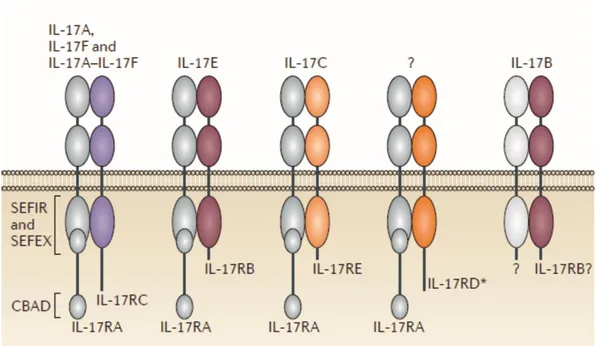
1.2 INTERLEUKIN 17 375
IL-17 and receptors 376
The IL-17 family consists of six cytokines, IL-17A (IL-17), IL-17B, IL-17C, 377
IL-17D, IL-17E (IL-25) and IL-17F (Figure 5). IL-17A and IL-17F are the most 378
closely related, since IL-17F has 50% sequence identity with IL-17A and its gene is 379
localized adjacent to the IL-17A gene on human chromosome 6 [64]. IL-17F is often 380
coproduced with IL-17A and together they can form an IL-17F/IL-17A heterodimer 381
[65]. Being the most potent one, IL-17A induces the expression of proinflammatory 382
27 mediators, including IL-1, IL-6, TNFα, CXCL8, G-CSF and GM-CSF by epithelial 383
cells and stromal cells, which together lead to the recruitment and activation of 384
neutrophils. IL-17B is expressed in various tissues, such as pancreas, small intestine 385
and brain [66], and has been shown to induce inflammatory cytokines such as IL-1 386
and TNF by monocyte cell line THP-1 [67] and IL-1, IL-6 and IL-23 by murine 387
fibroblast cell line 3T3 [68]. IL-17B is present in the rheumatoid arthritis synovium 388
and was shown to enhance the TNF-induced production of G-CSF and IL-6 in 389
fibroblasts [69]. Moreover, IL-17B can play an important role in lymphoid tissues by 390
regulating the trafficking of germinal center B cells by downregulation of the 391
expression of RGS16 protein [70]. Furthermore, IL-17B has been shown to have 392
potential roles in the pathogenesis of inflammatory and autoimmune diseases and 393
tumor progression [66]. Less is known regarding the other IL-17 members. IL-17C has 394
been shown to bind to IL-17RE and induce the activation of nuclear IkappaB family 395
member (IкBζ) in Th17 cells, hence leading to a potentiation of IL-17 production [71], 396
while IL-17E (also known as IL-25) has been correlated with the augmentation of 397
allergic responses, by enhancing Th2 memory cell function and suppressing the Th17 398
responses [72]. 399
The 17R group comprises five receptor subunits, 17RA, 17RB, IL-400
17RC, IL-17RD and IL-17RE. IL-17RA was the first to be described and is 401
ubiquitously expressed, particularly in hematopoietic cells (Figure 5). It is by far the 402
largest member of the family and functions as a common receptor subunit used by at 403
least four ligands, namely IL-17A, IL-17C, IL-17E and IL-17F[73]. For example, both 404
IL-17A and IL-17F, as either homodimers or heterodimers, induce signals through the 405
IL-17RA/IL17RC complex. It is important to note that IL-17RA is unique among the 406
interleukin receptors because it recruits an adaptor protein known as ACT1 [74, 75]. 407
Binding of IL-17A to IL-17R leads to the activation of canonical nuclear factor кB 408
(NF-кB) and mitogen-activated protein kinase (MAPK) pathways, which requires the 409
recruitment of ACT1 and the presence of TNF receptor associated factor 6 410
(TRAF6)[76]. Furthermore, IL-17A triggers the activation of the CCAAT/enhancer 411
binding proteins (C/EBPs) for the transcription of IL-17-target genes [77]. 412
28 414
Gaffen, S.L., et al., The IL-23-IL-17 immune axis: from mechanisms to therapeutic testing. Nat Rev
415
Immunol, 2014. 14(9): p. 585-600 [78].
416 417
Figure 5 | Members and related receptors of the IL-17 family.
418
CBAD, C/EBPβ activation domain; SEFEX, SEFIR extension; SEFIR, SEF/IL-17R.
*IL-419
17RD is also known as SEF.
420 421
IL-17 and Th17 cells 422
IL-17A is a cytokine cloned in 1993 that is important in host defence and 423
inflammation [79]. After the discovery of a subtype of CD4+ T helper (Th) expressing 424
IL-17A (currently known as Th17 cells), plenty of studies have been published 425
correlating Th17 with a wide range of physiological and pathological processes. 426
Although Th17 cells are considered the main sources of IL-17A and IL-17F, other 427
innate immune cells produce these cytokines, including T cells, natural killer T cells 428
(NKT), invariant natural killer cells (iNKT), Paneth cells, TCRβ+ natural Th17 cells,
429
lymphoid-tissue inducer-like cells (LTi), IL-17-expressing type 3 innate lymphoid cells 430
(ILC3s) and mast cells [80, 81]. Nonetheless, an exaggerated IL-17A production can 431
lead to the development of autoimmune diseases, such as psoriasis, multiple sclerosis 432
and rheumatoid arthritis, as well as cancer progression, which hence makes IL-17 as a 433
very important target for the development of new therapies [78]. 434
The expression of IL-17 by Th17 cells is regulated by specific transcription 435
factors and chromatin status. Specifically, the activation of the T cell receptor (TCR) 436
29 signalling pathway leads to the recruitment of pioneering transcription factors, namely 437
basic leucine zipper transcription factor ATF-like (BATF) and interferon-regulatory 438
factor 4 (IRF4), which cooperatively make accessible target genes on the chromatin. 439
When an inflammatory stimuli is present, such as IL-6 or activation of mTOR during 440
hypoxia, transcription factor signal transducer activator of transcription 3 (STAT3) is 441
recruited to the accessible chromatin, in turn promoting the transcription of genes 442
encoding retinoic acid receptor-related orphan receptor-γt (RORγt; encoded by Rorc) 443
and hypoxia-inducible factor 1 alpha (HIF1α; encoded by Hif1a) leading to lineage 444
specification of T helper 17 (Th17) cells. A further activation by IL-23 promotes the 445
recruitment of B lymphocyte-induced maturation protein 1 (BLIMP1) to the RORγt-446
STAT3 transcriptional complex enhancing the expression of Th17 induced-genes, 447
such as IL-17A, IL-17F, IL-23R, CSF2 and IL-2 [78]. 448
Although IL-17A and IL-17F are modest activators of signaling, a notable 449
feature of these cytokines is their strong synergistic effect with other pro-inflammatory 450
molecules, in particular with TNF, but also with IFNγ, IL-22, lymphotoxin, IL-1β and 451
LPS[82]. Such synergistic effect could occur at the level of promoter (for example, Il6 452
and Lnc2) and/or mRNA stability (for example, CXCL1 and CXCL2) [83, 84]. 453
Moreover, IкBζ is also upregulated upon IL-17 activation and in turn, promotes the 454
expression of some target genes [85]. 455
456
Neutrophils and IL-17 457
The relationship between human neutrophils and the IL-17 cytokines 458
indirectly begins with the demonstration that in vitro activated neutrophils release 459
chemoattractant molecules, including CCL2 and CCL20, that efficiently recruit Th17 460
cells to the inflamed tissue [86]. Th17 cells further produce IL17A and IL17F on one 461
side and CXCL8 on the other, which promote, respectively, the release of neutrophil 462
chemoattractant molecules by epithelial cells and the direct recruitment of neutrophils. 463
Moreover, activated Th17 cells release cytokines, such as TNF, GM-CSF and IFN, 464
that positively modulate survival and expression of activation markers by human 465
neutrophils [40, 87]. However, whether human neutrophils respond to IL-17 or not 466
appears to be still controversial. In fact, despite of our original data showing that 467
neutrophils cannot be directly activated by IL-17A and/or IL-17F due to their lack of 468
30 IL-17RC expression[87], confirming previous studies reporting the inability of IL-17 469
to directly affect neutrophil apoptosis[88], subsequent studies have claimed that IL-470
17A is instead capable to attenuate the anti-apoptotic effect of GM-CSF [89], as well 471
as increase the killing rate of pneumococcal when IL-17A is used at high 472
concentrations (> 1 µg/ml) [90]. 473
While Th17 cells are considered the major producers of IL-17 by far, whether 474
human neutrophils represent an IL-17 source again appears to be currently an unclear 475
issue, based on the controversial data reported in the literature. In such regard, our 476
group reported no detection of IL-17A or IL-17F mRNA/protein 477
expression/production by highly purified populations (> 99.7 %) of human 478
neutrophils incubated for up to 20 h with 100 U/ml IFN and/or 100 ng/ml LPS [87, 479
91-93]. By contrast, diverse studies have reported that neutrophils are capable of 480
transcribing/producing IL-17A. For instance, in a recent study, Taylor et al. [94] have 481
reported that a putative subset of human neutrophils express IL-17A mRNA after 1 h 482
stimulation with 20 µg/mL IL-6 and 2 µg/mL IL-23, as well as produce IL-17A after 483
3 h [94]. Moreover, neutrophils were shown to express RORt, which upon incubation 484
with supernatants containing IL-6 and IL-23 that were obtained from PBMCs treated 485
with Aspergillus fumigatus hyphae that, was found translocated into the nucleus and, 486
consequently, retained as responsible for the activation of IL-17A gene. When 487
neutrophils treated with PBMC supernatants were incubated with antibodies against 488
IL-6 or IL-23, their ability to express IL-17A mRNA vanished [94]. It is important to 489
note that Taylor et al. [94] have also reported that, in the presence of IL-6, IL-23 and 490
A. fumigatus hyphae, neutrophils are induced to express surface IL17RC, in turn
491
proposing that neutrophils can respond to IL-17A in an autocrine fashion, for instance 492
by producing ROS [94]. The same group has also shown that the JAK/STAT 493
signalling pathway activated by IL-6 plus IL-23 plays a very important role in the ROS 494
production and hyphal killing by neutrophils [95]. In a further study [96], the plasma 495
of both healthy controls and patients with fungal keratitis was found to contain IL-496
17A, IL-6 and IL-23, showing no significant differences between them. Peripheral 497
neutrophils from patients with fungal keratitis were analysed by intracellular flow 498
cytometry, immunofluorescence and RT-qPCR and found to express IL-17A, 499
suggesting a possible effect in the corneal inflammation and cytokine production in 500
response to growing hyphae [96]. Another group has instead recognized a subset of 501
31 peripheral human neutrophils expressing IL-17 during Mycobacterium tuberculosis (MTB) 502
infection [97]. According to the authors, such IL-17 production by neutrophils is due 503
to an autocrine activity of the IL-6 and IL-23 production during MTB infection [97]. 504
Furthermore, IL-6 and IL-23 have also been found in the microenvironment of human 505
gastric cancer, where neutrophils were, again, found to be IL-17A-, as well as MMP9-506
, positive by IHC and IF [98]. In the same study, neutrophils (> 96 % pure) isolated 507
from the whole blood of gastric cancer patients were shown to express IL-17A and 508
RORt mRNAs when stimulated with 20 µg/ml IL-6 and 2 µg/ml IL-23 [98]. 509
IL-17-producing neutrophils have been also found to be related with chronic 510
diseases, such as asthma, psoriasis and rheumatoid arthritis (RA). For instance, 511
neutrophils (> 95 % pure) isolated from the blood of severe asthma patients stimulated 512
with IL-21, IL-23, IL-21 plus IL-23 and IL-6 (all used at 20 ng/ml) were found to 513
express IL-17A and IL-17F mRNA after 4 h by RT-qPCR and after 12 h by 514
intracellular flow cytometry. In addition, the same authors observed a basal IL-17A 515
expression in resting neutrophils by western blot, which was further increased upon 516
stimulation with IL-21, IL-23 and IL-21 plus IL-23 for 18 h [99]. Consistently, a subset 517
of IL-17-positive neutrophils was found in the whole blood of patients with allergic 518
asthma, those one being allergic to fungi having a higher percentage of them [100]. 519
IL-17 has become a very interesting target for the development of therapies 520
towards autoimmune diseases, especially psoriasis, which displays a high infiltration of 521
neutrophils in the damaged tissue. Immunofluorescence and immunohistochemistry 522
experiments detected IL-17A in neutrophils present in biopsies from moderate-to-523
severe psoriasis patients [92]. Since no mRNA expression was observed in neutrophils 524
isolated from active plaques, the authors suggested that IL-17 might be stored (pre-525
formed) by neutrophils [92]. Furthermore, because treatment of these patients with 526
secukinumab, a human monoclonal anti-IL-17A antibody, was found to promote a 527
clearance of neutrophils in the psoriasis plaques and decrease CXCL8 mRNA 528
expression, authors proposed that IL-17A may have a role in the cross-talk between 529
neutrophils and keratinocytes leading to the release of CXCL8 and further neutrophils 530
recruitment [92]. A different study identified IL-17A-positive neutrophils and mast 531
cells in psoriasis biopsies by immunofluorescence, leading the authors to state that 532
both cell types represent the major sources of IL-17 in human skin [81]. Moreover, as 533
detected by immunofluorescence of psoriasis biopsies, neutrophils and mast cells were 534
32 found to release IL-17A during extrusion of extracellular traps, even though authors 535
reported that “mRNA from human neutrophils from several subjects showed either low or 536
undetectable levels of IL-17 using real time RT-PCR and Affymetrix gene array experiments
537
(M.J.Kaplan, data not shown)”, suggesting, similarly to Reich et al (ref), that IL-17A is
pre-538
formed and stored intracellularly by human neutrophils and mast cells[81]. However, 539
a recent report by Yamanaka et al. [91] contradicts the notion that human neutrophils 540
represent the major source of IL-17A in psoriasis. Accordingly, authors show no 541
evident IL-17A mRNA expression in highly purified populations of neutrophils 542
isolated from peripheral blood of psoriasis patients [91]. However, authors show that 543
IL-17A expression become evident if neutrophils are isolated by gradient 544
centrifugation only since they are contaminated by CD3+ lymphocytes, which generate
545
false-positive results for IL-17A mRNA expression[91]. In the case of RA and psoriatic 546
arthritis (PsA), a study demonstrated the presence of IL-17A expressing–neutrophils, 547
-mast cells and –T cells within the inflamed synovial membrane from these patients 548
[101]. Using immunohistochemistry for IL-17A and dual-immunofluorescence 549
staining for CD15 and IL-17A, authors found that the percentage positivity of IL-17A 550
was highest in neutrophils[101]. Opposite results were however reported by van 551
Baarsen et al. [93], who failed to detect a co-localisation of IL-17A with CD15 by dual-552
immunofluorescence staining of synovial biopsies from RA, PsA and inflammatory 553
osteoarthritis (OA) patients [93]. The contrasting results between the two latter studies 554
are likely explained by the different antibodies used, as Moran et al [101] utilized 555
polyclonal anti-IL-17A (polyclonal goat, R&D systems), while van Baarsen et al. [93, 556
101] performed the dual-immunofluorescence using a monoclonal antibody from the 557
same manufacturer. That is not trivial, taking into account in fact Tamarozzi et al.’s 558
paper [102]. These authors, in fact, initially reported the presence of IL-17A-positive 559
neutrophils in Wolbachia-positive Onchocerca volvulus nodules by 560
immunohistochemistry (IHC) [102]. Subsequently, they found that IL-17A protein is 561
not detectable in highly pure population of neutrophils (99.9 %) using alternative 562
assays, including western blot and ELISA or RT-qPCR for mRNA expression [102]. 563
Based on these premises, this study has the objective to clarify whether human 564
neutrophils express and/or produce IL-17. To do so, we have used highly pure 565
populations of human neutrophils (> 99.7 %) to evaluate the mRNA expression and 566
33 production of IL-17 members and related receptors, in response to various stimuli, 567
including those supposed to be very effective according to the literature. 568
34 570
35
2 MATERIALS AND METHODS
571 572
Cell purification and culture 573
Neutrophils were isolated from buffy coats of healthy donors and manipulated 574
under endotoxin-free conditions. In selected experiments, neutrophils were also 575
isolated from peripheral blood of patients with severe. After Ficoll-Paque gradient 576
centrifugation of buffy coats or peripheral blood, followed by dextran sedimentation 577
of granulocytes and hypotonic lysis of erythrocytes, neutrophils were isolated to reach 578
99.7 ± 0.2 % purity by positively removing all contaminating cells using the EasySep 579
neutrophil enrichment kit (StemCell Technologies, Vancouver, Canada) [17]. 580
Neutrophils were then suspended at 5×10⁶/ml in RPMI 1640 medium supplemented
581
with 10 % low (< 0.5 EU/ml) endotoxin FBS (BioWhittaker-Lonza, Basel, 582
Switzerland), incubated with or without 0.2-50 µM R848, 5-50 µM R837, 500 g/ml 583
particulate -glucan (Invivogen, San Diego, CA, USA), 0.1-100 µg/ml ultrapure LPS 584
(from E. coli 0111:B4 strain, Alexis, Enzo Life Sciences, Farmingdale, NY, USA), 1 585
µg/ml Pam3CSK4 (Invivogen), 50 µg/ml poly(I:C) (Invivogen), 1000 U/ml G-CSF 586
(Myelostim, Italfarmaco Spa, Milano, Italy), 100 U/ml IFN (R&D Systems, 587
Minneapolis, MN, USA), 10 ng/ml GM-CSF (Miltenyi Biotec), 5 ng/ml TNF 588
(Peprotech, Rocky Hill, NJ, USA), 2-20 µg/ml 6 (R&D Systems), 0.2-2 µg/ml IL-589
23 (R&D Systems), 100-500 ng/ml IL-17A (R&D Systems), 10 µg/ml anti-IL-17A 590
neutralizing Abs (secukinumab, Novartis, Basel, Switzerland) 100 nM fMLF, 500 591
g/ml curdlan (Sigma, Saint Louis, MO, USA), 20 µg/ml phorbol mysistate acetate 592
(PMA) (Sigma), 1 µg/ml Ionomycin (Sigma), 100 µg/ml CpG oligodeoxynucleotides 593
(ODN) (Invivogen), 1000 U/ml PEGylated IFNα-2a (Pegasys, Roche, Basel, 594
Switzerland). Inactivated conidia and hyphae from Aspergillus fumigatus were kindly 595
provided by prof. Luigina Romani (University of Perugia), and used at a neutrophil-596
fungi ratio of 1:5 for A. fumigatus conidia and 1:1 for A. fumigatus hyphae, as previously 597
described [103]. Neutrophils were plated either in 6/24-well tissue culture plates or in 598
polystyrene flasks (from Greiner Bio-One, Kremsmünster, Austria) for culture at 37°, 599
5 % CO2 atmosphere. After the desired incubation period, neutrophils were either
600
processed for chromatin immunoprecipitation (ChIP) experiments, or collected and 601
36 spun at 300 × g for 5 min for other types of assays. In the latter case, cell-free 602
supernatants were immediately frozen in liquid nitrogen and stored at – 80°, while the 603
corresponding cell pellets were either extracted for total RNA or lysed for protein 604
analysis. Th17 clones, kindly provided by prof. Francesco Annunziato (University of 605
Firenze), were stimulated for up 72 h with anti-CD3 and anti-CD28 mAbs (5µg/ml, 606
BD Biosciences) essentially as described [104]. 607
608
Flow Cytometry 609
For flow cytometry, 105 neutrophils were harvested after the desired treatment,
610
centrifuged and suspended in 100 µL PBS containing 10 % complemented-inactivated 611
human serum for FcR blocking. Neutrophils were then stained for 15 min at T room 612
with: APC anti-human IL-17RA/CD217 (clone 424LTS) and APC mouse IgG1к, as 613
isotype control (clone P3.6.2.8.1) from eBioscience (San Diego, CA, USA); PE anti-614
human IL-17RC (clone 309822) and mouse PE IgG2B isotype control from R&D 615
systems; PE-vio770 anti-human CD11b (clone ICRF44), FITC anti-human CD66b 616
(clone G10F5) and PerCP-Cy5.6 anti-human CD16 (clone 3G8) from Biolegend (San 617
Diego, California, USA); APC anti-human CD62L (clone 145/15 Miltenyi Biotec), all 618
at working dilutions specified in the corresponding datasheets. For intracellular 619
staining, neutrophils were incubated for 20 min in intracellular (IC) fixation buffer 620
(eBioscience), followed by permeabilization buffer (eBioscience) and 1h incubation 621
with antibody in the presence of Fc block. Sample fluorescence was then measured by 622
MACSQuant Analyzer (Miltenyi Biotec), while data analysis performed using FlowJo 623
software Version 10 from Tree Star (Ashland, OR, USA) [18]. For neutrophils within 624
the blood of psoriatic patients, 100 µl whole blood were stained with APC anti-human 625
IL-17RA and PE anti-human IL-17RC Abs in combination with the following mAbs: 626
VioBlue anti-human CD14 (clone TÜK4), PE anti-human CD56 (clone AF12-7H3), 627
PE-Vio770 anti-human CD3 (clone BW264/56), APC anti-human CD19 (clone LT19) 628
from Miltenyi; Brilliant Violet human CD45 (clone 2D1), PerCP-Cy5.5 anti-629
human CD16 (clone 3G8) and APC-Cy7 anti-human HLA-DR (clone L243) from 630
Biolegend. After red cells lysis by the ammonium chloride buffer, sample fluorescence 631
was immediately measured as previously described. 632
37 Respiratory burst activity
633
After isolation, neutrophils were suspended at the concentration of 2 x 10⁶
634
cells/ml in HBSS buffer containing 0.5 mM CaCl2 and 1 mg/ml glucose. Neutrophils
635
(100 µl/well) were then distributed in a 96-well plate and then incubated for 10 min at 636
37º prior to the addition of 80 µM cytochrome C, 2 mM NaN3 (Sigma) and the 637
indicated stimuli, including 20 ng/ml phorbol mysistate acetate (PMA) as control. 638
Plates were then incubated at 37º in an automated ELx808IU microplate reader 639
(BioTek Instruments, Inc., Winooski, VT) to record cytochrome C reduction (via 640
absorbance at 550 and 468 nm at intervals of 5 min for 90 min. O2- production was
641
finally calculated using an extinction coefficient of 24.5 mM [105]. 642
643
Immunohistochemistry 644
Cytospin preparations of neutrophils [106] cultured with the indicated stimuli 645
were stained by ematoxylin and eosin for morphological evaluation. After coverslip 646
removal, specimens were rehydrated through a scale of alcohols, with endogenous 647
peroxidase activity blocked by treatment with 0.3 % H202 in methanol for 20 min.
Anti-648
human IL-17A (AF-317-NA) and IL-17B (AF1248) goat IgG pAbs from R&D 649
Systems were 1:50 diluted, added to specimens for 60 min and then revealed using the 650
goat HRP-polymer (Biocare Medical, Pacheco, CA, USA) followed by 651
diaminobenzidine (DAB). Omission of the primary antibody was also performed as 652
negative control. For IL-17A and IL-17B tissue immunostaining, four-micron tissue 653
sections from two FFPE cases of pustular psoriasis were deparaffinised and rehydrated 654
through a scale of alcohols. Endogenous peroxidase activity was then blocked by 655
treatment with 0.3 % H202 in methanol for 20 min. Epitope retrieval in sections was
656
performed using a slide steamer in 1.0 mM ethylene diamine EDTA buffer (pH 8.0), 657
for 40 min at 98º C [107]. IL-17A and IL-17B antibodies were diluted 1:50 and revealed 658
using goat HRP-polymer (IHC) or horse anti-goat IgG biotinylated antibody (Vector 659
Laboratories, Peterborough, UK) followed by streptavidin-FITC (Southern Biotech, 660
Birmingham, AL, USA). DAPI was used for counterstaining. For double IHC, anti-661
IL-17A and IL-17B Abs were diluted 1:500 and after revelation (as detailed above), 662
anti-CD66b abs (diluted 1:80 from BioLegend) were added to the sections. Mach4 AP 663