1;
;
=
UNIVERSfl'Y OF CALABRIA
*
***
*
Department of CelI Biology
PhD in Operations Research - XXV
CVCLEBio
-
pathologic curriculum
(Disciplil1.ary field BIO-18)
Mitochondrial
DNA
a:nd
epigenetics.
unexpected
complex
interactions
Candidate Supervisor
Prof. Dina Bellizzi
~~ ~ét..
~A.~arCOGiOrdano
/fOO~
Sommario ... I Summary ... III
CHAPTER 1 - Introduction ... 1
1. DNA methylation: an epigenetic process ... 2
2. Structural and functional characteristics of DNA (cytosine-5-)-methyltransferases (DNMTs) ... 3
2.1 DNMT1 (DNA nucleotide methyltransferase1) ... 6
2.2 DNMT3 (DNA nucleotide methyltransferase3) ... 7
2.3 TET proteins are responsible for DNA demethylation ... 8
3. Non-CpG methylation ... 12
4. The heritability of DNA methylation changes ... 14
5. DNA methylation and aging ... 16
5.1 General features of DNA methylation throughout aging ... 16
5.2 Factors affecting DNA methylation changes during aging ... 18
6. Mitochondria, mitochondrial DNA replication and transcription ... 20
7. Mitochondria and aging ... 24
7.1 Mitochondrial dynamics and aging: autophagy, fusion, fission, biogenesis and apoptosis 27 8. Methylation of mitochondrial DNA ... 30
9. Plan of the thesis ... 33
CHAPTER 2 The control region of mitochondrial DNA shows an unusual CpG and non-CpG methylation pattern ... 34
Dina Bellizzi,Patrizia D’Aquila,Teresa Scafone, Marco Giordano, Giuseppe Passarino CHAPTER 3 Age and gender related pattern of methylation in the 12s rRNA mitochondrial gene 76 Marco Giordano, Patrizia D’Aquila, Alberto Montesanto, Teresa Scafone, Giuseppe Passarino, Dina Bellizzi CHAPTER 4 Global DNA methylation levels are modulated by mitochondrial DNA variants ... 93
APPENDIX
The genetic component of human longevity: analysis of the survival advantage of parents and siblings of Italian nonagenarians ... 123
Nell’ambito della ricerca sulle basi dell’invecchiamento e della longevità, un campo emergente è rappresentato dall’ “epigenetica dell’invecchiamento”. Molti studi hanno infatti dimostrato che i meccanismi epigenetici, quali la metilazione del DNA, hanno un ruolo importante nella determinazione di diversi fenotipi complessi incluso l’invecchiamento, che, in quanto tale, è causato dalla complessa interazione tra fattori genetici, epigenetici ed ambientali.
Il termine “epigenetica” si riferisce a quei cambiamenti che, pur non causando una variazione della sequenza del DNA, sono in grado di garantire l’espressione di un genotipo in fenotipo.
La metilazione del DNA è tra i processi epigenetici più studiati e si verifica in tutti gli organismi procarioti ed eucarioti. Sebbene la presenza di tale processo ed i suoi effetti nei meccanismi intracellulari è stato ampiamente documentato per il genoma nucleare, la presenza della metilazione anche nel DNA mitocondriale (mtDNA) è ampiamente dibattuta. Inoltre, nonostante nel processo di metilazione sia stato ipotizzato un ruolo giocato dalla disponibilità di ATP, poco è noto su un eventuale ruolo della variabilità del DNA mitocondriale nella regolazione dello stato di metilazione del genoma nucleare. Il lavoro qui presentato, ha affrontato le complesse interazioni che intercorrono tra DNA mitocondriale e cambiamenti epigenetici, sia determinando la presenza di metilazione nel genoma mitocondriale, sia analizzando gli effetti delle variazioni del mtDNA sulla metilazione globale del genoma nucleare. Pertanto, una serie di esperimenti in vivo e in
vitro sono riportati nel presente lavoro suddiviso in tre sezioni.
Nelle prime due sezioni, sono riportate le evidenze sperimentali da noi ottenute circa la presenza di citosine metilate sia all'interno della regione di controllo mitocondriale (D-loop), contenente elementi regolatori per la replicazione e trascrizione del mtDNA, che all'interno del gene codificante per l'RNA ribosomiale 12S (12S rRNA).
Lo stato di metilazione del D-loop è stata analizzata su DNA estratto sia da sangue di soggetti umani che in DNA estratto da cellule in coltura. Dopo trattamento con sodio bisolfito il DNA è stato sequenziato e, successivamente, analizzato mediante saggi di immunoprecipitazione. In tutti i campioni analizzati abbiamo riscontrato la presenza sia di citosine metilate (5mC) che di citosine idrossimetilate (5hmC). Sorprendentemente la metilazione del mtDNA si verifica in particolare nei dinucleotidi non-Cp. È altresì emerso che il pattern di metilazione del D-loop sono strettamente dipendenti dal tipo cellulare e ciò
potrebbe essere un esempio di metilazione diretta da RNA (RdDM). La metilazione del D-loop si verifica soprattutto nella regione del promotore del filamento pesante e nei blocchi di sequenza conservata (CSBI-III), indicando il coinvolgimento delle modificazioni epigenetiche nella regolazione della replicazione del mtDNA e/o della propria trascrizione. Il sequenziamento della regione 12S rRNA in campioni precedentemente trattati con sodio bisofito, ha evidenziato la presenza di metilazione in un sito CpG posizionato al nucleotide 931. Questo sito è stato poi analizzato mediante la real-time PCR MGB probe-based. Il DNA è stato estratto da sangue venoso periferico di soggetti con diversa età e classificati per fenotipo frailty d’invecchiamento. Abbiamo rilevato la co-presenza sia di citosine non metilate che citosine metilate in gran parte del campione. L’analisi statistica ha rivelato che la metilazione del 12S rRNA è correlata al sesso, all’età ed allo stato di salute degli individui analizzati.
Nella terza sezione, sono riportati i risultati che hanno evidenziato come i livelli di metilazione globale del DNA sono influenzati dalla variante del mtDNA ereditato, probabilmente attraverso una diversa regolazione del sistema delle OXPHOS. Abbiamo quindi dimostrato, avvalendoci dell’utilizzo della tecnologia dei cibridi, un metodo utile per rivelare possibili vie di comunicazione tra i genomi mitocondriale e nucleare, che i processi epigenetici sono modulati in risposta ai suddetti processi.
In appendice è riportato un articolo pubblicato (Montesanto et al., The genetic component
of human longevity: analysis of the survival advantage of parents and siblings of Italian nonagenarians, 2011, Eur J Hum Genet 19: 882-886.), sul quale ho lavorato durante il mio
SUMMARY
In the research of fundamental processes underlying aging and longevity, an emerging field is represented by "aging epigenetics". In fact, different experimental evidences demonstrate as the rate and quality of human aging depend on a complex interplay among genetic, epigenetic and environmental factors.
Epigenetics refers to the programmed changes, not involving alteration of DNA sequence, leading different genotypes to phenotypes.
DNA methylation is the most studied epigenetic modification occurring in all prokaryotic and eukaryotic organisms. Although the occurrence of this modification and its effects in intracellular processes has been extensively documented for the nuclear genome, conflicting reports regarding the possible presence of methylated cytosines within the mitochondrial DNA (mtDNA) have emerged. In addition, in spite of the hypothesized role of ATP availability on the methylation process , little is known about the role of mitochondrial DNA variability on the methylation status of nuclear genome.
The work presented here, has addressed the complex interactions between mitochondrial DNA and epigenetic changes, both investigating the methylation of mitochondrial genome and analyzing the effects of mtDNA variation on the Global methylation of nuclear genome. In fact, a series of in vivo and in vitro investigations are here reported in three sections.
In the first two sections, experimental evidences about the presence of methylated cytosines within the mitochondrial control region (D-loop), containing regulator elements for replication and transcription of mtDNA, and within the gene encoding for ribosomal RNA 12S (12S rRNA), are reported.
The methylation status of the D-loop was analyzed in both blood DNA collected from human subjects and in DNA from cultured cells by bisulfite sequencing and, consecutively, by methylated/hydroxymethylated DNA immunoprecipitation assays. We found the presence of methylated (5mC) and hydroxymethylated (5hmC) cytosines in all the samples analyzed. MtDNA methylation especially occurs within non-CpG sites. It also emerged that the methylation pattern of the D-loop is strictly cell type-dependent and that it might be an example of RNA-directed DNA methylation (RdDM). The methylation of D-loop occurred mainly in the promoter region of the heavy strand and in conserved sequence
blocks (CSBI-III), indicating the involvement of epigenetic modifications in regulating mtDNA replication and/or transcription.
Bisulfite sequencing of 12S rRNA region showed the methylation of one CpG site located at 931nt. This site was then analyzed by real-time MGB Probe-based PCR reactions in bisulfite-treated DNA extracted from peripheral venous blood collected from subjects of different age and classified for frailty phenotype. We detected the co-presence of both unmethylated and methylated cytosines in most sample analyzed. Statistical analyses revealed that 12S rRNA methylation displays sex- and age-specific differences, and it is correlated with the health status.
In the third section, it is reported that global DNA methylation levels are influenced by mitochondrial DNA inherited variants, probably via the different regulation of OXPHOS machinery. So that, we prove, through cybrid technology, which is an useful approach to reveal possible pathways of communication between mitochondrial and nuclear genomes, as epigenetics processes are modulated in response to the above pathways.
In the appendix an additional published paper (Montesanto et al., The genetic component of
human longevity: analysis of the survival advantage of parents and siblings of Italian nonagenarians, 2011, Eur J Hum Genet 19: 882-886.), which I worked to during my PhD
CHAPTER 1
1.
DNA methylation: an epigenetic process
The term epigenetics was initially referred to the study of how genotypes lead to phenotypes through programmed changes, not involving DNA sequence, during development [1,2,3]. Today, “epigenetics” refers to both hereditable changes in gene activity and expression and to stable not necessarily heritable alterations in transcriptional potential of a cell. Therefore, epigenetics include all mechanisms involved in performing the genetic program of many biological processes such as development, differentiation, stress response. Epigenetic modification can be affected by different factors, including physiological and pathological circumstances, as well as by environment. Constant progress are being made in the identification of epigenetic marks, i.e., the molecular marks on the chromosome that influence genome function, and while DNA methylation remains the most extensively studied, the importance of histone modifications as well as the contribution of ncRNAs has become increasingly clear.
In the genomes of vertebrates, one of the key mechanisms of epigenetic modification is the methylation of cytosine residues in DNA sequences, first discovered in calf thymus DNA by Hotchkiss in 1948 [4]. DNA methylation consists of the addition of a methyl group at 5-carbon position of deoxycytosines thus forming 5-deoxymethylcytosine (5mC). In particular, in humans this process accounts for 3-6% of the total cytosines [5]. The methylation of DNA in mammals occurs on cytosine residues located 5’ to a guanosine, the so called CpG dinucleotide. CpGs are not evenly distributed across the genome but are concentrated in short CpG-rich sequences, named CpG islands (CGI), usually located in the promoter regions of genes [6]. A CpG island refers to a region composed by at least 550 bp, having an observed CG/expected CG ratio>0.65 (in the whole genome it is around 0.1–0.2) [7]. CGI are found in the promoter regions of about 70% of all human genes, including most housekeeping and tissue-specific genes [8]. Normally, the amount of methylated cytosine in a gene control region is inversely correlated with gene activation [9,10]. As a consequence, active genes usually show hypomethylation in the promoter region around the transcriptional start site (TSS), and high levels of methylation in the gene body, which increase with gene expression [11,12,13]. The presence of methylation in gene body has an important functional role. Indeed, it blocks aberrant transcription initiation inside the gene and therefore contribute to avoiding the assembly of cut mRNAs and proteins. In addition, exons show an higher DNA methylation levels than introns suggesting a role of DNA methylation also in the splicing process [12,13].
DNA methylation is involved in regulating many cellular processes as: i) development and differentiation. In fact, it was demonstrated that the homozygous loss of DNMT1, 3a and 3b genes in the early stage of embryonal development results in embryonic death. Furthermore, somatic DNA methylation contributes to differentiation by repressing key genes in the germline and irreversibly forging the cell to differentiate; ii) X chromosome inactivation. During the embryogenesis of females, the inactivation of one of the X chromosomes is realized by DNA methylation in order to lead to the dosage compensation and thus to equalize gene expression between the sexes; iii) genomic imprinting. Through DNA methylation one copy of a gene is preferentially silenced and retains the same mono-allelic expression of its parental origin. Some studies demonstrated as DNMT3 family is implicated in maternal imprinting; iv) parasitic DNA suppression. Transposable sequences, including retrotrasposons and retroviral elements, are neutralized by DNA methylation, thus suggesting that this process can act as defense mechanism.
The regulation of the above processes is realized by DNA methylation acting on chromatin structure that induces gene silencing. This process takes place either inhibiting the transcription factor binding to DNA target sequences or facilitating the recruitment of methyl-binding proteins.
2. Structural and functional characteristics of DNA
(cytosine-5-)-methyltransferases (DNMTs)
DNA (cytosine-5-)-DNA methyltransferases (DNMTs) catalyze the transfer of the methyl group from a cofactor molecule S-adenosyl-L-methionine (SAM) to the C5 position of the cytosine residues. As a result, 5-methylcytosine is produced, and S-adenosylhomocysteine (AdoHcy, SAH) is released from the enzyme (Figure 1).
FIGURE 1. DNA methylation cycle.
The Watson and Crick pairing is not altered after the addition of the methyl group to cytosines because it is located in the major groove of DNA, a perfect place in which DNA-interacting proteins can identify it.
The human genome contains three active enzymes, called DNMT1, DNMT3a, and DNMT3b, and one related protein without any catalytic activity, called DNMT3L. After embryonic fertilization cells need a primary establishment of methylation patterns. This establishment is carried out by the de novo methyltransferases DNMT3a and DNMT3b, which have high affinity for unmethylated DNA [14,15]. The two enzymes are crucial for organism life and there is no other enzyme in the genome that can substitute their activity as demonstrated by knock out organisms in which the non expression of the above enzymes is fatal at the embryonic stage [16,17]. DNMT1, the most abundant form of DNMTs in mammalian cells, is highly expressed during the S phase and it works after each replication cycle acting as maintenance DNA methyltransferase [18].
In mammals, the initial methylation pattern (Figure 2) is decided by DNMT3a and DNMT3b enzymes. This pattern is perpetuated (with a fine tissue-specific regulation) by a mechanism first proposed by Riggs and Holliday in 1975 [19,20]. After each round of DNA replication, DNA is hemimethylated since the parental strand carries methylation marks meanwhile these marks are absent in de novo synthesized daughter strand. The unmethylated de novo strand is then methylated by DNMT1 recognizing specifically hemimethylated DNA. In case of altered activity of the maintenance methyltransferase and/or its suppression, some errors in the maintenance of methylation patterns take place
(i.e. passive demethylation). This generate a molecule of unmethylated DNA after another cycle of replication equivalent to loosing the 50% of the methylation per replication phase.
FIGURE 2. Dynamics and roles of DNA methylation in mammals.
Significant cooperation between the DNMTs is required to maintain DNA methylation patterns during the post-replication phase [21]. However, it seems that for maintenance of DNA methylation is necessary not only the identification of hemimethylated DNA by DNMT1 but also the recognition of the DNMT3a and DNMT3b enzymes of specific chromatin regions containing methylated DNA [22,23]. According to this hypotesis, errors made by DNMT1 can be corrected by DNMT3a and DNMT3b that complete the methylation process thereby assuring high-fidelity replication of DNA methylation patterns.
Mammalian DNA methyltransferases consist of two elements, a large multidomain N-terminal of variable size with regulatory functions, and a C-N-terminal catalytic domain (Figure 3). The N-terminal directs the nuclear localization of the enzymes and mediates their interactions with other proteins, DNA and chromatin. The smaller C-terminal part, conserved between eukaryotic and prokaryotic organisms, contain the active centre of the enzyme and ten amino acids motif diagnostic for all DNA C-5 cytosine methyltransferases [24]. A common core structure is shared by the catalytic domains of all enzymes, called the AdoMet-dependent MTase fold. It consists of a mixed seven-stranded β-sheet, formed by six parallel β strands and one strand in an antiparallel orientation, inserted into the sheet between strands 5 and 6. Six helices are folded around the central β-sheet [25]. This domain is involved both in cofactor binding and catalysis. The so called target recognition domain (TRD), a non-conserved region of the enzyme, is involved in DNA recognition and specificity [24,26].
FIGURE 3. Domain architecture of mammalian DNA methyltransferase s.
2.1 DNMT1 (DNA nucleotide methyltransferase1)
DNMT1 was the first mammalian DNA methyltransferase enzyme to be cloned and biochemically characterized [27]. As mentioned above DNMT1 shows a preference for hemimethylated DNA over unmethylated DNA and is localized at DNA replication foci during the S phase, properties that reflect its role as maintenance methyltransferase.
Human DNMT1 contains 1616 amino acid residues. The first three quarters of the protein contain regulatory domains; the DNA methyltransferase-associated protein 1 (DMAP1)-binding domain, the Replication Foci Targeting Sequence (RFTS) domain, the CxxC zinc binding domain and two Bromo-Adjacent Homology (BAH) domains (Figure 3). The remaining C-terminal region corresponds to the catalytic methyltransferase domain [28]. DNMT1 is highly expressed in proliferating cells. It has the major catalytic methyltransferase activity in somatic tissues all over mammalian development [29].
Ten motifs of the catalytic domain of eukaryotic DNMT1 are highly conserved also in prokaryotes. DNMT1 catalyzes methyl transfer according to the Michael addition reaction pathway as shown in Figure 4 [30].
FIGURE 4. DNMTs reaction mechanism.
Some post-translational modification like phosphorylation, methylation and sumoylation have DNMT1 as a target. Although initially the serine 515 was identified as the main site of the phosphorylation in insect cells, three phosphorylated serine residues (Ser717, Ser958 and Ser1108) have been then successively observed, through high-throughput proteomics approaches, within DNMT1 purified from human cells [31,32,33,34]. In addition, sumoylation of DNMT1 has been shown to activate the enzyme [31,32,33,34]. In addition, sumoylation of DNMT1 has been shown to activate the enzyme [31,32,33,34]. In addition, sumoylation of DNMT1 has been shown to activate the enzyme [35]. Moreover, it was recently reported that DNMT1 is degraded by proteasome system when is methylated by SET7/9 at level of Lys142 [36].
2.2 DNMT3 (DNA nucleotide methyltransferase3)
The mammalian DNMT3 family of DNA methyltransferases includes three members: DNMT3a, DNMT3b and DNMT3L. DNMT3a and DNMT3b are responsible for the establishment of DNA methylation patterns early in mammalian development and in germ cells. DNMT3L is catalytically inactive and acts as regulatory factor in germ cell. [21,37,38,39].
Because of their de novo activity DNMT3a and DNMT3b are highly expressed in tissues that still need to differentiate, like embryonic tissues and undifferentiated embryonic stem cells (ESC). On the contrary, these enzymes are under-represented in differentiated cells. As DNMT1, DNMT3a is ubiquitously expressed and can be detected in different tissues. Nevertheless, a shorter isoform of the enzyme exists, called DNMT3a2, tightly regulated. It is predominant in embryonic stem cells, germ cells and embryonal carcinoma cells and is also detectable in spleen and thymus, but silenced in adult tissues [40].
Similarly, DNMT3b isoforms show distinct expression profiles and localization patterns during development, raising the possibility that they might contribute to the methylation of different sets of sequences in the genome [41].
Similarly to DNMT1, the DNMT3 enzymes possess an N-terminal regulatory part and a C-terminal catalytic part harboring the conserved C-5 DNA methyltransferase motif. The catalytic domains of DNMT3a and DNMT3b share approximately 85% sequence similarity but, in contrast to the catalytic domain of DNMT1, they are enzymatically active in isolated form [42].
The third member of the mammalian DNMT3 family, DNMT3L, is catalytically inactive, even though is homologue to the DNMT3a and DNMT3b enzymes. In addition, it is not capable to bind the cofactor SAM and binds to DNA only in a very weakly way [43,44]. DNMT3L is expressed specifically in germ cells during gametogenesis and embryonic stages [45,46]. DNMT3L acts as a positive regulator of de novo methyltransferases. Indeed, it co-localizes with both DNMT3a and DNMT3b, and is able to directly interact with catalytic domains of the enzymes stimulating their activity both in vivo and in vitro. Likely, this is due to a conformational change induced by DNMT3L that favors DNA and AdoMet binding and catalysis [45,47,48,49].
2.3 TET proteins are responsible for DNA demethylation
Beside enzymes involved in DNA methylation, cells also have an antithetical family of enzymes prone to demethylate DNA.
As already discussed, CpG islands are often in transcriptional sites and 60–70% of known gene promoters, including most housekeeping genes, as well as tissue-specific and developmental genes. For these reasons, cytosine methylation can be considered the major regulation mechanism of gene expression. This epigenetic process reveals its importance along cells differentiation controlling the differential expression of specific genes in diverse kind of tissues. Hence, it emerges the importance to finely regulate DNA methylation patterns in order to have a correct embryogenesis and to prevent a pathological development. To date, mechanisms regulating methylation levels and avoiding the accumulation of DNA methylation at CpG islands are yet not understood. Notably, despite of intense searches, none direct DNA demethylase has been identified. However, Rao and colleagues recently found that the ten-eleven translocation 1 (TET1) protein can promote
the conversion of 5mC into 5-hydroxymethylcytosine (5hmC) suggesting a potential mechanism for active demethylation (see also Figure 2) [50].
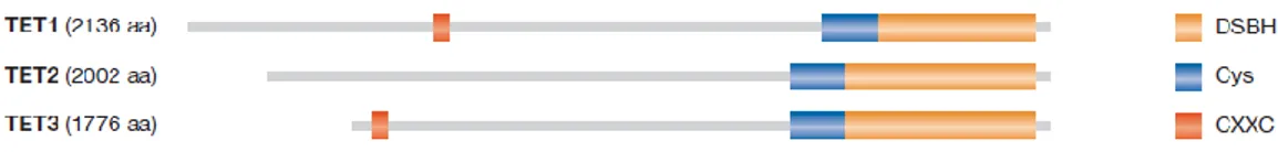
The TET protein family members TET1, TET2 and TET3 are 2-oxoglutarate and Fe-dependent dioxygenases [50,51,52]. The founding member of the TET family of DNA hydroxylases, the TET1 gene, was initially identified in acute myeloid leukemia (AML) as a fusion partner of the histone H3 Lys 4 (H3K4) methyltransferase MLL (mixed-lineage leukemia) [53,54]. TET proteins contain several conserved domains (Figure 5), including a CXXC domain that has high affinity for clustered unmethylated CpG dinucleotides, a cysteine rich region followed by the double-stranded β-helix (DSBH) fold required for catalytic activity [50]. In agreement with the known reaction mechanism of dioxygenases, mutation of putative iron-binding sites of TET proteins abolishes their enzymatic activities [50,51,55].
FIGURE 5. The domain structure of human TET proteins.
Contrarily to 5mC, 5hmC levels fluctuate significantly between tissues, with the highest levels reported in specific cell types of the brain and in ESCs, although these levels decrease during differentiation [50,56,57,58].
TET-mediated conversion of 5mC into 5hmC (Figure 6) could be realized by a passive mechanism but, in contrast to 5mC, 5hmC is not maintained through DNA replication. Alternatively, production of 5hmC could be determined, in non-dividing cells, by an active demethylation pathway during the repletion of 5mC into cytosine [59]. This hypothesis was recently supported by studies demonstrating the existence of formylcytosine and carboxylcytosine in mammalian DNA (Figure 6) [51,60,61]. These cytosine modifications can be generated by two successive oxidation reactions of 5hmC catalysed by the TET proteins, raising the possibility that the TET proteins might be involved in several steps in converting 5mC to cytosine [51,60]. As the TET proteins cannot convert carboxylcytosine to cytosine, a decarboxylase or a glycosylase might be involved in this step. In agreement with this, depletion of thymidine-DNA glycosylase (TDG) leads to accumulation of carboxylcytosine in mouse ESCs, and other studies have shown that TDG is required for DNA demethylation [60,62].
FIGURE 6. Different biological mechanisms of TET-mediated conversion of 5mC to 5hmC.
As to the regulatory role of TET proteins in determining gene transcription, many functions were postulated and some of them demonstrated. Both TET1 and 5hmC localize to TSSs supporting the TET proteins regulative role. However, in TET1-depleted ESCs was found that less than 10% of TET1 target genes change expression after TET1 depletion. Unexpectedly, it was also found that the number of genes down-regulated was similar to, or even lower than, the number of genes upregulated after TET1 depletion, indicating that TET1 could also have repressive effects on gene transcription. In agreement with this, TET1 shares many TET1-bound promoters with the Polycomb repression complex 2 (PRC2) [63,64].
The chromatin-binding ability of PRC2 members was decreased in TET1-depleted cells, suggesting a role of TET1 in PRC2 recruitment. TET1 might indirectly facilitate PRC2 chromatin binding by decreasing DNA methylation levels at PRC2 target genes (Figure 7A) [63,64].
TET1 transcriptional repression could be explained by its association with the Sin3A co-repressor complex (Figure 7B). Indeed, TET1 has been shown to physically interact with the Sin3A complex and dysplay a significant overlap with Sin3A on target genes. Moreover, the recruitment of Sin3A to a subset of these genes was dependent on TET1 expression [63].
Even if the transcriptional activator effects of TET1 are enough weak, the repressive functions are much effective. It seems that 5hmC is enriched at several promoters with intermediate CpG content (Fig 8a). These “weak CpG islands” have a major possibility to undergo methylation during differentiation, suggesting that TET1 ensures that these promoters remain unmethylated in the undifferentiated ESCs [65,66]. Thus, besides its main objective to prevent casual and useless methylation of housekeeping genes, TET1 also protects numerous weak CpG island genes that are predisposed to undergo meth-ylation. Weak CpG islands have been reported to become de novo DNA-methylated during differentiation. They often display high levels of 5hmC, indicating that TET1, by converting 5mC into 5hmC, ensures the opportune methylation and silencing of these target genes during differentiation (Fig 8b).
FIGURE 8. TET1 down-regulation, allows for the repressed genes activat ion.
Unlike the 5hmC, the pattern of 5mC can be maintained permitting the heritability of DNA methylation from a mother cell to its daughter cells.
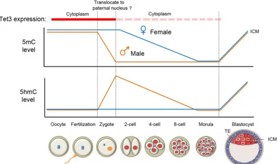
A rapid decrease of global DNA methylation has been observed in developing Primordial Germ Cells (PGCs) as well as zygotes [67,68,69]. In the one-cell zygotes state, the 5mC mark in the paternal pronucleus quickly vanishes (in orange in the top panel of Figure 9)
while the same phenomenon is not observed in the maternal pronucleus. TET3 is highly expressed in the oocyte and one-cell zygote. Instantly after fertilization, TET3 may potentially translocate from the cytoplasm into the paternal nucleus converting 5mC to 5hmC. Then, in paternal and maternal genomes a reduction of 5hmC and 5mC levels occurs due to the initiation of the replication process. Although the exact mechanism is currently unclear, either knockout of TET3 from the female germ cells or siRNA specific for TET3 in the zygote prevent the conversion of 5mC to 5hmC in the paternal genome supporting the hypothesis that TET3 could be responsible for 5mC oxidation in the paternal pronucleus [70,71]. Since the different marking of 5hmC in the paternal pronucleus is observed also in bovine and rabbit zygotes, a conserved role for oxidation of 5mC in the erasure of paternal 5mC after fertilization is suggested [71].
FIGURE 9. Dynamic changes of 5mC and 5hmC levels in the paternal and maternal genomes during pre-implantation development.
3.
Non-CpG methylation
In plants, DNA methylation occurs in both CpG and CpNpG contexts permitting a normal plant development, regulation of transcription and transposition [72]. Plants have two classes of DNA methyltransferases (DNMTs): the MET1 family of methyltransferases and chromomethylases (CMTs). MET1s preferentially methylate cytosine in CpG sites with de
novo and maintenance functions. CMTs are unique to plants and methylate cytosine in
CpNpG sites [72,73].
More recently, non-CpG methylation was found also in mammals, initially in embryonic stem cells then in the promoter regions of different genes. It was largely demonstrated that the amount of this methylation type within exons, introns and 3’ UTRs of H1 embrionic stem cells was double that the CpG one, although gene expression in the H1 cells seem not to be correlated with CG methylation density. The uniqueness of non-CpG methylation in stem cells (it is no longer observed in differentiated cells) is likely to be maintained by continual de novo methyltransferase activity, suggesting that it may play a key role in the origin and maintenance the pluripotent state [11]. BMP4-induced H1 cells, in fact, maintain canonical CpG methylation pattern but misses non-CpG methylation at several loci indicating that, at the time of differentiation, the widespread non-CG methylation is lost [11]. Recently, Ziller and colleagues, through a comprehensive analysis of non-CpG methylation in 76 genome-scale DNA methylation maps across pluripotent and differentiated human cell types, confirmed non-CpG methylation to be predominant in pluripotent cell types observing also a decrease upon differentiation and near complete absence in various somatic cell types [74].
As to non-CpG methylation locus-specific, three independent studies revealed the presence of methylated cytosines within CCWGG sequence (W= A or T) located in the promoter of the murine Sry gene (sex determining region on the Y chromosome) [75]. Barres and coll., using a whole-genome promoter methylation analysis of skeletal muscle from normal glucose-tolerant and type 2 diabetic subjects, identified cytosine hypermethylation of peroxisome proliferator-activated receptor γ (PPARγ) coactivator-1 α (PGC-1α) in diabetic subjects, in which the highest proportion of the above cytosines was found within non-CpG nucleotides. Methylation levels were negatively correlated with PGC-1α mRNA and mitochondrial DNA content [76].
Regarding the enzyme involved in non-CpG methylation, the de novo methyltransferase DNMT3a has been considered to be responsible for the establishment of non-CpG methylation patterns. Indeed, DNMT3a methylates to a lesser extent than CpG sites, both CpA and CpT dinucelotides in vivo after DNMT3a expression in Drosophila. Probably due to the high DNMT3a expression in ES cells rather than in differentiated tissues, the occurrence of non-CpG methylation in ES cells but not in tissues may be explained [77].
4.
The heritability of DNA methylation changes
The definition provided by Conrad Waddington in the 1940 did not take into account the molecular aspects of the stable nature of epigenetics, but the inheritance of environmentally induced phenotypes. These aspects were reconsidered successively by Robin Holliday and Art Riggs in two independent studies [78,79]. The first coined “epimutation” term to distinguish heritable changes due to DNA modifications from classical gene mutations. The second discussed the stable nature of epigenetic inheritance following cell proliferation or mitosis. Hence, it was derived the concept that the epigenetic marks are mitotically and meiotically stable [80].
“Mitotically stable” implies that the epigenetic patterns, during a cell division or proliferation, are replicated. If an epigenetic mark is mitotically stable, then all cells coming from the initial cell will have the same epigenome. On the contrary, an epigenetic mark not mitotically stable would only be relevant in the individual cell and would not be important outside that cell’s function. Mechanisms underlining the above stability are directly correlated to the replication of DNA, associated with mitosis, in which the methyltrasferase methylates the hemimethylated DNA to make the original strand epigenetic mark.
Mitotic stability of the epigenome highlights molecular mechanisms which allow to better understand somatic cell differentiation and function as well as the influence of environment on disease etiology and phenotypic variation.
The epigenome is programmed and maintained in a cell population as it further differentiates and is associated with the development of any tissue or organism.
An environmental factor occurring early in life could modify the epigenome of a somatic cell during a critical window of development and this, as is mitotically stable, would be replicated and influence the somatic cell differentiation and function throughout lifetime. Concerning meiotically stability of epigenetic marks, or better known as epigenetic transgenerational inheritance, the basic mechanisms of this aspect involves the action of an environmental factor (nutrition or chemical) during germline remethylation to permanently alter epigenome of this line, that becomes permanently programmed. Therefore, altered epigenome and phenotype become transgenerational and appear in subsequent progeny and generations in the absence of any further environmental exposure.
Of course, the majority of exposures at other times of development or altering somatic cells, previously described, does not have the capability to become transgenerational. Epigenetic transgenerational inheritance through the permanently altered epigenome of the germ line dramatically influences developmental biology, disease etiology as well as other areas of biology including evolutionary biology. One the best known examples of specific environmentally dependent epigenetic marks transmitted between generations is probable the agouti mice. Morgan et al. studied epigenetic inheritance at the agouti locus. In viable yellow (A(vy)/a) mice, transcription originating in an intracisternal A particle (IAP) retrotransposon, inserted upstream of the agouti gene (A), causes ectopic expression of agouti protein, resulting in yellow fur, obesity, diabetes and increased susceptibility to tumors. Avy mice exhibits variable phenotypes because they are epigenetic mosaics for activity of the retrotransposon: inbred Avy mice, presumably isogenic, have coats that vary in a continuous spectrum from full yellow, through variegated yellow/agouti, to full agouti (pseudoagouti). It was found that the phenotype of a mouse dam with the Avy allele was related to the phenotypes of the offspring, whereby yellow dams produced yellow and mottled offspring, but not pseudoagouti offspring. Interestingly, the passage of the allele through two generations of pseudoagouti females produced significantly more pseudoagouti offspring than through only one generation of pseudoagouti dams [81]. Wolff et al. described how the maternal diet enriched in methyl donors could affect the expression of the agouti gene in the offspring [82].
In humans, the first report of a possible epimutation was published by Buiting et al., who found that epimutations at the SNURF-SNRPN locus correlated with loss of imprinting in patients with Prader–Willi syndrome [83]. Subsequently, Suter et al. reported that germ line epimutation at the DNA mismatch repair gene MLH1 was associated with a greater risk of nonpolyposis colorectal cancer [84]. However, further work by the same group questioned the transmission of this epigenetic mark between generations [85]. Chan et al. also reported a stably inherited, allele-specific, mosaic methylation in the promoter of another DNA mismatch-repair gene (MSH2) in a family affected with nonpolyposis colorectal cancer, although, the presence of the epimutation was not studied in germinal cells [86].
Importantly, two recent studies in monozygotic and dizygotic twins show that specific DNA methylation signatures can be trans-generationally heritable by either genetic or epigenetic mechanisms [87,88].
5. DNA methylation and aging
A growing number of human pathological (such as cancer, muscular dystrophy, lupus) and physiological (such as aging) phenotypes have been found to be associated with peculiar DNA methylation [89,90].
Aging is a process of slow and gradual deterioration of the functional capacities that makes the individual particularly susceptible to environmental challenges and more prone to a variety of illnesses, leading to a dramatic reduction of the individual survival probability and, ultimately, to death. It was largely demonstrated that it is the result of concurrent oxidative stress damage, ion homeostasis deregulation, chromosomal instability and of the accumulation of nuclear and mitochondrial DNA mutations. More recently, evidence about the involvement of epigenomic alterations on inter-individual susceptibility to functional decline and vulnerability to diseases in the elderly people.
5.1 General features of DNA methylation throughout aging
DNA methylation patterns are not fixed; during various stages of mammalian development they are reprogrammed to ensure the normal mammalian embryogenesis and cell differentiation. In particular, in mammalian germ cells and in pre-implantation embryos two waves of genome-wide epigenetic reprogramming occur, generating cells with a broad developmental potential [91].
Historically, the role of epigenetic changes in aging emerges from two independent studies of Berdyshev and Vanyushin, demonstrating as genomic global DNA methylation decreases in spawning humpbacked salmon and in rat brain and heart during aging [92,93]. Subsequent studies have revealed that two specific alterations of DNA methylation occur during aging: a robust and progressive rise in DNA methylation levels across lifespan for several loci and an hypomethylation of ALU and other repetitive elements [94]. Among the above mentioned studies a significant contribution has been provided by the study of mono- (MZ) and dizygotic (DZ) twins, in which gene-specific and global epigenetic differences in monozygotic twins increase overtime within different tissue and cells. These differences are ascribed to stochastic or systematic events, in response to external factor and environment changes. DNA methylation changes in aging exhibit familiar clustering in
individuals that do not share house-holds, suggesting that the DNA methylation stability is genetically determined [95].
A number of specific loci have been described to become hypermethylated with aging. The first gene showing an association between aging and promoter DNA methylation was the one encoding for estrogen receptor (ER). Subsequently, hypermethylation was found in genes encoding for ribosomal DNA clusters as well as in those involved in DNA binding and regulation of transcription, thus affecting a large spectrum of intracellular pathways [96,97]. Genes for tumor suppression (COX7A1, LOX, RUNX3, TIG1, p16INK4A, RASSF1,
DUSP22), development and growth (IGF2, cFos), cell-cell adhesion (CDH1), metabolism
(ELOVL2, SLC38A4, SLC22A18,MGC3207, ECRG4, ATP13A4, AGPAT2, LEP), DNA repair (MLH1) and control of signal transmission (FZD1, FZD7) exhibited altered DNA methylation patterns in aging, displaying sometimes tissue- and cell type-specific features with consequent different functional outcomes [98,99,100]. On the whole, these studies demonstrated a loss of the epigenetic control in aging, suggesting its correlation with age-related pathological phenotypes and physiological processes of aging itself including psycophysical and immune decline, sarcopenia and frailty [101,102,103]. In particular, recently Bellizzi et al. the reported that global DNA methylation levels were correlated to the frailty status in middle/advanced-aged subjects but not with age. Consistently, 7-year follow-up study revealed that a worsening in the frailty status was associated to a significant decrease in the above levels [103]. A gradual loss of DNA methylation with age occurs in most vertebrate tissues and in humans as well [95,104,105,106]. This hypomethylation predominantly affects non island-CpGs and interspersed repetitive sequences (IRSs), such as Alu and human endogenous retrovirus K (HERV-K). More recently, Heyn et al. corroborated and extended the above findings demonstrating that the age-associated hypomethylation is present in all genomic compartments, including promoters, exonic, intronic and intergenic regions [107].
Epigenetic alterations during aging are also attributed to modifications in expression and/or functions of DNA methyltransferases. Indeed, an under-expression of DNMT1 and DNMT3A in human lymphocytes T from elderly individuals and in senescent fibroblasts as well as an increase of DNMT3B in fibroblast was described [108,109]. Moreover, insufficient DNA methylation in Dnmt1+/- mice has been reported to cause immunosenescence, autoimmunity thus affecting healthy aging.
5.2 Factors affecting DNA methylation changes during aging
Non-random mechanisms such as stochastic errors in maintaining established patterns of DNA methylation or environmental stimuli are able to induce changes in epigenetic profiles at both early and late life stages, being in most cases responsible for many processes occurring during the lifespan, including development, differentiation, stress response, and pathological conditions. [110,111]. However, the above mechanisms normally do not mutate DNA sequence nor alter genetic processes indelibly.
The human life expectancy experienced a remarkable increase during the last century with an unprecedented gain in the developed world [112]. At the same time, researches have shown even larger improvement in health expectancy than in lifespan [113,114]. These phenomena suggest the important role of improving environment in determining individual health status and survival. Social-economic development and consequent advances in biomedical technology together with improved healthcare and disease treatment can be considered in explaining these changes. Recent works in scientific literature have shown that the epigenome could acquire and undergoes the impact of environmental factors [115,116,117,118]. As previously described, studies on monozygotic twins have highlighted the contribution of environmental factors to inter-individual susceptibility to disease as well as to determine lifespan, independently from genetic components of aging [119,120,121,122,123] [122,123]. A recent study demonstrated that in Calabrian population there is a greater genetic component in longevity in males than in females [124]. So genetic and environmental factors work together to determine many phenotypes in which the latter could have a major importance than the former and vice versa depending on the considered phenotype.
Several evidence, in keeping with the so-called “the fetal basis of origins of adult-onset disease” theory, indicate the mother's behavior or diet can affect epigenetic patterns in her offspring. A study carried out on Agouti pregnant mice showed that feeding the mice with a diet rich in methyl donors influences coat color, body weight, and health of their progeny [125]. Other studies conducted in rats showed that demethylation of both glucocorticoid (GR) and estradiol (ER) receptors, in the absence of appropriate nurturing and following exposure to a high-fate diet, respectively, induced increased expression of the receptor genes later in life, thereby leading to an increased stress sensitivity and a higher incidence of cancer in offspring. A diet supplemented with antioxidants like folate, choline and vitamins B6 and B12 have been demonstrated very effective for aging prevention, through different mechanisms, in several animal models. Their activity could reduce
overproduction of free radicals, which are responsible of the conversion of 5mC into 5hmC, with consequent loss of DNA methylation.
Calorie restriction (CR) is by far the most effective environmental manipulation that can extend maximum lifespan in many different species [126,127]. Although it was reported that CR acts via reduction of oxidative stress or regulation of metabolic pathways, precise mechanisms are not very well understood. Recent data suggest that DNA methylation modifications involving specific genes play an important role in CR-dependent aging and longevity. Evidences suggest that the biological effects of CR are closely related to chromatin function [128]. In fact, acting as an important environmental intervention, CR is speculated to exert its aging-delaying effect through its capacity to increase genomic stability. Reversal of aberrant DNA methylation patterns during aging is believed to be much effective for CR to maintain chromatin function and thus to influence aging processes. Interestingly, CR is likely to recover the above aberrant patterns by specific gene control rather than globally [129]. Although the majority of CR research has been based on experimental animal studies, Li et al. have generated an in vitro mammalian cellular system to mimic CR-controlled longevity by the reduction of glucose, the main caloric resource in cell culture medium [130]. DNA hypermethylation was found in the promoter of the p16INK4a gene, an important tumor suppressor and aging-associated gene, in correspondence of an E2F-1 binding site. This DNA hypermethylation blocks access of E2F-1, an active transcription factor of p16INK4a, to the p16INK4a promoter, resulting in p16INK4a down regulation, which contributes to CR-induced lifespan extension. Similar mechanisms were described for Ras gene. In this regard, there is a strong tendency for the DNA methylation pathway to predominately control key cancer-related genes during CR, suggesting a close connection between aging and cancer.
What is more, CR leads to aberrant DNA methylation patterns likely by modulating DNMT function. Indeed, DNMT1 activity is significantly elevated in response to CR, likely to correct the decreased methylation level during aging [130]. Evidence indicate also that Dnmt3a level changes induced by CR in the mouse hippocampus may promote brain function during aging [131].
Lastly, elements including chromium, zinc, selenium or arsenic have been demonstrated to affect DNA methylation during the lifetime. These elements are capable of reducing methylation levels at genetic loci inhibiting the activity of DNA methyltransferases. Many chemical compounds and xenobiotics, such as diethylstilbestrol (used to prevent miscarriages), bisphenol A (used in plastic industry) and vinclozolin (a fungicide used in
vineyards), alter DNA methylation at a global and/or locus-specific level, acting as endocrine disruptors with consequent developmental disorders and tumourigenesis [132].
6.
Mitochondria, mitochondrial DNA replication and transcription
Mitochondria are essential cell organelles whose primary function consists in converting dietary calories in ATP via the electron transport chain (ETC) and the oxidative phosphorylation process (OXPHOS), thus implying that mitochondria have a central position between energy uptake and energy production [133]. Mitochondria are emerging to be involved in several cellular processes, including ion homeostasis, cell proliferation and differentiation, stress response and apoptosis.
Mitochondria are the sole cellular organelles that carry an own DNA (mtDNA). The human mtDNA is a 16569 bp closed-circular and double-stranded molecule, which sequence has been entirely determined in 1981 [134,135]. It has a very compact structure lacking of introns and intergenic spacing and with overlapping genes (ATPase 6 and 8, and ND4 and ND4L subunits).
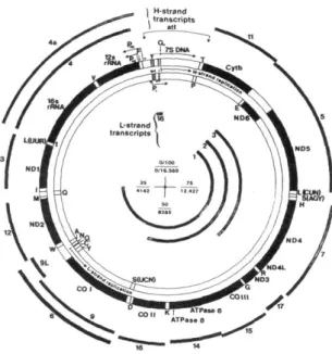
MtDNA contains 37 genes encoding two ribosomal RNAs (rRNA 12S and 16S), 22 transfer RNAs and 13 polypeptides, all of which are components of the OXPHOS system (Figure 10).
FIGURE 10. The gene products encoded by the L-strand are shown in the inner complete circle and the gene products of the H-strand in the outer complete circle.
Seven of the OXPHOS genes (MTND1, MTND2, MTND4L, MTND4, MTND5, and MTND6) encode subunits of respiratory Complex I (NADH dehydrogenase or NADH:ubiquinone oxidoreductase); one gene (MTCYB) encodes a component of Complex III (ubiquinol:cytochrome c oxidoreductase); three genes (MTCO1, MTCO2 and MTCO3) encode constituents of Complex IV (cytochrome c oxidase or COX); and two genes (MTATP6 and MTATP8) encode subunits of respiratory Complex V (ATP synthase). The remaining OXPHOS subunits, as well as all the factors required for maintenance, replication and expression of mtDNA, are nuclear-encoded, translated on cytoplasmic ribosomes and imported to their final mitochondrial compartment.
A series of distinctive features of mtDNA make it particularly interesting for genetic studies: i) cells are polyploid with respect to mtDNA: most mammalian cells contain hundreds of mitochondria and each mitochondrion contains several copies of mtDNA [136,137]. In a given individual, all mtDNA copies can be identical, a condition known as homoplasmy, although mutations can arise, be maintained or amplified to different levels and coexist with wild-type mtDNA, so called heteroplasmy; ii) the mitochondrial genome is maternally inherited, and does not undergo recombination; the few mitochondria from the sperm cell that could enter the oocyte during fertilization are actively eliminated by ubiquitin-dependent mechanisms [138,139]; iii) the evolution rate of mtDNA is much faster than that of the nuclear genome [140]. This aspect is attributable to several reasons: mtDNA is less protected by proteins, it is physically associated with the mitochondrial inner membrane where ROS are generated, and it appears to have less-efficient repair mechanisms than the nucleus [141]. This high mutation rate and the maternal inheritance pattern have made mtDNA sequence analysis an interesting tool in human population genetics and evolutionary studies [142]; iv) Mitochondrial genes are translated using a genetic code with some differences from the universal genetic code. In fact, in mammals UGA specifies tryptophan instead of a termination codon, AUA, AUC and AUU are used as initiation codons and AGA and AGG are termination codons instead of encoding arginine. In addition, a specific codon–anticodon pairing system allows translation to proceed with only 22 tRNAs. [134,135,143,144,145,146].
The two strands of the mtDNA have an asymmetric distribution of guanines and cytosines generating heavy (H) and light (L) strands, respectively. The H-strand encodes for the majority of the genes; the L-strand encodes only eight genes for tRNAs and the gene of the ND6 OXPHOS subunit. Each strand is transcribed from one predominant promoter, HSP (high strand promoter) and LSP (light strand promoter) both having a bipartite structure.
One element contains a consensus sequence motif of 15 bp surrounding the initiation points and is essential for transcription. A second element, located immediately upstream of the initiation point is required for optimal transcription and can be considered as an enhancer. In this element it is included the binding site for mitochondrial transcription factor A (mtTFA), main regulator of mtDNA expression. The two promoters are located in the control region which includes the displacement loop (so called D-loop). The D-loop is the sole non coding region, with a triple-stranded structure generated from a template-directed termination of H-strand DNA synthesis soon after initiation carrying out to the synthesis of a short piece of DNA, the 7S DNA. This fragment remains stably hybridized to the parental molecule, thus forming the triple-strand structure characterized by the displaced parental H-strand.
Transcription occurs from the HSP or the LSP in opposite directions around the entire genome circle. The H-strand is transcribed by two overlapping units. One starts at the initiation site H1, located upstream of the tRNAPhe gene and ends at the 16S rRNA 3’ end. This unit operates much more frequently than the second one and is responsible for the synthesis of the two ribosomal RNAs, tRNAPhe and tRNAVal. The activity of this unit is linked to a transcription termination event taking place immediately downstream from 16S rRNA, inside the gene for tRNA Leu. The second transcription unit, operating with a frequency about 20 times lower, starts at the initiation site H2, close to the 12S rRNA 5’ end and originates a polycistronic molecule covering almost the whole H-strand [147,148]. Therefore, a differential regulation of rRNA versus mRNA transcription can be operated through the initiation of H-strand transcription at the two alternative sites.
The L-strand gives rise to a single polycistron starting at the 5’ end of 7S RNA, about 150 bp away from the H1 initiation point, from which the eight tRNAs and the ND6 mRNA are derived. To date it has not been clearly established whether transcription occur from both promoters at the same time on the same molecule or two independent events take place. A single-subunit mitochondrial RNA polymerase (mtRNAP), that is distantly related to the RNAP of bacteriophage T7, the pol I family of DNA polymerases, and single-subunit RNAPs from chloroplasts, is responsible of both the transcription for RNA synthesis and for initiation of replication. mtRNAP, which X-structure was recently characterized, requires the factors mtTFA and mtTFB2 for binding and melting promoter DNA [149]. Long polycistronic RNA chains are produced and subsequently cleaved on both sides at tRNA level into distinct mature RNA molecules. Additional transcript processing intermediates have been observed in selected tissues and in association with various
pathogenic mtDNA mutations [150,151,152,153]. Polyadenylation of mRNAs occurs during or immediately after cleavage.
mtDNA replication starts at OH (origin of H-strand), located in the D-loop region
downstream of the LSP, and proceeds along the parental L-strand to produce a daughter H-strand circle. When H-H-strand replication reaches the origin of L-H-strand replication (OL),
approximately located at two thirds of the genome, within a cluster of five tRNA genes, the parental H-strand is displaced, OL is exposed and its replication starts and proceeds in the
opposite direction producing a daughter L-strand. Initiation of H-strand replication needs short RNA primers, which are originated by the processing of L-strand promoter transcripts. Therefore, replication of mammalian mtDNA is functionally linked to mitochondrial transcription and, hence, depends on the activity of regulatory factors required for L-strand transcription. Briefly, during the replication initiation mtRNA polymerase starts transcription from the L-strand promoter producing the primer precursor. This RNA remains hybridized with a region of DNA upstream of OH containing the CSBs,
highly conserved sequences, forming a stable R-loop structure. A mitochondrial RNA-processing endoribonuclease (RNase MRP) cuts the RNA primer precursor generating the mature primer(s) for replication. Finally, the mitochondrial DNA polymerase starts H-strand replication through the extension of an RNA primer. Then, H-H-strand synthesis events or are arrested around the TAS sequences, short (15 bp) sequences conserved in vertebrates, creating the triplex D-loop structure or replication proceeds over the entire length of the genome. The precise mechanisms regulating the two events are not yet fully understood. This initiation of replication from OL requires a specific primase capable of
generating short RNA primer molecules. Once initiated, L-strand replication proceeds over the entire length of the strand and ends after the H-strand.
Both transcription and replication of mtDNA are regulated mainly by mtTFA transcription factor, that it is not only required for transcription initiation, but also seems to have a direct role in mtDNA maintenance.
mtTFA binds throughout the D-loop with a 40 to 50 base periodicity, with MTCSB2 and MTCSB3 being unbound and MTCSB1 being strongly bound. The mtTFA phasing downstream from MTCSB1 corresponds to DNA synthesis initiation sites, thus suggesting that mtTFA play a role in defining the transition from RNA to DNA [154]. Results from in
vitro transcription assays suggested that the amount of TFAM bound to DNA dictates
whether LSP or H1 is activated; this promoter-switching activity was recently confirmed quantitatively with an assay utilizing recombinant mitochondrial polymerase and
mitochondrial transcription factor 2B. At low TFAM concentration LSP is preferentially activated, and as TFAM concentration increases, transcription activity switches to H1. In addition, it was demonstrated that TFAM alone is sufficient to compact DNA in vitro, likely through DNA looping to reduce contour length, and supercoiling to impart structural rigidity. The rules governing TFAM–mtDNA interactions should allow for a fine-tuning of TFAM activity that integrates specific and non-specific DNA binding functions to ensure proper maintenance of mtDNA [155]. However, controversial data emerged about the activity of mtTFA, since it was demonstrated that the reduction in its levels in heterozygous knock-out mice was accompanied in all tissues by a drop in mtDNA levels but not in mtRNA levels [156].
7. Mitochondria and aging
The role of mitochondria in the aging process has been a topic of intense interest for many years; in fact age-related changes in mitochondrial content, structure and function, as well as in mitochondrial DNA have been extensively documented. A series of studies have demonstrated a decline of the mitochondrial respiration efficiency with age in human and primate and that this decline can be attributed either to a progressive down regulation of genes encoding for mitochondrial proteins such as several subunits of cytochrome-c oxidase, NADH dehydrogenase and ATP synthase, or to the decline of mitochondrial biogenesis with aging [157,158,159,160,161,162].
It was also documented that the decline in OXPHOS activity, and in turn in the overall mitochondrial function, is correlated with an accumulation, in post-mitotic tissues during human aging, of mitochondrial DNA mutations, including point mutations, large scale deletions and duplications. Among the variety of mtDNA alterations, the most prevalent age-associated point mutations of mtDNA are A3243G and A8344G transition, while the most common mtDNA deletion in aging human tissues is the 4977 bp deletion [163,164]. The creation of mtDNA-mutator mice, that are knock-in mutant mice expressing a proofreading-deficient version of the mitochondrial DNA polymerase γ gene (POLG), has provided the first direct evidence that accelerating the mtDNA mutation rate can result in premature ageing, consistent with the view that loss of mitochondrial function is a major causal factor in ageing. Mitochondria accomplish numerous tasks crucial for cellular and
organism good health status in eukaryotes. Many cell lines that are genetically modified and constructed to study pathological alterations show an altered energetic metabolism or a defective mitochondria. For this reason mitochondrion is an important cellular organelle that permit to maintain a subtle regulation of cellular energy metabolism and so has a significant role in some complex phenotype or disease. As a complex phenotype, during aging some mitochondrial functions are altered. However, for many years if is mitochondrial dysfunction that cause aging or if the natural process of aging that altered normal mitochondria remained an unsolved question.
Since mitochondria is the main source of reactive oxygen species (ROS) and free radicals, molecules both involved in cellular age-dependent damage, researchers proposed the connection between mitochondria and aging [165]. Mitochondrial role in aging was refined by Miquel and colleagues that in 1980 stated that these organelles begin cellular processes leading to aging since they are in the main source of ROS and the principal target of their injury [166]. In fact, the age-related accumulation of free radicals in the mitochondrion has heavy effects on the integrity of the organelle itself that are indeed oxidized by these reactive species [133].
A healthy mitochondrion has the ability to produce the amount of ATP proportional to the energy demand. Thus, the equilibrium between mitochondrial functions and dysfunction is necessary for retarding or avoiding cellular senescence and extend an organism survival probability.
Mitochondrial theory of cellular aging relies on the fact that mitochondrial DNA (mtDNA) has a high rate of mutation and limited capacity for repair. Essential for mitochondrial function is the integrity of the mtDNA. Nonetheless, over time an accumulation of mtDNA mutations compromises mitochondrial genome [167]. Thus, while continue to release higher quantity of ROS the organelle progressively lose their ability to generate energy [168]. During the aging of tissues the amount of cells containing mitochondria whit altered functions raises. This caused cellular injure and/or death that induce degeneration of tissues and organism disorders, which are present in age-related diseases that could lead to the individual death. Interesting is the definition of the “aging clock” that considers mtDNA and its mutation like a clock that marks the organism’s time and initiate the aging events determining the organism lifespan [169].
Rates of mtDNA mutation have been reported to be 15-fold higher than that of nuclear DNA (nDNA). This induce abnormal expression of electron transport chain (ETC) proteins unbalancing the oxidative phosphorylation process [162,170]. ETC dysfunction decreases
ATP production and raises ROS production that lead to impair nucleotide synthesis, that, in turn, affects nDNA genes [171].
Human mtDNA contain 37 genes encoding 13 polypeptides that compose subunits within the OXPHOS complexes (see paragraph 7). Histone are not present in mtDNA and so mitochondrial nucleic acid does not has its protection. In addition, mtDNA and has a less effective repair mechanisms than nDNA [171,172]. As mentioned above, mutations in mtDNA lead to mitochondrial dysfunction and increased ROS production. As its location in the mitochondrial matrix, mtDNA is directly involved into harmful consequence of ROS production making it highly prone to oxidative damage [171]. Indeed, in rat liver cells was found that the total dosage of 8-hydroxydeoxyguanosine, a marker of DNA oxidative damage, was 16 times higher than that of nDNA [172]. Together, this damage results in numerous pathological anomalies, which are related to the degree of mtDNA alterations, quantity of altered mitochondria and the kind of tissue involved [173,174]. The age-related tissues deterioration in heart, in nervous system and in kidney has been associated with the gradual accumulation of mtDNA mutations [175].
The employment of mice with a proof-reading-deficient version of mtDNA polymerase γ (POLG) permitted to shed light on the connection between mutated mtDNA and aging. This defective POLG lead to an increase three to five folds of point mutations along with broken linear mtDNA and deletions. Up to their early adolescence these “mutant mice” were in a good status of health, but in the course of time they showed the onset of aging phenotypes such as osteoporosis, weight loss, heart enlargement, anemia, curvature of the spine, hair loss, and reduced fertility, associated with significant reduction of life span [176].
Miquel and colleagues in 1980 stated that mitochondria are involved in regulating cellular processes leading to aging since they are the main source of Reactive Oxygen Species (ROS) as well as principal targets of their injury [166]. A progressive and irreversible accumulation of the above species is realized during aging with consequent heavy effects on the integrity of the organelle itself that are indeed oxidized by these molecules, as emerged in the well known theory of aging [133]. Mitochondrial DNA is especially susceptible to attack by ROS, for several reasons: i) its close proximity to the electron transport chain, the major site of ROS production; ii) the size and the compactness of the genome; iii) the lack of protective histones; iv) the absence of adequate mitochondrial DNA repair systems.