High-sensitivity assay for
monitoring ESR1 mutations in
circulating cell-free DNA of breast
cancer patients receiving endocrine
therapy
Laura Lupini
1, Anna Moretti
1,2, Cristian Bassi
1, Alessio Schirone
2, Massimo Pedriali
3,
Patrizia Querzoli
1,3, Roberta Roncarati
4, Antonio Frassoldati
1,2& Massimo Negrini
1Approximately 70% of breast cancers (BCs) express estrogen receptor alpha (ERα) and are treated with endocrine therapy. However, the effectiveness of this therapy is limited by innate or acquired resistance in approximately one-third of patients. Activating mutations in the ESR1 gene that encodes ERα promote critical resistance mechanisms. Here, we developed a high sensitivity approach based on enhanced-ice-COLD-PCR for detecting ESR1 mutations. The method produced an enrichment up to 100-fold and allowed the unambiguous detection of ESR1 mutations even when they consisted of only 0.01% of the total ESR1 allelic fraction. After COLD-PCR enrichment, methods based on next-generation sequencing or droplet-digital PCR were employed to detect and quantify ESR1 mutations. We applied the method to detect ESR1 mutations in circulating free DNA from the plasma of 56 patients with metastatic ER-positive BC. Fifteen of these patients were found to have ESR1 mutations at codons 536–538. This study demonstrates the utility of the enhanced-ice-COLD-PCR approach for simplifying and improving the detection of ESR1 tumor mutations in liquid biopsies. Because of its high sensitivity, the approach may potentially be applicable to patients with non-metastatic disease.
Breast cancer (BC) is the most commonly diagnosed neoplastic disease in women worldwide and has a high inci-dence in Western countries where it is the second leading cause of cancer-related death1.
Approximately 70% of breast tumors express estrogen receptor alpha (ERα) at diagnosis; proliferation and survival of neoplastic cells are dependent on estrogen stimulation2. Patients with these cancers are administered
endocrine-based therapies that stop or slow tumor growth via various mechanisms of action. Therapeutic agents include tamoxifen, a specific estrogen antagonist; aromatase inhibitors (AIs), which suppress estrogen produc-tion; and fulvestrant, which promotes ERα degradation.
Antiestrogenic drugs produce survival benefits in patients with BC; however, one-third of patients develop resistance to therapy. Missense mutations in the ESR1 gene, which encodes ERα, represent an important mech-anism leading to endocrine resistance3. Most mutations of the ESR1 gene are found in codons 536–538. These
mutations have been shown to promote ERα transcriptional activity in an estrogen-independent manner4.
Among the mutations, Y537S, Y537N, Y537C, and D538G represent more than 80% of the abnormalities found in resistant cases5,6.
Such mutations have been identified in approximately 15–20% of ER-positive (ER+) metastatic lesions from patients treated with endocrine therapy, but rarely in primary tumors. It is therefore believed that these altera-tions are selected from rare mutant clones to confer resistance to therapy and possibly favor the development of
1Universit di Ferrara, Dipartimento di Morfologia, Chirurgia e Medicina Sperimentale, Via Luigi Borsari, 46, 44121,
Ferrara (FE), Italy. 2Azienda Ospedaliero Universitaria di Ferrara, Divisione di Oncologia clinica, via Aldo Moro, 8,
44124, Cona (FE), Italy. 3Azienda Ospedaliero Universitaria di Ferrara, Unità di Anatomia Patologica, via Aldo Moro,
8, 44124, Cona (FE), Italy. 4Institute of Genetics and Biomedical Research, Consiglio Nazionale delle Ricerche,
Milano (MI), Italy. Laura Lupini and Anna Moretti contributed equally to this work. Correspondence and requests for materials should be addressed to A.F. (email: [email protected]) or M.N. (email: [email protected])
Received: 16 January 2018 Accepted: 19 February 2018 Published: xx xx xxxx
metastatic disease6. It is thus essential to detect these mutations as soon as possible to select the best therapeutic
options.
Tissue biopsy is generally not a suitable approach for the frequent monitoring of disease because the inva-sive nature of the required procedures; moreover, mutation could be missed because of tumor heterogeneity. These limitations can be overcome by a liquid biopsy approach, based on analysis of circulating cell-free DNA (cfDNA) to monitor patients with advanced cancer during clinical follow-up. Such patients have cfDNA that is often enriched with tumor DNA (ctDNA), which makes it possible to pinpoint genetic or epigenetic changes that are present in tumor cells7–9. Assessing ctDNA is minimally invasive and, more importantly, can detect mutations
from hidden metastases and genetically heterogeneous tumors.
The technical challenges of this type of analysis are related to the low amount of cfDNA present in plasma as well as the low proportion of ctDNA. Therefore, high sensitivity of detection is essential. Several studies have been published in recent years using next-generation sequencing (NGS), real-time PCR, or droplet digital PCR (ddPCR) to perform liquid biopsy tests aimed at revealing specific cancer-associated changes in cfDNA10–14. Some
of these studies have been aimed at identifying ESR1 mutations in the cfDNA of patients with endocrine-resistant breast cancer4,15–19.
To improve the sensitivity of mutation detection, methods have been developed to enrich low-frequency allelic variants (COLD-PCR and its derivatives)20–22. In particular, such approaches have been reported to enrich the BRAF and KRAS variants associated with cancer21,23. In this study, we developed an enhanced-ice-COLD-PCR
(E-ice-COLD-PCR)-based method for the enrichment of ESR1 gene mutations at codons 536–538. We demon-strated that the use of this approach consistently improved detection of ESR1 alterations compared to other cur-rently employed methods. We tested this approach in a large group of patients with metastatic BC to investigate its potential clinical applicability.
Results
ESR1 gene mutations in primary and metastatic breast cancer lesions.
With the aim of estab-lishing a sensitive method for the detection of ESR1 mutations in cfDNA, we identified ESR1 mutant alleles by investigating tumor tissue samples from a cohort of 40 patients with metastatic BC (Table 1). All patients were diagnosed with ER+ BC and treated with endocrine therapy. None of the patients had metastasis at diagnosis. Primary tumor samples (N = 40) and metastatic lesions (N = 47) were from matched patients. In these sam-ples, we examined mutations to codons 536–538 of the ESR1 gene using Sanger sequencing. We identified ESR1 mutations in 6 metastases (none of which were in primary tumors): Y537S was found in 3 samples, D538G in 2 samples, and Y537C in 1 (Table 1).DNA samples with ESR1 mutations were employed to develop a method for the specific enrichment of mutant alleles present in the ESR1 536–538 codons. Based on the E-ice-COLD-PCR method21,23,24, we designed
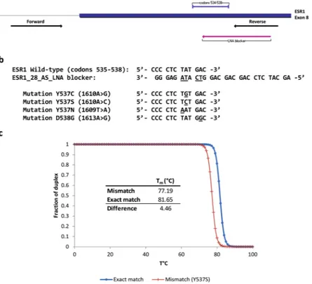
prim-ers for PCR amplification as well as a partially overlapping oligonucleotide blocker (Fig. 1a,b). The blocker was designed to include Locked nucleic acid (LNA) modified-nucleotides at the putative mutant codons and a phos-phate group at the 3′-end to block its extension. The melting temperature of the blocker was 81.7 °C if matched to a wildtype sequence, but lower (77.2 °C for Y537S) if a mutation was present (Fig. 1c). The different melt-ing temperatures functioned to block or limit the amplification of the wildtype sequence and thereby favor the enrichment of any present mutant allele.
To test the ability of the method to enrich mutant alleles, DNA from mutant samples (Y537S, Y537C, and D538G) was diluted in normal DNA to achieve allelic frequencies of 1% and 0.5%. After performing E-ice-COLD-PCR, amplicons were analyzed by NGS to measure the achieved frequency of mutation. All 3 muta-tions were found to be considerably enriched (9–70-fold). No mutated ESR1 was amplified in SW480 colorectal cancer cell DNA, which was used as a negative control (Table 2). The concentration of the blocker that produced the highest Y537S mutation enrichment was 80 nM (Supplementary Figure 1).
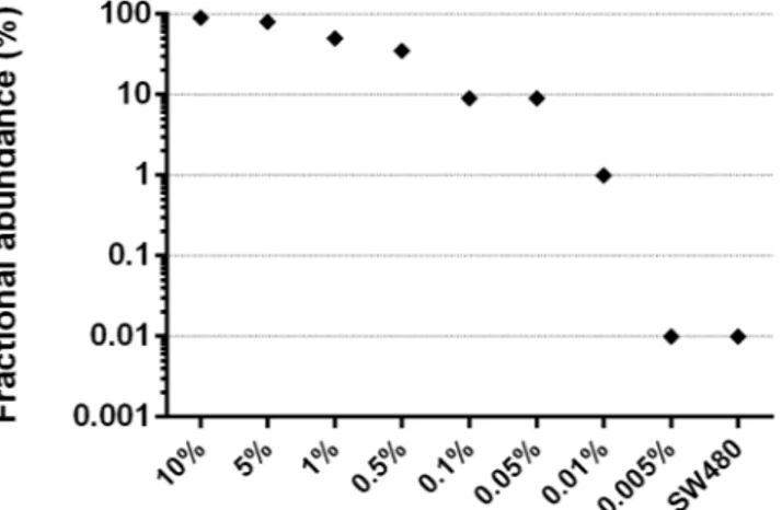
After demonstrating the potential of the method to increase the frequency of ESR1 mutant alleles, we next evaluated its lower limit of detection by designing fluorescent probes capable of discriminating the Y537S mutant from wild type DNA. We serially diluted the Y537S mutant DNA in normal DNA; the smallest dilution was 0.005% (1 mutant among 20,000 molecules). All dilutions were subjected to E-ice-COLD-PCR in duplicate, and the resultant amplicons were quantified by droplet-digital PCR (ddPCR) using fluorescence-specific probes for either the Y537S or wild type allele. The mutant allele was detected at a minimum original dilution of 0.01% (i.e., detected at 1%, with 100-fold enrichment) after the application of E-ice-COLD-PCR (Fig. 2).
ESR1 gene mutation in plasma of breast cancer patients.
To test the assay in a clinical setting, we analyzed DNA from the plasma of 56 patients with metastatic ER+ breast cancer. We performed E-ice-COLD amplification in the hotspot region of the ESR1 gene. The resulting amplicons were analyzed using both ddPCR (for the Y537S variant) and NGS for all mutation types. Overall, 15 patients (27%) were found to have a muta-tion in codons 536–538 (Table 3 and Supplementary Figure 2). The results for the detection of the Y537S variant obtained with both methods were consistent (Table 3). Additionally, the experiment also proved that specificity of the method of detection based on ddPCR labeled-probe was 100%, since not only negative samples remained negative but also mutants other than Y537S ESR1 were negative when assayed for the Y537S mutation.In 6 patients (S-26, S-27, S-28, S-31, S-51, and S-60), tumor and metastasis tissues were available among samples initially analyzed for the mutational status of ESR1. Four of these patients (S-26, S-27, S-28, and S-60) did not show any ESR1 mutation, while the remaining 2 patients (S-31 and S-51) showed mutations in samples derived from metastases. Analysis of the corresponding cfDNA revealed what follows: in 3 cases, the results were consistent in that 2 patients (S-27 and S-60) had no mutations while 1 (S-31) had the same mutation in both the metastasis and cfDNA (Y537S). In the remaining 3 cases, 2 patients (S-26 and S-28) exhibited ESR1 mutations in
Patient Tumor tissue Metastasis site Date ER PR HER2 MIB1 ESR1 status (codons 536–538) S-26 primary 23/01/2013 93% 72% 1+ 42% WT metastasis liver 17/03/2015 95% 10% 1+ 20% WT S-27 primary 19/04/2001 86% 84% 0 7% WT metastasis skin 16/09/2008 94% 56% 0 24% WT S-28 primary 23/12/2006 100% 90% 1+ 20% WT metastasis liver 28/10/2013 99% 30% 1+ 35% WT S-30 primary 16/01/2014 56% 35% 1+ 45% WT metastasis skin 31/03/2015 95% 41% 2+ 48% WT S-31 primary 17/05/2013 96% 82% 1+ 15% WT
metastasis skin 14/09/2015 99% 98% 2+ 24% Y537S
S-32 primary 31/05/2011 64% 41% 0 22% WT metastasis skin 04/03/2015 85% 0% 0 2% WT S-33 primary 12/05/2010 22% 0% 1+ 84% WT metastasis skin 11/05/2011 25% 0% NA NA WT S-34 primary 11/02/2008 60% 10% 3+ 50% WT metastasis skin 05/05/2012 4% 0% 3+ 57% WT S-35 primary 05/08/2009 96% 8% 1+ 50% WT metastasis brain 31/07/2014 94% 0% 1+ 27% WT S-36 primary 07/10/2010 75% 23% 1+ 2% WT metastasis 1 ovary 31/05/2013 90% 71% 1+ 19% WT metastasis 2 ovary 31/05/2013 NA NA NA NA WT S-37 primary 16/10/2007 100% 100% 0 1% WT
metastasis ovary 05/10/2011 95% pos NA NA WT
S-38 primary 04/04/2013 78% 2% 1+ 39% WT metastasis liver 14/09/2015 30% 2% 1+ 43% WT S-39 primary 04/04/2006 100% 76% 3+ 17% WT metastasis 1 liver 18/04/2013 98% 0% 3+ 15% WT metastasis 2 liver 24/02/2014 99% 0% 3+ 20% D538G S-40 primary 15/06/2006 99% 80% 0 26% WT metastasis liver 10/12/2014 0% 25% 0 40% WT S-41 primary 25/02/2009 48% 46% 3+ 42% WT metastasis liver 07/07/2014 91% 79% 3+ 48% WT S-42 primary 03/04/2012 53% 4% 3+ 19% WT metastasis liver 26/03/2014 58% 0% 3+ 30% WT S-43 primary 13/11/2009 88% 26% 2+ 16% WT metastasis liver 26/11/2012 12% 0% 1+ 40% WT S-44 primary 23/02/2009 45% 48% 1+ 25% WT metastasis liver 13/05/2011 74% 69% 1+ 65% WT S-45 primary 10/03/2009 98% 56% 1+ 12% WT
metastasis liver 26/04/2011 pos NA NA NA WT
S-46 primary 18/12/2006 99% 0% 3+ 25% WT
metastasis brain 26/02/2011 42% 0% 3+ 38% WT
S-47 primary 30/05/2002 99% 40% 0 5% WT
metastasis ovary 20/09/2013 99% 99% 1+ 10% Y537S
S-51 primary 28/03/2007 99% 99% 0 5% WT
metastasis lung 27/12/2013 98% 95% 0 18% Y537C
S-52 primary 13/03/2008 89% 0% 1+ 16% WT
metastasis liver 20/04/2009 15% NA NA NA WT
S-53
primary 25/03/2005 NA NA NA NA WT
metastasis 1 ovary 20/04/2011 55% 21% 0 5% WT
metastasis 2 liver 31/10/2012 98% 15% 0 60% Y537S
S-54
primary 30/12/2010 31% 30% 3+ 45% WT
metastasis 1 brain 04/10/2012 0% pos pos NA WT
metastasis 2 brain 03/09/2014 5% 3% 3+ 30% WT
metastasis 3 brain 16/04/2015 1% 0% 3+ 35% WT
their cfDNA but not in the original metastasis biopsy, while the remaining patient (S-51) exhibited the opposite situation.
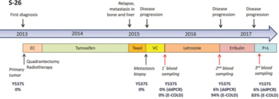
Patient S-26 had a markedly long interval between biopsy and blood withdrawal. In this patient, multiple blood samplings made it possible to monitor the evolution of the cancer’s status over time. Analysis of metastatic tissue and liquid biopsy samples collected in the spring of 2015 showed that both samples were negative for ESR1 mutations. One year later (May 2016), the liquid biopsy was positive for the ESR1 Y537S mutation. The patient was administered letrozole therapy between Spring 2015 and Spring 2016, raising the possibility that this therapy was responsible for selecting the mutant neoplastic clone (Fig. 3).
For patient S-28, a similar situation can be envisioned, since 33 months had elapsed between the liver metas-tasis biopsy (October 2013) and blood withdrawal (May 2016). Considering that the patient underwent several consecutive lines of endocrine therapy, it is plausible that the ESR1 mutation was not detectable in October 2013 but was subsequently selected.
Patient S-51 showed an opposite pattern: the liquid biopsy obtained in September 2016 was mutation-negative, while the metastasis as evaluated in December 2013 was positive for the Y537C mutation. The patient was admin-istered AIs until December 2013, and was on fulvestrant therapy starting from January 2014, suggesting that the latter was effective in eliminating cells with ESR1 mutations.
Discussion
In recent years, liquid biopsy technology has evolved rapidly because of its great potential and minimal invasive-ness. cfDNA can be used to monitor the evolution of mutations associated with neoplastic disease in real time, reflecting subclonal dynamics linked to the heterogeneity of neoplasms or the development of new cancer cell clones and metastases25.
However, technical challenges that are mainly related to the small amount of cancer DNA found in cfDNA remain. The use of technologies, such as NGS and ddPCR, partially overcome this problem and allow for the detection of mutations that are present in DNA at fractions as low as 1%. Such technologies have also been used to detect ESR1 mutations in metastatic BC15,17,19,26–28. However, the sensitivity of these technologies is dependent on
the quality and quantity of DNA isolated from plasma, making difficult to identify mutations that are present at
Patient Tumor tissue Metastasis site Date ER PR HER2 MIB1 ESR1 status (codons 536–538)
S-55 primary 25/05/2006 99% 63% 0 41% WT
metastasis skin 04/09/2007 90% 45% NA NA WT
S-56 primary 22/04/2008 99% 99% 0 2% WT
metastasis ovary 30/11/2013 pos pos 0 NA WT
S-57 primary 04/04/2001 87% 93% 0 40% WT
metastasis lung 19/01/2015 98% 35% 1+ 60% WT
S-58 primary 16/05/2012 75% 46% 3+ 33% WT
metastasis brain 12/11/2014 pos NA NA NA WT
S-59 primary 19/12/2008 55% 36% 1+ 27% WT metastasis liver 16/04/2010 52% 27% 1+ 14% WT S-60 primary 12/12/2006 98% 0% 2+ 45% WT metastasis liver 17/11/2015 98% 0% 2+ 45% WT S-61 primary 06/09/2006 99% 91% 0 26% WT metastasis liver 21/03/2014 99% 30% 1+ 35% WT S-62 primary 20/11/2001 NA NA NA NA WT
metastasis ovary 06/07/2010 pos pos NA NA WT
S-63 primary 13/06/2001 98% 96% 0 17% WT
metastasis lung 23/11/2009 75% NA 1+ NA WT
S-64 primary 20/02/2013 1% 0% 1+ 51% WT
metastasis lung 22/04/2015 5% neg 1+ NA WT
S-65 primary 05/10/2011 26% 0% 3+ 35% WT
metastasis liver 28/03/2014 21% 0% 3+ 47% WT
S-66 primary 21/07/2004 53% 46% NA 61% WT
metastasis lung 28/12/2009 pos pos 0 NA WT
S-67 primary 29/05/2000 96% 66% 0 61% WT
metastasis lung 06/08/2010 pos NA NA NA WT
S-68 primary 19/04/2010 96% 87% 1+ 33% WT
metastasis skin 31/05/2012 pos pos NA NA WT
S-69 primary 04/05/2010 98% 12% 0 42% WT
metastasis liver 03/06/2014 pos NA 0 NA D538G
Table 1. ESR1 variations in codons 536–538 in primary tumors and metastasis of 40 BrCa patients. pos: > 10%
very low frequencies. Methods aimed at enriching mutant alleles are aimed at overcoming this limitation; to that end, E-ice-COLD-PCR was developed for the enrichment of mutated KRAS and BRAF21,23.
In the present study, we employed NGS or ddPCR methods for the detection of mutant ESR1 alleles after performing an E-ice-COLD-PCR approach, designed to enrich ESR1 mutations at codons 536–538. The ESR1 region was suitable for this type of approach because most of the mutations of interest fall within a small region of 9 nucleotides where the oligonucleotide blocker was designed. E-ice-COLD-PCR was able to enrich mutant
ESR1 alleles up to 100-fold, enabling mutations to be detected even if present at only 0.01% in the initial sample.
By using this strategy, we could detect ESR1 alterations in cfDNA at a sensitivity that could not be achieved by any of the currently employed approaches4,15–19, which can reveal the presence of ESR1 mutations in cfDNA at
allelic frequencies generally greater than 5%, with few cases showing mutant allele frequencies at 1–2%, a limit that is mainly related to the high error rates at detection frequencies below 1%. One additional advantage of this approach, compared to using ddPCR or NGS alone, is in the interpretation of the results. The approach makes easier to distinguish positive samples from background noise, even in cases of low-frequency mutations, thus allowing the attainment of clear results also in challenging samples with low frequency alterations or little cfDNA.
Figure 1. ESR1 enhanced-ice-COLD PCR assay design. (a) Location of ESR1_109F (forward) and ESR1_109R
(reverse) primers, as well as the ESR1_28_AS_LNA blocker in the ESR1 gene (exon 8). (b) Nucleotide sequence of the ESR1_28_AS_LNA blocker. Locked nucleic acid (LNA) nucleotides (underlined letters) correspond to the most frequently mutated nucleotides in the Y537 and D538 codons (underlined letters). (c) Theoretical melting curves and melting temperatures (Tm) of the LNA-blocker/wildtype ESR1 and LNA-blocker/Y537S ESR1 duplex.
Sample ESR1 status
Before E-Ice-COLD After E-Ice-COLD Fold
enrichment Sequencing depth Variant frequency % Variant frequency %
S-31 Y537S 1.0 44.2 44.2 1060 S-31 Y537S 0.5 35.4 70.8 1959 S-51 Y537C 1.0 7.6 7.6 396 S-51 Y537C 0.5 13.3 26.6 2033 S-39 D538G 1.0 9.5 9.5 1006 S-39 D538G 0.5 6.6 13.2 665 SW480 WT 0.0 0.0 0.0 1603
By applying the approach to the analysis of clinical samples, we demonstrated the efficacy of our method by detecting ESR1 mutations in the plasma DNA of patients with metastatic BC. From the analysis of 56 cfDNAs, we found different ESR1 mutations (L536H, Y537S, D538G, Y537N, Y537H and L536Q) in 15 samples (27% of the total). This confirmed that our method, coupled with NGS, is effective in enriching and detecting all possible alterations present in ESR1 codons 536–538 without requiring prior knowledge of these alterations.
Notably, ESR1 mutations were more frequently detected in cfDNA than in biopsies (27% vs. 15%, respec-tively). Similar results were also obtained in previous studies18,27, and the ESR1 mutation frequency in our
inves-tigation was consistent with that reported in a similar study2, suggesting that the analysis of tissue biopsies cannot
fully represent the heterogeneity of primary tumors or of metastatic lesions; rather, such heterogeneity is more faithfully represented in the ctDNA present in plasma.
In 6 of the patients, it was possible to analyze and compare the mutational status of ESR1 in both metastatic samples and cfDNA. In other cases, either patient was not alive, precluding the possibility to obtain plasma sam-ples, or only primary tumor biopsy was available. Data from matched biopsies and cfDNAs revealed identical results in 3 patients, but exhibited heterogeneity in the other 3. In the 2 patients (S-28 and S-26) who showed a wildtype ESR1 according to biopsies but a mutated gene in cfDNAs, the differences were related to the hetero-geneity of the tumor sample, or the evolution of the neoplasm over time. Such evolution was clearly shown for patient S-26, where the appearance of the ESR1 mutation was observed over the 1-year period while the patient was on AIs. Conversely, patient S-51 showed a Y537C mutation in her metastasis biopsy sample, but not in cfDNA that was obtained approximately 3 years later. This patient was treated with fulvestrant during that period, pre-sumably leading to the elimination of the mutant subclone, consistent with the evidence that the Y537C mutation has a modest effect in inducing resistance to fulvestrant and AZD949629. These results illustrate the clinical
ben-efits of cfDNA analysis to monitor ESR1 gene mutation status in patients with BC. As opposed to single biopsies, cfDNA analysis allows the observation of multiclonal evolution across all lesions.
In conclusion, we report a new approach for a highly sensitive detection of mutations at ESR1 codons 536–538 in plasma DNA. The method is highly sensitive and specific and can achieve the detection of mutant alleles even when tiny amounts of ctDNA is present in plasma. Here, we have shown that this liquid biopsy approach could be used to monitor patients with metastatic ER+ BC and follow their disease in real time in order to eventually adjust therapies. Given its high sensitivity, this method can also potentially be applied to the monitoring of ER+ non-metastatic BC patients for the early detection of tumor clones that develop resistance to endocrine therapy.
Materials and Methods
Patients.
Primary tumors and their matched metastases were collected from 40 patients with ER+ metastatic breast cancer who underwent surgical excision of their tumors between 2000 and 2015 at the St. Anna Hospital (Ferrara, Italy). The clinicopathological features of the patients are summarized in Table 1. None of the patients had metastases at diagnosis; however, all patients developed metastasis and recurrence during the course of endo-crine therapy. Pathological features were all assessed at the Clinical Pathology Unit of the St. Anna Hospital (Ferrara, Italy) using standard criteria.Plasma samples were collected from 56 ER+ metastatic breast cancer patients. Among these, 6 were from the first cohort of 40 patients. The study protocol was approved by the Comitato Etico Unico della Provincia di Ferrara ethical committee, and written informed consent was obtained from all patients. All participants included in the study were anonymized by using sample identifiers that could not be associated with any individual.
DNA extraction.
Archival formalin-fixed and paraffin-embedded (FFPE) tissue blocks were retrieved, whereupon 10 μm sections were stained with hematoxylin and eosin and were then macrodissected to obtainFigure 2. Sensitivity of ESR1 enhanced-ice-COLD-PCR assay. Y537S alteration was diluted in wild type DNA
to attain mutation fractions ranging from 10% to 0.005%. All dilutions and SW480 cell wild type DNA were subjected to enhanced-ice-COLD-PCR followed by droplet digital PCR analysis. The detected Y537S fractional abundance was enriched 100-fold compared to the initial abundance in templates, allow the detection of the Y537S mutation at a 0.01% dilution (i.e., 1 mutant in 10,000 molecules).
Sample Blood sampling date
Mutation in plasma after E-Ice-COLD
Metastasis
biopsy date Mutation in Metastasis Varianta Sequencing depth NGS (Freq%) ddPCR Y537S (Freq%)
S-26 19-Jun-15 None 2695 — — 17-Mar-15 None
S-26 13-May-16 p.Y537S c.1610A > C 1995 94 93.8
S-26 28-Mar-17 p.Y537S c.1610A > C 1994 83 80
S-27 17-May-16 None 1984 — — 16-Sep-08 None
S-28 17-May-16 p.L536H c.1607T > A 1881 34 — 28-Oct-13 None
S-29 27-May-16 None 1970 — — —
S-31 15-Sep-16 p.Y537S c.1610A > C 1534 23 25 14-Sep-15 Y537S
S-51 1-Sep-16 None 1988 — — 27-Dec-13 Y537C
S-60 29-Aug-16 None 525 — — 17-Nov-15 None
S-74 20-Sep-16 None 1993 — — —
S-80 2-Nov-16 None 1936 — — —
S-81 2-Nov-16 p.Y537S c.1610A > C 1443 81 84 —
S-84 15-Nov-16 None 1991 — — —
S-85 15-Nov-16 None 1476 — — —
S-86 15-Nov-16 p.Y537S c.1610A > C 1988 16 16 —
S-87 15-Nov-16 p.Y537S c.1610A > C 1232 87 88 —
S-88 16-Nov-16 p.Y537S c.1610A > C 200 18 34 —
S-89 16-Nov-16 None 1987 — — — S-90 16-Nov-16 None 1300 — — — S-91 16-Nov-16 p.D538G c.1613A > G 1988 12 — — S-94 30-Nov-16 p.L536H c.1607T > A 201 18 — — S-96 12-Dec-16 None 1888 — — — S-97 21-Dec-16 None 1997 — — — S-98 16-Dec-16 p.D538G c.1613A > G 1984 29 — — S-99 16-Dec-16 None 1996 — — — S-100 3-Mar-17 None 113 — — — S-101 7-Mar-17 p.D538G c.1613A > G 1977 12 — — S-102 7-Mar-17 p.D538G c.1613A > G 1992 35 — — S-103 16-Mar-17 None 1982 — — — S-104 16-Mar-17 p.Y537H c.1609T > C 1459 17 — — S-105 24-Mar-17 None 1142 — — — S-106 24-Mar-17 None 1995 — — — S-107 24-Mar-17 None 1991 — — — S-108 24-Mar-17 None 1995 — — — S-109 24-Mar-17 None 376 — — — S-110 28-Mar-17 None 135 — — — S-111 28-Mar-17 None 252 — — —
S-112 29-Mar-17 p.Y537S c.1610A > C 1734 40 39 —
S-113 29-Mar-17 None 1931 — — — S-114 29-Mar-17 None 1427 — — — S-115 29-Mar-17 None 851 — — — S-116 4-Apr-17 None 990 — — — S-117 4-Apr-17 p.L536Q c.1607_1608TC > AG 1889 65 — — S-118 5-Apr-17 None 1976 — — — S-119 5-Apr-17 None 495 — — — S-120 6-Apr-17 None 1921 — — — S-121 6-Apr-17 None 1857 — — — S-122 6-Apr-17 None 1811 — — — S-123 7-Apr-17 None 1990 — — — S-124 11-Apr-17 None 1983 — — — S-125 11-Apr-17 None 1999 — — — S-126 11-Apr-17 None 1992 — — — S-127 11-Apr-17 None 1822 — — — S-128 4-May-17 None 1960 — — — Continued
>70% of the tumor. DNA was extracted from 2 sections of 10 μm using the Maxwell rapid sample concen-trator (RSC) instrument (Promega) and Maxwell RSC DNA FFPE kit (Promega), following the manufacturer’s instructions.
We collected 5 mL of blood in EDTA tubes and processed within 4 hours. Plasma was prepared by centrifuga-tion at 1,000 g for 10 min and stored at −80 °C. DNA was extracted from 1 mL of plasma using the Maxwell RSC instrument (Promega) and Maxwell RSC ccfDNA plasma kit (Promega), according to the manufacturer’s instruc-tions. DNA was quantified using the Qubit dsDNA HS Assay Kit (Thermo Fisher) on the Qubit 2.0 Fluorimeter (Thermo Fisher Scientific).
Capillary Sequencing.
Sanger sequencing was performed by IGA Technology Services (Udine, Italy) according to standard procedures. Amplicons were generated using the following primers: ESR1_192F: GCTCGGGTTGGCTCTAAAGT and ESR1_192R: CTTTGGTCCGTCTCCTCCA. Sequences for both the for-ward and reverse strands were obtained.E-ice-COLD-PCR.
Primers for E-ice-COLD-PCR were as follows: ESR1_109F: AGTCCTTTCTGTG TCTTCCCA and ESR1_109R: TCCAGCATCTCCAGCAGC, which amplified a 109 bp PCR product. The oligonu-cleotides were synthesized by Integrated DNA Technologies (IDT). The 28-nuoligonu-cleotides locked nucleic acid (LNA) blocker had the following sequence (LNA nucleotides are marked with a + ; 3Phos is a phosphate group added to the 3′ end of the molecule): ESR1_28_AS_LNA: AGCATCTCCAGCAGCAGG+T+CA+T+AGAGGG/3Phos/. The blocker was synthesized by Exiqon (Denmark). The reverse primer (ESR1_109R) and LNA-blocker had an overlap of 15 nucleotides. Theoretical melting temperatures of LNA-blocker/wild-type ESR1 and LNA-blocker/ mutated ESR1 duplex were determined by using the IDT Biophysics calculator (Integrated DNA Technologies,http://biophysics.idtdna.com/).
E-ice-COLD-PCR was performed in duplicate on 10 ng of genomic DNA from tissues or on 6 μL of cfDNA (from 1 to 10 ng, mean = 2.95 ng, median = 1.8 ng) in a 12.5 μL reaction composed of 1 × Precision Melt Supermix (Bio-rad Laboratories), 100 nM of each primer ESR1_109F and ESR1_109R, and 80 nM ESR1_28_AS_LNA. The reaction was performed on a CFX real-time thermocycler (Bio-Rad), using the following protocol: 2 minutes at 95 °C, 6 cycles of pre-amplification (10 seconds at 95 °C, 30 seconds at 59 °C, and 30 seconds at 72 °C), 49 cycles of the E-ice-COLD-PCR protocol (10 seconds at 95 °C, 30 seconds at 70 °C, 20 seconds at the critical temperature of 80.3 °C, 30 seconds at 59 °C, and 10 seconds at 72 °C), and a final melting curve analysis from 65 °C to 95 °C (5 acquisitions per degree).
ddPCR.
ddPCR was used to evaluate the frequency of Y537S mutations in genomic DNA or E-ice-COLD amplicons. Fluorescent LNA-probes specific for Y537S (56-FAM/CCC+CT+C+T+C+TGAC/3IABkFQ) and wildtype ESR1 (5HEX/C+CT+C+T+ATG+A+CC/3IABkFQ) sequences were designed and synthesized by IDT (LNA nucleotides are marked with a + ). Droplet digital PCR reactions were set up in a multiplex assaySample Blood sampling date
Mutation in plasma after E-Ice-COLD
Metastasis
biopsy date Mutation in Metastasis Varianta Sequencing depth NGS (Freq%) ddPCR Y537S (Freq%)
S-129 9-May-17 None 1930 — — —
S-130 29-May-17 None 987 — — —
S-131 29-May-17 None 1561 — — —
S-132 29-May-17 None 1362 — — —
Table 3. ESR1 variants in plasma cfDNA of metastatic BC patients. aReference genome: NC_000006.11
(GRCh37/hg19); ESR1 transcript: NM_000125.3.
Figure 3. Clinical timeline for patient S-26. The timeline extends from January 2013 (first diagnosis) to March
2017 (last recorded checkup). Lines of treatment, tissue and liquid biopsies, and corresponding results of ESR1 Y537S mutation analyses (after a regular droplet digital PCR [ddPCR] or after enhanced-ice-COLD-PCR [E-COLD]) are displayed. The percentages of mutations in tissues (primary and metastasis) were assessed by next-generation sequencing and ddPCR analyses. EC: Epirubicin/Cyclophosphamide; VC: Vinorelbine/ Capecitabine; P + L: Palbociclib + Letrozole
in 20 μL using 1 × ddPCR Supermix as probes (Bio-Rad), 450 nM of each primer ESR1_109F and ESR1_109R, 100 nM of each probe for Y537S and wildtype ESR1, and 20 ng of genomic DNA or 10–7-diluted E-ice-COLD-PCR
amplicons as templates. Emulsions were created by using a QX200 droplet generator (Bio-Rad), according to the manufacturer’s instructions. Emulsified PCRs were run on a T100 thermal cycler (Bio-Rad) using the following settings: 10 minutes at 95 °C, 40 cycles of amplification (30 seconds at 94 °C, 1 minute at 59 °C, and 10 minutes at 98 °C) setting the temperature ramp increment to 2 °C/second for all steps. Samples were read on a Bio-Rad QX200 droplet reader (Bio-Rad) with QuantaSoft v1.7.4.0917 software (Bio-Rad). The fraction of Y537S muta-tions was calculated considering the number of Y537S-positive droplets/total number of positive droplets.
NGS.
NGS using an Ion Torrent Personal Genome Machine (PGM) was performed to evaluate the frequency of ESR1 mutations in FFPE genomic DNA and E-ice-COLD amplicons. Ten nanograms of FFPE genomic DNA were first amplified using Accuprime Taq DNA polymerase (Thermo Fisher Scientific) in a 10 μL reaction usingESR1_109F and ESR1_109R primers (400 nM final, each). ESR1 amplicons from each sample were linked to Ion
Torrent-specific oligonucleotide motifs to prepare the sample library. Equimolar amounts of each library were pooled and sequencing was performed using an Ion PGM Hi-Q Sequencing Kit (Thermo Fisher Scientific) on an Ion 314 chip, according to the manufacturer’s protocol. Sequencing data analysis was performed as previously described30. A sequencing depth of at least 1,500 reads per segment was achieved.
Data availability statement.
Data are all presented in the manuscript. Primary data are available upon request.References
1. Siegel, R. L., Miller, K. D. & Jemal, A. Cancer statistics, 2016. CA: a cancer journal for clinicians 66, 7–30, https://doi.org/10.3322/ caac.21332 (2016).
2. Jeselsohn, R., De Angelis, C., Brown, M. & Schiff, R. The Evolving Role of the Estrogen Receptor Mutations in Endocrine Therapy-Resistant Breast Cancer. Current oncology reports 19, 35, https://doi.org/10.1007/s11912-017-0591-8 (2017).
3. Jeselsohn, R., Buchwalter, G., De Angelis, C., Brown, M. & Schiff, R. ESR1 mutations-a mechanism for acquired endocrine resistance in breast cancer. Nature reviews. Clinical oncology 12, 573–583, https://doi.org/10.1038/nrclinonc.2015.117 (2015).
4. Schiavon, G. et al. Analysis of ESR1 mutation in circulating tumor DNA demonstrates evolution during therapy for metastatic breast cancer. Science translational medicine 7, 313ra182, https://doi.org/10.1126/scitranslmed.aac7551 (2015).
5. Toy, W. et al. ESR1 ligand-binding domain mutations in hormone-resistant breast cancer. Nature genetics 45, 1439–1445, https://doi. org/10.1038/ng.2822 (2013).
6. Jeselsohn, R. et al. Emergence of constitutively active estrogen alpha mutations in pretreated advanced estrogen receptor-positive breast cancer. Clinical cancer research: an official journal of the American Association for Cancer Research 20, 1757–1767,
https://doi.org/10.1158/1078-0432.CCR-13-2332 (2014).
7. Alix-Panabieres, C., Schwarzenbach, H. & Pantel, K. Circulating tumor cells and circulating tumor DNA. Annual review of medicine
63, 199–215, https://doi.org/10.1146/annurev-med-062310-094219 (2012).
8. Dawson, S. J. et al. Analysis of circulating tumor DNA to monitor metastatic breast cancer. The New England journal of medicine 368, 1199–1209, https://doi.org/10.1056/NEJMoa1213261 (2013).
9. Diaz, L. A. Jr & Bardelli, A. Liquid biopsies: genotyping circulating tumor DNA. Journal of clinical oncology: official journal of the American Society of Clinical Oncology 32, 579–586, https://doi.org/10.1200/JCO.2012.45.2011 (2014).
10. Forshew, T. et al. Noninvasive identification and monitoring of cancer mutations by targeted deep sequencing of plasma DNA. Science translational medicine 4, 136ra168, https://doi.org/10.1126/scitranslmed.3003726 (2012).
11. Vanni, I. et al. Next-Generation Sequencing Workflow for NSCLC Critical Samples Using a Targeted Sequencing Approach by Ion Torrent PGM Platform. International journal of molecular sciences 16, 28765–28782, https://doi.org/10.3390/ijms161226129 (2015). 12. Couraud, S. et al. Noninvasive diagnosis of actionable mutations by deep sequencing of circulating free DNA in lung cancer from never-smokers: a proof-of-concept study from BioCAST/IFCT-1002. Clinical cancer research: an official journal of the American Association for Cancer Research 20, 4613–4624, https://doi.org/10.1158/1078-0432.CCR-13-3063 (2014).
13. Takai, E. et al. Clinical utility of circulating tumor DNA for molecular assessment in pancreatic cancer. Scientific reports 5, 18425,
https://doi.org/10.1038/srep18425 (2015).
14. Frenel, J. S. et al. Serial Next-Generation Sequencing of Circulating Cell-Free DNA Evaluating Tumor Clone Response To Molecularly Targeted Drug Administration. Clinical cancer research: an official journal of the American Association for Cancer Research 21, 4586–4596, https://doi.org/10.1158/1078-0432.CCR-15-0584 (2015).
15. Guttery, D. S. et al. Noninvasive detection of activating estrogen receptor 1 (ESR1) mutations in estrogen receptor-positive metastatic breast cancer. Clinical chemistry 61, 974–982, https://doi.org/10.1373/clinchem.2015.238717 (2015).
16. Chandarlapaty, S. et al. Prevalence of ESR1 Mutations in Cell-Free DNA and Outcomes in Metastatic Breast Cancer: A Secondary Analysis of the BOLERO-2 Clinical Trial. JAMA oncology 2, 1310–1315, https://doi.org/10.1001/jamaoncol.2016.1279 (2016). 17. Takeshita, T. et al. Clinical significance of monitoring ESR1 mutations in circulating cell-free DNA in estrogen receptor positive
breast cancer patients. Oncotarget 7, 32504–32518, https://doi.org/10.18632/oncotarget.8839 (2016).
18. Wang, P. et al. Sensitive Detection of Mono- and Polyclonal ESR1 Mutations in Primary Tumors, Metastatic Lesions, and Cell-Free DNA of Breast Cancer Patients. Clinical cancer research: an official journal of the American Association for Cancer Research 22, 1130–1137, https://doi.org/10.1158/1078-0432.CCR-15-1534 (2016).
19. Chung, J. H. et al. Hybrid capture-based genomic profiling of circulating tumor DNA from patients with estrogen receptor-positive metastatic breast cancer. Annals of oncology: official journal of the European Society for Medical Oncology 28, 2866–2873, https://doi. org/10.1093/annonc/mdx490 (2017).
20. Li, J. et al. Replacing PCR with COLD-PCR enriches variant DNA sequences and redefines the sensitivity of genetic testing. Nature medicine 14, 579–584, https://doi.org/10.1038/nm1708 (2008).
21. How Kit, A. et al. Sensitive detection of KRAS mutations using enhanced-ice-COLD-PCR mutation enrichment and direct sequence identification. Human mutation 34, 1568–1580, https://doi.org/10.1002/humu.22427 (2013).
22. Tost, J. The clinical potential of Enhanced-ice-COLD-PCR. Expert review of molecular diagnostics 16, 265–268, https://doi.org/10.1 586/14737159.2016.1123623 (2016).
23. How-Kit, A. et al. Ultrasensitive detection and identification of BRAF V600 mutations in fresh frozen, FFPE, and plasma samples of melanoma patients by E-ice-COLD-PCR. Analytical and bioanalytical chemistry 406, 5513–5520, https://doi.org/10.1007/s00216-014-7975-5 (2014).
24. How-Kit, A. & Tost, J. Pyrosequencing(R)-Based Identification of Low-Frequency Mutations Enriched Through Enhanced-ice-COLD-PCR. Methods Mol Biol 1315, 83–101, https://doi.org/10.1007/978-1-4939-2715-9_7 (2015).
25. Murtaza, M. et al. Multifocal clonal evolution characterized using circulating tumour DNA in a case of metastatic breast cancer. Nature communications 6, 8760, https://doi.org/10.1038/ncomms9760 (2015).
26. Page, K. et al. Next Generation Sequencing of Circulating Cell-Free DNA for Evaluating Mutations and Gene Amplification in Metastatic Breast Cancer. Clinical chemistry 63, 532–541, https://doi.org/10.1373/clinchem.2016.261834 (2017).
27. Chu, D. et al. ESR1 Mutations in Circulating Plasma Tumor DNA from Metastatic Breast Cancer Patients. Clinical cancer research: an official journal of the American Association for Cancer Research 22, 993–999, https://doi.org/10.1158/1078-0432.CCR-15-0943
(2016).
28. Gyanchandani, R. et al. Detection of ESR1 mutations in circulating cell-free DNA from patients with metastatic breast cancer treated with palbociclib and letrozole. Oncotarget, https://doi.org/10.18632/oncotarget.11383 (2016).
29. Toy, W. et al. Activating ESR1 Mutations Differentially Affect the Efficacy of ER Antagonists. Cancer discovery 7, 277–287, https:// doi.org/10.1158/2159-8290.CD-15-1523 (2017).
30. Lupini, L. et al. Prediction of response to anti-EGFR antibody-based therapies by multigene sequencing in colorectal cancer patients. BMC cancer 15, 808, https://doi.org/10.1186/s12885-015-1752-5 (2015).
Acknowledgements
We wish to thank EDITAGE for the English language editing. The work was supported by funds from the University of Ferrara (FAR 2015–2016) and from the Italian Association for Cancer Research to MN and from the Lega Italiana contro i Tumori to AF.
Author Contributions
L.L. designed E-ice-COLD-PCR assay and performed most of the experiments, A.M. collected samples and clinical data, C.B. performed NGS analyses, A.S. collected clinical data, M.P. and P.Q. performed histopathological analyses, R.R. performed sequencing analyses, A.F. and M.N. designed the study and wrote the manuscript. All authors reviewed the manuscript.
Additional Information
Supplementary information accompanies this paper at https://doi.org/10.1038/s41598-018-22312-x.
Competing Interests: The authors declare no competing interests.
Publisher's note: Springer Nature remains neutral with regard to jurisdictional claims in published maps and
institutional affiliations.
Open Access This article is licensed under a Creative Commons Attribution 4.0 International
License, which permits use, sharing, adaptation, distribution and reproduction in any medium or format, as long as you give appropriate credit to the original author(s) and the source, provide a link to the Cre-ative Commons license, and indicate if changes were made. The images or other third party material in this article are included in the article’s Creative Commons license, unless indicated otherwise in a credit line to the material. If material is not included in the article’s Creative Commons license and your intended use is not per-mitted by statutory regulation or exceeds the perper-mitted use, you will need to obtain permission directly from the copyright holder. To view a copy of this license, visit http://creativecommons.org/licenses/by/4.0/.