PHARMACOLOGY OF ADENOSINE RECEPTORS:
THE STATE OF THE ART
Pier Andrea Borea, Stefania Gessi, Stefania Merighi, Fabrizio Vincenzi, and Katia Varani
Department of Medical Sciences, University of Ferrara, Ferrara, Italy
L
Borea PA, Gessi S, Merighi S, Vincenzi F, Varani K. Pharmacology of Adenosine
Receptors: The State of the Art. Physiol Rev 98: 1591–1625, 2018. Published May
30, 2018; doi:10.1152/physrev.00049.2017.—Adenosine is a ubiquitous
endoge-nous autacoid whose effects are triggered through the enrollment of four G
protein-coupled receptors: A
1, A
2A, A
2B, and A
3. Due to the rapid generation of adenosine from
cellular metabolism, and the widespread distribution of its receptor subtypes in almost all organs
and tissues, this nucleoside induces a multitude of physiopathological effects, regulating central
nervous, cardiovascular, peripheral, and immune systems. It is becoming clear that the expression
patterns of adenosine receptors vary among cell types, lending weight to the idea that they may be
both markers of pathologies and useful targets for novel drugs. This review offers an overview of
current knowledge on adenosine receptors, including their characteristic structural features,
molecular interactions and cellular functions, as well as their essential roles in pain, cancer, and
neurodegenerative, inflammatory, and autoimmune diseases. Finally, we highlight the latest
find-ings on molecules capable of targeting adenosine receptors and report which stage of drug
development they have reached.
I.
INTRODUCTION
1591
II.
ADENOSINE: ORIGIN AND METABOLISM 1592
III.
MOLECULAR STRUCTURE OF...
1594
IV.
DISTRIBUTION, PHYSIOLOGICAL...
1595
V.
ADENOSINE RECEPTORS AND...
1600
VI.
DISCUSSION AND PERSPECTIVES
1612
I. INTRODUCTION
The first evidence of a role for adenosine in cellular
physi-ology dates back to 1927, when the presence of an adenine
compound able to slow the heart rhythm and rate was
dis-covered in extracts from cardiac tissues (90). Fifty years
later, this finding led to the introduction of adenosine in the
diagnosis and treatment of supraventricular tachycardia
(31, 81). Since then, scientists from different
areas—span-ning physiology, biochemistry, pharmacology, chemistry
and immunology— have been focusing their efforts on
in-vestigating adenosine’s many roles in health and disease,
thereby generating a new field of research.
Thanks to these studies, we now know that adenosine is an
ubiquitous endogenous molecule that affects almost all
as-pects of cellular physiology, including neuronal activity,
vascular function, platelet aggregation, and blood cell
reg-ulation. To early investigators, adenosine behavior
ap-peared to resemble that of hormones or second messengers,
but its particular mechanism of generation during
condi-tions of stress suggested that it was in fact a novel kind of
cell regulator, which was accordingly granted a new term:
“retaliatory metabolite” (288).
Adenosine mediates its effects mainly through its
interac-tion with four G protein-coupled receptors (GPCR); these,
named A
1, A
2A, A
2B, and A
3adenosine receptors (ARs), are
expressed in several cells and tissues throughout the body
(37). Their presence was demonstrated in the cerebral
cor-tex, for example, by observing the specific antagonism of
adenosine-induced cAMP accumulation induced by
meth-ylxanthines caffeine and theophylline (348). Interestingly,
caffeine is the most widely misused psychoactive substance
worldwide (22).
The understanding that ARs are implicated in numerous
pathological functions crucial in severe human diseases
prompted researchers to search for novel potential drugs
exploiting ARs (117). These efforts have led to the
identifi-cation of several useful ligands—from agonists/partial
ago-nists, to antagoago-nists, allosteric enhancers, and enzyme
mod-ulators—which now offer a wide spectrum of activity (310).
Nevertheless, there is still only a limited number of
adenos-inergic drugs on the market
(TABLE 1)
. This is due to the
complexity of AR signaling; indeed, AR receptors are
widely distributed throughout the body, which may lead to
redundancy of effect. Among the commercially available
AR-mediated drugs, in addition to adenosine itself, an
A
2AAR agonist is used for coronary artery imaging, and
there is an A
2AAR antagonist for the treatment of
Parkin-son’s disease (PD), but this is only used in Japan. Great
efforts are being concentrated on the clinical development
of A
3AR agonists, which show potential in the treatment of
various high-impact pathologies, including autoimmune
diseases and cancer (37).
With the intention of ultimately advancing the field of
aden-osine research, this review is designed to shed light on the
pharmacological role of adenosine and ARs, and their
rel-evance in the onset of human diseases. We describe the
origin and metabolism of adenosine, and the classification,
structure, distribution, and function of ARs, focusing on
their physiological aspects in major organ systems (nervous,
cardiovascular, immune) as well as their pathological
ef-fects in inflammation, pain, and cancer. We then discuss the
therapeutic applications of AR ligands, addressing the state
of the art in clinical trials, highlighting gaps in our
knowl-edge and points of controversy throughout
(TABLE 2)
.
II. ADENOSINE: ORIGIN AND
METABOLISM
From a phylogenetic point of view, the earliest evidence of
adenosine’s role as life-preserving molecule was published
in 1981, when excreted adenosine was identified as a
cell-density signal able to induce the formation of fruiting
bod-ies, following starvation, in the bacterium Myxococcus
xanthus (359). Subsequently, its production was linked to
energy metabolism, thanks to physiological evidence of an
increase in adenosine generation in leukocytes and heart
cells during ATP catabolism. Indeed, adenosine has been
observed to play a “helper” role in the protection of
work-ing cells, like neurons and cardiomyocytes, against stressful
conditions by enabling them to adjust their energy intake
and adapt their activity to reduce ATP requirement. This
effect is mainly brought about by reducing
energy-consum-ing activities, such as the heart inotropic effect, and by
increasing nutrients/oxygen support through vasodilation
(FIGURE 1)
. This disproved the existing hypothesis of its
origin as a second messenger from the cAMP pathway, and
later prompted the introduction of the term “retaliatory
metabolite” to describe this useful nucleoside. Under
nor-mal physiological conditions, extracellular adenosine levels
are between 20 and 300 nM, rising to a low micromolar
range under extreme physiological situations—like
inten-sive exercise or low atmospheric oxygen levels (e.g., at high
altitude)—and high micromolar levels (30
M) in
patho-logical conditions such as ischemia (288).
The principal mechanism responsible for the extracellular
generation of adenosine is dephosphorylation of its
precur-sor entities: ATP, ADP, and AMP. These are released by
several cell types under stressful conditions through specific
hydrolyzing enzymes termed ectonucleoside triphosphate
diphosphohydrolase (CD39) and ecto-5=-nucleotidase
(CD73), without which nucleotide concentrations would be
relatively stable (117, 455). However, under physiological
conditions, adenosine is principally originated
intracellu-larly, from hydrolysis of AMP and S-adenosylhomocysteine
(SAH) through the endo-5=-nucleotidase, and SAH
hydro-lase, respectively (56). Once generated, extracellular
adeno-sine is captured at the intracellular level via the SLC28 family
of cation-linked concentrative nucleoside transporters (CNTs)
and the SLC29 family of energy-independent, equilibrative
nucleoside transporters (ENTs), which allow free passage of
adenosine across the cell membrane. The direction of
adeno-sine uptake or release from cells is determined by the
concen-tration difference across the membrane. The role of ENTs in
this transfer is more critical than that of CNTs. Indeed, the
four isoforms of ENT (1– 4) transport nucleosides into or out
of cell membranes on the basis of adenosine concentrations,
while the three isoforms of CNT (1–3) facilitate adenosine
influx against a concentration gradient, using the sodium ion
gradient as a source of energy. Normally the flux is from
ex-tracellular to inex-tracellular milieu, while during hypoxia, it is
reversed, as nicely reported (83– 85).
After intracellular uptake, adenosine undergoes
deamina-tion to inosine by adenosine deaminase (ADA) or
phos-phorylation to AMP through adenosine kinase (AK), giving
adenosine a physiological half-life of
⬍1 s. The respective
Michaelis constant (K
m) values of these enzymes are 2
M
(AK) and 17– 45
M (ADA), which suggests that AK is the
principal means of adenosine clearance in the physiological
milieu, while deamination occurs preferentially under
path-ological conditions featuring raised adenosine levels. In
such situations, deamination through ecto-ADA or influx
through ENTs may occur to reduce the extracellular
aden-osine concentration
(FIGURE 2)
. In addition to its enzymatic
activity, ecto-ADA is also able to modulate the ligand
bind-ing to ARs. Specifically, A
1ARs, A
2AARs, and A
2BARs
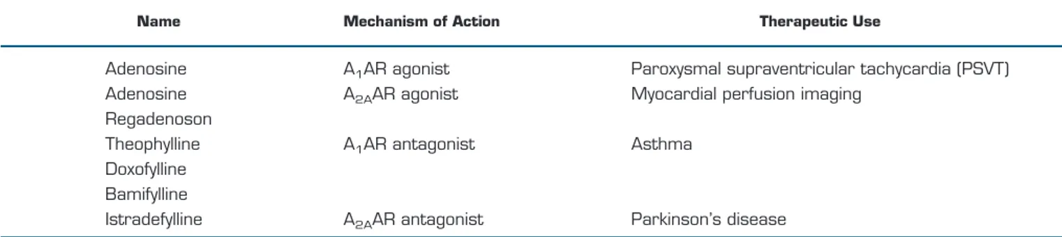
rep-Table 1.
List of clinically approved adenosine receptors drugs
Name Mechanism of Action Therapeutic Use
Adenosine
A
1AR agonist
Paroxysmal supraventricular tachycardia (PSVT)
Adenosine
A
2AAR agonist
Myocardial perfusion imaging
Regadenoson
Theophylline
A
1AR antagonist
Asthma
Doxofylline
Bamifylline
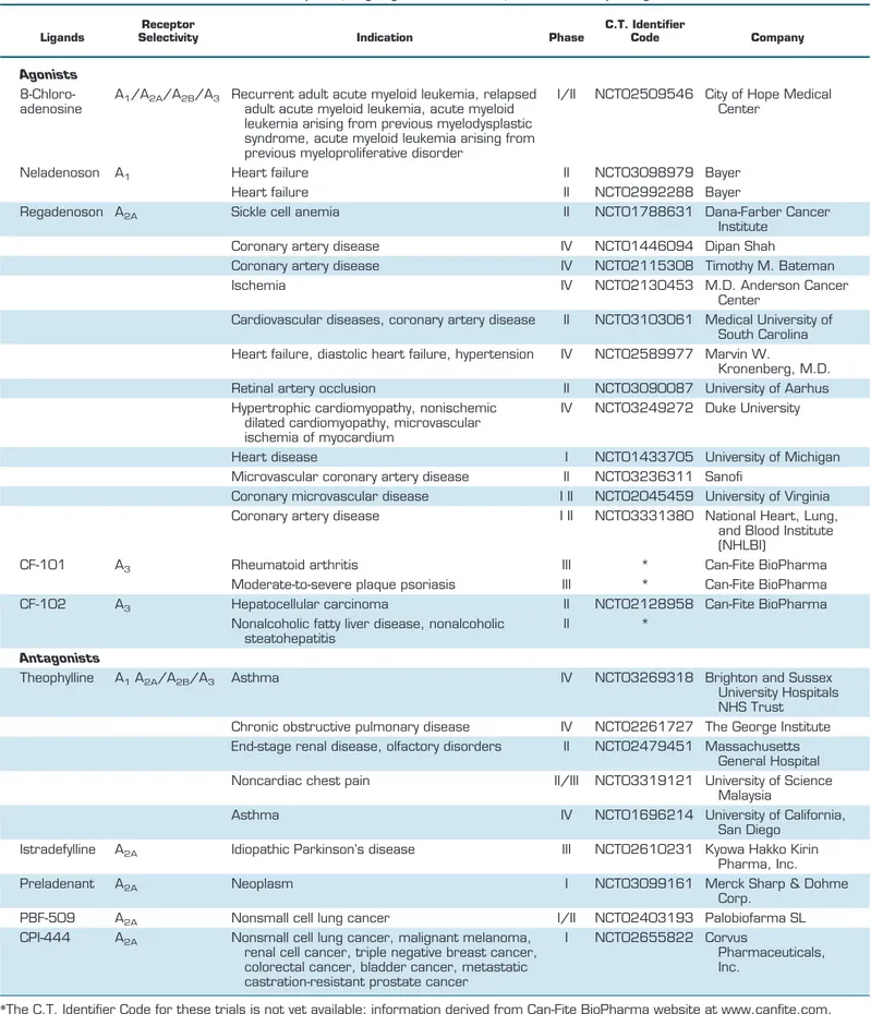
Table 2.
Examples of ongoing clinical studies of adenosine receptor ligands
Ligands
Receptor
Selectivity Indication Phase
C.T. Identifier
Code Company
Agonists
8-Chloro-adenosine
A
1/A
2A/A
2B/A
3Recurrent adult acute myeloid leukemia, relapsed
adult acute myeloid leukemia, acute myeloid
leukemia arising from previous myelodysplastic
syndrome, acute myeloid leukemia arising from
previous myeloproliferative disorder
I/II
NCT02509546 City of Hope Medical
Center
Neladenoson
A
1Heart failure
II
NCT03098979 Bayer
Heart failure
II
NCT02992288 Bayer
Regadenoson A
2ASickle cell anemia
II
NCT01788631 Dana-Farber Cancer
Institute
Coronary artery disease
IV
NCT01446094 Dipan Shah
Coronary artery disease
IV
NCT02115308 Timothy M. Bateman
Ischemia
IV
NCT02130453 M.D. Anderson Cancer
Center
Cardiovascular diseases, coronary artery disease
II
NCT03103061 Medical University of
South Carolina
Heart failure, diastolic heart failure, hypertension
IV
NCT02589977 Marvin W.
Kronenberg, M.D.
Retinal artery occlusion
II
NCT03090087 University of Aarhus
Hypertrophic cardiomyopathy, nonischemic
dilated cardiomyopathy, microvascular
ischemia of myocardium
IV
NCT03249272 Duke University
Heart disease
I
NCT01433705 University of Michigan
Microvascular coronary artery disease
II
NCT03236311 Sanofi
Coronary microvascular disease
I II
NCT02045459 University of Virginia
Coronary artery disease
I II
NCT03331380 National Heart, Lung,
and Blood Institute
(NHLBI)
CF-101
A
3Rheumatoid arthritis
III
*
Can-Fite BioPharma
Moderate-to-severe plaque psoriasis
III
*
Can-Fite BioPharma
CF-102
A
3Hepatocellular carcinoma
II
NCT02128958 Can-Fite BioPharma
Nonalcoholic fatty liver disease, nonalcoholic
steatohepatitis
II
*
Antagonists
Theophylline
A
1A
2A/A
2B/A
3Asthma
IV
NCT03269318 Brighton and Sussex
University Hospitals
NHS Trust
Chronic obstructive pulmonary disease
IV
NCT02261727 The George Institute
End-stage renal disease, olfactory disorders
II
NCT02479451 Massachusetts
General Hospital
Noncardiac chest pain
II/III
NCT03319121 University of Science
Malaysia
Asthma
IV
NCT01696214 University of California,
San Diego
Istradefylline
A
2AIdiopathic Parkinson’s disease
III
NCT02610231 Kyowa Hakko Kirin
Pharma, Inc.
Preladenant
A
2ANeoplasm
I
NCT03099161 Merck Sharp & Dohme
Corp.
PBF-509
A
2ANonsmall cell lung cancer
I/II
NCT02403193 Palobiofarma SL
CPI-444
A
2ANonsmall cell lung cancer, malignant melanoma,
renal cell cancer, triple negative breast cancer,
colorectal cancer, bladder cancer, metastatic
castration-resistant prostate cancer
I
NCT02655822 Corvus
Pharmaceuticals,
Inc.
resent binding sites for ecto-ADA, and its interaction with
them has been reported to increase receptor affinity and
signaling (143, 301). The relation of ADA with ARs has an
important role in immune cells. In particular, the
intercel-lular interaction made by ARs on dendritic cells, ADA, and
CD26 on CD4-T cells, increases immune responses,
sug-gesting the role of ADA as a bridge between cells expressing
ARs and cells expressing CD26.
III. MOLECULAR STRUCTURE OF
ADENOSINE RECEPTORS
Adenosine mediates its physiological effects through the
activation of four ARs. These are characterized by different
tissue distribution and effector coupling and by either high
(A
1, A
2A, A
3) or low (A
2B) affinity for the parent molecule.
All four ARs have been well identified, cloned and
pharma-cologically studied, and present a common structure: each
possesses a core domain which crosses the plasma
mem-brane seven times, in which each helix is 20 –27 amino acids
long and linked by three intracellular and three
extracellu-lar loops (115). The extracelluextracellu-lar NH
2terminus contains
one or more glycosylation sites, while the intracellular
COOH terminus provides sites for phosphorylation and
palmitoylation, thereby playing a role in receptor
desensi-tization and internalization mechanisms. Different AR
sub-types present different numbers of amino acids. For
in-stance, a longer COOH terminus, with 122 amino acids, is
found on A
2AAR, whereas A
1AR, A
2BAR, and A
3AR bear
COOH-terminal tails consisting of ~30 – 40 amino acids
(116). Details of the structures of human A
1AR and A
2AAR
have been provided by crystallization studies (51, 95, 139,
170, 213, 433), which will ultimately aid in the
structure-based drug design of A
1AR and A
2AAR ligands (139, 377).
The generation of selective ligands is particularly desirable, as
ARs present a sequence homology of 80 –95% (there is 70%
homology in their amino acids between human and rat). The
exception to this rule is A
3AR, which differs significantly
among species, with the A
1AR sequence being the most
con-served (323). ARs have been cloned from several species, with
A
3AR being the only subtype isolated before its
pharmacolog-ical characterization (270), and the chromosome location of
human and mouse ARs genes is reported in
TABLE 3
.
Interest-ingly, a comparison between human (h) A
1AR/A
3AR and
hA
2AR/hA
2BR shows overall amino acid sequence identities of
46.5% and 46.6%, respectively.
Recent evidences document the presence of several GPCRs
including ARs in homomer, oligomer, and heteromer forms
(43, 101, 102, 285–287). GPCR heteromers appear as new
signaling entities characterized by different functional
properties when compared with homomers. In this field, the
adenosine A
1AR-A
2AAR unit represents the first reliable
structure of a macromolecular complex, including two
dif-Adenosine
O
2demand
Angiogenesis
Ischemic
preconditioning
O
2supply
Inflammation
O
2demand
Chemokines
Cytokines
O
2VEGF
supply
Neurons
Cardiomyocytes
Vascular
endothelial cells
Immune
cells
ATP
ATP
Physiological conditions
(e.g. physical activity)
Pathological conditions
(e.g. ischemia/hypoxia)
A
1
A
3
A
2A
A
2B
FIGURE 1.
Physiological role of adenosine through interaction with
A
1, A
2A, A
2B, and A
3adenosine receptors (ARs). Adenosine is an
endogenous ubiquitous mediator, highly increased following hypoxia,
ischemia, or physical activity due to ATP consumption. It exerts body
surveillance and protection by different mechanisms triggered by
ARs activation, resulting in decreased oxygen demand and
inflam-mation, increased oxygen supply and angiogenesis, as well as
isch-emic preconditioning.
Extracellular
Intracellular
CD73
ADA
CD39
ATP
Inosine
ADA
AK
Cytosolic
5’-nucleotidase
SAH hydrolase
Inosine
SAH
Adenosine
Adenosine
5’-AMP
5’-AMP
ENTs
FIGURE 2.
Adenosine metabolism and transport in the
extra-intra-cellular milieu. At the intraextra-intra-cellular level, adenosine derives from
S-adenosylhomocysteine (SAH) hydrolase or cytosolic
5=-nucleoti-dase and is degraded by adenosine deaminase (ADA) and adenosine
kinase (AK). Extracellularly, it is generated by CD73 and converted by
ADA. Equilibrative nucleoside transporters (ENTs) allow adenosine free
flux through cell membrane, following gradient concentration.
ferent receptors plus two different G proteins coupled to
them
(FIGURE 3)
(43, 285). Indeed A
1AR is coupled to G
iand A
2AAR to G
s, thus rendering heteromer able to trigger
opposite signals affecting the cAMP-dependent
intracellu-lar pathway. Specifically, this unit represents a cell surface
sensor of adenosine concentration, able to discriminate
be-tween low and high nucleoside level (285). When adenosine
levels are low, its interaction occurs preferentially with
A
1AR protomer of the heteromer and activates G
i/oprotein,
thus reducing adenylate cyclase (AC), protein kinase A
(PKA), and GABA uptake. Instead, when adenosine levels
are higher, its binding is favored to A
2AR component of the
complex, which reduces A
1AR activation and, through G
sprotein, associates with the AC/cAMP/PKA cascade,
re-sulting in the increase of GABA uptake (68). Therefore,
adenosine depending on its concentration may affect a
number of other physiological process, including the
re-lease of glutamate (63). Interestingly, the
heteromeriza-tion phenomenon appears as a general mechanism
affect-ing also A
3ARs, forming homodimers and A
1AR-A
3AR
heterodimers (157, 190). This opens up new horizons in
drug development (102); in particular, A
2AAR-D2
dopa-mine receptor heterodimers have been detected in the
striatum and may be a viable therapeutic target in PD
(121, 122, 283).
IV. DISTRIBUTION, PHYSIOLOGICAL
EFFECTS, AND SIGNAL
TRANSDUCTION
ARs are found throughout the nervous, cardiovascular,
respi-ratory, gastrointestinal, urogenital, and immune systems as
well as in bone, joints, eyes, and skin (310)—a pattern of
distribution that denotes their significant control of neuronal,
cardiac, metabolic, and renal activities (3). Each AR is
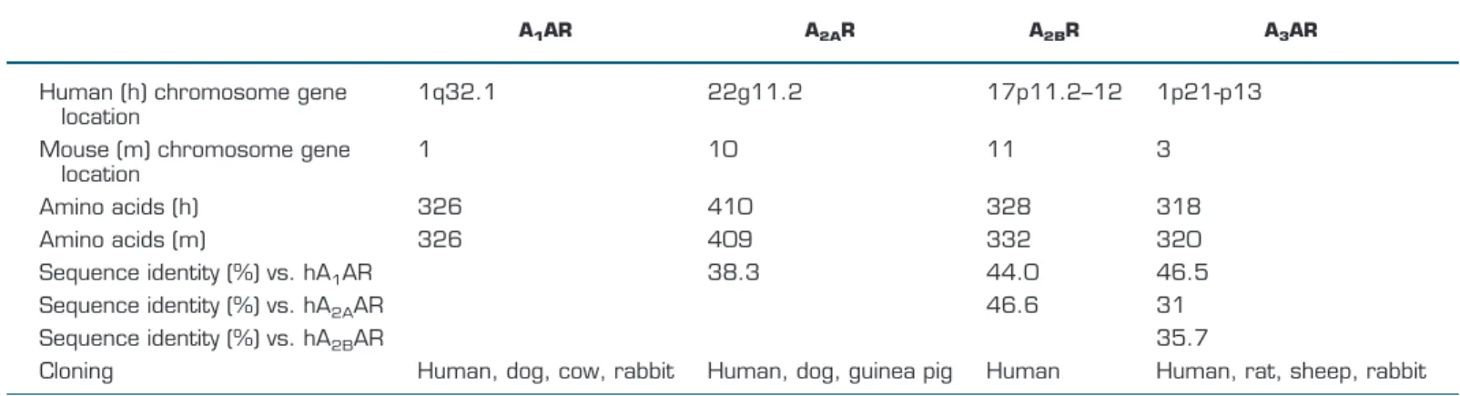
charac-Table 3.
Molecular characteristics of adenosine receptors
A1AR A2AR A2BR A3AR
Human (h) chromosome gene
location
1q32.1
22g11.2
17p11.2–12
1p21-p13
Mouse (m) chromosome gene
location
1
10
11
3
Amino acids (h)
326
410
328
318
Amino acids (m)
326
409
332
320
Sequence identity (%) vs. hA
1AR
38.3
44.0
46.5
Sequence identity (%) vs. hA
2AAR
46.6
31
Sequence identity (%) vs. hA
2BAR
35.7
Cloning
Human, dog, cow, rabbit
Human, dog, guinea pig
Human
Human, rat, sheep, rabbit
Low adenosine concentration
Striatal GABAegic
efferent neuron
ATP
Glu
ATP
Glu
ATP
Glu
ATP
Glu
Adenosine
A
1A
2AHigh adenosine concentration
Striatal GABAegic
efferent neuron
ATP
Glu
ATP
Glu
ATP
Glu
ATP
Glu
Adenosine
A
1A
2AFIGURE 3.
Schematic representation of
A
1AR–A
2AAR heteromer as adenosine
sen-sor. Low adenosine concentration
prefer-entially stimulates the A
1AR protomer of
the heteromer, which would inhibit
gluta-matergic transmission. On the other hand,
high adenosine concentration activates
adenosine A
2AAR that blocks adenosine
A
1AR-mediated effects and results in
terized by unique cell and tissue distribution, secondary
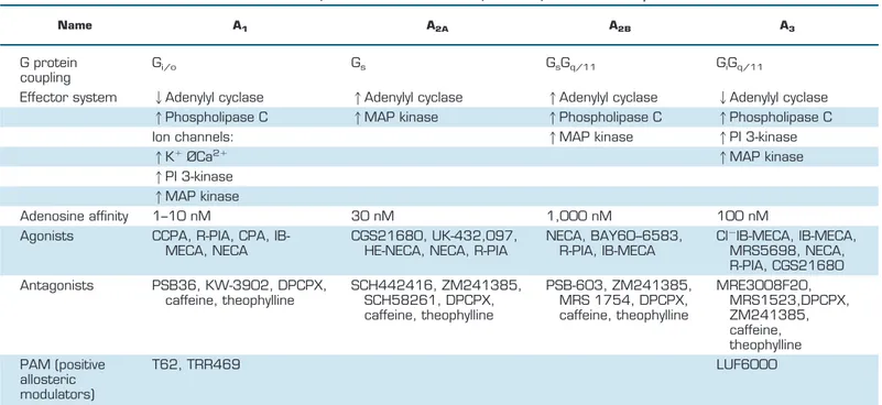
signal-ing transductors
(TABLE 4)
, and physiological effects
(TABLE
5)
. A
1AR and A
3AR signals are mediated through G
iand G
omembers of the G protein family, through which they reduce
AC activity and cAMP levels, while A
2AARs and A
2BARs are
coupled to G
sproteins, through which they stimulate AC and
increase cAMP levels, thereby leading to the activation of a
plethora of mediators, depending on the signaling triggered by
cAMP in specific cells (116).
A. A
1AR and A
3AR G
iand G
o-Coupled
Receptors
The A
1AR subtype is expressed in the central nervous
sys-tem (CNS), mainly in the brain cortex, cerebellum,
hip-pocampus, autonomic nerve terminals, spinal cord, and
glial cells (56). This broad distribution reflects the wide
range of physiological functions regulated by A
1AR,
span-ning neurotransmitter release, dampespan-ning of neuronal
ex-citability, control of sleep/wakefulness, pain reduction, as
well as sedative, anticonvulsant, anxiolytic, and locomotor
depressant effects (131, 349, 375). This subtype is also
pres-ent at high levels in the heart atria, kidney, adipose tissue,
and pancreas, where it induces negative chronotropic,
ino-tropic, and dromotropic effects, reduces renal blood flow
and renin release, and inhibits lipolysis and insulin
secre-tion, respectively (86, 263, 319, 322, 378, 397, 410). It is
also located on airway epithelial and smooth muscle cells,
where it stimulates a bronchoconstrictory response, and in
several immune cells such as neutrophils, eosinophils,
mac-rophages, and monocytes, where it promotes essentially
proinflammatory effects (165, 317, 422).
A
1AR also induces phospholipase C (PLC)-
 activation,
thereby increasing inositol 1,4,5-trisphosphate (IP
3) and
in-tracellular Ca
2⫹levels, which stimulate calcium-dependent
protein kinases (PKC) and/or other calcium-binding
pro-teins.
Table 4.
Classification and mechanism of action of adenosine receptors
Name A1 A2A A2B A3
G protein
coupling
G
i/oG
sG
sG
q/11G
iG
q/11Effector system
2Adenylyl cyclase
1Adenylyl cyclase
1Adenylyl cyclase
2Adenylyl cyclase
1Phospholipase C
1MAP kinase
1Phospholipase C
1Phospholipase C
Ion channels:
1MAP kinase
1PI 3-kinase
1K
⫹ØCa
2⫹1MAP kinase
1PI 3-kinase
1MAP kinase
Adenosine affinity
1–10 nM
30 nM
1,000 nM
100 nM
Agonists
CCPA, R-PIA, CPA,
IB-MECA, NECA
CGS21680, UK-432,097,
HE-NECA, NECA, R-PIA
NECA, BAY60–6583,
R-PIA, IB-MECA
Cl
⫺IB-MECA, IB-MECA,
MRS5698, NECA,
R-PIA, CGS21680
Antagonists
PSB36, KW-3902, DPCPX,
caffeine, theophylline
SCH442416, ZM241385,
SCH58261, DPCPX,
caffeine, theophylline
PSB-603, ZM241385,
MRS 1754, DPCPX,
caffeine, theophylline
MRE3008F20,
MRS1523,DPCPX,
ZM241385,
caffeine,
theophylline
PAM (positive
allosteric
modulators)
T62, TRR469
LUF6000
BAY60 – 6583, 2-[[6-amino-3,5-dicyano-4-[4-(cyclo propylmethoxy)phenyl]-2-pyridinyl]thio]-acetamide; CCPA, 2-chloro-N-cyclopentyladenosine;
CGS21680, 4-[2-[[6-amino-9-(N-ethyl-
-
D-ribofuranuronamidosyl)-9H-purin-2-yl]amino]ethyl]benzenepropanoic acid hydrochloride; Cl
⫺IB-MECA,
CF102, 2-chloro-N6-(3-iodobenzyl)-adenosine-5=-N-methyluronamide; DPCPX, 8-cyclopentyl-1,3-dipropylxanthine; MRS5698, (1S,2R,3S,4R,
5S)-4-[6-[[(3-chlorophenyl)methyl]amino]-2-[2-(3,4-difluorophenyl)-ethynyl]-9H-purin-9-yl]-2,3-dihydroxy-N-methylbicyclo[3.1.0]hexane-1-carboxamide; KW-3902, 8-(hexahydro-2,5-methanopentalen-3a (1H)-yl)-3,7-dihydro-1,3-dipropyl-1H-purine-2,6-di one; LUF6000,
N-(3,4-dichloro-phenyl)-2-cyclohexyl-1H-imidazo[4,5-c]quinolin-4-amine; MRS 1754,
N-(4-cyanophenyl)-2-[4-(2,3,6,7-tetrahydro-2,6-dioxo-1,3-dipropyl-1H-purin-8-yl)phenoxy]-acetamide; MRE 3008F20,
N-[2-(2-furanyl)-8-propyl-8H-pyrazolo[4,3-e][1,2,4]triazolo[1,5-c]pyrimidin-5-yl]-N’-(4-methoxyphenyl)urea; MRS1523, 3-propyl-6-ethyl-5-[(ethylthio)carbonyl]-2 phenyl-4-propyl-3-pyridine carboxylate; PAM, positive allosteric
modu-lators; PSB36, 1-butyl-8-(hexahydro-2,5-methanopentalen-3a(1H)-yl)-3,7-dihydro-3-(3-hydroxypropyl)-1H-purine-2,6-dione; PSB-603,
8-[4-[4-(4-chlorophenzyl)piperazide-1-sulfonyl)phenyl]]-1-propylxanthine; SCH442416,
2-(2-furanyl)-7-[3-(4-methoxyphenyl)propyl]-7H-pyrazolo[4,3-e][1,2,4]triazolo[1,5-c]pyrimidin-5-amine; SCH 58261,
2-(2-furanyl)-7-(2-phenylethyl)-7H-pyrazolo[4,3-e][1,2,4]triazolo[1,5-c]pyrimidin-5-amine; T62, 2-amino-4,5,6,7-tetrahydrobenzo[b]thiophen-3-yl)-(4-chlorophenyl)-methanone; TRR469,
2-amino-4-[(4-(phenyl)piperazin-1-yl)methyl]-5-(4-fluorophenyl)thiophen-3-yl)-(4-chlorophenyl)methanone; UK-432,097,
6-[2,2-di(phenyl)ethylamino]-9-[(2R,3R,4S,5S)-5-(ethylcarbamoyl)-3,4-dihydroxyoxolan-2-yl]-N-[2-[(1-pyridin-2-ylpiperidin-4-yl)-carbamoylamino]-ethyl]-purine-2-carboxamide; ZM 241385,
4-(2-[7-amino-2-(2-furyl)[1,2,4]triazolo[2,3-a][1,3,5]triazin-5-ylamino]ethyl)phenol.
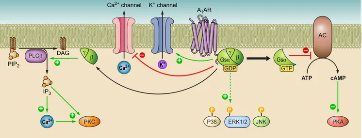
At the neuronal and myocardial level, A
1AR stimulates
po-tassium (K) pertussis toxin-sensitive and K
ATPchannels,
while reducing Q-, P-, and N-type Ca
2⫹channels.
Further-more, the involvement of A
1AR in the intracellular
phos-phorylative cascade of the mitogen-activated protein kinase
(MAPK) family—including extracellular signal-regulated
kinase (ERK), p38, and Jun NH
2-terminal kinase (JNK)—
has been reported (351, 352)
(FIGURE 4)
.
Pharmacological agents that increase the activation of
A
1AR in response to adenosine would be useful for the
treatment of CNS, cardiovascular, and inflammatory
pa-thologies. A
1AR drawback effects, due to their wide
distri-bution, broad spectrum of physiological effects, and
pro-miscuous signaling pathway transduction, can fortunately
be mitigated through allosteric enhancers, which stabilize
the ternary complex formed by agonist-A
1AR-G protein
molecules. This enhances the agonist action only at the site
affected by injury, where adenosine concentrations are
in-creased (330).
The A
3AR subtype is widely expressed in a variety of
primary cells, tissues, and cell lines. Low levels have been
reported in the brain, where it is located in the thalamus,
hypothalamus, hippocampus, cortex, and retinal
gan-glion cells, as well as at motor nerve terminals and the
pial and intercerebral arteries. A
3ARs are also expressed
in microglia and astrocytes, and the inhibition of a
neu-roinflammatory response in these cells has been
associ-ated with their induction of an analgesic effect (175).
Although A
3AR is also known to have cardioprotective
effects, and to be greatly expressed in the coronary and
carotid artery, its precise location in the heart has not yet
been reported. At the peripheral level, however, A
3AR
has been found in enteric neurons, as well as epithelial
cells, colonic mucosa, lung parenchyma, and bronchi.
Furthermore, A
3AR has a broad distribution in
inflam-matory cells like mast cells, eosinophils, neutrophils,
monocytes, macrophages, foam cells, dendritic cells,
lymphocytes, splenocytes, bone marrow cells, lymph
nodes, synoviocytes, chondrocytes, and osteoblasts,
where it mediates anti-inflammatory effects (37).
Inter-estingly, A
3AR is overexpressed in several cancer cells
and tissues and is therefore likely to have an important
antitumoral role (39).
A
3ARs trigger a variety of intracellular signaling by
pref-erentially coupling to G
iproteins, by which they
reduce cAMP levels, and, at high concentrations of A
3AR
agonists, to G
qproteins or G
␥ subunits, thereby
induc-ing an increase in both PLC and calcium. A reduction in
cAMP results in PKA inhibition, which leads to an
in-crease in glycogen synthase kinase-3
 (GSK-3);
down-regulation of beta-catenin, cyclin D1, and c-Myc; and
reduction of nuclear factor (NF)-
B DNA-binding ability
(108). A different pathway from GPCR
signaling—in-volving monomeric G protein RhoA and phospholipase
D—is important for A
3AR-mediated neuro- and
cardio-protection. A
3ARs are also known to regulate MAPK,
PI3K/Akt, and NF-
B signaling pathways, by which
Table 5.
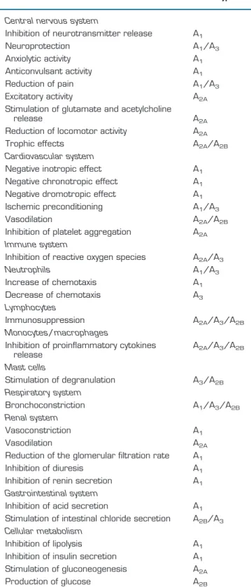
Biological effects of adenosine
Effects
Receptor Subtype
Central nervous system
Inhibition of neurotransmitter release
A
1Neuroprotection
A
1/A
3Anxiolytic activity
A
1Anticonvulsant activity
A
1Reduction of pain
A
1/A
3Excitatory activity
A
2AStimulation of glutamate and acetylcholine
release
A
2AReduction of locomotor activity
A
2ATrophic effects
A
2A/A
2BCardiovascular system
Negative inotropic effect
A
1Negative chronotropic effect
A
1Negative dromotropic effect
A
1Ischemic preconditioning
A
1/A
3Vasodilation
A
2A/A
2BInhibition of platelet aggregation
A
2AImmune system
Inhibition of reactive oxygen species
A
2A/A
3Neutrophils
A
1/A
3Increase of chemotaxis
A
1Decrease of chemotaxis
A
3Lymphocytes
Immunosuppression
A
2A/A
3/A
2BMonocytes/macrophages
Inhibition of proinflammatory cytokines
release
A
2A/A
3/A
2BMast cells
Stimulation of degranulation
A
3/A
2BRespiratory system
Bronchoconstriction
A
1/A
3/A
2BRenal system
Vasoconstriction
A
1Vasodilation
A
2AReduction of the glomerular filtration rate
A
1Inhibition of diuresis
A
1Inhibition of renin secretion
A
1Gastrointestinal system
Inhibition of acid secretion
A
1Stimulation of intestinal chloride secretion
A
2B/A
3Cellular metabolism
Inhibition of lipolysis
A
1Inhibition of insulin secretion
A
1Stimulation of gluconeogenesis
A
2Athey exert anti-inflammatory effects. Stimulation or
inhibition of HIF-1 has been also demonstrated to
have protumoral and neuromodulatory effects in
cancer
cells
and
astrocytes,
respectively
(39)
(FIGURE 5)
.
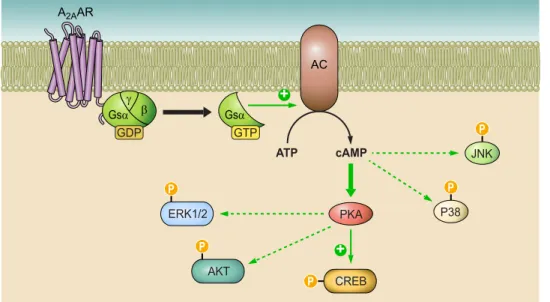
B. A
2AAR and A
2BAR G
s-Coupled Receptors
The A
2AAR subtype occurs both centrally and peripherally,
but its greatest expression is in the striatum, the olfactory
tubercle, and the immune system, while lower levels are
–
P
P
+
+
+
+
–
PKC
P38
PKA
P
JNK
PLC
E
ERK1/2
AC
A
1AR
K
+channel
Ca
2+channel
J
Gs
D E
J
E
GDP
Gs
D
GTP
ATP
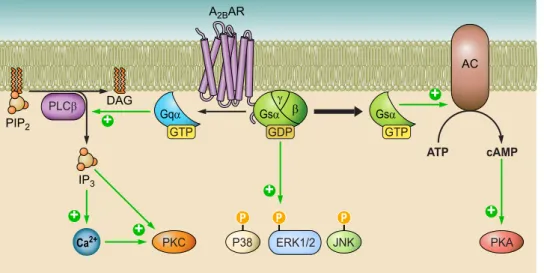
cAMP
+
PIP
2DAG
IP
3Ca
2+Ca
2+K
+–
FIGURE 4.
Overview of A
1AR intracellular signaling pathways. A
1AR stimulation decreases adenylate cyclase
(AC) activity and cAMP production, thus inhibiting protein kinase A (PKA), while activated phospholipase C
(PLC)- and Ca
2⫹. K
⫹and Ca
2⫹channels are opened and closed, respectively, by A
1