OncoImmunology
ISSN: (Print) 2162-402X (Online) Journal homepage: https://www.tandfonline.com/loi/koni20
Evaluating the role of FAMIly history of cancer and
diagnosis of multiple neoplasms in cancer patients
receiving PD-1/PD-L1 checkpoint inhibitors: the
multicenter FAMI-L1 study
Alessio Cortellini, Sebastiano Buti, Melissa Bersanelli, Raffaele Giusti,
Fabiana Perrone, Pietro Di Marino, Nicola Tinari, Michele De Tursi, Antonino
Grassadonia, Katia Cannita, Alessandra Tessitore, Federica Zoratto, Enzo
Veltri, Francesco Malorgio, Marco Russano, Cecilia Anesi, Tea Zeppola, Marco
Filetti, Paolo Marchetti, Andrea Botticelli, Gian Carlo Antonini Cappellini,
Federica De Galitiis, Maria Giuseppa Vitale, Francesca Rastelli, Federica
Pergolesi, Rossana Berardi, Silvia Rinaldi, Marianna Tudini, Rosa Rita Silva,
Annagrazia Pireddu, Francesco Atzori, Daniela Iacono, Maria Rita Migliorino,
Alain Gelibter, Mario Alberto Occhipinti, Francesco Martella, Alessandro
Inno, Stefania Gori, Sergio Bracarda, Cristina Zannori, Claudia Mosillo,
Alessandro Parisi, Giampiero Porzio, Domenico Mallardo, Maria Concetta
Fargnoli, Marcello Tiseo, Daniele Santini, Paolo A Ascierto & Corrado
Ficorella
To cite this article: Alessio Cortellini, Sebastiano Buti, Melissa Bersanelli, Raffaele Giusti, Fabiana Perrone, Pietro Di Marino, Nicola Tinari, Michele De Tursi, Antonino Grassadonia, Katia Cannita, Alessandra Tessitore, Federica Zoratto, Enzo Veltri, Francesco Malorgio, Marco Russano, Cecilia Anesi, Tea Zeppola, Marco Filetti, Paolo Marchetti, Andrea Botticelli, Gian Carlo Antonini Cappellini, Federica De Galitiis, Maria Giuseppa Vitale, Francesca Rastelli, Federica Pergolesi, Rossana Berardi, Silvia Rinaldi, Marianna Tudini, Rosa Rita Silva, Annagrazia Pireddu, Francesco Atzori, Daniela Iacono, Maria Rita Migliorino, Alain Gelibter, Mario Alberto Occhipinti, Francesco Martella, Alessandro Inno, Stefania Gori, Sergio Bracarda, Cristina Zannori, Claudia Mosillo, Alessandro Parisi, Giampiero Porzio, Domenico Mallardo, Maria Concetta Fargnoli, Marcello Tiseo, Daniele Santini, Paolo A Ascierto & Corrado Ficorella (2020) Evaluating the role of FAMIly history of cancer and diagnosis of multiple neoplasms in cancer patients receiving PD-1/PD-L1 checkpoint inhibitors: the multicenter FAMI-PD-1/PD-L1 study, OncoImmunology, 9:1, 1710389, DOI: 10.1080/2162402X.2019.1710389
To link to this article: https://doi.org/10.1080/2162402X.2019.1710389
© 2020 The Author(s). Published with license by Taylor & Francis Group, LLC. View supplementary material
Full Terms & Conditions of access and use can be found at
https://www.tandfonline.com/action/journalInformation?journalCode=koni20 Submit your article to this journal
Article views: 173
View related articles
ORIGINAL RESEARCH
Evaluating the role of FAMIly history of cancer and diagnosis of multiple neoplasms
in cancer patients receiving PD-1/PD-L1 checkpoint inhibitors: the multicenter
FAMI-L1 study
Alessio Cortellini a,b, Sebastiano Buti c, Melissa Bersanellic,d, Raffaele Giustie, Fabiana Perronec, Pietro Di Marinof,
Nicola Tinarig, Michele De Tursig, Antonino Grassadoniag, Katia Cannitaa, Alessandra Tessitoreb, Federica Zorattoh,
Enzo Veltrih, Francesco Malorgioi, Marco Russanoj, Cecilia Anesij, Tea Zeppolaj, Marco Filettie, Paolo Marchettie,k,l,m,
Andrea Botticellik, Gian Carlo Antonini Cappellinim, Federica De Galitiism, Maria Giuseppa Vitalen, Francesca Rastellio,
Federica Pergolesio, Rossana Berardip, Silvia Rinaldip, Marianna Tudiniq, Rosa Rita Silvaq, Annagrazia Pireddur,
Francesco Atzorir, Daniela Iaconos, Maria Rita Migliorinos, Alain Gelibterl, Mario Alberto Occhipintil,
Francesco Martellat, Alessandro Innou, Stefania Goriu, Sergio Bracardav, Cristina Zannoriv, Claudia Mosillov,
Alessandro Parisia,b, Giampiero Porzioa,b, Domenico Mallardow, Maria Concetta Fargnolib,x, Marcello Tiseoc,d,
Daniele Santinij, Paolo A Ascierto w, and Corrado Ficorellaa,b
aMedical Oncology, St. Salvatore Hospital, L’Aquila, Italy;bDepartment of Biotechnological and Applied Clinical Sciences, University of L’Aquila, L’Aquila, Italy;cMedical Oncology, University Hospital of Parma, Parma, Italy;dDepartment of Medicine and Surgery, University of Parma, Parma, Italy;eU.O.C. Oncologia Medica, Azienda Ospedaliero Universitaria Sant’Andrea, Rome, Italy;fClinical Oncology Unit, S.S. Annunziata Hospital, Chieti, Italy;gDepartment of Medical, Oral & Biotechnological Sciences, University G. D’Annunzio, Chieti-Pescara, Italy;hMedical Oncology, Santa Maria Goretti Hospital, Latina, Italy;iMedical Oncology,“Santo Spirito” Hospital, Pescara, Italy;jMedical Oncology, Campus Bio-Medico University, Rome, Italy;kMedical Oncology, Sapienza University of Rome, Rome, Italy;lMedical Oncology (B), Policlinico Umberto I, Rome, Italy;mMedical Oncology, IDI-IRCCS, Roma, Italy;nMedical Oncology, University Hospital of Modena, Modena, Italy;oMedical Oncology, Fermo Area Vasta 4, Fermo, Italy; pOncology Clinic, Università Politecnica delle Marche, Ancona, Italy;qMedical Oncology, AV2 Fabriano ASUR Marche, Italy;rMedical Oncology Unit, University Hospital of Cagliari, Cagliari, Italy;sPulmonary Oncology Unit, St. Camillo Forlanini Hospital, Rome, Italy;tMedical Oncology,“G. Mazzini” Hospital, Teramo, Italy;uMedical Oncology Unit, IRCCS, Sacro Cuore Don Calabria Hospital, Verona, Italy;vMedical Oncology, Azienda Ospedaliera S. Maria, Terni, Italy;wMelanoma, Cancer Immunotherapy and Development Therapeutics Unit, Istituto Nazionale Tumori-IRCCS Fondazione“G. Pascale”, Naples, Italy;xDermatology, San Salvatore Hospital, L’Aquila, Italy
ABSTRACT
Background: We investigate the role of family history of cancer (FHC) and diagnosis of metachronous and/or synchronous multiple neoplasms (MN), during anti-PD-1/PD-L1 immunotherapy.
Design: This was a multicenter retrospective study of advanced cancer patients treated with anti-PD-1/ PD-L1 immunotherapy. FHC was collected in lineal and collateral lines, and patients were categorized as follows: FHC-high (in case of cancer diagnoses in both the lineal and collateral family lines), FHC-low (in case of cancer diagnoses in only one family line), and FHC-negative. Patients were also categorized according to the diagnosis of MN as follows: MN-high (>2 malignancies), MN-low (two malignancies), and MN-negative. Objective response rate (ORR), progression-free survival (PFS), overall survival (OS), and incidence of immune-related adverse events (irAEs) of any grade were evaluated.
Results: 822 consecutive patients were evaluated. 458 patients (55.7%) were FHC-negative, 289 (35.2%) were FHC-low, and 75 (9.1%) FHC-high, respectively. 29 (3.5%) had a diagnosis of synchronous MN and 94 (11.4%) of metachronous MN. 108 (13.2%) and 15 (1.8%) patients were MN-low and MN-high, respectively. The median follow-up was 15.6 months. No significant differences were found regarding ORR among subgroups. FHC-high patients had a significantly longer PFS (hazard ratio [HR] = 0.69 [95%
CI: 0.48–0.97], p = .0379) and OS (HR = 0.61 [95% CI: 0.39–0.93], p = .0210), when compared to
FHC-negative patients. FHC-high was confirmed as an independent predictor for PFS and OS at multivariate analysis. No significant differences were found according to MN categories. FHC-high patients had
a significantly higher incidence of irAEs of any grade, compared to FHC-negative patients (p = .0012).
Conclusions: FHC-high patients seem to benefit more than FHC-negative patients from anti-PD-1/PD-L1 checkpoint inhibitors. ARTICLE HISTORY Received 5 October 2019 Revised xx xxx xxxx Accepted 2 December 2019 KEYWORDS
Family history of cancer; multiple neoplasms; DDR genes; immune checkpoint inhibitors; immunotherapy; PD-1
Introduction
After the advent of immune checkpoint inhibitors (ICIs), oncol-ogy clinical practice radically changed, leading to an unprece-dented improvement of cancer patients' clinical outcomes.
Nevertheless, we are still a long way from predicting ICI efficacy in each patient. PD-L1 (programmed death ligand-1) protein expression, evaluated in both tumor and immune cells, is the most investigated predictive biomarker1; on the other hand, other factors such as tumor mutational burden, body mass
CONTACTAlessio Cortellini [email protected] Medical Oncology Unit, St. Salvatore Hospital, L’Aquila, Via Vetoio 67100, Italy Supplemental data for this article can be accessed on thepublisher’s website.
ONCOIMMUNOLOGY
2020, VOL. 9, NO. 1, e1710389 (10 pages)
https://doi.org/10.1080/2162402X.2019.1710389
© 2020 The Author(s). Published with license by Taylor & Francis Group, LLC.
This is an Open Access article distributed under the terms of the Creative Commons Attribution-NonCommercial License (http://creativecommons.org/licenses/by-nc/4.0/), which permits unrestricted non-commercial use, distribution, and reproduction in any medium, provided the original work is properly cited.
index, and gut microbiota, have been investigated as predictors of clinical benefit from immunotherapy across different tumor types.2–5
Mismatch repair (MMR) deficiency, which leads to the con-dition of genetic hypermutability known as microsatellite instability (MSI), is related to the number of somatic mutations (especially in MSI-high cases); many studies have already con-firmed its positive predictive role (MSI-high) for ICI treatment, particularly with anti-PD-1 (programmed death-1) antibodies.6,7 MSI is known to be the hallmark of Lynch syndrome (LS), a familial clustering of colorectal and endometrial cancers. LS is caused by several germline mutations, which result in a defective MMR and is inherited as dominant autosomal char-acter. Similarly,BRCA 1 and 2 (Breast Cancer 1/2) mutations, which are associated with hereditary breast-ovarian cancer syn-drome (HBOC), may correlate with the mutational landscape of the tumors, because of the homologous recombination repair deficiency.8 Moreover, patients with inherited cancer suscept-ibility syndromes are more likely to develop multiple primary tumors during their life.9“BRCA-like” phenotype may be more sensitive to anti-PD-1/PD-L1 agents10; thus prospective clinical trials with anti-PD-1 for patients with germlineBRCA 1/2 muta-tions are currently ongoing.11LS and HBOC syndrome are just two of the forms of inherited cancer susceptibility. Even though notoriously only about 5% to 10% of all cancers result directly from germline mutations,12we can hypothesize that much about family cancer syndromes and cancer predisposition is still unknown. Starting from this hypothesis and from the suggestion that tumors related to inherited cancer susceptibility syndromes seem to have an“immune sensitive phenotype,” we investigated if positive family history of cancer (FHC) and diagnosis of metachronous and/or synchronous multiple neoplasms (MN) could be somehow related to clinical outcomes with anti-PD-1/ PD-L1 treatment.
In the preliminary analysis of the “FAMI-L1” study (211 patients), we found that patients with a positive FHC had higher objective response rate (ORR) and disease control rate (DCR), and prolonged time to treatment failure and overall survival (OS), while patients with diagnosis of MN only had a significantly higher DCR.13Our first hypothesis has been that the underlying mechan-isms to our findings might be DNA damage repair (DDR) gene alterations.14
Here, we present the updated results of the FAMI-L1 study, implemented in the study population, in order to con-firm our preliminary findings.13
Results
Patients’ characteristics
822 consecutive, stage IV cancer patients underwent a treatment with anti-PD-1/PD-L1. 458 patients (55.7%) were FHC-negative, while 364 (44.3%) were FCH-positive: 289 (35.2%) were FHC-low and 75 (9.1) were high patients, respectively. Among FHC-positive patients, 270 (32.8%) were lineal line FHC-positive and 167 (20.3%) were collateral line positive. 123 patients (14.9%) had diagnosis of MN: 29 (3.5%) synchronous MN and 94 (11.4%) metachronous MN. 108 patients (13.2%) were MN-low, while 15
(1.8%) were MN-high. All patient features are summarized in
Table 1.
Among FHC-positive and FHC-negative patients, 61 (16.8%) and 62 (13.5%) had a diagnosis of MN (p = .1987).
Efficacy analysis
Among 822 patients, 775 were evaluable for activity; the other 47 had not yet evaluated the disease at the time of the data cutoff analysis or were lost to follow-up/death without evaluation of clinical response. ORR in the overall population was 34.8% (95% CI: 30.8–39.2, 270 responses). As summarized in Table 2, no significant differences were found regarding ORR among subgroups.
The median follow-up was 15.6 months; in the overall population, median PFS was 9.2 months (95% CI: 8.2–10.6; 479 events) and median OS was 20.5 months (95% CI: 16.2– 27.8; 477 censored patients).Tables 3and4report univariate and multivariate analyses of PFS and OS in detail.
Median PFS in FHC-negative, FHC-low, and FHC-high patients was 9.3 months (95% CI: 7.5–10.6; 277 events), 8.4 months (95% CI: 7–11.4; 166 events), and 20.5 months (95% CI: 8.7–26.4; 36 events), respectively (Figure 1). As reported in
Table 3, FHC-high patients had a significantly longer PFS when compared to FHC-negative patients (HR = 0.69 [95% CI: 0.48–0.97], p = .0379); at multivariate analysis, FHC-high
Table 1.Patient features.
N° (%) 822 Age (years) Median Range Elderly (≥ 70) 68 21–92 359 (43.7) Sex Male Female 552 (67.1) 270 (32.9) ECOG PS 0– 1 ≥2 689 (83.8)133 (16.2) Primary tumor NSCLC Melanoma Renal cell carcinoma Others
475 (57.8) 190 (23.1) 133 (16.2) 24 (2.9) Number of metastatic sites
≤2 > 2
407 (49.5) 415 (50.5) Type of anti-PD-1/PD-L1 agent
Pembrolizumab Nivolumab Atezolizumab 239 (29.1) 559 (68) 24 (2.9) Treatment line of immunotherapy
First Nonfirst 214 (26) 608 (74) FHC Negative FHC-low FHC-high 458 (55.7) 289 (35.2) 75 (9.1) FHC-straight line 270 (32.8) FHC-collateral line 167 (20.3) MN Negative MN-low MN-high 699 (85.1) 108 (13.1) 15 (1.8) MN-synchronous 29 (3.5) MN-methacronous 94 (11.4)
was confirmed an independent predictor for PFS (compared to FHC-negative).
Median OS in FHC-negative, FHC-low, and FHC-high patients was 18.2 months (95% CI: 14.9–23.9; 250 cen-sored patients), 20.8 months (95% CI: 15.4–20.9; 176 cen-sored patients), and 31.6 months (95% CI: 26.2–31.6; 51 censored patients), respectively (Figure 1). As reported in
Table 4, FHC-high patients had a significantly longer OS when compared to FHC-negative patients (HR = 0.61 [95% CI: 0.39–0.93], p = .0210); at multivariate analysis, FHC-high was confirmed an independent predictor for OS (compared to FHC-negative).
Median PFS in MN-negative, MN-low, and MN-high patients was 8.7 months (95% CI: 7.6–10.2; 414 events), 12.3 months (95% CI: 8.3–28.9; 58 events), and 14.4 months (95% CI: 3.6–14.5; 7 events), respectively (Figure 2). As reported in
Table 3, no significant differences were found regarding PFS, according to MN categories.
Median OS in MN-negative, MN-low, and MN-high patients was 20.5 months (95% : 15.7–27.1; 43 censored patients), 26.2 months (95% CI: 18.7–48.9; 66 censored patients), and 15.9 months (95% CI: 10.5–15.9; 8 censored patients), respectively
Table 2.Activity data for overall population and subgroups. ORR analysis
Variable (comparator) Response ratio ORR (%) (95% CI) p-value Overall 270/775 34.8 (30.8–39.2) -FHC Positive Negative 130/347 140/428 37.5 (31.3–44.5) 32.7 (27.5–38.6) 0.1675 FHC-Straight line Positive Negative 101/256 169/519 39.5 (32.1–47.9) 32.6 (27.8–37.8) 0.0584 FHC-Collateral line Positive Negative 56/161 214/614 34.8 (35.3–56.3) 34.9 (26.3–45.2) 0.9866 FHC (FHC-negative) FHC-low FHC-high 101/275 29/72 36.7 (29.9–44.6) 40.3 (26.9–57.8) 0.3288 Multiple neoplasm Yes No 46/116 224/659 39.7 (29.0–52.8) 34.0 (29.6–38.7) 0.2380 MN (no MN) MN-low MN-high 41/104 5/12 39.4 (28.2–53.4) 41.7 (13.5–97.2) 0.4922 MN (no MN) MN-synchronous MN-metachronous 7/27 39/89 25.9 (10.4–53.4) 43.8 (31.2–59.9) 0.1156
Table 3.Univariate and multivariate analyses for PFS.
Progression-free survival
Univariate analysis Multivariate analysis Variable (comparator) HR (95% CI); p-value HR (95% CI); p-value FHC Positive vs negative 0.92 (0.76–1.10); p = .3705 -FHC-Straight line Positive vs negative 0.87 (0.72–1.06); p = .1790 -FHC-Collateral line Positive vs negative 0.91 (0.73–1.15); p = .4722 -FHC (FHC-negative) FHC-low FHC-high 0.98 (0.81–1.19); p = .9116 0.69 (0.48–0.97); p = .0379 0.94 (0.78–1.14); p = .5845 0.64 (0.45–0.91); p = .0148 Multiple neoplasm Yes vs no 0.78 (0.61–1.02); p = .0771 -MN (MN-negative) MN-low MN-high 0.79 (0.60–1.04); p = .1060 0.73 (0.34–1.55); p = .4170 -MN (MN-negative) MN-synchronous MN-metachronous 0.84 (0.51–1.38); p = .4939 0.77 (0.58–1.04); p = .0912 -Primary tumor (NSCLC) Melanoma Kidney Others 0.60 (0.47–0.76); p < .0001 0.79 (0.62–1.02); p = .0716 1.34 (0.81–2.22); p = .2516 0.70 (0.54–0.90); p = .0053 0.65 (0.51–0.84); p = .0012 1.11 (0.66–1.84); p = .6911 Sex Male vs female 1.15 (0.95–1.40); p = .1309 -Age Elderly vs nonelderly 1.02 (0.85–1.22); p = .7982
-Treatment lineNonfirst vs first 1.46 (1.16–1.84); p = .0011 1.33 (1.03–1.71); p = .0261 N° of metastatic sites >2 vs≤2 1.71 (1.43–2.06); p < .0001 1.62 (1.35–1.95); p < .0001 ECOG PS ≥2 vs 0–1 2.14 (1.72–2.67); p < .0001 2.14 (1.72–2.69); p < .0001 ONCOIMMUNOLOGY e1710389-3
(Figure 2). As reported in Table 4, no significant differences were found regarding OS, according to MN categories.
Immune-related adverse events
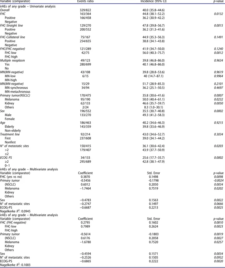
In the overall population, 329 patients experienced any grade immune-related adverse events (irAEs) (40%). Table 5
summarizes the univariate and multivariate analysis of irAEs of any grade. Overall, FHC-positive patients had a significantly higher incidence of irAEs of any grade (p = .0132) compared to FHC-negative patients; this also occurs when considering lineal line exclusively (p = .0015), but not when considering collateral line exclusively (p = .1491). FHC-high patients had a significantly higher incidence of irAEs of any grade, compared
Table 4.Univariate and multivariate analyses for OS.
Overall survival
Univariate analysis Multivariate analysis Variable (comparator) HR (95% CI); p-value HR (95% CI); p-value FHC Positive vs negative 0.81 (0.65–1.01); p = .0612 -FHC-Straight line Positive vs negative 0.79 (0.63–1.01); p = .0572 -FHC-Collateral line Positive vs negative 0.82 (0.62–1.08); p = .8207 -FHC(FHC-negative) FHC-low FHC-high 0.87 (0.69–1.10); p = .2652 0.61 (0.39–0.93); p = .0210 0.84 (0.67–1.06); p = .1600 0.57 (0.37–0.88); p = .0114 MN Yes vs no 0.86 (0.63–1.17); p = .3403 -MN (MN-negative) MN-low MN-high 0.83 (0.62–1.15); p = .2837 1.05 (0.49–2.23); p = .8909 -MN (MN-negative) MN-synchronous MN-metachronous 1.01 (0.57–1.75); p = .9753 0.82 (0.58–1.16); p = .2624 -Primary tumor (NSCLC) Melanoma Kidney Others 0.46 (0.35–0.62); p < .0001 0.56 (0.44–0.82); p = .0014 1.34 (0.75–2.39); p = .3239 0.54 (0.40–0.74); p = .0001 0.49 (0.36–0.68); p < .0001 1.03 (0.57–1.85); p = .9233 Sex Male vs female 1.51 (1.19–1.92); p = .0006 1.30 (1.02–1.65); p = .0317 Age Elderly vs nonelderly 1.15 (0.93–1.42); p = .1972 -Treatment line Nonfirst vs first 1.41 (1.07–1.84); p = .0129 1.19 (0.88–1.61); p = .2361 N° of metastatic sites >2 vs≤2 1.66 (1.34–2.06); p < .0001 1.52 (1.22–1.89); p = .0001 ECOG PS ≥2 vs 0–1 3.09 (2.43–3.92); p < .0001 3.05 (2.39–3.89); p < .0001 a b 0 20 40 60 80 100
Progression Free Survival
0 10 20 30 40 50 60 Months ) %( yti li ba b or p la vi vr u S Number at risk Group: FHC-negative 458 167 63 21 3 0 0 Group: FHC-low 289 93 29 15 3 1 0 Group: FHC-high 75 33 16 3 2 2 0 FHC-negative FHC-low FHC-high 0 20 40 60 80 100 Overall Survival 0 10 20 30 40 50 60 Months Number at risk Group: FHC-negative 458 221 85 33 7 0 0 Group: FHC-low 289 127 40 19 6 2 0 Group: FHC-high 75 45 20 6 2 2 0 FHC-negative FHC-low FHC-high ) %( yti li ba b or p la vi vr u S
Figure 1.Kaplan–Meier survival curves according to FHC. (a) Progression-free survival. FHC-negative: 9.3 months (95% CI: 7.5–10.6; 277 events); FHC-low: 8.4 months (95% CI: 7–11.4; 166 events); FHC-high: 20.5 months (95% CI: 8.7–26.4; 36 events). (b) Overall survival. FHC-negative: 18.2 months (95% CI: 14.9–23.9; 250 censored patients); FHC-low: 20.8 months (95% CI: 15.4–20.9; 176 censored patients); FHC-high: 31.6 months (95% CI: 26.2–31.6; 51 censored patients).
to FHC-negative patients (p = .0012), while FHC-low did not (p = .1240). FHC overall (positive vs negative) and FHC-high (vs negative) were confirmed as independent predictors for higher incidence of irAEs of any grade at the multivariate analysis.
Discussion
It is well known that a small percentage (5 – 10%) of cancers are related to inherited mutations, which usually occurs with typical familial patterns.11 Syndromes of inherited cancer predisposition are also one of the under-lying mechanisms of MN development.9 In our popula-tion, 44.3% and 14.9% of the patients had a positive FHC and diagnosis of MN, respectively; these findings are quite aligned to what was previously reported among cancer patients.9,15,16
In the preliminary analysis of the FAMI-L1 study, including the first 211 patients, FHC-positive patients had significantly higher ORR/DCR and significantly longer time to treatment failure and OS, when compared to FHC-negative patients.13 No significant association was found between diagnosis of MN (all metachronous tumors) and clinical outcomes, with the exception of a higher DCR compared to MN-negative patients.13 In this update, no significant associations were found between FHC, MN, and ORR; however from a speculative point of view, looking at the ORRs for FHC-negative, FHC-low, and FHC-high (32.7%, 36.7%, and 40.3%, respectively), we can notice that there is a trend to a direct proportionality, between the number of the positive familial lines and the ORR. Moreover, we can now confirm that MN does not affect PFS and OS, even considering the different analyses according to“burden of MN” and to synchronous/metachronous diagnosis of MN. Interestingly, only FHC-high patients had a significantly longer PFS and OS, when compared to FHC-negative patients, while no significant differences were found between low and negative, nor between FHC-positive and FHC-negative patients (analyzed overall, for lineal line only and for collateral line only, see Tables 3
and 4). The aim of the preliminary analysis was
exploratory and purely descriptive. We did not compute the sample size nor performed subgroup analyses accord-ing to the “FHC burden.” In our opinion, the present results are more reliable, thanks to the bigger sample size and to the more appropriate analysis.
Although our preliminary results seem now mitigated,13 this update seems to confirm our hypothesis that there is at least an association between the “FHC burden” and immunotherapy clinical outcomes, as if the more positive family lines, the greater the benefits. Looking at the hazard ratios, it is noticeable that they are concordantly higher in each comparison between FHC-high and FHC-negative patients than in those between low and FHC-negative. Intriguingly, adding the irAE analysis, we found a significantly higher incidence of any grade irAEs among FHC-positive patients (overall and for lineal line only) when compared to FHC-negative patients. Moreover, FHC-high patients had a significantly higher incidence of irAEs of any grade, when compared to FHC-negative patients, while FHC-low patients did not. It is also notice-able that the highest incidence of irAEs of any grade was reported among FHC-high patients (56%). In light of the emerging association between the development of irAEs and improved clinical outcomes with ICIs across different tumor types,17–20 these findings would bear our hypothesis.
As previously stated, a history of MN is one of the clinical hallmarks of inherited cancer susceptibility, just as a positive FHC. Despite that, in our population, FHC and diagnosis of MN are not significantly related, and this is reflected in the different correlations that they have with clinical outcomes. Nevertheless, it is noticeable that patients with metachronous MN and MN-high ones had the highest ORRs (43.8% and 41.5%, respectively, seeTable 2). Moreover, MN-high patients had at the same time the longest PFS and the shortest OS (compared to MN-negative and MN-low). We can thus spec-ulate that a history of MN may underlie a kind of“immune sensitiveness,” demonstrated by good ORR and PFS to treat-ment, which is, however, outclassed by the prognostic weight that further malignancies have. We could assume that under-lying mechanisms of MN and FHC are the same and lead to
a b 0 20 40 60 80 100
Progression Free Survival
0 10 20 30 40 50 60 Months ) %( yti li ba b or p la vi vr u S Number at risk Group: MN-negative 699 236 91 34 6 3 0 Group: MN-low 108 49 16 4 2 0 0 Group: MN-high 15 8 1 1 0 0 0 MN-negative MN-low MN-high 0 20 40 60 80 100 Overall Survival 0 10 20 30 40 50 60 Months Number at risk Group: MN-negative 699 326 122 49 12 4 0 Group: MN-low 108 58 21 7 3 0 0 Group: MN-high 15 9 2 2 0 0 0 MN-negative MN-low MN-high ) %( yti li ba b or p la vi vr u S
Figure 2.Kaplan–Meier survival curves according to MN. (a) Progression-free survival. MN-negative: 8.7 months (95% CI: 7.6–10.2; 414 events); MN-low: 12.3 months (95% CI: 8.3–28.9; 58 events); MN-high: 14.4 months (95% CI: 3.6–14.5; 7 events). (b) Overall survival. MN-negative: 20.5 months (95% CI: 15.7–27.1; 43 censored patients); MN-low: 26.2 months (95% CI: 18.7–48.9; 66 censored patients); MN-high: 15.9 months (95% CI: 10.5–15.9; 8 censored patients).
the same “immune sensitiveness,” but, on the other hand, patients developing MN surely have some negative prognostic features compared to FHC-positive patients.
The possible relationships between somatic alterations of genes belonging to DNA repair systems (such as homologous recombination, MMR, nucleotide excision repair, cell cycle
Table 5.Univariate and multivariate analysis for incidence of irAEs of any grade.
Variable (comparator) Events ratio Incidence (95% CI) p-value irAEs of any grade– Univariate analysis
Overall 329/822 40.0 (35.8–44.6) FHC Positive Negative 163/364 166/458 44.8 (38.1–52.2) 36.2 (30.9–42.2) 0.0132 FHC-Straight line Positive Negative 129/270 200/552 47.8 (39.8–56.7) 36.2 (31.3–41.6) 0.0015 FHC-Collateral line Positive Negative 75/167 254/655 44.9 (35.3–56.3) 38.8 (34.1–43.8) 0.1491 FHC(FHC-negative) FHC-low FHC-high 121/289 42/75 41.9 (34.7–50.0) 56.0 (40.3–75.7) 0.1240 0.0012 Multiple neoplasm Yes No 49/123 280/699 39.8 (46.8–86.0) 40.1 (46.8–86.0) 0.9634 MN(MN-negative) MN-low MN-high 43/108 6/15 39.8 (28.8–53.6) 40 (14.7–87.1) 0.9619 0.9964 MN(MN-negative) MN-synchronous MN-metachronous 15/29 34/94 51.7 (28.9–85.3) 36.2 (25.1–50.5) 0.2101 0.4697 Primary tumor(NSCLC) Melanoma Kidney Others 170/475 95/190 62/133 2/24 35.8 (30.6–41.6) 50.0 (40.4–61.1) 46.6 (35.7–59.7) 8.3 (1.0–30.1) 0.0007 0.0232 0.0050 Sex Male Female 196/552 133/270 35.5 (30.7–40.8) 49.3 (41.2–58.3) 0.0002 Age Elderly Non-elderly 186/463 143/359 40.2 (34.6–46.3) 39.8 (33.6–46.9) 0.9215 Treatment line First Nonfirst 92/214 237/608 43.0 (34.6–52.7) 39.0 (34.1–44.2) 0.3034 N° of metastatic sites >2 ≤2 150/415 179/407 36.1 (30.6–42.4) 43.9 (37.7–50.9) 0.0203 ECOG PS ≥2 0–1 34/133 295/689 25.6 (17.7–35.7) 42.8 (38.1–47.9) 0.0002
irAEs of any grade– Multivariate analysis
Variable (comparator) Coefficient Std. Error p-value
FHC (yes vs no) 0.3870 0.1498 0.0098 Primary tumor (NSCLC) Melanoma Kidney Others -0.5456 0.6012 –1.7464 -0.1798 0.2050 0.7519 -0.0024 0.0034 0.0202 Sex −0.4783 0.1563 0.0022 N° of metastatic sites −0.2747 0.1497 0.0666 ECOG-PS −0.6687 0.2213 0.0025 Nagelkerke R2: 0.0945
irAEs of any grade– Multivariate analysis
Variable (comparator) Coefficient Std. Error p-value FHC (FHC-negative) FHC-low FHC-high 0.2795 0.7989 0.1602 0.2624 0.0810 0.0023 Primary tumor (NSCLC) Melanoma Kidney Others -0.5614 0.6176 –1.6780 -0.1803 0.2058 0.7520 0.0019 0.0027 0.0257 Sex −0.4594 0.1571 0.0034 N° of metastatic sites −0.2526 0.1505 0.0932 ECOG-PS −0.6865 0.2222 0.0020 Nagelkerke R2: 0.1003
checkpoints, Fanconi anemia DNA repair pathway, and others), “immune-sensitiveness,” and ICI clinical outcomes have been already explored.21,22 Teo et al. reported a significant association between better clinical outcomes and somatic DDR gene alterations in a cohort of advanced urothelial cancer patients treated with atezolizumab.23 Importantly, a higher response rate was found not only in patients whose tumors harbored known or likely deleterious DDR gene alterations but also in patients with DDR altera-tions of unknown significance when compared to patients whose tumors were wild-type for DDR genes.23 In a study of single-agent pembrolizumab in docetaxel-refractory meta-static castration-resistant prostate cancer patients (mCRP), those with somatic mutations in BRCA1/2 or ATM (ataxia telangiectasia mutated) had higher response rates.24
That being said, if we are demonstrating that there is a proportional relationship between better clinical out-comes with anti-PD-1/PD-L1 inhibitors and “burden of familiarity,” we are allowed to think that DDR gene altera-tions (even of unknown clinical significance) might repre-sent the underlying mechanism, which would make the cancer more “immune-sensitive,” maybe throughout an increased production of neo-antigens. However, assuming that FHC is a surrogate of DDR gene alterations, such alterations should not be found exclusively with somatic assays (on the tumor specimen), but also with germline assays. In a recent study of mCRP patients, treated with Durvalumab (an anti-PD-L1 checkpoint inhibitor) and olaparib, patients harboring somatic DDR gene alterations were more likely to benefit from the treatment.25 Interestingly, four out of nine responders harbored germ-line alterations in DDR genes: one had a known deleter-ious mutation in NBN (nibrin) and 3 had frameshift BRCA2 indels.25 We must, however, recognize that
pros-tate cancer might be associated with specific syndromes of inherited cancer susceptibility;26thus it does not represent the most appropriate model to be extended to all other cancers. Nevertheless, it is conceivable that in case of a nonspecific high “burden of familiarity,” even without a peculiar familial pattern of cancers (as in the LS and HBOC syndrome), germline DDR gene alterations might be the substrate which explains the better outcomes with immunotherapy.
Among the limitations of the present study, we must cite the retrospective design, which exposes to selection biases, and the lack of centralized data review (imaging and toxici-ties). Our cohort was made of patients who received anti-PD -1/PD-L1 as different treatment lines; thus, we are not able to balance the expected immunosuppression induced by pre-vious treatments. Even if the discussion about LS and HBOC syndrome was only a presupposition for our study, which“generated the hypothesis,” we must recognize that our patients were not affected by breast/ovarian cancers nor by colorectal cancer. Moreover, we do not have sufficient data for a proper counseling (e.g., age at diagnosis and type/number of malignancies among the relatives), nor regarding inherited cancer predisposition syndrome diagnosis and DDR gene alteration (including germline BRCA mutations or LS diagnosis).
Collecting family history is one of the first steps in filling each patient medical record. Even though the role of this information is often underestimated, it should be taken into consideration to properly evaluate the development risk of a wide range of diseases, including cancer.27,28We are a long way from saying that FHC could be used as a selection method for anti-PD-1/PD-L1 treat-ments. However, our study gives rise to interesting insights, which we intend to validate prospectively.
Conclusion
Thanks to the great sample size, this update confirms our preliminary findings. Particularly, FHC-high patients seem to benefit more than FHC-negative patients from PD-1/PD-L1 checkpoint inhibitors, suggesting that FHC might be the surrogate of some biological features related to the immune-sensitiveness. However, further investigations on the topic are still required.
Materials and methods Patient eligibility
This multicenter retrospective observational study evaluated advanced cancer patients consecutively treated with single-agent anti-PD-1/PD-L1 immunotherapy from April 2015 to July 2018, regardless of the treatment line, at 17 Italian insti-tutions (Supplementary file 1). Patients were eligible if they had histologically confirmed diagnosis of measurable stage IV cancer, with availability of records about FHC and history of eventual metachronous or synchronous MN. All patients pro-vided written informed consent to the treatment with immunotherapy.
Study design
The primary endpoint of this analysis was to confirm the correlations between FHC and clinical outcomes; the sec-ondary endpoint was to further investigate the relation-ships between diagnosis of MN and clinical outcomes. ORR, progression-free survival (PFS), OS, and incidence of any grade irAEs were evaluated. Patients were assessed with radiological imaging every 8–12 weeks using the RECIST (v. 1.0) criteria29 according to the local clinical practice and national guidelines required by the Agenzia Italiana del Farmaco (AIFA). ORR was defined as the portion of patients experiencing an objective response (complete or partial response) as best response to immu-notherapy. PFS was defined as the time from ICI treat-ment’s start to disease progression or death whichever occurred first; OS as the time from the beginning of treatment to death. For PFS as well as for OS, patients without events were considered as censored at the time of the last follow-up.
On the basis of our previous results,13 and what was reported in other studies,15,16,30 we hypothesized that 48% of the evaluated patients were FHC-positive, and 52% were FHC-negative. With a probability of Type I error of 0.05 and of Type II error of 0.20 and assuming a possible
survival benefit for FHC-positive patients with a reduction of the risk of death by 70%, 247 total events were neces-sary and at least 712 patients had to be included. Univariate and multivariate analyses were performed using the following covariates: age (<70 vs ≥70 years old),31–34 sex (male vs. female), primary tumor (NSCLC, melanoma, renal cell carcinoma, and others), Eastern Cooperative Oncology Group Performance Status (ECOG-PS) (0–1 vs ≥2), number of metastatic sites (≤2 vs >2), and treatment line (first vs nonfirst). χ2 test was used to correlate ORR and incidence of any grade irAEs with patient features.35 χ2 test was also used to evaluate the correlation between FHC (yes vs no) and diagnosis of MN (yes vs no). Logistic regression was used for the multi-variate analysis of ORR and incidence of irAEs of any grade.36 Median PFS and median OS were evaluated using the Kaplan–Meier method.37 The median period of
follow-up was computed according to the reverse Kaplan– Meier method.38 Cox proportional hazards model39 was used to evaluate predictor variables in univariate and multivariate analysis for median PFS and median OS. Data cutoff period was October 2018. All statistical ana-lyses were performed using MedCalc Statistical Software version 18.11.3 (MedCalc Software bvba, Ostend, Belgium;
https://www.medcalc.org; 2019).
Definition of FHC and MNs
Given the lack of data availability in medical records, we did not use the traditional designations of first and second degree of relatedness for family history. Family history was collected in lineal (descendants or ascendants) and collateral lines (not-descentants/ascendants) till the second degree of relatedness (grandparents for lineal line and brothers/sisters for the col-lateral line). FHC was defined as“positive” with at least one diagnosis of cancer among the considered relatives. Patients were also categorized according to their FHC as follows: FHC-high (in case of cancer diagnoses in both the lineal and collateral family lines), FHC-low (in case of cancer diagnoses in only one family lines, lineal or collateral), and FHC-negative. Diagnosis of metachronous and/or synchronous MN was defined according to the international association of cancer registry (IARC/IACR) rules.40 Patients were also categorized according to the diagnosis of MN as follows: MN-high (in case of more than two cancer diagnoses in their medical history), MN-low (in case of two cancer diagnoses in their medical history), and MN-negative. A further analysis was performed categorizing patients into synchronous MN, metachronous MN, and MN-negative.
Acknowledgments
This work was supported by the Consorzio Interuniversitario Nazionale per la Bio-Oncologia (CINBO).
Disclosure of Potential Conflicts of Interest No potential conflicts of interest were disclosed.
Funding
No funding was received.
Ethics approval and consent to participate
All patients provided written informed consent to treatment with immu-notherapy. All patients alive at the time of data collection provided an informed consent for the present retrospective analysis. The procedures followed were in accordance with the precepts of Good Clinical Practice and the declaration of Helsinki. The study was approved by the respective local ethical committees on human experimentation of each institution, after previous approval by the coordinating center (University of L’Aquila, Internal Review Board protocol number 32865, approved on July 24, 2018).
Authors contributions
All authors contributed to the publication according to the ICMJE guidelines for the authorship as follows:
Study conception and design: AC, PAA, CF, SB, MT, MB.
Acquisition of data: AC, RG, FP, PDM, NT, MDT, AG, KC, AT, FZ, EV, FM, MR, CA, TZ, MF, PM, AB, GCAC, FDG, MGV, FR, RRS, AP, SR, MT, MRM, AG, MAO, FM, AI, SG, SB, CZ, CM, AP, GP, DM, MCF.
Analysis and interpretation of data: AC, SB MB, PAA, CF. Drafting of manuscript: PAA, CF, SB, MB, RB, DS. Critical revision: PAA, CF, MT, RB, DS, MCF.
All authors read and approved the submitted version of the manu-script (and any substantially modified version that involves the author’s contribution to the study). Each author has agreed both to be personally accountable for the author’s own contributions and to ensure that ques-tions related to the accuracy or integrity of any part of the work, even ones in which the author was not personally involved, are appropriately investigated, resolved, and the resolution documented in the literature.
Availability of data and materials
The datasets used during the present study are available from the corre-sponding author upon reasonable request.
Conflicts of interest
Dr Alessio Cortellini received grants as speaker by MSD, Astra-Zeneca and Boehringer Ingelheim, grant consultancies by BMS, Roche, Novartis, Istituto Gentili, and Ipsen; Dr Marcello Tiseo received grant as speaker and advisory role by Astra-Zeneca, Pfizer, Eli-Lilly, BMS, Novartis, Roche, MSD, Boehringer Ingelheim, Otsuka, Pierre Fabre; Dr Maria Giuseppa Vitale received travel grants and speaker fees by BMS, Ipsen Astellas, Janssen, Novartis, and Pfizer; Dr Sebastiano Buti received grants as speaker and advisory role by BMS, Pfizer, MSD, Ipsen, Novartis, Astra-Zeneca, and research funding from Novartis; Dr. Melissa Bersanelli received honoraria as speaker at scientific events and as con-sultant for advisory role by BMS, Novartis, and Pfizer and research funding by Seqirus; Dr Paolo A. Ascierto has/had a consultant/advisory role for Bristol Myers-Squibb, Roche-Genentech, Merck Sharp & Dohme, Array, Novartis, Merck Serono, Pierre Fabre, Incyte, NewLink Genetics, Genmab, Medimmune, AstraZeneca, Syndax, SunPharma, Sanofi, Idera, Ultimovacs, Sandoz, Immunocore, he also received research funds from Bristol Myers-Squibb, Roche-Genentech, Array, and travel support from MSD.
ORCID
Alessio Cortellini http://orcid.org/0000-0002-1209-5735
Sebastiano Buti http://orcid.org/0000-0003-0876-0226
References
1. Taube JM, Klein A, Brahmer JR, Xu H, Pan X, Kim JH, Chen L, Pardoll DM, Topalian SL, Anders RA, et al. Association of PD-1, PD-1 ligands, and other features of the tumor immune microenvir-onment with response to anti-PD-1 therapy. Clin Cancer Res.
2014;20(19):5064–5074. doi:10.1158/1078-0432.CCR-13-3271. 2. Rizvi NA, Hellmann MD, Snyder A, Kvistborg P, Makarov V,
Havel JJ, Lee W, Yuan J, Wong P, Ho TS. Mutational landscape determines sensitivity to PD-1 blockade in non–small cell lung cancer. Science. 2015;348(6230):124–128. doi:10.1126/science. aaa1348.
3. Hellmann MD, Ciuleanu T-E, Pluzanski A, Lee JS, Otterson GA, Audigier-Valette C, Minenza E, Linardou H, Burgers S, Salman P, et al. Nivolumab plus ipilimumab in lung cancer with a high
tumor mutational burden. N Engl J Med. 2018 May 31;378
(22):2093–2104. doi:10.1056/NEJMoa1801946. Epub 2018 Apr 16. 4. Cortellini A, Bersanelli M, Buti S, Cannita K, Santini D, Perrone F, Giusti R, Tiseo M, Michiara M, Di Marino P, et al. A multicenter study of body mass index in cancer patients treated with anti-PD-1/PD-L1 immune checkpoint inhibitors: when over-weight becomes favorable. J Immuno Ther Cancer.2019Feb 27;7 (1):57. doi:10.1186/s40425-019-0527-y.
5. Routy B, Le Chatelier E, Derosa L, Duong CPM, Alou MT, Daillère R, Fluckiger A, Messaoudene M, Rauber C, Roberti MP, et al. Gut microbiome influences efficacy of PD-1-based immu-notherapy against epithelial tumors. Science. 2018 Jan 5;359 (6371):91–97. doi:10.1126/science.aan3706.
6. Le DT, Uram JN, Wang H, Bartlett BR, Kemberling H, Eyring AD, Skora AD, Luber BS, Azad NS, Laheru D, et al. PD-1 blockade in tumors with mismatch-repair deficiency. N Engl J Med.2015;372:2509–2520. doi:10.1056/NEJMoa1500596. 7. Viale G, Trapani D, Curigliano G. Mismatch repair deficiency as
a predictive biomarker for immunotherapy efficacy. Biomed Res Int.2017;4719194:2017.
8. Dai Y, Sun C, Feng Y, Sun C, Jia Q, Zhu B. Potent immunogeni-city in BRCA1-mutated patients with high-grade serous ovarian
carcinoma. J Cell Mol Med. 2018 May 31;22:3979–3986.
doi:10.1111/jcmm.13678. [Epub ahead of print].
9. Vogt A, Schmid S, Heinimann K, Frick H, Herrmann C, Cerny T, Omlin A. Multiple primary tumours: challenges and approaches, a review. ESMO Open. 2017 May 2;2(2):e000172. doi:10.1136/ esmoopen-2017-000172. eCollection 2017.
10. Strickland KC, Howitt BE, Shukla SA, Rodig S, Ritterhouse LL, Liu JF, Garber JE, Chowdhury D, Wu CJ, D’Andrea AD, et al. Association and prognostic significance of BRCA1/2-mutation status with neoantigen load, number of tumor-infiltrating lym-phocytes and expression of PD-1/PD-L1 in high grade serous
ovarian cancer. Oncotarget. 2016;7(12):13587–13598.
doi:10.18632/oncotarget.v7i12.
11. Pembrolizumab in advanced BRCA-mutated breast cancer. Identifier: NCT03025035. [accessed 2019 Mar 9]. https://clinical trials.gov/ct2/show/NCT03025035.
12. Garber JE, Offit K. Hereditary cancer predisposition syndromes.
J Clin Oncol. 2005 Jan 10;23(2):276–292. doi:10.1200/
JCO.2005.10.042.
13. Cortellini A, Bersanelli M, Buti S, Gambale E, Atzori F, Zoratto F, Parisi A, Brocco D, Pireddu A, Cannita K et al. Family history of cancer as surrogate predictor for immunotherapy with anti-PD1/PD-L1 agents: preliminary report of the FAMI-anti-PD1/PD-L1 study. Immunotherapy.
2018Jun;10(8):643–655. doi:10.2217/imt-2017-0167.
14. Cortellini A, Bersanelli M, Ficorella C, Buti S. Family history of cancer and DNA damage response genes: two sides of the same coin? Thorac Cancer. 2019 Feb;10(2):401. doi: 10.1111/1759-7714.12926.
15. Ogawa H, Kato I, Tominaga S. Family history of cancer among cancer patients. Jpn J Cancer Res GANN.1985;76:113–118. 16. Gaughan EM, Cryer SK, Yeap BY, Jackman DM, Costa DB.
Family history of lung cancer in never smokers with non-small-cell lung cancer and its association with tumors harboring
EGFR mutations. Lung Cancer. 2013 Mar;79(3):193–197.
doi:10.1016/j.lungcan.2012.12.002. Epub 2012 Dec 27.
17. Freeman-Keller M, Kim Y, Cronin H, Richards A, Gibney G, Weber JS. Nivolumab in resected and unresectable metastatic melanoma: characteristics of immune-related adverse events and association with outcomes. Clin Cancer Res. 2016 Feb 15;22 (4):886–894. doi:10.1158/1078-0432.CCR-15-1136.
18. Haratani K, Hayashi H, Chiba Y, Kudo K, Yonesaka K, Kato R, Kaneda H, Hasegawa Y, Tanaka K, Takeda M, et al. Association of immune-related adverse events with nivolumab efficacy in non-small-cell lung cancer. JAMA Oncol.2018Mar 1;4(3):374–378. doi:10.1001/jamaoncol.2017.2925.
19. Teraoka S, Fujimoto D, Morimoto T, Kawachi H, Ito M, Sato Y, Nagata K, Nakagawa A, Otsuka K, Uehara K, et al. Early immune-related adverse events and association with outcome in advanced non-small cell lung cancer patients treated with nivolu-mab: a prospective cohort study. J Thorac Oncol.2017 Dec;12 (12):1798–1805. doi:10.1016/j.jtho.2017.08.022. Epub 2017 Sep 20. 20. Cortellini A, Chiari R, Ricciuti B, Metro G, Perrone F, Tiseo M, Bersanelli M, Bordi P, Santini D, Giusti R. Correlations between the immune-related adverse events spectrum and efficacy of anti-PD1 immunotherapy in NSCLC patients. Clin Lung Cancer.
2019 Feb 21:pii: S1525-7304(19)30025–7. doi:10.1016/j.
cllc.2019.02.006. [Epub ahead of print].
21. Rizvi NA, Hellmann MD, Snyder A, Kvistborg P, Makarov V, Havel JJ, Lee W, Yuan J, Wong P, Ho TS, et al. Cancer immunology. Mutational landscape determines sensitivity to PD-1 blockade in non-small cell lung cancer. Science. 2015
Apr 3;348(6230):124–128. doi:10.1126/science.aaa1348. Epub 2015 Mar 12.
22. Wang Z, Zhao J, Wang G, Zhang F, Zhang Z, Zhang F, Zhang Y, Dong H, Zhao X, Duan J, et al. Comutations in DNA damage response pathways serve as potential biomarkers for immune
checkpoint blockade. Cancer Res. 2018 Nov 15;78
(22):6486–6496. doi:10.1158/0008-5472.CAN-18-1814. Epub 2018 Aug 31.
23. Teo MY, Seier K, Ostrovnaya I, Regazzi AM, Kania BE, Moran MM, Cipolla CK, Bluth MJ, Chaim J, Al-Ahmadie H, et al. Alterations in DNA damage response and repair genes as potential marker of clinical benefit from PD-1/PD-L1 blockade in advanced urothelial cancers. J Clin Oncol.2018Jun 10;36(17):1685–1694. doi:10.1200/ JCO.2017.75.7740. Epub 2018 Feb 28.
24. De Bono J, Goh J, Ojamaa K, Piulats Rodriguez JM, Drake CG, Hoimes CJ, Wu H, Poehlein CH, Antonarakis ES. KEYNOTE-199: pembrolizumab (pembro) for docetaxel-refractory metastatic castration-resistant prostate cancer (mCRPC). J Clin Oncol.
2018;36(suppl):abstr5007. doi:10.1200/
JCO.2018.36.15_suppl.5007.
25. Karzai F, VanderWeele D, Madan RA, Owens H, Cordes LM, Hankin A, Couvillon A, Nichols E, Bilusic M, Beshiri ML, et al.
Activity of durvalumab plus olaparib in metastatic
castration-resistant prostate cancer in men with and without
DNA damage repair mutations. J Immuno Ther Cancer. 2018
Dec 4;6(1):141. doi:10.1186/s40425-018-0463-2.
26. Wang G, Zhao D, Spring DJ, DePinho RA. Genetics and biology of prostate cancer. Genes Dev. 2018Sep 1;32(17–18):1105–1140. doi:10.1101/gad.315739.118.
27. Walter FM, Emery J.‘Coming down the line’– patients’ under-standing of their family history of common chronic disease.
Ann Fam Med. 2005 Sep-Oct;3(5):405–414. doi:10.1370/
afm.368.
28. Nathan PA, Johnson O, Clamp S, Wyatt JC. Time to rethink the capture and use of family history in primary care. Br J Gen Pract.
2016Dec;66(653):627–628. doi:10.3399/bjgp16X688273.
29. Eisenhauer EA, Therasse P, Bogaerts J, Schwartz LH, Sargent D, Ford R, Dancey J, Arbuck S, Gwyther S, Mooney M, et al. New response evaluation criteria in solid tumours: revised RECIST guideline (version 1.1). Eur J Cancer. 2009;45:228–247. doi:10.1016/j.ejca.2008.10.026.
30. Song JL, Chen C, Yuan JP, Li -J-J, Sun S-R. Family history of cancer other than breast or ovarian cancer in first-degree relatives is associated with poor breast cancer prognosis. Breast. 2017
Apr;32:130–134. doi:10.1016/j.breast.2017.01.016. Epub 2017 Feb 5.
31. Minana B, Cozar JM, Palou J, Unda Urzaiz M, Medina-Lopez RA, Subirá Ríos J, de la Rosa-kehrmann F, Chantada-Abal V, Lozano F, Ribal MJ, et al. Bladder cancer in Spain 2011: population-based study. J Urol. 2014 Feb;191(2):323–328. doi:10.1016/j.juro.2013.08.049.
32. Ciocan D, Barbe C, Aubin F, Granel-Brocard F, Lipsker D, Velten M, Dalac S, Truchetet F, Michel C, Mitschler A, et al. Distinctive features of melanoma and its management in elderly patients: a population-based study in France. JAMA Dermatol. 2013Oct;149 (10):1150–1157. doi:10.1001/jamadermatol.2013.706.
33. Gridelli C, Balducci L, Ciardiello F, Di Maio M, Felip E, Langer C, Lilenbaum RC, Perrone F, Senan S, de Marinis F, et al. Treatment of elderly patients with non-small-cell lung cancer: results of an international expert panel meeting of the italian association of thoracic oncology. Clin Lung Cancer.2015Sep;16(5):325–333. doi:10.1016/j.cllc.2015.02.006.
34. Azawi NH, Joergensen SM, Jensen NV, Clark PE, Lund L. Trends in kidney cancer among the elderly in Denmark, 1980–2012. Acta Oncol. 2016;55(Suppl 1):79–84. doi:10.3109/ 0284186X.2015.1115121.
35. Mantel N. Chi-square tests with one degree of freedom: exten-sions of the Mendel-Haenszel procedure. J Am Stat Assoc.
1963;58:690–700.
36. Hosmer DW Jr, Lemeshow S. Sturdivant RX applied logistic regression. Third ed. New Jersey: John Wiley & Sons;2013. 37. Kaplan EL, Meier P. Nonparametric estimation of incomplete
observations. J Am Stat Assoc. 1958;53:457–481. doi:10.1080/ 01621459.1958.10501452.
38. Schemper M, Smith TL. A note on quantifying follow-up in studies of failure time. Control Clin Trials. 1997;17:343–346. doi:10.1016/0197-2456(96)00075-X.
39. Cox DR. Regression models and life tables (with discussion). J R Stat Soc Ser B.1972;74:187–200.
40. International rules for multiple primary cancers ICD-O third edition. Internal Report No.2004/02. Lyon: International Agency for Research on Cancer. IARC (2004).
![Table 4 , FHC-high patients had a significantly longer OS when compared to FHC-negative patients (HR = 0.61 [95% CI: 0.39 –0.93], p = .0210); at multivariate analysis, FHC-high was confirmed an independent predictor for OS (compared to FHC-negative).](https://thumb-eu.123doks.com/thumbv2/123dokorg/4967359.53442/5.890.60.435.95.468/patients-significantly-multivariate-analysis-confirmed-independent-predictor-compared.webp)