P
H.D
.T
HESIS IN MOLECULAR BIOPHYSICSAptamers against transferrin receptor:
rational engineering of nanoscale platforms
for targeted delivery of molecular payloads
CANDIDATE
David Porciani
ADVISORSFabio Beltram
Giovanni Signore
2015/16
Table of Contents
Introduction... v
Nucleic acid aptamers: new tools for targeted drug delivery applications... 1
1.1 Brief overview on targeted therapy: passive and targeting drug delivery ...1
1.2 Cell-surface markers: the gate at the entrance of diseased cells ...4
1.3 Targeting ligands: the keys to open the gates of the diseased cells ...11
1.3.1 Antibodies ...12
1.3.2 Aptamers ...14
1.4 Aptamers as novel “keys of the castle” ...16
1.5 In vitro selection of aptamers ...24
1.6 Selection of aptamers recognizing cell-surface markers ...29
1.7 Cell-specific aptamer applications in targeted drug delivery...33
1.7.1 Aptamer-chemotherapeutic conjugates...33
1.7.2 Aptamer-therapeutic oligonucleotide conjugates...37
1.7.3 Aptamer-conjugated nanomaterials ...40
Aptamer-mediated codelivery of doxorubicin and NF-kB decoy enhances chemosensitivy of pancreatic tumor cells... 47
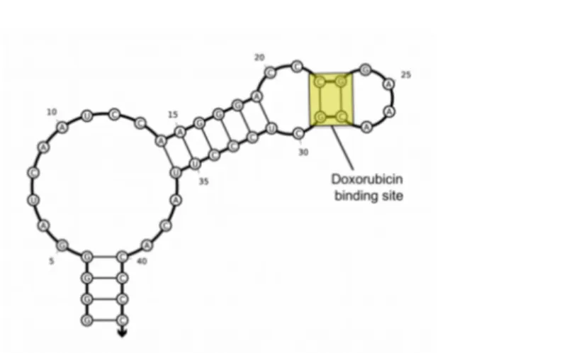
2.1 Rational design of aptamer–Dox conjugate...51
2.2 Aptamer-mediated tumor targeting and Dox release mechanism ...58
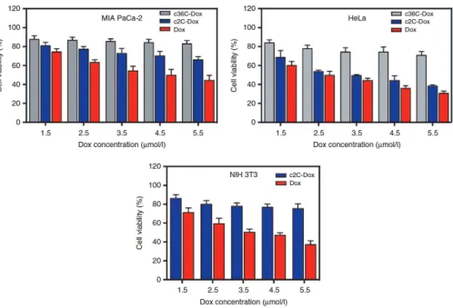
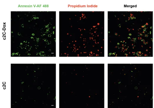
2.3 Antitumor efficacy of c2C-Dox...61
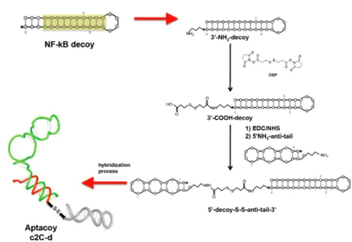
2.4 Design, synthesis and Dox-loading of a novel aptacoy chimera ...65
2.5 Codelivery of Dox and NF-κB decoy via anti-TfR aptamer in living cells ...70
2.6 Concluding remarks ...75
2.7 MATERIALS AND METHODS ...77
2.7.1 Materials...77
2.7.2 Oligonucleotide sequences...77
2.7.3 Absorption and fluorescence measurements...78
2.7.4 Cell culture...78
2.7.5 HPLC analyses...78
2.7.6 Confocal imaging of cells ...79
2.7.7 Secondary structure and hybridization predictions...79
2.7.9 Oligonucleotide annealing protocol ...80
2.7.10 Doxorubicin intercalation in double helix region of the hybridized aptamer ...80
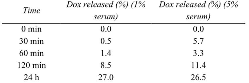
2.7.11 Doxorubicin release from aptamer in serum-containing media...80
2.7.12 Assessment of cellular uptake by confocal microscopy ...81
2.7.13 WST-8 Cell viability assay ...81
2.7.14 Statistical analysis ...82
2.7.15 Apoptosis assay...82
2.7.16 pH- and nuclease-dependent drug controlled release ...83
2.7.17 Synthesis of the NF-κB decoy-anti-tail conjugate ...83
2.7.18 Detection of NF-κB by immunofluorescence staining of p65 ...84
2.7.19 Western Blot analysis...85
An enhanced aptamer sequence as tool for targeted drug delivery: rational engineering of a DNA aptamer against transferrin receptor... 87
3.1 Folding conformation analysis...90
3.2 Fluorescence anisotropy assay...96
3.3 Internalization assay in living cells...101
3.4 Rational engineering of GS24 aptamer...109
3.5 Concluding remarks ...120
3.6 MATERIALS AND METHODS ...122
3.6.1 Materials...122
3.6.2 Chromatographic analyses, and purification of labeled folds...123
3.6.3 Fluorescence anisotropy assay ...124
3.6.4 Fluorescence Anisotropy Data Analysis ...125
3.6.5 Secondary and tertiary structure generation...126
3.6.6 Simulated annealing...126
3.6.7 Molecular dynamics simulations ...127
3.6.8 Electrostatic properties...127
3.6.9 Endocytosis assays in living cells ...128
Aptamer-mediated delivery of large functional RNA ... 131
4.1 Design and synthesis of a plug-and-play platform for targeted delivery...136
4.1.1 Assembly of aptamer-aptamer complexes: annealing reaction with equimolar concentration of aptamer modules and excess of bridge...139
4.1.2 Assembly of aptamer-aptamer complexes: annealing reaction with an excess of delivery aptamers and bridge...141
4.2 Aptamer functional assays ...144
4.2.1 Spinach2 fluorescence assay...144
4.2.2 Endocytosis assay ...148
4.3 An improved fluorescent aptamer sequence: rational multimerization of Baby Spinach ...154
4.4 Concluding remarks ...164
4.5.1 Reagents, oligonucleotides, and transcription templates ...166
4.5.2 Aptamer sequences ...166
4.5.3 Secondary structure and hybridization predictions...169
4.5.4 In vitro RNA transcription ...169
4.5.5 Denaturing PAGE and RNA purification ...169
4.5.6 Preparation of self-assembled aptamer-aptamer complexes...170
4.5.7 Electrophoretic Mobility Shift Assay ...171
4.5.8 Measurement of Spinach Fluorescence...172
4.5.9 Endocytosis in living cells and cell imaging...172
Concluding remarks and research perspectives ... 175
Introduction
Cancer remains one of the leading causes of death worldwide with high morbidity and mortality. Though significant advances have been made in the fundamental understanding of cancer biology, the treatment of some cancer forms remains elusive, and in general cancer treatments suffer from severe side effects. Cancer cells share many common features with the normal host cells from which they derive. As a consequence, traditional anticancer agents do not achieve the specific toxicity shown by bacterial and viral chemotherapeutics due to their lack of selectivity, and ultimately they lead to significant off-target effects.
Interestingly, cancer cells show different composition of cell membrane compared to healthy cells, with an enhanced expression of specific proteins (i.e. cell-surface markers) required to increase the provision of nutrients and to enhance cell-cell signaling and communication. Thus, a valid strategy to improve the selectivity of the treatments relies on the development of molecules able to discriminate between diseased and healthy cells by interacting with these cell-surface markers. Over the past decades an exciting trend in life sciences and biotechnology has been the rapid development of highly specific structures recognizing these cell-surface markers. Nanomedicine aims at exploiting these ligands to generate nanoscale platforms for targeted cancer therapy and diagnosis. In this way, non-selective therapeutics are localized and concentrated at the targeted site via ligands that recognize tumor-associated markers, thus leading to negligible off-target effect.
Aptamers are short single-chained DNA or RNA molecules able to recognize molecular targets with high affinity and specificity owing to their exclusive spatial conformation that are emerging as a class of biocompatible ligands with huge potential in diagnostics and therapeutics. Despite the rapid development of a wide variety of targeted delivery platforms exploiting aptamers as nanocarriers against cell-surface receptors, these strategies show some limitations that could hamper
the transition to clinical application, including elaborated design, difficult scale-up, and complex aptamer-payload conjugate preparation. Furthermore, aptamers as targeting moieties have been used mainly to deliver small drugs and small interfering RNAs. However, a modular aptamer-based platform to deliver large therapeutic payloads, such as therapeutic aptamers recognizing cancer-associated protein, was not developed yet, likely due to limitations in designing suitable assemblies in which the oligonucleotide modules retain their proper activity. Therefore, the work reported in this thesis addresses these restrictions in aptamer technology for targeted delivery applications.
In this thesis, I shall describe novel designs and molecular engineering strategies that lead to aptamer-based nanoassemblies with enhanced and innovative features as tools for targeted cancer therapy. These assemblies exploit known RNA and DNA aptamers recognizing a cell-surface protein, the transferrin receptor (TfR). Particularly, TfR was chosen as biomolecular target owing to (i) its upregulation in many solid tumors (e.g. breast, prostate, pancreatic, ovarian, lung) and (ii) its constitutive endocytic pathway. Thus, aptamers binding TfR may discriminate between tumor and healthy cells and aptamer-based platforms may selectively deliver into target cells therapeutic payloads upon binding to TfR via receptor-mediated endocytosis. To achieve these goals, this thesis work exploits the synergy among different studies of aptamer properties, in terms of folding stability, recognition of target cell population and efficiency of internalization into target cells.
The outline of the thesis is the following:
- Chapter 1 provides a review of aptamers an emerging class of targeting ligands for cancer therapy. Emphasis on the advantages of aptamers compared to antibodies will be reported together with their drawbacks and the innovative solutions to overcome these limitations. The in vitro evolutionary selection process used for the aptamer generation will be described. Then, I shall introduce the main approaches and the most representative applications of cell-specific aptamers. Particularly, I shall focus on the conjugation designs used to deliver traditional anticancer drugs and functional therapeutic oligonucleotides.
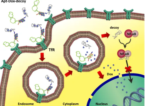
- In Chapter 2 I shall describe a molecular-engineering strategy to develop a drug-delivery system that is able to (i) target human tumor cells via an anti-transferrin receptor RNA aptamer and (ii) perform selective co-delivery of a chemotherapeutic drug (doxorubicin) and of an inhibitor of a cell-survival factor, the NF-κB decoy oligonucleotide. I shall demonstrate that this platform shows selective cytotoxicity activity towards tumor cells, owing to the targeting properties of the aptamer. Upon internalization, the κB decoy does inhibit NF-κB activity, reducing tumor chemoresistance towards doxorubicin and enhancing its therapeutic efficacy specifically in target cells.
- In Chapter 3 a biophysical study to unveil the conformation-dependent activity of an anti-mouse TfR DNA aptamer will be reported. I shall demonstrate the presence of competing folding pathways, which sensibly affected the aptamer activity. Indeed, two main aptamer conformers were identified and their activities investigated both in vitro and in living cells. Actually, only one conformer was able to bind mouse TfR. Starting from these observations I rationally designed variations of the parent sequence aimed at stabilizing the active conformation. This strategy led to an enhanced aptamer sequence with higher affinity and improved biological activity, which emerges as a valid targeting ligand both for in
vitro and in vivo applications.
- In Chapter 4, I shall report on the design and synthesis of a modular platform for aptamer-mediated cellular delivery of increasingly large RNA payloads (~50-60 kDa). In order to demonstrate the versatility of this platform, anti-TfR RNA and DNA aptamers, previously investigated in Chapter 2 and 3, will be exploited as targeting and delivery tools, and fluorescent RNA aptamers (known as RNAs mimic of GFP) as large functional RNA payloads. These aptamers were chosen as RNA sensors to monitor both their intracellular localization and to evaluate folding alterations within the oligonucleotide platform because their fluorescence depends on the retaining of the proper fold. Taking inspiration from the design of the delivery system described in Chapter 2, the assembly is generated through straightforward self-assembly by means of a short oligonucleotide bridge that contains regions complementary to both aptamers. In this chapter, I shall demonstrate the possibility to selectively deliver to living cells large functional
RNAs using different combinations of targeting and payload aptamers. This “proof of concept” is the first evidence of an aptamer-mediated aptamer delivery.
- In Chapter 5 I shall summarize the main findings of this research activity and discuss some perspectives of the aptamer-based nanoscale platforms proposed in this thesis.
Chapter
1
Nucleic acid aptamers: new tools for targeted
drug delivery applications
1.1 Brief overview on targeted therapy: passive and targeting
drug delivery
One hundred years ago, Paul Ehrlich popularized the “magic bullet” concept as an ideal strategy for targeted therapy against virtually any disease. Theoretically, a magic bullet is a therapeutic agent able to overcome biological barriers, distinguish between diseased and healthy tissues and selectively kill only target cells, avoiding off-target effect1. Despite extensive research carried out in this field, safe and efficient drug delivery for the treatment of most disorders (e.g. cancer and infectious diseases) remains a major challenge both for clinical translation and the development of new therapies2. Conventional therapeutic agents are administered systemically and are unable to target the site of the disease, ultimately leading to unwanted side effects and to a reduction of the therapeutic index (i.e. the ratio between the dosage of a drug that causes a lethal effect and the dosage that causes a therapeutic effect). Nanomedicine aims at overcoming these drawbacks, allowing the construction of nanosized molecular devices (10-200 nm) for targeted delivery of therapeutic and/or diagnostic agents3.
During the last decade, the rapid expansion of new nanotechnologies tailored to cancer treatment has been an exciting trend in nanomedicine. Compared with conventional therapy, nanomedicine offers significant advantages and new perspectives in which novel drug-delivery platforms exploit passive and active targeting to improve their therapeutic index4. Passive targeting takes advantage of the unique physiology and structure of the tumor microenvironment (e.g. highly permeable vasculature, and poorly-defined lymphatic system), which results in drug accumulation through the enhanced permeability retention (EPR) effect (Fig.1). This strategy can be achieved by modulating the size, shape, and surface properties of the drug delivery platform. To date, dozens of nanosized devices, named first-generation nanomedicines, were approved by the Food and Drug Administration (FDA) and proved to be well tolerated in patients5,6. However, several factors mainly dependent on patient and/or stage of disease (such as blood pressure, structure of neo-vasculature, and disease type and location) can adversely affect passive transport, often leading to inadequate drug concentration into target districts and lower therapeutic efficacy3,7 (Fig.1).
Fig 1 Physiological properties of tumor tissue and vasculatures that influence cancer drug delivery. Reprinted from Kobayashi et al.8; Copyright © Ivyspring International
Ultimately, passive targeting strategy does not discriminate satisfactorily between normal and cancerous cells; this in turn leads to modest improvements compared to conventional therapeutic strategies9.
A common strategy to enhance selectivity relies on targeting specific biomolecules overexpressed on the surface of tumor cells. To this end, many ligands were developed and exploited to deliver potent nonspecific cytotoxic agents selectively to malignant cells. The general structure of these ligand-payload platforms involves a targeting moiety linked to a therapeutic payload via a spacer that often contains a cleavable bond10. Alternatively, specific targeting ligands can be conjugated and displayed on the surface of nanosized devices (or nanocarriers) designed for passive targeting drug delivery. When the suitable recognition motif is included in these nanocarriers, it endows the entire system with selectivity towards target cells, leading to more efficient “actively targeted” nanostructures, also called second-generation nanodevices11 (Fig. 2).
Fig. 2 Schematic representation of several mechanisms by which nanocarriers can deliver drugs to tumors. Nanocarriers are shown as blue circular structures. Passive tissue targeting is achieved by extravasation of nanoparticles through increased permeability of the tumor vasculature and ineffective lymphatic drainage (EPR effect). Active cellular targeting (inset) can be achieved by functionalizing the surface of nanoparticles with ligands that promote cell-specific recognition and binding. The
nanoparticles can (i) release their contents in close proximity to the target cells; (ii) adhere to cell membrane and act as an extracellular sustained-release drug depot; or (iii) internalize into the cell. Adapted from Peer et al.12 Reprinted by permission from
Macmillan Publishers Ltd: Nature Nanotechnology (Ref. 12), Copyright © 2007
1.2 Cell-surface markers: the gate at the entrance of diseased
cells
Several genetic mutations lead to the deregulation of specific signaling pathways involved into cell proliferation, survival, or differentiation, causing uncontrolled cell growth and acquisition of neoplastic phenotype13. As a consequence of this mutated signal transduction, specific cell-surface and intracellular receptors show high expression levels in diseased tissue compared with other tissues (for example tumors versus healthy cells)13,14. An ideal target molecule is overexpressed on the surface of cancer cells rather than within its intracellular compartment (e.g cytoplasm or nucleus). Although several intracellular receptors (such as steroid receptors and retinoic acid receptors) are upregulated in cancer, a drug-delivery platform must be cell permeable to target and interact with them at an intracellular level. This would likely cause unwanted side effects due to nonspecific internalization10. In contrast, ligands that target
cell-surface receptors can be engineered to be membrane-impermeable, reducing their nonspecific cell uptake10. Therefore cell-surface markers represent ideal targets for the specific delivery of therapeutics to malignant cells avoiding off-target effects. The two main features of the off-targeted cell-surface receptor that must be considered while designing a targeted drug delivery device are (i) the extent of its overexpression in cancer cells relative to normal cells and (ii) its ability to perform a receptor-mediated endocytosis of the drug delivery platform, allowing intracellular accumulation of therapeutic quantities. This latter property is tightly dependent on the type of targeted therapy. Internalization might be essential for optimal results in therapies such as antibody-drug conjugates or immunoliposomes15,16. In other cases, such as antibody-directed-enzyme-prodrug
therapy (ADEPT)17, no internalization is required for the targeted drug delivery tool used in this approach: i.e. an antibody-enzyme conjugate. Indeed, after
antibody recognition of a non-internalizing receptor, the antibody-enzyme conjugate must be located on the cell-surface to convert non-active prodrugs into active cell-permeable cytotoxic molecules through the enzyme portion18. In this strategy, however, the activated drugs that are released in the tumor interstitial space might diffuse and non-selectively internalize into the nearby normal cells that do not express the cell-surface marker, causing off-target effects17. Thus, internalization of the targeted receptor appears to be the preferential way to achieve a desired therapeutic effect with negligible side effects.
With regard to the high level of expression of the targeted receptor in tumor cells, a threefold upregulation is commonly considered sufficient to perform efficient targeted drug delivery, although higher expression enhancement is obviously preferred10. Note that many healthy tissues are quiescent (or non-mitotic): they do not show cell proliferation and are thus less sensitive to antimitotic chemotherapeutic drugs. In this context, even a threefold overexpression leads to a significant improvement of the selectivity compared with the corresponding non-targeted therapy. Notably, several targeted receptors currently under clinical investigation are expressed in two- to five-fold excess in malignant tissues10.
Some tumor processes lead to cleavage of extracellular domains of cell-surface markers, shedding them into circulation18. These circulating domains may compete with the intact cell-surface markers for the binding of the ligand-payload platform, leading to accumulation in a body district away from the tumor tissue and reducing the therapeutic efficacy. The choice of candidate receptors should therefore avoid proteins undergoing this processing.
It is worth mentioning that in receptor-mediated drug delivery, the ligand-payload platform internalizes into target cells upon binding with its target molecule by a receptor-mediated endocytosis and then traffics through different intracellular vesicular compartments, depending on the receptor is exploited for the internalization. Some of the most common endocytic compartments encountered during intracellular trafficking include: endosomes (early, late and recycling) and lysosomes (Fig. 3).
Fig. 3 Internalization pathway of a targeted therapeutic agent. The ligand-payload conjugate includes within its structure: a targeting ligand; a spacer; a cleavable bridge that is stable in circulation but that permits drug release following endocytosis into a target cell; and a therapeutic ‘warhead’. Upon binding with the targeted receptor, the ligand–drug conjugate is internalized into the cells. During the intracellular trafficking of the ligand-payload conjugate several compartments are encountered including early endosome, compartments for uncoupling of receptor and ligand (CURLs), recycling endosome, and lysosomes. Adapted from Srinivasarao et al.10
Reprinted by permission from Macmillan Publishers Ltd: Nature Reviews Drug Discovery (Ref. 10), Copyright © 2015.
Particularly, during the endocytosis pathway the intravesicular environment of the endosome undergoes alterations of pH, ion composition, and redox potential. According to these different features it is possible to distinguish early and late endosomes19. Overall, these environmental variations may lead to the dissociation of the ligand-payload platform from the receptor. Note that the compartment
where the dissociation occurs is termed compartment for uncoupling of receptor and ligand (CURL) and usually corresponds to the early endosome20. After that, the unbound receptor and the free ligand-payload can be sorted into separate vesicular compartments, allowing the receptor to be either degraded in the lysosomes or recycled on the plasma membrane for another round of endocytosis21.
Importantly, the delivery platform can be engineered to release the therapeutic payload at the endolysosomal stage, where conventional chemotherapeutic drugs can freely diffuse to the cytoplasm and perform their anticancer activity22,23.
A critical aspect in the receptor-mediated targeted delivery is the availability of receptors on the surface of cancer cells that can interact with the excess of ligand-payload platforms present on the extracellular side, in order to allow continuous drug uptake. In this context, an ideal targeted receptor will be one that either recycles frequently or is resynthesized rapidly following degradation10. Importantly, most cell-surface markers show enhanced receptor-recycling rate or resynthesis rate in malignant cells compared with healthy cells, thus allowing increased receptor-mediated delivery of therapeutics in cancer cells10,24.
Here, I shall discuss the properties of three cell-surface receptors that were extensively investigated in the last decade. One of these proteins, the transferrin receptor, is also the target receptor exploited in this thesis for our drug delivery studies.
PSMA. Prostate-specific membrane antigen (PSMA) is a 100 kDa type II
membrane glycoprotein expressed in all types of prostatic tissues, including normal epithelial cells25, benign prostatic hyperplasia26, prostatic intraepithelial neoplasia27, and cancerous tissue28. Furthermore, very low levels of PSMA were
detected in kidney, proximal small intestine, salivary gland, and brain29. In addition to full-length PSMA, several splice variants exist in prostatic tissue, namely PSM’, PSM-C, PSM-D, and PSM-E. Notably, the N-terminally truncated PSMA variant, termed PSM', is the most frequently described alternatively spliced variant of PSMA and is the most prevalent form in healthy prostatic cells where it is located cytosolically30. During malignant progression, prostatic epithelial cells
express the transmembrane/extracellular form of the protein, PSMA. Therefore, the ratio of PSMA/PSM' mRNA was shown to correlate with cancer progression31. Note that, almost 95% of prostate cancers express PSMA, with the highest level of expression found in the most aggressive cancers32.
Interestingly, besides being expressed in prostatic tissues, PSMA is also expressed in tumor-associated neovasculature of many solid cancers including prostate, lung, breast and colon33. Thus, targeting neoangiogenesis through PSMA may represent a valid diagnostic and therapeutic option in several solid tumors.
Although a PSMA-endogenous ligand was not discovered yet, PSMA can constitutively internalize into cells by receptor-mediated endocytosis via clathrin-coated pits34. In the last decade, several PSMA targeted ligands such as
antibodies35, aptamers36 and peptides37 were developed and exploited to perform targeted delivery of chemotherapeutics and imaging agents into PSMA-positive tumor cells both in vitro and in vivo38. Particularly, these ligands were internalized upon binding with PSMA on the cell membrane even without possessing agonist-like function.
HER2. The human epidermal growth factor receptor-2 (EGFR2 or HER2) is a
member of the human epidermal growth factor receptor (EGFR) family, a class of Receptor Tyrosine Kinase (RTK). In fact, HER2 is a 185 kDa transmembrane receptor with an extracellular binding domain and an intracellular tyrosine kinase domain. Upon binding with specific extracellular ligands, receptor dimerization occurs with the formation of homodimers or heterodimers with HER3 or HER4 that triggers intracellular signaling cascades. The most activated downstream signaling pathways lead to increased proliferation and invasiveness. Notably, overexpression of HER2 is associated with subsets of multiple cancer types such as breast, ovarian, cervical, uterine, and gastric39. Particularly, overexpression in 25-30% of all breast cancers coincides with an aggressive phenotype and poor prognosis. This HER2-upregulation is due to a remarkable gene amplification (e.g. 25-50 copies of HER2 gene) that may lead up to a 40-to 100-fold enhanced protein expression40,41.
Unlike other RTKs of this family whose extracellular domains shuttle between a closed inactive state and a ligand activated open state, HER2 exhibited a constitutively active open state. Additionally, HER2 has the strongest catalytic kinase activity and heterodimers containing HER2 exhibit the most effective downstream signaling activation. This feature suggests to target HER2 both for inhibiting this receptor and carrying therapeutic or diagnostic agents.
Interestingly, HER2 targeted agents were extensively used to treat HER2-positive breast cancer41 Particularly, Trastuzumab (Herceptin®), a human monoclonal anti-HER2 antibody with inhibitory property, recently received FDA approval for the treatment of HER2 overexpressing breast cancer42. Trastuzumab can be used alone or in combination with chemotherapy drugs such as doxorubicin, cisplatin, cyclophosphamide, and either paclitaxel or docetaxel43.
Although Trastuzumab is the first treatment for HER2 positive-breast cancer, several intracellular resistance mechanisms against this antibody were recently discovered that lead to a reduced therapeutic efficacy or insensitivity to Trastuzumab44,45. To date, novel targeted therapies and new systems to overcome Trastuzumab resistance are under investigation46.
Finally, recent findings showed that HER2-targeted therapeutic agents also inhibited the growth of breast cancer stem cells found in HER2-negative patients suggesting the importance of HER2 as target for multiple types of breast cancer47.
TfR. Transferrin receptor (also known as CD71) is a type II transmembrane
homodimer glycoprotein (180 kDa) involved in the cellular uptake of iron and in the regulation of cell growth. The TfR monomer contains three domains: a large extracellular C-terminal domain, a single-pass transmembrane domain, and a short intracellular N-terminal domain. Iron uptake occurs via the internalization of iron-loaded transferrin (Tf) mediated by the interaction with the TfR (Fig. 4). Tf is a monomeric glycoprotein (called apo-Tf) that can transport one (monoferric Tf) or two (diferric Tf) iron atoms. Diferric Tf (also known as holo-Tf) has the highest affinity for the TfR; being 10- to 100-fold more affine to TfR compared with apo-Tf at physiological pH48. Upon binding TfR, the Tf/TfR complex is internalized in clathrin-coated pits through receptor-mediated endocytosis48. Particularly in the
endosomal compartment, the transferrin structure undergoes a conformational change due to pH decrease and iron is released from the protein. Tf still remains bound to its receptor at this pH and the apo-Tf/TfR complex is recycled back to the cell surface where apo-Tf is released ready to bind iron atoms in the extracellular milieu. Interestingly, TfR undergoes recycling constitutively, with or without binding to its ligand, transferrin49.
Fig. 4 Schematic representation of transferrin receptor-mediated endocytosis. The TfR is constiuitively internalized via receptor-mediated endocytosis. Upon binding of iron-loaded transferrin to TfR, internalization of the tripartite complex occurs. This complex is delivered into endosomes where the decrease in pH triggers the release of iron from the Tf/TfR complex. The bipartite complex is sorted to either its corresponding degradation or recycling pathways in the early endosome. The restoration of neutral pH following recycling to the cell surface induces the dissociation of the Tf/TfR complex Adapted from Hsu et al.49 Reprinted by permission from Macmillan Publishers Ltd: Nature Reviews Molecular Cell Biology (Ref. 49), Copyright © 2012.
Iron is a co-factor involved in many enzymatic reactions including metabolism, respiration, and DNA synthesis48. The TfR is ubiquitously expressed on normal
cells and its expression is sensitively increased on cells with a high proliferation rate that require large amounts of iron. Therefore, TfR is significantly upregulated in a variety of tumor cells (e.g. breast, prostate, pancreatic, ovarian, lung, chronic lymphocytic leukemia and non-Hodgkin’s lymphoma) and in many cases increased expression correlates with tumor stage and is associated with poor
prognosis48. Note that, the expression of TfR in cancer cells can be up to 100-fold higher than the average expression of normal cells50,51. Moreover, high levels of TfR were found on the brain capillary endothelium of the blood-brain barrier (BBB) where it plays a crucial role in the receptor-mediated transcytosis of the iron-loaded Tf across the BBB52. As a consequence, TfR is one of the most widely investigated targeted receptor for drug delivery across the blood–brain barrier53.
Owing to its enhanced expression in specific districts of the body (such as BBB) or in malignant tumor cells, its extracellular accessibility, and its ability to internalize, transferrin receptor represents an attractive target for targeted drug delivery applications and is an ideal marker in cancer diagnosis. Particularly, both monoclonal antibodies-54 and transferrin- drug conjugates55 have been extensively
investigated as nanocarriers for the treatment of several solid tumors and for delivering therapeutic cargoes to the brain compartment56. However both these strategies display several limitations due to (i) the intrinsic drawbacks of monoclonal antibody technology and (ii) the competition between endogenous Tf and the exogenous Tf-drug conjugates. Specifically, transferrin receptors are nearly saturated by endogenous Tf under physiological conditions (concentration of circulating Tf is about 2.4 mg/ml i.e. ~25 mM)57, and exceedingly high concentrations of exogenous Tf derivatives are necessary to ensure adequate delivery of payloads to target tissues58.
1.3 Targeting ligands: the keys to open the gates of the diseased
cells
An insightful comparison has been recently introduced between the cell and the castle structure59: if the plasma membrane represents the walls of the cellular fortress, and its overexpressed receptors are the fortress gates, the ligands recognizing these markers and internalizing into the cells will be the “keys of the castle”.
To date, there are several ways to unlock the fortress gates in the form of proteins (mainly antibodies and their fragments), oligonucleic acids (DNA and RNA aptamers), and other receptor ligands (peptides, vitamins, carbohydrates, and
protein-endogenous ligands such as transferrin, or epidermal growth factor)4,12. Here I shall describe the properties of the most investigated class of targeting ligands, the monoclonal antibodies (mAbs), shedding light on the antibody advantages and drawbacks. Then, I shall introduce a class of binders, the nucleic acid aptamers, which represents a promising alternative to the antibodies and that offers improved options for targeted delivery applications.
1.3.1 Antibodies
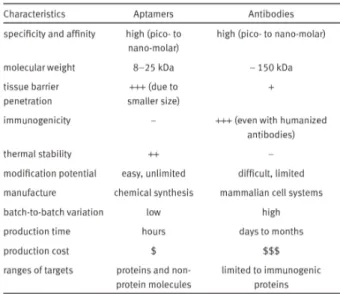
Over the past two decades the potential of mAbs as high-affinity binders for targeted therapy has been clinically demonstrated60 with several therapeutic mAbs approved by FDA61. To date, more than 200 antibody-based targeted platforms were successfully translated into the clinical environment and are currently in preclinical and clinical trials62. However, this class of ligands shows several limitations. For example they are produced through a complex and expensive process that is difficult to scale-up to large-scale manufacturing without affecting product properties18. They have limited stability, are rapidly inactivated under
acidic conditions, in the presence of proteases or at elevated temperatures63. Moreover, their large size (150-160 kDa and 12-15 nm) results in slow tissue penetration64 and prevents access to many biological compartments, ultimately leading to an increase toxicity due to a long blood residence65. The most
significant restriction, however, is related to their immunogenicity, which was observed mainly for first-generation of rodent antibodies. Indeed, injection of mouse mAbs into humans results in the production of human anti-mouse antibodies (HAMA), thus leading to serious immune reactions66. To overcome this limitation, recent developments in the field of antibody engineering have led to the production of antibodies that contain animal and human domains, called chimeric mAbs, then humanized mAbs, and fully human mAbs60 (Fig. 5). Overall, these
structures present a gradually reduced immunogenicity, with fully human and humanized antibodies carrying lower risk to induce immune response in humans than mouse or chimeric antibodies67. However, other problematic immune responses still exist, such as idiotypic responses, with the production of anti-idiotypic antibodies, also called anti-drug antibodies, against humanized or fully
human antibodies that ultimately lead to the neutralization of their therapeutic effect67.
Interestingly, the last frontier in the field of the antibody engineering led to the development of antibody fragments18, which include monomer of antigen-binding
fragments, dimers of antigen-binding fragments, and single chain variable (Fig. 5).
Fig. 5 Antibodies and antibody fragments. Targeting antibodies are normally monoclonal immunoglobulin G (IgG) (Aa) or IgG fragments (B–D). F(ab′)2 (B) or Fab′ (C) fragments can be made by enzymatic cleavage of the whole monoclonal antibody (mAb) (Aa) or by molecular biological techniques — for example, Fab′ (C), scFV (Da), bivalent (Db) or recombinant fragments (Dc). mAbs that are made from the traditional hybridoma technique are murine in origin. Recent developments have led to the production of chimeric, humanized or fully human antibodies or fragments (Ab–d). Fc, fragment crystallizable region; VH, variable heavy chain; VL, variable light chain. Adapted from T. M. Allen.18 Reprinted by permission from Macmillan
As shown in Fig. 5, only the antigen recognition domain is included in the antibody fragment structures, while the fragment crystallizable (Fc) region is removed in order to reducing their immunogenicity. Indeed the Fc domain is a double-edged sword: when mAb is bound to a tumor cell surface antigen, its Fc region can interact with Fc receptors expressed on the surface of effector cells of the immune system, such as natural killer cells,68 evoking antibody-dependent cellular cytotoxicity and complement-dependent-cytotoxicity18. Note that, these effects play a significant role in enhancing the anticancer effect69. However, interaction between the Fc region and Fc receptors expressed on normal cells, as occurs with macrophages, can elicit immune response leading to increased immunogenicity62,70.
Although antibody fragments are less immunogenic than whole mAbs, unfortunately they are less stable and tend to aggregate due to the absence of Fc region, which plays a role in the structural stability of mAbs63,71. Furthermore, as shown in Fig. 5 most antibody fragments have only one epitope-binding site included in their structure, which reduce their binding avidity18,70.
Despite many advancements mAbs and antibody fragments still suffer limitations that hamper their applicability. Particularly, the use of mAbs and antibody fragments is often restricted to conditions resembling their physiological environment. In the presence of organic solvents or in other non-physiological conditions (e.g. elevated temperature, high pH and high salt concentration), they generally lose their function63. Moreover, mAbs are not easily conjugated to therapeutic molecules, reporter tags, or engineered scaffolds to create complex biomolecular architectures that may be used as targeting delivery platforms. Indeed, the synthetic approaches required for antibody conjugation to molecular partners are stochastic and lead to product mixtures and reduced antibody activity72,73.
1.3.2 Aptamers
An alternative to antibodies should possess high stability, long-term storage, and ease of conjugation to molecular partners7475. In this context, it is worth noting
information assuming simple base pairing structures, but they can also fold into complex tertiary structures, and perform different functions including gene-regulation76, catalytic activity77, and ligand-binding78. Although this is true for biological nucleic acids, in 1990 a series of technological advances allowed the development of an in vitro evolutionary method for the selection and identification of synthetic oligonucleic acids, called aptamers, that bind specifically to molecular targets79,80.
Nucleic acid aptamers are composed of short structured RNA or single-stranded DNA sequences, and are emerging as attractive targeting tools with huge potential in diagnostics and therapeutics. Aptamers are able to recognize and bind a definite target molecule with high affinity and selectivity, owing to their exclusive spatial conformation. In this regard, they are often termed “chemical antibodies”9. In general, aptamers may act similarly to antibodies and they can be used as activating ligands81, therapeutic antagonists82, or as vectors to deliver therapeutic and diagnostic payloads, especially in the field of targeted cancer therapy9.
Compared with antibodies, aptamers are non-immunogenic, easy to synthesize, modify, and manipulate. Furthermore, their small size -usually 8-20 kDa and 3-5 nm- allows more efficient penetration into target tissues72,83. Importantly, aptamers do not include the Fc region of mAbs, thus avoiding undesired interactions with Fc receptors expressed on immune cells and other certain types of cells, which may result in immune stimulation or other unwanted side effects.
Aptamer technology was born twenty-five years ago; despite being a relatively new technology, one therapeutic aptamer is already available in the clinic and various other aptamer-based drugs and aptamer nanocarriers are currently in different stages of preclinical and clinical trial for diseases that span from cancer84 and heart disease85 to type II diabetes86.
The first FDA-approved aptamer is called pegaptanib and it was approved in 2004 for the treatment of age-related macular degeneration (AMD) in the U.S. and the EU82. Pegaptanib (Macugen®; Pfizer and Eyetech) is an RNA aptamer that specifically recognizes human VEGF with high affinity (Kd~50 pM) and selectivity87. VEGF promotes angiogenesis and may be up regulated under certain
binds to the heparin-binding site of VEGF and blocks the interaction between VEGF and its receptors, which results in the prevention or reduction of the neovascularization. In 2004, pegaptanib became the first anti-angiogenesis drug for wet AMD therapy under intravitreal administration with a dosage of 0.3 mg per eye every 6 weeks82,89.
The FDA approval of Pegaptanib represents a landmark in the development of aptamers as biological agents with applications in therapeutics, diagnostics and basic research.
In the next section, I shall mention the main groundbreaking studies that led to the aptamer discovery, and then I shall report an insightful analysis of the aptamer properties and the recent progresses in the development of targeted drug delivery platforms based on aptamer-drug conjugates or aptamer-nanomaterial assemblies2.
1.4 Aptamers as novel “keys of the castle”
The first evolutionary experiments involving nucleic acids were reported in the 1960s by Spiegelman and his collaborators, which described the use of an RNA-dependent RNA replicase for the replication of a given RNA species90. Several years later, in 1986, the innovative discovery of the polymerase chain reaction (PCR) by Kary Mullis and co-workers has led to a technological revolution of the most biological fields (i.e. molecular biology, biochemistry) including evolutionary biology91. In 1990, three independent research groups described in
vitro evolutionary methods for the selection of small nucleic acid ligands as
binding partners for defined molecular targets. Particularly, Ellington and Szostak isolated short RNA molecules that bind to a small organic dye79. These small
nucleic acid ligands were called aptamers (from the Latin aptus – to fit and the Greek meros – part or region). Tuerk and Gold selected RNA molecules able to bind the bacteriophage T4 DNA polymerase and they named SELEX (systematic evolution of ligands by exponential enrichment) the iterative selection process they used80. Robertson and Joyce described the application of in vitro selection to convert a group I ribozyme having RNase activity into an enzyme able to preferentially cleave DNA than single stranded RNA92. These groundbreaking
of functional nucleic acids with desired properties. Indeed, for the first time, it was demonstrated that artificial nucleic acids could be used as functional moieties besides as blueprints of the genetic code or as rigid scaffold for nanoarchitectures. Interestingly, Nature already makes use of several functional nucleic acids, such as noncoding RNA and riboswitches as regulatory elements to control gene expression in bacteria, eukaryotes, and higher organisms91.
It is worth mentioning that few years later the aptamer discovery, the term “aptamers” was extended to include peptides with protein-binding properties93,94. However for the most part and for the purposes of this thesis the term is used to describe oligonucleotide molecules.
Since the invention of SELEX technology, a plethora of high-affinity RNA and DNA aptamers have been selected towards a wide range of different targets ranging from small molecules (i.e. metal ions, organic dyes, amino acids, and peptides)95,96 to proteins72 to complex targets (whole cells2, viruses or bacteria97). Thus, during the last two decades aptamers found broad application across various biomedical fields: biomarker discovery, in vitro diagnosis, in vivo imaging, and targeted therapy9.
Aptamers fold into distinct three-dimensional (3D) structures able to bind and recognize a particular molecular target with high affinity and specificity (Fig. 6a), with dissociation constants typically within the pico- to nanomolar range3. In fact, aptamers are not just linear nucleic acid molecules that simply carry genetic information, but rather they can assume a wide variety of secondary structures, such as hairpin loop, pseudoknot and G-quadruplex (Fig. 6b) generated by a combination of Watson–Crick and non-canonical intramolecular interactions that ultimately lead to complex spatial structures.
Fig. 6 Schematic representation of (a) aptamer–target interaction and (b) aptamer secondary structures. Figure (a) is reprinted from Biotechnology Advance 33 (2015)
1141–1161, Darmostuk et al.98; Copyright © (2015), with permission from Elsevier. Figure
(b) is reprinted from Biotechnology Advances 31 (2013) 1260–1274, Radom et al.99;
Copyright © (2015), with permission from Elsevier.
In contrast with other nucleic acid probes (such as antisense oligonucleotides and ribozymes), aptamers bind their targets through 3D structural recognition involving non-covalent interactions, similarly to the antigen-antibody reaction. Therefore, aptamers are also known as “chemical antibodies”9. However, aptamers possess significant advantages relative to their protein counterpart in terms of virtually no immunogenicity, thermal stability, low-cost chemical synthesis, and ease of conjugation or modification with different functional moieties3 (Table 1). Compared with antibodies, aptamer production exploits a chemical technology with high batch fidelity, which is not prone to viral or bacterial contamination100.
of non-nucleotide linkers (e.g. hexaethylene glycol), or chemical functionalities useful for conjugation (such as primary amines, thiol precursors and aldehyde precursors), and the addition of fluorescent or other reporter moieties72.
Moreover, thanks to their small size (aptamers: 25-70 nt equals 8-25 kDa; antibodies ∼150–160 kDa), aptamers can penetrate into biological compartments more efficiently than antibodies, thus accessing target domains that might otherwise be inaccessible to bulky ligands.
Table 1. Comparison of aptamers and antibodies. Adapted from Sun et al.3
Reprinted by permission from John Wiley and Sons Copyright © 2015; Small 11 (2015) 2352–2364 (Ref. 3)
Lastly, the nature of nucleic acids provides another important advantage compared with protein ligands. Since the function of the aptamer is tightly dependent from their 3D structures, it can be modulated for desired therapy and drug delivery application simply by using the complementary oligo sequence. The complementary base pairing can alter the aptamer spatial structure and ultimately disrupt the interaction with its target. This approach, known as aptamer antidote
strategy, allows a precise control of the aptamer activity by a rational design of the antidote sequence, and was firstly introduced by Sullenger and coworkers in 2002. Briefly, they developed an aptamer that targets the blood coagulation factor IXa101, which induces anticoagulation in pigs and inhibits thrombosis in murine
models102. However, a major risk associated with anticoagulant therapy is uncontrollable bleeding if reversal of the anticoagulation is not achieved103. Therefore, they also developed an antisense strand, complementary to 17 nucleotides of the aptamer, which was able to interfere with the aptamer structure (Fig. 7), inhibiting its activity both in animal models of cardiovascular disease and surgical trauma and humans102,103.
Fig. 7 Aptamer–antidote pair. The sequence and structure of the anti-factor IXa aptamer and its antidote are shown. Binding of the antidote to the aptamer inactivates the aptamer by disrupting its secondary and tertiary structure. Abbreviations: Ch, cholesterol; idT, inverse deoxythymidine. Reprinted from Current
Opinion in Chemical Biology 10 (2006) 282–289, Lee et al.104; Copyright © (2006), with
permission from Elsevier.
The therapeutic potential of aptamers is largely influenced by their pharmacokinetic property. As small nucleic acid molecules, natural RNA and DNA aptamers are highly susceptible to the nuclease-mediated degradation and
size-dependent clearance via renal filtration. However, both limitations can be addressed with appropriate chemical modifications.
Evidence shows that firstly the 2’-OH group of RNA and secondarily the phosphodiester bonds are the preferentially target sites of nucleases hydrolysis9.
Note that, DNA molecules are naturally resistant to 2’-endonucleases owing to the lack of the 2’-OH group. Thus, the main approaches to overcome the nuclease sensitivity are aimed at increasing the in vivo stability of RNA aptamers. As seen for other therapeutic oligonucleotide agents105, nuclease resistance can be achieved with substitutions of the 2’-OH RNA group, internucleotide linkage substitutions of the phosphodiester backbone with either boranophosphate or phosphorothioate, conformationally restricted ribose (locked nucleic acids), generation of mirror RNA sequences (Spiegelmers), introduction of functional groups at the nucleobase level, and the combination of these modifications106 (Fig. 8). Since serum nucleases responsible for RNA turnover are largely directed towards pyrimidine nucleotides107, the most prominent modification is the
substitutions of 2’-OH group of pyrimidines by 2’-fluoro, 2’-amino, or 2’-O-methyl (Fig. 8b) that lead to nuclease-stabilized RNA molecules. Particularly, whereas natural RNA aptamers exhibit a very short serum half-life (estimated at 10 s), the widely used 2’ fluoropyrimidine-containing RNAs have a sensibly longer serum half-lives (estimated at 80 h)108. This approach relies on the effective incorporation into RNA transcript of 2’-modified nucleotides using mutant forms of T7 RNA polymerase during the in vitro transcription109,110.
Fig. 8 Subset of modified nucleotides used for increase nuclease-resistance of aptamers. (a) In addition to the unmodified nucleotides of DNA and RNA, a wide variety of modified nucleotides have been used for improving stability, chemical diversity, and target binding potential of selected aptamers. (b) The most commonly used modified nucleotides contain modifications of the ribose sugar such as 2′-Fluoro, 2′-Amino, or 2′-OMethyl. (c) Locked nucleic acids (LNAs) contain modifications to the sugar phosphate backbone, and are more stable than DNA and RNA. (d) SOMAmers contain modifications (i.e., benzyl, napthyl, tryptamino, or isobutyl) on deoxyuridine nucleotide (dUTP), which improve the chemical diversity and thus the target interaction capacity of the aptamers. (e) Spiegelmers are derived from mirror-image (enantiomer) ribonucleotides. Spiegelmers are not recognized by natural nucleases that degrade wild type oligonucleotides. Adapted from Ozer et al. 111
Reprinted by permission from Nature Publishing Group: Mol. Ther. Nucleic Acids (Ref. 111), Copyright © 2014 The American Society of Gene & Cell Therapy.
Other approaches to the development of nuclease-resistant aptamers relies on modifications of the ribose motif using locked nucleic acid technology or generate “mirror” RNA sequence structures, termed Spiegelmers.
Locked nucleic acids (LNAs) bear a methylene ether bridge between the 2’-oxygen atom and 4’-carbon atom (Fig. 8c). Thus, in the LNA ribose the 3’carbon atom is locked in an endo (N-type) conformation with a consequent lower flexibility of the sugar motif and a high degree of nuclease resistance112. Spiegelmers are aptamers in which all the sugars are the enantiomers of wild-type nucleic acids (Fig. 8e). In the case of RNA Spiegelmers, the backbone is composed by L-riboses, instead of D-riboses present in native RNA, linked by phosphodiester bonds. As a consequence, nucleases are not able to recognize these molecules as substrates of their hydrolytic degradation. The first functional Spiegelmers were designed to bind to the small molecules arginine and adenosine113,114, and they exhibited the expected biostability, as demonstrated for the D-adenosine specific L-RNA spiegelmer. Indeed, no evidence of degradation in human serum were detected over 60 hours of incubation at 37°C 114.
Modifications of nucleobases have also been reported, with derivatization at the C5-position of uridine and deoxyuridine being the most prevalent (see Fig. 8). Notably, the introduction of amino acid side chain-like groups (Fig. 8d) at this position conferred an increased chemical complexity together with a higher hydrophobic character of the aptamers, thus increasing both the range of available binding epitopes on the target proteins and the success rate of SELEX115. A class of DNA aptamers bearing these modifications is known as SOMAmer (Slow Off-rate Modified Aptamers) owing their high affinity towards their biomolecular targets116.
When appropriate modifications to control nuclease-mediated degradation are included in the aptamer sequences, elimination via renal filtration becomes the main limitation to aptamer pharmacokinetics. The molecular mass cutoff for the renal glomerulus is 30-50 KDa, thus aptamers (8-25 kDa) can be conjugated to bulky groups in this size range to reduce the renal filtration rate. The most common strategy is the conjugation of either 5’-end or 3’-end of the aptamers to high molecular mass polyethylene glycol (PEG) (from 5 kDa up to 60 kDa)108.
This method is better known as PEGylation and represents a typical modification when aptamers should be employed for in vivo applications. Importantly, nuclease-stabilized aptamer conjugated to 40 kDa-PEG can have circulating half-lives up to 1 day in mice117 (Fig. 9).
Alternatively, other bulky groups that have been directly conjugated to the aptamers are cholesterol derivates, streptavidin (via biotinylated aptamer), and liposomes (through conjugation to lipid tags).
Fig. 9 The pharmacokinetics of aptamers conjugated to different molecular mass PEGs. Pharmacokinetic profiles of 39-mer 2′-deoxy purine, 2′-O-methyl pyrimidine composition aptamers. These aptamers were unconjugated or conjugated to either 20 kDa polyethylene glycol (PEG) or 40 kDa PEG and administered intravenously to CD-1 mice (n = 3 per time point) at 10 mg per kg. Adapted from Keefe et al.72 Reprinted by permission from Macmillan Publishers Ltd: Nature Reviews Drug Discovery (Ref. 72), Copyright © 2010
1.5 In vitro selection of aptamers
SELEX technology is based on an iterative process of amplification and enrichment, in which a very large (~1014) number of random oligonucleotide
(either RNA or single-stranded DNA) is exposed to the target of interest for several repetitive rounds. During this process, the library is subjected to a selection pressure in parallel that allow an enrichment of the molecules with binding properties against the desired target. Typically, four main steps can be recognized into basic SELEX process (Fig. 10).
Fig. 10 Schematic illustration of aptamer selection procedures by SELEX. Generally, in a typical SELEX procedure, the initial single-stranded DNA/RNA pool contains a 20–60-nt random sequence to provide a sequence space that facilitates presence of structures with high binding affinity to the target protein. By repeating selection rounds, aptamers against any given targets can be routinely isolated from an initial combinatorial oligonucleotide library. A typical SELEX process consists of four main steps (1) binding to the target protein, (2) selective partitioning, (3) recovery of target-bound sequences, and (4) re-amplification of recovered species. Reprinted from RNA
Interference from Biology to Therapeutics Cap. 10 “Aptamer-Mediated siRNA Targeting” 207–220, Zhou and Rossi118 Copyright © (2013), with permission from Springer.
- The first step is the generation of the nucleic acid library. A single-stranded DNA library sequence that includes a random region composed by 20-50 residues in length, and flanking conserved sequences required for enzymatic manipulation is first designed and then synthesized72. PCR amplification of
this single-stranded DNA library generates a double-stranded PCR product with high sequence variability. The initial oligonucleotide selection pool (for
either DNA or RNA aptamers) usually contains 1013-1015 unique random molecules, although pools with a lower variability yielded functional aptamers119,120. In the case of DNA aptamer SELEX, the starting library of single-stranded DNA molecules is obtained after strand separation of double-stranded PCR product. In the case of RNA aptamer SELEX, the RNA library is prepared by in vitro transcription of double stranded DNA templates, using recombinant wild type or mutant T7 RNA polymerase, as previously described.
- The second step is the incubation of the oligonucleotide library with the target of interest for specific binding enrichment. Importantly, before this incubation the library is thermally denatured and cooled at room temperature to ensure proper oligonucleotide folding and consequently promote the formation of 3D structures.
- Next, the unbound fraction is discarded and separated from the target bound species.
- Finally, these binding oligonucleotide sequences are collected, purified and amplified by either PCR (for DNA aptamers) or by RT-PCR (for RNA aptamers) to generate a fresh oligonucleotide library ready for the next round of the in vitro selection.
SELEX process usually requires 4-20 rounds of amplification and enrichment9 to obtain high affinity aptamers. The number of SELEX rounds is strongly affected by the nature of the aptamer target (such as a purified protein or a living cell line), by the type of library used, as well as by the evolution of the aptamer library during each selection cycle. After the last round of selection, a rational strategy deeply examines the oligonucleotide sequences to disclose the ligands with the best properties. First, the enriched library is cloned and, after sequencing, candidate aptamers are classified in several groups on the basis of their sequence homology. High-throughput sequencing methods (Next-Generation Sequencing – NGS) and bioinformatics analysis (such as FASTAptamer121) allow the identification of candidate aptamers. Once the candidates are identified, they are explored in terms of binding affinity, specificity, and desired properties such as target inhibition, cell internalization, and stability. These latter steps are so far the
most time-consuming part of the aptamer generation process. Lastly, the best selected aptamers and their relative secondary structures are rationally investigated (by computer simulation, and both chemical and enzymatic probing) in order to find out the minimal aptamer binding sequence required for the specific recognition of the target. Post-SELEX modifications can also be introduced to increase the aptamer properties in terms of nuclease resistance, in vivo biodistribution, and other parameters affecting the aptamer pharmacokinetic behavior (as described in the previous section).
Early SELEX protocols targeting proteins usually required several months to identify aptamers able to bind the desired target protein. Recently, the traditional SELEX method has been modified to improve the outcome of this technology. Particularly, some innovative SELEX strategies have been developed in order to (i) maximize affinity and selectivity (e.g. slow-off rate modified aptamers – SOMAmer)116, (ii) to improve the evolution of the library and consequently the speed of selection and success rate (e.g. capillary gel electrophoresis-122,
microfluidic-123, bead-based SELEX124 and FACS-SELEX125), (iii) to provide additional properties to the selected ligands (e.g. cell-internalization SELEX126), (iv) to obtain aptamers against more complex targets (e.g Cell-SELEX127, in vivo SELEX128)111. A fruitful approach for increasing the activity of selected sequences
is the “doped-SELEX”, in which an original, successful aptamer sequence is partially randomized at some or all positions and reselected129. For example, each position in a doped library could contain 55% of the initial selected nucleotide, and 15% each of the other three nucleotides (45% doping)130. Moreover, the biomolecular target of this second selection could be the same of the original SELEX or closely related.
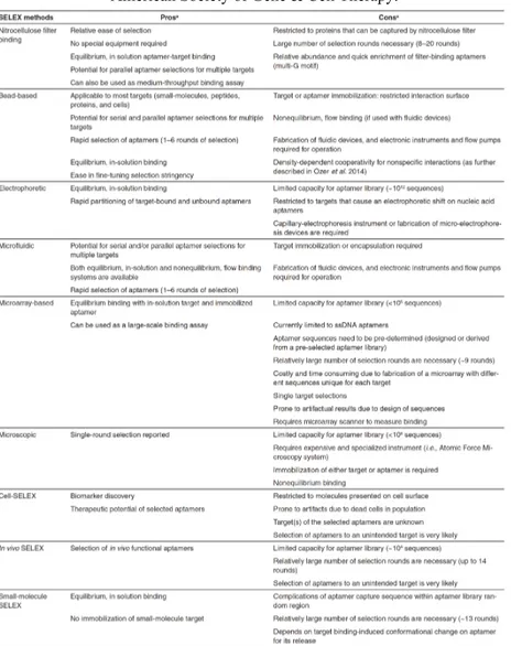
Table 2 shows a detailed scheme of the SELEX methods and offers a better comparison among these techniques. To date, thanks to these strategies, an in vitro selection takes only several days and aptamers are emerging as valid tools for basic research and biomedical applications.
The main drawback of the SELEX technology is the uncertain prediction of the success of the selection. It is not possible to judge the ability of a target molecule
to be suitable for the selection process and whether an aptamer can be successfully raised against the target (aptamerogenicity) a priori91
Table 2 Comparison of SELEX methods. Reprinted by permission from Nature
Publishing Group: Mol. Ther. Nucleic Acids (Ref. 111), Copyright © 2014 The American Society of Gene & Cell Therapy.
1.6 Selection of aptamers recognizing cell-surface markers
As mentioned in the previous section, in the recent years innovative strategies have been introduced to enhance both performance and outcome of the conventional SELEX process. Here, I shall focus on the in vitro selection methods to generate aptamers targeting cell-surface markers or specific cell-types to exploit as targeted drug delivery agents. Two main approaches are typically employed: (i) traditional purified membrane protein-based SELEX and (ii) live cell-based SELEX.
In a typical protein-based SELEX the nucleic acid library is mixed with a target purified membrane protein, a cell surface antigen overexpressed and/or mutated in diseased cells. Traditional bead-, resin-, membrane-, or chip-based partition approaches are employed as affinity matrixes to separate target-bound sequences from unbound species. In this context, the immobilization of the recombinant purified protein on the affinity surface is the first step before the incubation of the target with the nucleic acid library. Protein-SELEX is a method in which low nonspecific binding and easy control of the conditions allow optimal enrichment of specific ligands. To date, most aptamers as targeting ligands have been successfully generated using purified membrane protein-based SELEX2. A strong limitation of this method is the conformational instability of target proteins, especially under the conditions of the selection process. Protein conformational changes can lead to a misleading selection with enrichment of oligonucleotide sequences that recognize a false 3D protein structure. Alternatively, a reduction of enrichment can also occur from one selection cycle to the other, in which the enriched pool suddenly is not more able to bind the target protein due to the conformational changes91. Additionally, purified recombinant proteins are generated by in vitro expression systems, which often not include the presence of post-translational modifications, such as glycosylation (N- and O-linked), phosphorilation of tyrosine, threonine or serine residues and addition of glycosylphosphatidylinositol (GPI), typically present in cell-surface receptors. Furthermore, in many cases cell-surface proteins are insoluble in their recombinant form, or they are functional only when tightly associated with other
cell components (e.g. G protein-coupled receptor) or when associated to form multimeric and/or multivalent structures131. Thus, RNA and DNA aptamers selected through a protein-SELEX approach might not be able to recognize the same target when embedded in a physiological milieu132. To overcome these
limitations, an innovative SELEX method that uses whole living cells as target, termed live cell-based SELEX (or cell-SELEX) has been introduced127. This approach allows the identification of aptamers that bind cell-surface specific antigens in their native environment133. In contrast to protein-SELEX, cell-SELEX does not need information regarding native conformation, identity, and biological function of target proteins, and aptamers against unknown cell-surface antigens can be also generated. This method relies on the difference between the expression pattern of cell-surface receptors in target cell population (e.g cancer cells) and the receptor pattern in control cell line (e.g. healthy cells). Therefore, cell-SELEX involves two different selection steps (Fig. 11): (i) a negative selection (or counter selection) step to suppress nonspecific binding sequences, in which the oligonucleic acid library is incubated with non-target cells (or negative cells). Unbound sequences are recovered and (ii) a positive selection is performed with target cells (or positive cells) and target-bound ligands are recovered and amplified for the subsequent round of SELEX.
Fig. 11 Schematic illustration of cell-SELEX procedure. A live cell-based SELEX procedure consists of four main steps each round of selection: (1) counter-selection by incubating library with negative cells that do not express the target antigen, (2) positive selection by incubating recovered unbound sequences with positive cells expressing cell-surface receptors of interest, (3) recovery of target-bound sequences, and finally (4) re-amplification of recovered species.
Overall, cell-SELEX provides aptamer sequences able to bind a pool of different cell-surface antigens overexpressed or exclusively present in target cells. Using this approach several groups generated aptamers able to discriminate a particular cell population2. Tan group introduced for the first time this new
strategy to select DNA aptamers able to discriminate T cells (CCRF-CEM cells, a human precursor T-cell acute lymphoblastic leukemia cell line) from a B cell line (Ramos cells, human Burkitt’s lymphoma cells), in patient samples127. Despite the remarkable success of cell-based SELEX, one important drawback is related to the nonspecific binding to dead cells that can lead to low target-bound sequences