Red flags for appropriate referral to the gastroenterologist and the
rheumatologist of patients with inflammatory bowel disease and
spondyloarthritis
C. Felice,* P. Leccese,
†L. Scudeller,
‡E. Lubrano,
§F. Cantini,
¶F.
Castiglione,
**P. Gionchetti,
††A.
Orlando,
‡‡C. Salvarani,
§§R. Scarpa,
¶¶M. Vecchi,
***I. Olivieri
†1and
A. Armuzzi
*
1and on behalf of the
Italian SpA-IBD Expert Panel Group
* IBD Unit, Presidio Columbus, Fondazione Policlinico Universitario A. Gemelli IRCCS Università Cattolica del Sacro Cuore, Rome, Italy, † Rheumatology Institute of Lucania (IRel) and the Rheumatology Department of Lucania, San Carlo Hospital of Potenza and Madonna delle Grazie Hospital of Matera, ‡ Clinical Epidemiology Unit, IRCCS Policlinico San Matteo Foundation, Pavia, Italy, § Academic Rheumatology Unit, Dipartimento di Medicina e Scienze della Salute ‘Vincenzo Tiberio’, Università degli Studi del Molise, Campobasso, Italy, ¶ Division of Rheumatology, Hospital of Prato, Italy, ** Gastroenterology Unit, Department of Clinical Medicine and Surgery, University Federico II, Naples, Italy, †† IBD Unit, Department of Medical and Surgical Sciences, S. Orsola-Malpighi Hospital, University of Bologna, Italy, ‡‡ IBD Unit, A.O. Ospedali Riuniti ‘Villa Sofia-Cervello’, Palermo, Italy, §§ Azienda USL - IRCCS di Reggio Emilia e, Università di Modena e Reggio Emilia, Italy, ¶¶ Rheumatology Unit, Department of Clinical Medicine and Surgery, University Federico II, Naples, Italy, and *** Gastroenterology and Endoscopy Unit, Fondazione IRCCS Ca’ Granda Ospedale Maggiore Policlinico, Department of Pathophysiology and Organ Transplantation, University of Milan, Italy Accepted for publication 21 November 2018 Correspondence: A. Armuzzi, IBD Unit, Presidio Columbus, Fondazione Policlinico Universitario A. Gemelli IRCCS Università Cattolica del Sacro Cuore, Via Moscati 31-33, 00168, Rome, Italy.
E-mail: [email protected]
1Deceased.
Summary
Collaboration between gastroenterologists and rheumatologists is
recom-mended for the correct management of patients with associated
spondy-loarthritis (SpA) and inflammatory bowel disease (IBD). We aimed to
establish the appropriateness of several red flags for a prompt specialist
referral. A systematic review of the literature was performed using the
GRADE method to describe the prevalence of co-existing IBD-SpA and
the diagnostic accuracy of red flags proposed by a steering committee.
Then, a consensus among expert gastroenterologists and rheumatologists
(10 in the steering committee and 13 in the expert panel) was obtained
using the RAND method to confirm the appropriateness of each red flag
as ‘major’ (one sufficient for patient referral) or ‘minor’ (at least three
needed for patient referral) criteria for specialist referral. The review of
the literature confirmed the high prevalence of co-existing IBD-SpA.
Posi-tive and negaPosi-tive predicPosi-tive values of red flags were not calculated, given
the lack of available data. A consensus among gastroenterology and
rheu-matology specialists was used to confirm the appropriateness of each red
flag. Major criteria to refer patients with SpA to the gastroenterologist
included: rectal bleeding, chronic abdominal pain, perianal fistula or
ab-scess, chronic diarrhoea and nocturnal symptoms. Major criteria to refer
patients with IBD to the rheumatologist included: chronic low back pain,
dactylitis, enthesitis and pain/swelling of peripheral joints. Several major
and minor red flags have been identified for the diagnosis of co-existing
IBD-SpA. The use of red flags in routine clinical practice may avoid
diagnostic delay and reduce clinic overload.
Introduction
Inflammatory bowel diseases (IBD, including Crohn’s
disease (CD) and ulcerative colitis (UC) and
spondyloar-thritis (SpA), are chronic disorders which may co-exist
in the same subject, worsening the disability and the
quality of life of the patient and making the clinical
man-agement of the diseases more complicated. It is noteworthy
that SpA is reported in the literature as the most common
extra-intestinal manifestation in IBD patients [1–5].
However, this still represents an underestimated clinical
problem, as demonstrated by Stolwijk et al. in a Dutch
study: among 350 IBD patients, 129 (39·6%) reported
articular symptoms suggestive of SpA, but only half of
them were referred to a rheumatologist [5]. A prompt
and correct diagnosis of these disorders may have a
sig-nificant impact on their therapeutic management,
influ-encing the type and duration of therapies [6,7] and possibly
preventing the complications related to progressive and
potentially irreversible intestinal and articular tissue
dam-age. Conversely, symptoms not specifically related to
inflammatory conditions may induce inappropriate referral,
causing clinic overload. Therefore, direct collaboration
between gastroenterologists and rheumatologists is
fun-damental, and may benefit from the identification of ‘red
flags’ (disease-specific signs and symptoms) for easier and
more appropriate patient referral.
Recently, several red flags have been proposed to
facili-tate early referral of patients with Crohn’s disease from
primary to specialist care and thus avoid diagnostic delay
[8]. Moreover, a six-item questionnaire (DETAIL) has
been developed to screen patients with IBD for the
diag-nosis of SpA, but it needs to be validated in larger cohorts
of patients [9].
This study aimed to obtain a consensus among
gas-troenterology and rheumatology specialists on the
ade-quateness of several ‘red flags’ for a correct referral of
patients with IBD and SpA from the gastroenterologist
to the rheumatologist (and vice versa).
Methods
The entire process was developed throughout several
meet-ings, from December 2016 to October 2017. ‘Red flags’ were
defined as signs or symptoms which may alert to a possible
diagnosis of IBD in a patient with axial or peripheral SpA,
or (analogously) may alert to a possible diagnosis of axial
or peripheral SpA in a patient with IBD, allowing a prompt
referral to the relevant clinical specialist (Table 1).
Project management
The steering committee was the same as previous projects
already published concerning the management of this
par-ticular clinical setting [6,7] and included 10 Italian
rheu-matologists and gastroenterologists with definitive expertise
in the field of SpA and IBD identified according to their
publication record, participation in national meetings and
clinical trials and/or senior academic rank. Two clinical
fellows (C.F. and P.L.) performed the systematic review
of the literature. The expert panel was composed of 13
gastroenterologists and rheumatologists from different
Table 1. Definition of gastrointestinal and rheumatological red flags selected by the Steering Committee‘Red flag’: sign or symptom suggestive of a specific disease
Red flags for IBD Red flags for SpA
Chronic diarrhoea (change in the bowel habit with loose stools and/or increase of bowel movements per day lasting >4 weeks)
Chronic low back pain (>3 months)
Chronic abdominal pain (>3 months) [8] Family history of SpA (presence in first-degree or second-degree relatives of any of the following: AS, psoriasis, acute uveitis, reactive arthritis, IBD [73]
Rectal bleeding (not from haemorrhoids) Peripheral joint pain*/swelling
Weight loss (>5% in the last 3 months [8], involuntarily) Dactylitis (past or present, diagnosed by a doctor) [73] Fever (no otherwise explained and associated to raised inflammatory markers) Enthesitis (heel enthesitis: past or present spontaneous pain or
tenderness at examination at the site of the insertion of the Achilles tendon or plantar fascia at the calcaneus) [73]
Family history of IBD Psoriasis (past or present, diagnosed by a doctor) [73]
Anaemia (no otherwise explained) Anterior uveitis (past or present, and confirmed by an ophthal-mologist) [73]
Perianal fistula or abscess (past or current) Urethritis/cervicitis (within 1 month before the onset of arthritis/ enthesitis/dactylitis) [73]
Nocturnal symptoms (diarrhoea or abdominal pain) Chest pain Oral aphtosis (recurrent)
IBD = inflammatory bowel disease; SpA = spondyloarthritis; AS = ankylosing spondylitis. *Recurrent or lasting >3 months.
Italian regions (Supporting information, Appendix 1). A
clinical epidemiologist with expertise in the GRADE
frame-work and consensus methods was also involved (L.S.), as
well as an experienced medical librarian.
Systematic literature review
The GRADE framework for diagnostic tests was used to
formulate the search questions (Table 2), with the
defini-tion of patients, diagnostic test (in our case, ‘red flag’),
comparison (in our case, the absence of ‘red flag’) and
outcomes of interest [10].
To estimate the positive and negative predictive value
(post-test probability) of each red flag, information about
the prevalence of the disease (pre-test probability) and
test accuracy (sensitivity and specificity) would be needed.
Therefore, different systematic reviews were performed to
address the following issues:
• the prevalence of SpA in patients with an established
di-agnosis of IBD;
• the prevalence of IBD in patients with an established
diagnosis of SpA;
• the diagnostic accuracy of rheumatological red flags in
IBD patients; and
• the diagnostic accuracy of gastrointestinal red flags in
SpA patients.
PubMed and EMBASE were interrogated for the search,
without initial date limit, until January 2017. Only
English papers were included, and abstracts without full
text were excluded. Details of search terms for
prevalence data are available in the Supporting
information.
Statistical analyses
Abstract and full texts were assessed for eligibility, and data
were extracted by the clinical fellows (C.F. and P.L.) in two
dedicated spreadsheets (one for SpA and one for IBD), in
duplicate. The metan suite of commands in stata version
14 was used for data synthesis, using random effect models.
Heterogeneity was assessed by means of the I
2statistic. The
sources of heterogeneity that were explored were specific
diagnosis (AxSpA/pSpA, CD/UC/IBD) in all population/
outcome combinations (i.e. all possible scenarios) and (for
diagnostic accuracy) in each individual red flag.
To obtain a rough estimate of the positive and negative
predictive value of each red flag for the
population/out-come combination of interest, the Bayes formula was
applied, informed with estimates obtained in the
meta-analysis.
RAND method
Given the results of the systematic review (see Results),
the RAND method [11] was used to define the
appro-priateness of patient referral from the gastroenterologist
to the rheumatologist and vice versa, when specific signs
or symptoms (red flags) suggest co-existing IBD-SpA in
a number of clinical scenarios.
Expert opinion
Based on the results of the systematic review of the
lit-erature and their personal opinion, the gastroenterology
Table 2. GRADE model for diagnostic tests used for the literature searchPopulation Rheumatological patient Gastroenterological patient
• AxSpA
• pSpA • IBD• UC
• CD
Diagnostic test (‘red flag’) Gastrointestinal signs or symptoms Rheumatological signs or symptoms
• Family history of IBD • Rectal bleeding • Weight loss
• Chronic abdominal pain • Anaemia
• Perianal fistula or abscess • Fever
• Chronic diarrhoea • Nocturnal symptoms • Oral apthosis
• Family history of SpA • Chronic low back pain • Psoriasis
• Dactylitis • Heel/knee enthesitis • Anterior uveitis • Urethritis/cervicitis • Peripheral joint swelling/pain • Chest pain
Control (absence of ‘red flag’) Absence of gastrointestinal symptoms Absence of rheumatological symptoms Outcome Diagnosis of gastrointestinal disease Diagnosis of rheumatological disease:
• IBD • UC • CD
• AxSpA • pSpA
IBD = inflammatory bowel disease; CD = Crohn’s disease; UC = ulcerative colitis; SpA = spondyloarthritis; AxSpA = axial spondyloarthritis; pSpA = peripheral spondyloarthritis.
and rheumatology specialists participated in two rounds
of an online survey (the first in June 2017 and the second
in August 2017) to define the appropriateness of
gastro-enterology or rheumatology referral for each red flag. A
procedure (in this case: referral to a clinical specialist)
should be considered appropriate when ‘the expected health
benefit […] exceeds the expected negative consequences
[…] by a sufficiently wide margin that the procedure is
worth doing, exclusive of cost’ [11–13].
A nine-point scale was used to quantify appropriateness
of referral, considering ‘1’ as absolutely inappropriate, ‘5’
as uncertain and ‘9’ as absolutely appropriate. The median
score was used to classify appropriateness (1–3
inappro-priate, 4–6 probably approinappro-priate, 7–9 always appropriate),
and the 30–70th interpercentile range corrected for
asym-metry (IPRAS) was used to assess disagreement. After
viewing the results of the first round, in which their
responses were highlighted, panel members were asked
to review their choices in the second round.
Final consensus
The final meeting was held on 10 October 2017 in Rome,
Italy. The goal was to reach consensus on the final
clas-sification of each red flag as ‘major’ (1 red flag sufficient
for patient referral) or ‘minor’ (>1 red flag needed for
patient referral) and to establish how many minor red
flags are required to justify patient referral in both
gas-troenterological and rheumatological settings. All questions
were formulated as: ‘Do you agree to consider this red
flag as “major” criteria for referral in this scenario?’ or
‘do you agree that a minimum of three minor red flags
are needed for referral?’, allowing ‘yes’, ‘no’ or ‘no opinion’
as responses. All votes were expressed electronically and
anonymously. Consensus was defined as >70% of the panel
agreeing for ‘yes’ with <15% of the panel responding
‘no’.
Results
Systematic review
A total of 28 384 non-duplicate records were screened
at the abstract level: 939 for the prevalence of
co-existing IBD-SpA (Fig. 1); 15 954 for the diagnostic
accuracy of gastrointestinal red flags; and 11 491 for
rheumatological red flags (Supporting information, Figs.
S1 and S2). Then, 378 full texts were assessed for
eli-gibility and, finally, 78 papers were included in the
qualitative and quantitative analysis: 67 for the analysis
of the prevalence of co-existing IBD-SpA [2–4,14–77]
(Tables 3 and 4) and only 11 for the diagnostic
accu-racy of red flags [8,78–87].
Fig. 1. Flowchart of study selection for prevalence of co-existing inflammatory bowel diseases (IBD) and spondyloarthritis (SpA) (‘SpA in IBD’:
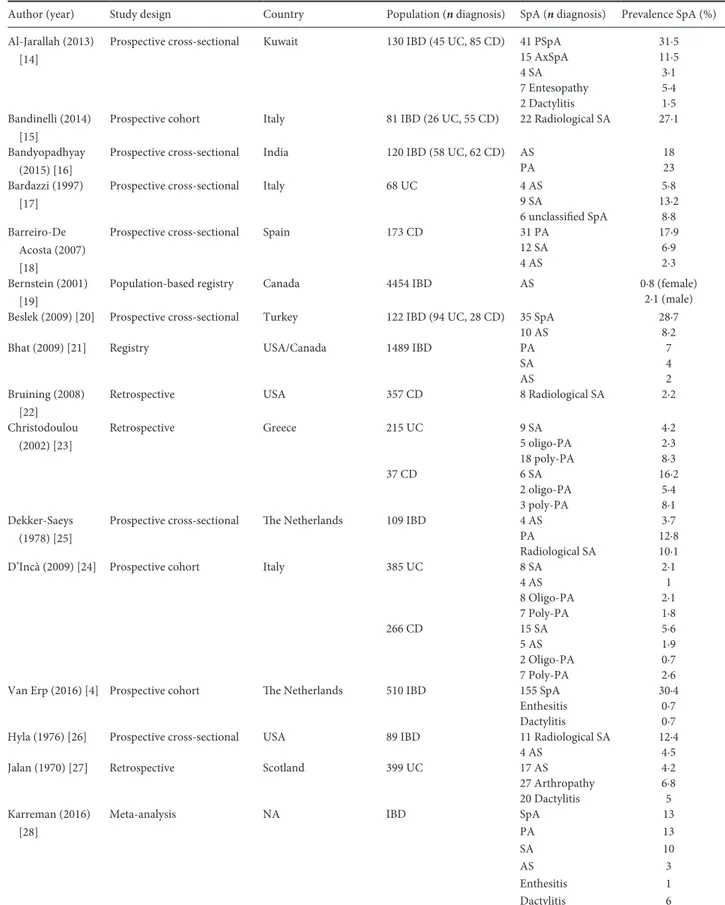
Table 3. Characteristics of studies included in the analysis of prevalence of SpA in IBD patients
Author (year) Study design Country Population (n diagnosis) SpA (n diagnosis) Prevalence SpA (%) Al-Jarallah (2013)
[14]
Prospective cross-sectional Kuwait 130 IBD (45 UC, 85 CD) 41 PSpA 31·5
15 AxSpA 11·5 4 SA 3·1 7 Entesopathy 5·4 2 Dactylitis 1·5 Bandinelli (2014) [15]
Prospective cohort Italy 81 IBD (26 UC, 55 CD) 22 Radiological SA 27·1
Bandyopadhyay (2015) [16]
Prospective cross-sectional India 120 IBD (58 UC, 62 CD) AS 18
PA 23
Bardazzi (1997) [17]
Prospective cross-sectional Italy 68 UC 4 AS 5·8
9 SA 13·2
6 unclassified SpA 8·8 Barreiro-De
Acosta (2007) [18]
Prospective cross-sectional Spain 173 CD 31 PA 17·9
12 SA 6·9
4 AS 2·3
Bernstein (2001) [19]
Population-based registry Canada 4454 IBD AS 0·8 (female)
2·1 (male) Beslek (2009) [20] Prospective cross-sectional Turkey 122 IBD (94 UC, 28 CD) 35 SpA 28·7
10 AS 8·2
Bhat (2009) [21] Registry USA/Canada 1489 IBD PA 7
SA 4
AS 2
Bruining (2008) [22]
Retrospective USA 357 CD 8 Radiological SA 2·2
Christodoulou (2002) [23] Retrospective Greece 215 UC 9 SA 4·2 5 oligo-PA 2·3 18 poly-PA 8·3 37 CD 6 SA 16·2 2 oligo-PA 5·4 3 poly-PA 8·1 Dekker-Saeys (1978) [25]
Prospective cross-sectional The Netherlands 109 IBD 4 AS 3·7
PA 12·8
Radiological SA 10·1
D’Incà (2009) [24] Prospective cohort Italy 385 UC 8 SA 2·1
4 AS 1 8 Oligo-PA 2·1 7 Poly-PA 1·8 266 CD 15 SA 5·6 5 AS 1·9 2 Oligo-PA 0·7 7 Poly-PA 2·6
Van Erp (2016) [4] Prospective cohort The Netherlands 510 IBD 155 SpA 30·4
Enthesitis 0·7
Dactylitis 0·7
Hyla (1976) [26] Prospective cross-sectional USA 89 IBD 11 Radiological SA 12·4
4 AS 4·5
Jalan (1970) [27] Retrospective Scotland 399 UC 17 AS 4·2
27 Arthropathy 6·8
20 Dactylitis 5
Karreman (2016) [28]
Meta-analysis NA IBD SpA 13
PA 13 SA 10 AS 3 Enthesitis 1 Dactylitis 6 (Continued)
Author (year) Study design Country Population (n diagnosis) SpA (n diagnosis) Prevalence SpA (%) Kochhar (1991)
[29]
Prospective cross-sectional India 150 UC 16 SA 10·7
21 PA 14
Lanna (2008) [30] Prospective cross-sectional Brasil 130 IBD 8 AS 6·2
7 Enthesitis 5·4 12 Radiological SA 9·2 21 PA 16·2 59 UC AS 0 2 Enthesitis 3·4 Radiological SA 3·4 PA 11·9 71 CD AS 11·3 5 Enthesitis 7 Radiological SA 14·1 PA 19·7 Leclerc-Jacob (2014) [31]
Retrospective France 186 IBD 31 Radiological SA 16·7
Liu (2016) [32] Retrospective China 195 CD 8 AS 4·12
McEniff (1994) [33]
Prospective case series USA 65 IBD 21 Radiological SA 32
Mocelin (2015) [34]
Retrospective Brasil 100 CD 6 SpA 6
Modena (1988) [35]
Prospective case series Italy 51 CD 6 AS 11·7
8 oligo-PA 15·7
2 poly-PA 3·9
6 Radiological SA 11·7
Münch (1986) [36] Prospective cross-sectional Germany 167 CD 73 SpA 44
15 AS 9 24 SA 14 23 PA 14 11 SA + arthritis 7 Orchard (2008) [37]
Prospective case series UK 44 CD 17 Radiological SA 39
Palm (2002) [38] Population-based cohort Norway 521 IBD 15 AS 3·7
2 Dactylitis 1 Enthesitis
SpA 22
Radiological SA 2
Paparo (2012) [39] Retrospective Italy 221 CD 53 Radiological SA 24
Peeters (2004) [40] Prospective cross-sectional Belgium 102 CD 23 Radiological SA 23
9 AS 8·8
17 PA 16·6
11 Enthesopathy 10·4 Peeters (2008) [41] Prospective cross-sectional Belgium 244 CD 65 Radiological SA 27
16 AS 6·5 Pezerović (2013) [42] Retrospective population-based Croatia 150 IBD 32 PA 21·3 6 SA 4 8 AS 5·3 119 UC 24 PA 20·2 3 SA 2·5 4 AS 3·4 31 CD 8 PA 25·8 3 SA 9·7 4 AS 12·9 Pokharna (2004) [43]
Prospective cross-sectional India 46 UC 1 PA 2
Table 3. (Continued)
Author (year) Study design Country Population (n diagnosis) SpA (n diagnosis) Prevalence SpA (%)
Queiro (2000) [44] Prospective cross-sectional Spain 62 IBD 15 Radiological SA 24
19 PA 30
2 AS 3
Rodriguez (2008) [45]
Prospective cross-sectional Puerto Rico 100 IBD 42 SpA 42
2 AS 2 13 SA 13 5 PA 5 3 Dactylitis 3 2 Enthesitis 2 Salvarani (2001) [2] Population-based inception cohort
Italy, the Netherlands 160 IBD 29 SpA 18·1
5 AS 3·1
23 Unclassified SpA
14·4
Scarpa (1992) [46] Prospective cross-sectional Italy 79 UC 20 AS 25·3
15 PA 19
14 Unclassified SpA
17·7
Scott (1990) [47] Prospective cross-sectional USA 86 CD 25 Radiological SA 29
Sofia (2014) [49] Retrospective USA 513 Caucasian UC 10 AS/SA 1·6
28 African American UC 2 AS/SA 7·1
1127 Caucasian CD 2.9 AS/SA 2·9
108 African American CD
3 AS/SA 2·8
Steer (2003) [50] Prospective cross-sectional UK 134 CD 31 Radiological SA 23
Suh (1998) [51] Retrospective Korea 129 IBD 20 PA 15
Radiological SA 6·2
AS 1·6
77 UC 15 PA 19·6
52 CD 5 PA 9·6
Sung (1994) [52] Retrospective China 15 CD 2 AS 13·3
1 SA 6·6
1 Colitic arthritis 6·6 Turkcapar (2006)
[3]
Prospective cross-sectional Turkey 162 IBD 74 SpA 45·7
16 AS 9·9 24 PA 14·8 81 Enthesitis 50 74 Bilateral SA 45·7 22 Radiological SA 13·6 84 UC 36 SpA 42·8 7 AS 8·3 12 PA 14·3 39 Enthesitis 46·4 36 Bilateral SA 42·8 12 Radiological SA 14·3 78 CD 38 SpA 48·7 9 AS 11·5 12 PA 15·4 42 Enthesitis 53·8 38 Bilateral SA 48·7 10 Radiological SA 12·8 Vavricka (2011) [53]
Prospective cohort Swiss 950 IBD 272 Arthritis 28·6
39 AS 4·1 370 UC 79 Arthritis 21·3 6 AS 1·6 580 CD 193 Arthritis 33·3 33 AS 5·7 (Continued) Table 3. (Continued)
Results of this exercise indicated high heterogeneity
of prevalence estimates across studies and clinical
sce-narios (I
2statistics: 90·3% for prevalence of IBD in
AxSpA, 89·1% in pSpA, 96·3% for SpA in CD, 94·7%
in UC, 98·1% in IBD, all P < 0·001) and low reliability
in the estimates of accuracy due to poor quality of
evidence. Therefore, the results were not pooled into a
summary estimate but used only in a qualitative
manner.
There were no studies specifically focused on the
diag-nostic accuracy of gastroenterological red flags in
rheu-matological patients, and vice versa. Therefore, the review
and the subsequent data analysis included the sensitivity
and specificity of red flags in the general population as
the best surrogate. For some red flags, there were no
specific data on diagnostic accuracy. Considering the
impossibility to pool results, and to obtain a reliable
summary estimate of the prevalence of co-existing
IBD-SpA and diagnostic accuracy of individual red flags,
positive and negative predictive values were not
calculated.
RAND online surveys
The response rate to the online survey was 100% in both
rounds. Nno disagreement was reported after the second
round, and all red flags were judged as ‘absolutely
appro-priate’ or ‘probably approappro-priate’. Moreover, there was a
general overlap between rheumatological (axial and peripheral
SpA) and gastrointestinal (IBD, UC and CD) scenarios.
Based on the results of the online survey, red flags were
categorized into two possible clinical scenarios:
gastroin-testinal or rheumatological signs or symptoms in patients
with SpA and IBD, respectively. In fact, a more accurate
diagnosis (axial or peripheral SpA and CD or UC) is the
result of the process guided by the specialist after patient
referral and, therefore, was considered out of the scope of
this paper.
Final consensus
A total of 22 specialists participated in this final session
of the consensus (attendance rate 92%).
The participants were called to vote on the
appropri-ateness of each red flag to confirm the classification as
minor or major criteria. Major criteria for the referral
of a patient with SpA to the gastroenterologist included:
rectal bleeding, chronic abdominal pain, perianal fistula
or abscess, chronic diarrhoea and nocturnal symptoms.
Major criteria for the referral of a patient with IBD to
the rheumatologist included: chronic low back pain,
dac-tylitis, enthesitis and pain/swelling of peripheral joints.
All remaining red flags were confirmed to be minor criteria
(Table 5). Urethritis/cervicitis was removed from the list
of red flags due to its inclusion in other three major
criteria (arthritis/enthesitis/dactylitis) [86].
Author (year) Study design Country Population (n diagnosis) SpA (n diagnosis) Prevalence SpA (%) Vavricka (2015)
[54]
Registry Swiss 1249 IBD 60 AS/SA 16·4
256 Arthritis 70 483 UC 14 AS/SA 13·4 62 Arthritis 59·1 735 CD 45 AS/SA 18·2 184 Arthritis 74·2 de Vlam (2000) [48]
Prospective cross-sectional 103 IBD 10 AS 10
10 Synovitis 10 7 Enthesopathy 7 33 SA 32 36 SpA 35 25 UC 3 AS 12 3 Synovitis 12 2 Enthesopathy 4 6 SA 24 11 SpA 44 78 CD 7 AS 9 7 Synovitis 9 5 Enthesopathy 8 27 SA 35 25 SpA 32
Yi (2012) [55] Retrospective China 153 CD 7 Arthritis 4·6
1 AS 0·65
IBD = inflammatory bowel disease; CD = Crohn’s disease; UC = ulcerative colitis; SpA = spondyloarthritis; AxSpA = axial spondyloarthritis; pSpA = peripheral spondyloarthritis; PA = peripheral arthritis; AS = ankylosing spondylitis; SA = sacroiliitis.
The participants also approved the need for at least
three minor criteria for specialist referral in both cases
(rheumatological referral of patients with IBD to, and
gastroenterological referral of, patients with SpA).
Discussion
The identification of patients with co-existing IBD and
SpA may have important implications for their clinical
management by influencing treatment, preventing possible
complications and, thus, improving clinical outcomes and
quality of life. Multi-disciplinary collaboration between
gastroenterologists and rheumatologists represents the best
way to improve the therapeutic approach to such complex
clinical scenarios.
This study identified several red flags for prompt and
appropriate referral between gastroenterologists and
rheumatologists, which might potentially facilitate the
diagnosis of co-existing IBD-SpA.
The results from our systematic review first confirmed
the high prevalence of co-existing IBD-SpA, particularly
in the gastroenterological population (Table 3). The
impact of this association is particularly relevant for
the clinical management of IBD patients because articular
involvement often requires more expensive or aggressive
therapeutic approaches, including biological agents or
combination treatment with immunosuppressants (i.e.
methotrexate). Moreover, van der Have and colleagues
recently showed that the presence of joint pain might
significantly and negatively affect the quality of life and
the work productivity of IBD patients [88]. The
preva-lence of IBD in the rheumatological setting seems to
be lower, but clinically significant even so (Table 4).
The importance of identifying co-existing IBD among
Table 4. Characteristics of studies included in the analysis of prevalence of IBD in SpA patientsAuthor (year) Study design Country
Population
(n diagnosis) IBD (n diagnosis) Prevalence IBD (%)
Mitulescu (2015) [68] Retrospective Romania 70 SA 1 IBD 1·4
39 PsA 0 IBD 0
17 USPA 2 IBD 11·8
Rudwaleit (2011) [73] Prospective cohort Multi-national 176 pSpA 6 IBD 3·4
Essers et al. (2015) [66] Prospective cohort The Netherlands 216 SA 15 IBD 23·6
Belgium France
Dougados (2015) [63] Prospective cohort France 708 AxSpA 35 IBD 4·9
Deesomchok (1985) [61] Retrospective cohort Thailand 46 SA 0 0
Eliakim (2005) [65] Prospective cross-sectional Israel 20 SpA 6 CD-like lesions endoscopic findings
30
Dean (2016) [60] Registry Scotland 1964 AS primary care 118 IBD 6
1700 secondary care 204 IBD 12
Perez Alamino (2011) [70] Retrospective Multi-national 1274 AS 45 IBD 3·6
Collantes (2007) [58] Registry Spain 1385 SpA 13 IBD 0·3
Buschiazzo (2011) [57] Prospective cohort Argentina 402 SpA 10 IBD 2·5
Sampaio-Barros (2011) [75] Prospective cohort Italy 1036 SpA 10 IBD 1
Peluso (2015) [69] Retrospective Italy 387 PsA 63 IBD 16·2
15 CD 3·8
10 UC 2·5
38 Non-specific colitis 9·8
Rojas-Vargas (2009) [71] Retrospective Spain 150 SpA 4 IBD 2·6
Costello (1980) [59] Prospective USA 55 SA 9 IBD 16·3
3 CD 5·45
6 UC 10·9
Edmunds (1981) [64] Prospective UK 1331 SA 82 IBD 6
Tayel (2012) [77] Registry Egypt 75 SpA 1 IBD 1·3
del Río-Martínez (2016) [62] Registry Spain 291 AxSpA 9 IBD 3·1
86 pSpA 1o IBD 11·6
Stolwijk (2015) [76] Registry UK 4101 SA 151 IBD 3·7
Said-Nahal (2000) [74] Retrospective France 329 SpA 17 IBD 5
10 CD 3
7 UC 2
García-Vicuña (2016) [67] Prospective cohort Spain 513 SpA 13 IBD 2·5
IBD = inflammatory bowel disease; CD = Crohn’s disease; UC = ulcerative colitis; SpA = spondyloarthritis; AxSpA = axial spondyloarthritis; pSpA = peripheral spondyloarthritis; USpA = unclassified SpA; AS = ankylosing spondylitis; SA = sacroiliitis; PsA = psoriatic arthritis.
these patients also derives from the possibility of
devel-oping chronic intestinal inflammation during treatment
with etanercept, a tumour necrosis factor (TNF) inhibitor
specifically used in rheumatology and dermatology [89].
Moreover, the diagnosis of co-existing IBD-SpA may
influence the dosage and infusion regimen of most
bio-logical agents, because gastroenterobio-logical diseases
require higher doses in comparison with those used for
the treatment of isolated SpA [7].
Very scarce evidence emerged from the literature
search concerning the diagnostic accuracy of red flags.
No studies could be found specifically in the
gastroen-terological or rheumatological setting, and a very limited
number of studies performed in the general population
were identified. In particular, sensitivity and specificity
of gastrointestinal red flags for the diagnosis of IBD
have been described in only three papers [8,78,79]. Ford
et al. [78] prospectively enrolled 1981 consecutive patients
attending the gastroenterological clinic of two Canadian
hospitals because of gastrointestinal symptoms. All
sub-jects underwent a full colonoscopy and were invited to
describe their intestinal symptoms among a list selected
from the Rome III diagnostic questionnaire [90]. Three
hundred and two patients were diagnosed with IBD,
whereas all the others (n = 1679) served as controls.
The items that resulted in independent predictors of
IBD were: positive family history, younger age, the
pas-sage of stools more than four times a day >75% of the
time, urgency most of the time and anaemia. However,
the authors concluded that individual items were not
useful to predict a diagnosis of IBD, because most of
them had low sensitivity and specificity values [78].
Danese and colleagues [8] identified several red flags
to be included in a 21-item questionnaire and
admin-istered it to 85 CD patients, 80 subjects with IBS
(irri-table bowel syndrome) and 36 healthy controls, asking
to select the symptoms they had had during the
12 months before the diagnosis (for CD) or at the time
of the visit (for IBS and controls). Interestingly, all red
flags included in our study were significantly more
fre-quent in the CD patients evaluated by Danese et al.
[8] Finally, the authors proposed an index with high
predictive value for CD diagnosis, based on the eight
items that resulted independent at the multivariate
analysis, to be validated in prospective studies [8]. The
study published by Lisciandrano et al. described the
pattern of oral lesions in IBD patients and controls,
without showing any statistically significant difference
among groups [79].
The ASAS (Assessment of SpondyloArthritis International
Society Group) developed sets of criteria for the
classifica-tion of peripheral and axial (with and without definite
radiographic sacroiliitis) SpA. The clinical history included
features of inflammatory back pain (IBP) and extraspinal
manifestations such as arthritis, enthesitis, uveitis, dactylitis,
psoriasis, Crohn’s/ulcerative colitis, good response to
non-steroidal anti-inflammatory drugs (NSAIDs), family history
for SpA, human leucocyte antigen (HLA)-B27 and elevated
C-reactive protein (CRP). In accordance with these criteria,
a patient with chronic back pain (>3 months) and age at
onset less than 45 years can be classified in the presence
of sacroiliitis plus at least one typical SpA feature, or in
the presence of HLA-B27 plus at least two other SpA
features. Patients with arthritis and/or enthesitis and/or
dactylitis plus one or more of the following
parame-ters – psoriasis, inflammatory bowel disease, preceding
infection, HLA-B27, uveitis, sacroiliitis on imaging – or
two or more other parameters – arthritis, enthesitis,
dac-tylitis, inflammatory back pain in the past, family history
of SpA – can be classified as ‘peripheral SpA’. In the
entire ASAS population of 975 patients, sensitivity and
specificity of the combined use of the two sets of criteria
for peripheral SpA were 79·5 and 83·3%, respectively [86].
Tomero et al. analysed the performance of the ASAS
cri-teria for the classification of SpA in early SpA clinics. The
sensitivity and specificity of the ASAS criteria set were 65
and 93%, respectively, suggesting how these criteria are
limited to detection of early SpA forms, especially in
Table 5. Classification of red flags as ‘major’ or ‘minor’ criteria for specialist referralRed flags in SpA Criteria classification Red flags in IBD Criteria classification
Chronic diarrhoea Major Chronic low back pain Major
Rectal bleeding Major Dactylitis Major
Perianal fistula/abscess Major Enthesitis Major
Chronic abdominal pain Major Peripheral joint pain/swelling Major
Nocturnal symptoms Major Family history of SpA Minor
Oral aphtosis Minor Psoriasis Minor
Fever Minor Anterior uveitis Minor
Anaemia Minor Chest pain Minor
Family history of IBD Minor Urethritis/cervicitis Removed
Weight loss Minor
populations in which magnetic resonance imaging (MRI)
is not routinely available or in populations with a low
prevalence of HLA-B27 [87].
For the early diagnosis of axSpA, the Berlin diagnostic
algorithm has been proposed in patients with IBP. This
algorithm is completely based on the sensitivity and
speci-ficity of typical SpA features and considers the probability
of SpA by calculating the likelihood ratio-product of SpA
features for each patient. Although the algorithm consists
of different steps, the presence of IBP is mandatory. It
means that this algorithm is not helpful in the detection
of the disease in patients with axSpA but without IBP
[85,91]. For this reason, Van der Berg et al. validated a
modified algorithm for diagnosing axSpA in which IBP is
excluded as obligatory entry criterion and added as an SpA
feature [80].
Although such scarce data represent an important
limi-tation for any possible evidence-based recommendation,
the appropriateness of each red flag was assessed by expert
opinion and expressed and quantified using the RAND/
UCLA method [11]. After the two rounds of the online
survey, there was no disagreement among the participants.
However, the interspecialist referral was judged as always
appropriate for some red flags and possibly appropriate
for others, leading to their classification as ‘major’ or
‘minor’ criteria, respectively. In fact, the establishment of
some rules for referral may avoid diagnostic delay, improve
quality of care and decrease the possibility of
complica-tions, but it is also fundamental to avoid clinic overflow
with unnecessary referrals and the possible consequent
increase of health-care costs. In this regard, we recognize
as a limitation of this study the inclusion of some general
symptoms in the list of red flags: in particular, our
defi-nition of low back pain intentionally excluded any
inflam-matory characteristic (such as onset before the age of 45,
morning stiffness, pain relieved by movements), as well
as peripheral joint pain without swelling. However, patients
should be referred to the rheumatologist only when these
symptoms recur or last for at least 3 months (Table 1).
In fact, IBD patients with back/joint pain have a
signifi-cantly lower quality of life and work productivity [88],
therefore these symptoms, when persistent, are worthy
of referral to a rheumatologist, regardless of the presence
of articular inflammation.
During the last meeting, it was established that at least
three minor criteria are required for specialist referral.
However, most of the participants argued that some minor
red flags have different importance in clinical practice.
For example, the family history of IBD is not enough to
refer a patient with SpA to the gastroenterologist, but it
should be considered more clinically relevant than isolated
fever or oral aphthosis; in this case, non-invasive tests
such as fecal calprotectin or bowel ultrasound may be
indicated to investigate the presence of intestinal
inflammation, with subsequent referral to the
gastroen-terologist only in the case of altered test results.
Similar considerations emerged from minor criteria for
rheumatological referral. Non-infectious anterior uveitis,
confirmed by the ophthalmologist, may be itself an
extra-intestinal manifestation in patients with IBD, regardless
of the presence of SpA, and thus should be considered
with more caution in comparison with family history of
SpA or chest pain.
The strength of our project was the joint involvement
of rheumatologists and gastroenterologists in the
manage-ment of patients with both diseases, and a structured
method to collect expert opinion. The main limitation is
the lack of reliable evidence to elaborate a solid decision
strategy: prospective and multi-centre studies are needed
to formally validate these symptoms and signs as diagnostic
tools and to combine individual red flags into a ‘diagnostic
score’. Moreover, the participants in our project came
from an Italian setting only; the appropriateness of these
criteria may be different in other populations and
health-care systems. In this regard, a Spanish study has been
published recently, describing some major or minor criteria
for diagnosis of SpA or IBD [92]: the results differ slightly
from ours. However, this confirms the need for a shared
strategy to diagnose such complicated diseases early.
In conclusion, our study suggests that several signs and
symptoms should be closely monitored to improve the
clinical management of patients with a suspected
associa-tion of IBD and SpA. Prospective validaassocia-tion of these red
flags is necessary before their routine use in clinical
practice.
Acknowledgements
Funding of the project was provided by an unrestricted
contribution from MSD Italia Srl. The funding source had
no role in selecting the participants, reviewing the
litera-ture, defining consensus statements, drafting or reviewing
the paper, or in the decision to submit the manuscript.
All views expressed are solely those of the authors. The
authors thank Editamed Srl – Torino for technical support
during all stages of this project and Chiara Formigoni,
the medical librarian, for the help with the literature search.
Author contributions
C. F. and P. L. performed the literature review. L. S.
performed the statistical analysis. C. F., P. L. and L. S.
wrote the manuscript. E. L., F. C., F.Cast., P. G., A. O.,
C. S., R. S., M. V., I. O. and A. A. are members of the
steering committee of the Italian SpA-IBD Expert Panel
Group and critically reviewed the manuscript. All authors
approved the final version of the manuscript.
Dedication
We remember with fondness Professor Ignazio Olivieri
for his professionalism and compassion.
Disclosures
C. F. has served as a consultant or advisory member for
AbbVie and MSD. F. Cast. has served as a consultant or
advisory member for AbbVie, Ferring, Janssen, MSD, Sofar
and Takeda. P. G. received honoraria or consultation fees
from Janssen, Abbvie, Pfizer, Celgene, Takeda, Ferring,
MSD, Alfa Wasserman and Amgen; and participated in
a company-sponsored speaker’s bureau for Abbvie, Janssen,
Takeda, Ferring, Msd, Sofar and Chiesi. A. O. served as
advisory board member for AbbVie, Janssen-Cilag, MSD,
Pfizer and Takeda Pharmaceuticals, and received lecture
grants from AbbVie, Chiesi, Janssen-Cilag, MSD, Sofar
and Takeda Pharmaceuticals. M. V. participated in the
Advisory Board and received lecture fees or support for
research from MSD, Hospira, Mundipaharma, Takeda,
Abbvie, Chiesi, Zambon, Amgen, Biogen, Jannsen, Pfizer,
Sofar and Giuliani. A. A. has served as a consultant or
advisory member for AbbVie, Allergan, Amgen, Biogen,
Celgene, Celltrion, Ferring, Hospira, Janssen, Lilly, MSD,
Mundipharma, Pfizer, Samsung Bioepis, Sofar and Takeda,
has received lecture fees from AbbVie, AstraZeneca, Chiesi,
Ferring, Hospira, Medtronic, MSD, Mitsubishi Tanabe,
Mundipharma, Nikkiso, Otsuka, Pfizer, Samsung Bioepis,
Takeda, Tigenix, and Zambon, and has received research
funding from MSD and Takeda. All other authors have
no conflicts of interest to declare.
References
1 Orchard TR, Wordsworth BP, Jewell DP. Peripheral arthropathies in inflammatory bowel disease: their articular distribution and natural history. Gut 1998; 42:387–91. 2 Salvarani C, Vlachonikolis IG, van der Heijde DM et al.
Musculoskeletal manifestations in a population-based cohort of inflammatory bowel disease patients. Scand J Gastroenterol 2001; 36:1307–13.
3 Turkcapar N, Toruner M, Soykan I et al. The prevalence of extraintestinal manifestations and HLA association in patients with inflammatory bowel disease. Rheumatol Int 2006; 26: 663–8.
4 van Erp SJ, Brakenhoff LK, van Gaalen FA et al. Classifying back pain and peripheral joint complaints in inflammatory bowel disease patients: a prospective longitudinal follow-up study. J Crohns Colitis 2016; 10:166–75.
5 Stolwijk C, Pierik M, Landewe R, Masclee A, van Tubergen A. Prevalence of self-reported spondyloarthritis features in a cohort of patients with inflammatory bowel disease. Can J Gastroenterol 2013; 27:199–205.
6 Olivieri I, Cantini F, Castiglione F et al. Italian Expert Panel on the management of patients with coexisting spondyloarthritis and inflammatory bowel disease. Autoimmun Rev 2014;
13:822–30.
7 Armuzzi A, Felice C, Lubrano E et al. Multidisciplinary management of patients with coexisting inflammatory bowel disease and spondyloarthritis: a Delphi consensus among Italian experts. Dig Liver Dis 2017; 49:1298–305.
8 Danese S, Fiorino G, Mary JY et al. Development of red flags index for early referral of adults with symptoms and signs suggestive of Crohn’s disease: an IOIBD initiative. J Crohns Colitis 2015; 9:601–6.
9 Di Carlo M, Luchetti MM, Benfaremo D et al. The DETection of Arthritis in Inflammatory boweL diseases (DETAIL) questionnaire: development and preliminary testing of a new tool to screen patients with inflammatory bowel disease for the presence of spondyloarthritis. Clin Rheumatol 2018; 37:1037–1077. 10 Schunemann HJ, Oxman AD, Brozek J et al. Grading quality
of evidence and strength of recommendations for diagnostic tests and strategies. BMJ 2008; 336:1106–10.
11 Fitch K, Bernstein SJ, Aguilar MD et al. The RAND/UCLA appropriateness method user’s manual. Santa Monica, CA: RAND Corporation; 2001.
12 Brook RH, Chassin MR, Fink A, Solomon DH, Kosecoff J, Park RE. A method for the detailed assessment of the appropriateness of medical technologies. Int J Technol Assess Health Care 1986; 2:53–63.
13 Park RE, Fink A, Brook RH et al. Physician ratings of appropriate indications for six medical and surgical procedures. Am J Public Health 1986; 76:766–72.
14 Al-Jarallah K, Shehab D, Al-Attiyah R et al. Antibodies to mutated citrullinated vimentin and anti-cyclic citrullinated peptide antibodies in inflammatory bowel disease and related arthritis. Inflamm Bowel Dis 2012; 18:1655–62.
15 Bandinelli F, Terenzi R, Giovannini L et al. Occult radiological sacroiliac abnormalities in patients with inflammatory bowel disease who do not present signs or symptoms of axial spondylitis. Clin Exp Rheumatol 2014; 32:949–52.
16 Bandyopadhyay D, Bandyopadhyay S, Ghosh P et al. Extraintestinal manifestations in inflammatory bowel disease: prevalence and predictors in Indian patients. Ind J Gastroenterol 2015; 34:387–94.
17 Bardazzi G, Mannoni A, d’Albasio G et al. Spondyloarthritis in patients with ulcerative colitis. Ital J Gastroenterol Hepatol 1997; 29:520–4.
18 Barreiro-De Acosta M, Enrique Domínguez-Muñoz J, Concepcion Núñez-Pardo De Vera et al. Relationship between clinical features of Crohn’s disease and the risk of developing extraintestinal manifestations. Eur J Gastro Hepatol 2007;
19:73–8.
19 Bernstein CN, Blanchard JF, Rawsthorne P, Yu N. The prevalence of extraintestinal diseases in inflammatory bowel disease: a population-based study. Am J Gastroenterol 2001; 96: 1116–22.
20 Beslek A, Onen F, Birlik M et al. Prevalence of spondyloarthritis in Turkish patients with inflammatory bowel disease. Rheumatol Int 2009; 29:955–7.
21 Bhat M, Nguyen GC, Pare P et al. Phenotypic and genotypic characteristics of inflammatory bowel disease in French Canadians: comparison with a large North American repository. Am J Gastroenterol 2009; 104:2233–40.
22 Bruining DH, Siddiki HA, Fletcher JG, Tremaine WJ, Sandborn WJ, Loftus EV Jr. Prevalence of penetrating disease and extraintestinal manifestations of Crohn’s disease detected with CT enterography. Inflamm Bowel Dis 2008; 14:1701–6. 23 Christodoulou DK, Katsanos KH, Kitsanou M, Stergiopoulou
C, Hatzis J, Tsianos EV. Frequency of extraintestinal manifestations in patients with inflammatory bowel disease in Northwest Greece and review of the literature. Dig Liver Dis 2002; 34:781–6.
24 D’Inca R, Podswiadek M, Ferronato A, Punzi L, Salvagnini M, Sturniolo GC. Articular manifestations in inflammatory bowel disease patients: a prospective study. Dig Liver Dis 2009;
41:565–9.
25 Dekker-Saeys BJ, Meuwissen SG, Van Den Berg-Loonen EM, De Haas WH, Agenant D, Tytgat GN. Ankylosing spondylitis and inflammatory bowel disease. II. Prevalence of peripheral arthritis, sacroiliitis, and ankylosing spondylitis in patients suffering from inflammatory bowel disease. Ann Rheum Dis 1978; 37:33–5.
26 Hyla JF, Franck WA, Davis JS. Lack of association of HLA B27 with radiographic sacroiliitis in inflammatory bowel disease. J Rheumatol 1976; 3:196–200.
27 Jalan KN, Prescott RJ, Walker RJ, Sircus W, McManus JP, Card WI. Arthropathy, ankylosing spondylitis, and clubbing of fingers in ulcerative colitis. Gut 1970; 11:748–54.
28 Karreman MC, Luime JJ, Hazes JMW, Weel AEAM. The prevalence and incidence of axial and peripheral spondyloarthritis in inflammatory bowel disease: a systematic review and meta-analysis. J Crohns Colitis 2017;11: 631–42.
29 Kochhar R, Mehta SK, Nagi B, Bhatia V, Goenka MK, Malik AK. Extraintestinal manifestations of idiopathic ulcerative colitis. Ind J Gastroenterol 1991; 10:88–9.
30 LannaCCD, Ferrari et al. A cross-sectional study of 130 Brazilian patients with Crohn’s disease and ulcerative colitis: analysis of articular and ophthalmologic manifestations. Clin Rheumatol 2008; 27:503–9.
31 Leclerc-Jacob S, Lux G, Rat AC et al. The prevalence of inflammatory sacroiliitis assessed on magnetic resonance imaging of inflammatory bowel disease: a retrospective study performed on 186 patients. Aliment Pharmacol Ther 2014;
39:957–62.
32 Liu S, Ding J, Wang M, Zhou W, Feng M, Guan W. Clinical features of Crohn disease concomitant with ankylosing spondylitis: a preliminary single-center study. Medicine 2016;
95:e4267.
33 McEniff N, Eustace S, McCarthy C, O’Malley M, O’Morain CA, Hamilton S. Asymptomatic sacroiliitis in inflammatory bowel disease. Assessment by computed tomography. Clin Imaging 1995; 19:258–62.
34 Mocelin V, Nisihara RM, Utiyama SRR, Kotze LMS, Ramos O, Messias-Reason I. Anti-CCP antibodies and rheumatological findings in Brazilian patients with Crohn’s disease. Digestion 2015; 91:303–6.
35 Modena V, Amoroso A, Frattasio C et al. HLA antigens and clinical manifestations in Crohn’s disease. Clin Exp Rheumatol 1988; 6:221–5.
36 Münch H, Purrmann J, Reis HE et al. Clinical features of inflammatory joint and spine manifestations in Crohn’s disease. Hepatogastroenterology 1986; 33:123–7.
37 Orchard TR, Holt H, Bradbury L et al. The prevalence, clinical features and association of HLA-B27 in sacroiliitis associated with established Crohn’s disease. Aliment Pharmacol Ther 2009;
29:193–7.
38 Palm O, Moum B, Ongre A, Gran JT. Prevalence of ankylosing spondylitis and other spondyloarthropathies among patients with inflammatory bowel disease: a population study (the IBSEN study). J Rheumatol 2002; 29:511–5.
39 Paparo F, Bacigalupo L, Garello I et al. Crohn’s disease: prevalence of intestinal and extraintestinal manifestations detected by computed tomography enterography with water enema. Abdom Imaging 2012; 37:326–37.
40 Peeters H, Vander Cruyssen B, Laukens D et al. Radiological sacroiliitis, a hallmark of spondylitis, is linked with CARD15 gene polymorphisms in patients with Crohn’s disease. Ann Rheum Dis 2004; 63:1131–4.
41 Peeters H, Vander Cruyssen B, Mielants H et al. Clinical and genetic factors associated with sacroiliitis in Crohn’s disease. J Gastroenterol Hepatol 2008; 23:132–7.
42 Pezerović D, Zulj M, Klarin I, Majnarić L, Vcev I, Vcev A. Clinical expression of inflammatory bowel diseases – a retrospective population-based cohort study; Vukovarsko-Srijemska County, Croatia, 2010. Coll Antropol 2013;
37:919–27.
43 Pokharna RK, Kabra PK, Sharma R, Kochar DK. Extraintestinal manifestations of idiopathic ulcerative colitis in northwestern India. Ind J Gastroenterol 2004; 23:89–90.
44 Queiro R, Maiz O, Intxausti J et al. Subclinical sacroiliitis in inflammatory bowel disease: a clinical and follow-up study. Clin Rheumatol 2000; 19:445–9.
45 Rodriguez VE, Costas PJ, Vazquez M et al. Prevalence of spondyloarthropathy in Puerto Rican patients with inflammatory bowel disease. Ethn Dis 2008; 18:S2–225-9.
46 Scarpa R, del Puente A, D’Arienzo A et al. The arthritis of ulcerative colitis: clinical and genetic aspects. J Rheumatol 1992;
19:373–7.
47 Scott WW, Fishman EK, Kuhlman JE et al. Computed tomography evaluation of the sacroiliac joints in Crohn disease. Radiologic/ clinical correlation. Skeletal Radiol 1990; 19:207–10.
48 de Vlam K, Mielants H, Cuvelier C, De Keyser F, Veys EM, De Vos M. Spondyloarthropathy is underestimated in inflammatory bowel disease: prevalence and HLA association. J Rheumatol 2000; 27:2860–5.
49 Sofia MA, Rubin DT, Hou N, Pekow J. Clinical presentation and disease course of inflammatory bowel disease differs by race in a large tertiary care hospital. Dig Dis Sci 2014;
59:2228–35.
50 Steer S, Jones H, Hibbert J et al. Low back pain, sacroiliitis, and the relationship with HLA-B27 in Crohn’s disease. J Rheumatol 2003; 30:518–22.
51 Suh CH, Lee CH, Lee J et al. Arthritic manifestations of inflammatory bowel disease. J Korean Med Sci 1998;
13:39–43.
52 Sung JJ, Hsu RK, Chan FK, Liew CT, Lau JW, Li AK. Crohn’s disease in the Chinese population. An experience from Hong Kong. Dis Colon Rectum 1994; 37:1307–9.
53 Vavricka SR, Brun L, Ballabeni P et al. Frequency and risk factors for extraintestinal manifestations in the Swiss inflammatory bowel disease cohort. Am J Gastroenterol 2011;
106:110–9.
54 Vavricka SR, Rogler G, Gantenbein C et al. Chronological order of appearance of extraintestinal manifestations relative to the time of IBD diagnosis in the Swiss inflammatory bowel disease cohort. Inflamm Bowel Dis 2015; 21: 1794–800.
55 Yi F, Chen M, Huang M et al. The trend in newly diagnosed Crohn’s disease and extraintestinal manifestations of Crohn’s disease in central China: a retrospective study of a single center. Eur J Gastro Hepatol 2012; 24:1424–9.
56 Bakland G, Nossent HC, Gran JT. Incidence and prevalence of ankylosing spondylitis in Northern Norway. Arthritis Rheum 2005; 53:850–5.
57 Buschiazzo E, Maldonado-Cocco JA, Arturi P et al. Epidemiology of spondyloarthritis in Argentina. Am J Med Sci 2011;
341:289–92.
58 Collantes E, Zarco P, Muñoz E et al. Disease pattern of spondyloarthropathies in Spain: description of the first national registry (REGISPONSER) extended report. Rheumatology (Oxf) 2007; 46:1309–15.
59 Costello PB, Alea JA, Kennedy AC, McCluskey RT, Green FA. Prevalence of occult inflammatory bowel disease in ankylosing spondylitis. Ann Rheum Dis 1980; 39:453–6.
60 Dean LE, Macfarlane GJ, Jones GT. Differences in the prevalence of ankylosing spondylitis in primary and secondary care: only one-third of patients are managed in rheumatology. Rheumatology (Oxf) 2016; 55:1820–5.
61 Deesomchok U, Tumrasvin T. Clinical study of Thai patients with ankylosing spondylitis. Clin Rheumatol 1985; 4:76–82. 62 del Río-Martínez P, Navarro-Compán V, Díaz-Miguel C et al.
Similarities and differences between patients fulfilling axial and peripheral ASAS criteria for spondyloarthritis: results from the Esperanza Cohort. Semin Arthritis Rheum 2016; 45:400–3.
63 Dougados M, Etcheto A, Molto A et al. Clinical presentation of patients suffering from recent onset chronic inflammatory back pain suggestive of spondyloarthritis: The DESIR cohort. Joint Bon, Spine 2015; 82:345–51.
64 Edmunds L, Elswood J, Kennedy LG, Calin A. Primary ankylosing spondylitis, psoriatic and enteropathic spondyloarthropathy: a controlled analysis. J Rheumatol 1991; 18:696–8.
65 Eliakim R, Karban A, Markovits D et al. Comparison of capsule endoscopy with ileocolonoscopy for detecting small-bowel lesions in patients with seronegative spondyloarthropathies. Endoscopy 2005; 37:1165–9.
66 Essers I, Ramiro S, Stolwijk C et al. Characteristics associated with the presence and development of extra-articular manifestations in ankylosing spondylitis: 12-year results from OASIS. Rheumatology (Oxf) 2015; 54:633–40.
67 García-Vicuña R, Zarco P, González CM, Vanaclocha F, Marín-Jiménez I, Cea-Calvo L. Two-year incidence of psoriasis, uveitis and inflammatory bowel disease in patients with spondyloarthritis: a study in the AQUILES cohort. Reumatologia Clinica 2016; 12:22–6.
68 Mitulescu TC, Popescu C, Naie A et al. Acute anterior uveitis and other extra-articular manifestations of spondyloarthritis. J Med Life 2015; 8:319–25.
69 Peluso R, Iervolino S, Vitiello M, Bruner V, Lupoli G, Di Minno MND. Extra-articular manifestations in psoriatic arthritis patients. Clin Rheumatol 2015; 34:745–53.
70 Pérez Alamino R, Maldonado Cocco JA, Citera G et al. Differential features between primary ankylosing spondylitis and spondylitis associated with psoriasis and inflammatory bowel disease. J Rheumatol 2011; 38:1656–60.
71 Rojas-Vargas M, Muñoz-Gomariz E, Escudero A et al. First signs and symptoms of spondyloarthritis – data from an inception cohort with a disease course of two years or less (REGISPONSER-Early). Rheumatology (Oxf) 2009; 48:404–9. 72 Roussou E, Sultana S. Early spondyloarthritis in multiracial
society: differences between gender, race, and disease subgroups with regard to first symptom at presentation, main problem that the disease is causing to patients, and employment status. Rheumatol Int 2012; 32:1597–604.
73 Rudwaleit M, van der Heijde D, Landewé R et al. The assessment of SpondyloArthritis International Society classification criteria for peripheral spondyloarthritis and for spondyloarthritis in general. Ann Rheum Dis 2011; 70:25–31.
74 Said-Nahal R, Miceli-Richard C, Berthelot JM et al. The familial form of spondylarthropathy: a clinical study of 115 multiplex families. Groupe Français d’Etude Génétique des Spondylarthropathies. Arthritis Rheum 2000; 43:1356–65. 75 Sampaio-Barros PD. Epidemiology of spondyloarthritis in Brazil.
Am J Med Sci 2011; 341:287–8.
76 Stolwijk C, Essers I, van Tubergen A et al. The epidemiology of extra-articular manifestations in ankylosing spondylitis: a population-based matched cohort study. Ann Rheum Dis 2015;
77 Tayel MY, Soliman E, El Baz WF, El Labaan A, Hamaad Y, Ahmed MH. Registry of the clinical characteristics of spondyloarthritis in a cohort of Egyptian population. Rheumatol Int 2012; 32:2837–42.
78 Ford AC, Moayyedi P, Bercik P et al. Lack of utility of symptoms and signs at first presentation as predictors of inflammatory bowel disease in secondary care. Am J Gastroenterol 2015;
110:716–24.
79 Lisciandrano D, Ranzi T, Carrassi A et al. Prevalence of oral lesions in inflammatory bowel disease. Am J Gastroenterol 1996;
91:7–10.
80 van den Berg R, de Hooge M, Rudwaleit M et al. ASAS modification of the Berlin algorithm for diagnosing axial spondyloarthritis: results from the SPondyloArthritis Caught Early (SPACE)-cohort and from the Assessment of SpondyloArthritis international Society (ASAS)-cohort. Ann Rheum Dis 2013; 72:1646–53.
81 Sieper J, van der Heijde D, Landewe R et al. New criteria for inflammatory back pain in patients with chronic back pain: a real patient exercise by experts from the Assessment of SpondyloArthritis international Society (ASAS). Ann Rheum Dis 2009; 68:784–8.
82 Bakland G, Alsing R, Singh K, Nossent JC. Assessment of SpondyloArthritis International Society criteria for axial spondyloarthritis in chronic back pain patients with a high prevalence of HLA-B27. Arthritis Care Res (Hoboken) 2013;
65:448–53.
83 Hermann J, Giessauf H, Schaffler G, Ofner P, Graninger W. Early spondyloarthritis: usefulness of clinical screening. Rheumatology (Oxf) 2009; 48:812–6.
84 Lin Z, Liao Z, Huang J et al. Evaluation of Assessment of Spondyloarthritis International Society classification criteria for axial spondyloarthritis in Chinese patients with chronic back pain: results of a 2-year follow-up study. Int J Rheum Dis 2014; 17:782–9.
85 Rudwaleit M, Landewe R, van der Heijde D et al. The development of Assessment of SpondyloArthritis international Society classification criteria for axial spondyloarthritis (part I): classification of paper patients by expert opinion including uncertainty appraisal. Ann Rheum Dis 2009; 68:770–6. 86 Rudwaleit M, van der Heijde D, Landewe R et al. The Assessment
of SpondyloArthritis International Society classification criteria for peripheral spondyloarthritis and for spondyloarthritis in general. Ann Rheum Dis 2011; 70:25–31.
87 Tomero E, Mulero J, de Miguel E et al. Performance of the Assessment of Spondyloarthritis International Society criteria for the classification of spondyloarthritis in early spondyloarthritis clinics participating in the ESPERANZA programme. Rheumatology (Oxf) 2014; 53:353–60.
88 van der Have M, Brakenhoff LK, van Erp SJ et al. Back/joint pain, illness perceptions and coping are important predictors of quality of life and work productivity in patients with inflammatory bowel disease: a 12-month longitudinal study. J Crohns Colitis 2015; 9:276–83.
89 O’Toole A, Lucci M, Korzenik J. Inflammatory Bowel disease provoked by Etanercept: report of 443 possible cases combined from an IBD referral center and the FDA. Dig Dis Sci 2016;
61:1772–4.
90 WhiteheadWE. The Validation Working Team Committee in association with the Rome Questionnaire C. Development and validation of the Rome III diagnostic questionnaire. In: III. DDAeR, ed. Funct Gastrointest Dis 2006:835-53.
91 Rudwaleit M, van der Heijde D, Khan MA, Braun J, Sieper J. How to diagnose axial spondyloarthritis early. Ann Rheum Dis 2004; 63:535–43.
92 Sanz SanzJ, Juanola Roura et al. Screening of inflammatory bowel disease and spondyloarthritis for referring patients between rheumatology and gastroenterology. Gastroenterol Hepatol 2018; 41:54–62.
Supporting Information
Additional supporting information may be found in the
online version of this article at the publisher’s web site:
Fig. S1. Flowchart of study selection for diagnostic
accu-racy of gastrointestinal red flags.
Fig. S2. Flowchart of study selection for diagnostic
accu-racy of rheumatologic red flags.
APPENDIX 1