324
Research | Dermatol Pract Concept 2018;8(4):16DERMATOLOGY PRACTICAL & CONCEPTUAL
www.derm101.com
Impact of clinical and personal data in the
dermoscopic differentiation between early
melanoma and atypical nevi
Linda Tognetti
1,2, Elisa Cinotti
1, Elvira Moscarella
3,4, Francesca Farnetani
5,
Josep Malvehy
6,
Aimilios Lallas
7, Giovanni Pellacani
5, Giuseppe Argenziano
3,4,
Gabriele Cevenini
2, Pietro Rubegni
11 Dermatology Division, Department of Medical, Surgical and NeuroSciences, University of Siena, Siena, Italy 2 Department of Medical Biotechnologies, University of Siena, Siena, Italy
3 Skin Cancer Unit, Arcispedale Santa Maria Nuova, IRCCS, Reggio Emilia, Italy 4 Dermatology Unit, University of Campania, Naples, Italy
5 Department of Dermatology, University of Modena and Reggio Emilia, Modena Italy 6 Melanoma Unit, Department of Dermatology, University of Barcelona, Barcelona, Spain 7 First Department of Dermatology, Aristotele University, Thessaloniki, Greece
Key words: melanoma, atypical nevi, dermoscopy, clinical and personal data
Citation: Tognetti L, Cinotti E, Moscarella E, Farnetani F, Malvehy J, Lallas A, Pellacani G, Argenziano G, Cevenini G, Rubegni P. Impact of clinical and personal data in the dermoscopic differentiation between early melanoma and atypical nevi. Dermatol Pract Concept. 2018;8(4):324-327. DOI: https://doi.org/10.5826/dpc.0804a16
Received: March 26, 2018; Accepted: May 23, 2018; Published: October 31, 2018
Copyright: ©2018 Tognetti et al. This is an open-access article distributed under the terms of the Creative Commons Attribution License, which permits unrestricted use, distribution, and reproduction in any medium, provided the original author and source are credited. Funding: None.
Competing interests: The authors have no conflicts of interest to disclose.
All authors have contributed significantly to this publication. LT and PR contributed equally to this study.
Corresponding author: Linda Tognetti, MD, Hospital S. Maria alle Scotte, Viale Bracci 16, 53100 Siena, Italy. Email: l.tognetti@ studentunisi.it
Background: Differential diagnosis of clinically atypical nevi (aN) and early melanomas (eMM) still
represents a challenge even for experienced dermoscopists, as dermoscopy alone is not sufficient to adequately differentiate these equivocal melanocytic skin lesions (MSLs).
Objectives: The objectives of this study were to investigate what were the most relevant parameters
for noninvasive differential diagnosis between eMM and aN among clinical, personal, and dermo-scopic data and to evaluate their impact as risk factors for malignancy.
Methods: This was a retrospective study performed on 450 MSLs excised from 2014 to 2016 with a
suspicion of malignancy. Dermoscopic standardized images of the 450 MSLs (300 aN and 150 eMM) were collected and evaluated. Patients’ personal data (ie, age, gender, body site, maximum diameter) were also recorded. Dermoscopic evaluations were performed by 5 different experts in dermoscopy blinded to histopathological diagnosis. Fleiss’ κ was calculated to measure concordance level between experts in the description of dermoscopic parameters for each MSL. The power of the studied vari-ables in discriminating malignant from benign lesions was also investigated through F-statistics.
Research | Dermatol Pract Concept 2018;8(4):16
325
liar dermoscopic pattern. After selectionfor image quality, availability of patient data, and agreement of 3/3 experts on histopathological diagnosis, the final database consisted of 450 standardized dermoscopic micrographs—300 aN and 150 eMM—acquired at 17× magni-fication. Dermoscopic evaluations were independently performed by 5 experts in dermoscopy. They were asked to assess the presence/absence of a series
Introduction
Dermoscopy is a useful noninvasive diagnostic method for differentiating benign from malignant melanocytic skin lesions (MSLs) [1]. In clinical practice, equivocal MSLs, including early mela-nomas (eMM), that do not yet exhibit clear-cut atypical features and atypical nevi (aN) showing clinical and dermo-scopic features usually associated with malignancy are seen frequently. Early diagnosis of these equivocal MSLs can be challenging even for experienced dermoscopists [2-5]. In daily practice, dermatologists consider a patient’s risk factors that together form a basis for the decision “to leave or to excise” that include lesion dimension, localization, evolution in time, number of nevi, per-sonal/familial history of melanoma, and skin phototype [6-8]. However, only 4 criteria—body site, maximum diameter, age, and sex—represent objec-tive and standardized variables to assess for malignancy.
The objective of this study was to define which clinical and personal data are the most relevant risk factors for malignancy and to investigate their impact in the dermoscopic differential diagnosis between eMM and aN.
Methods
A total of 493 atypical MSLs were excised from 2014 to 2016 with sus-pected malignancy (Figure 1). MSLs localized on the face, palms, and soles were excluded a priori due to their
pecu-Results: The variables age and maximum diameter supplied the highest discriminant power (F =
253 and 227, respectively). Atypical network, blue white veil and white shiny streaks were the most significant dermoscopic patterns suggestive of malignancy (F = 110, 104 and 99.5, respectively). Shiny white streaks was the only dermoscopic parameter to obtain satisfactory concordance value. Gender was not a discriminant factor. The specific statistical weight of clinical and personal data (ie, “patient’s age” and “lesion diameter”) surpassed those of atypical dermoscopic features.
Conclusions: The objective clinical and personal data collected here could supply a fundamental
con-tribution in the correct diagnosis of equivocal MSLs and should be included in diagnostic algorithms along with significant dermoscopic features (ie, atypical network, blue-white veil, and shiny white streaks).
ABSTRACT
of 18 dermoscopic structures designed to include only the features most com-monly associated with atypical MSLs according to the current in literature. To ensure a thorough, blinded pattern recognition analysis, all experts were unaware of the histopathological diag-nosis, clinical and personal data. Then, each one of the 18 selected dermoscopic structures was defined as absent/present within a lesion when 5/5 experts agreed. Figure 1. Examples of dermoscopically and clinically equivocal MSLs from the case study (polarized dermoscopy, 20×) diagnosed histologically. Atypical nevi exhibiting atypical net-work (A, B), blue-white veil and shiny white streaks (B). Early melanomas (C, D) showing only irregular dots and globules (C) and irregular pigmented blothes (D). Nevi were ex-cised from the abdomen of a 43-year-old woman (A) and the arm of a 51-year-old man (B). Melanomas were excised from the upper back of an 83-year-old man (C) and a 79-year-old woman (D).
A B
326
Research | Dermatol Pract Concept 2018;8(4):16 tomical criteria and further groupedinto 4 body areas according to UV exposure, ie, Group A, chronically pho-toexposed body sites (head, neck, arms/ hands); Group B, frequently photoex-posed body sites (thighs, legs, ankle, back of the feet); Group C, seldom photoexposed body sites (shoulders, chest/breast, back); and Group D, rarely photoexposed body sites (abdomen, bottom, side).
Results
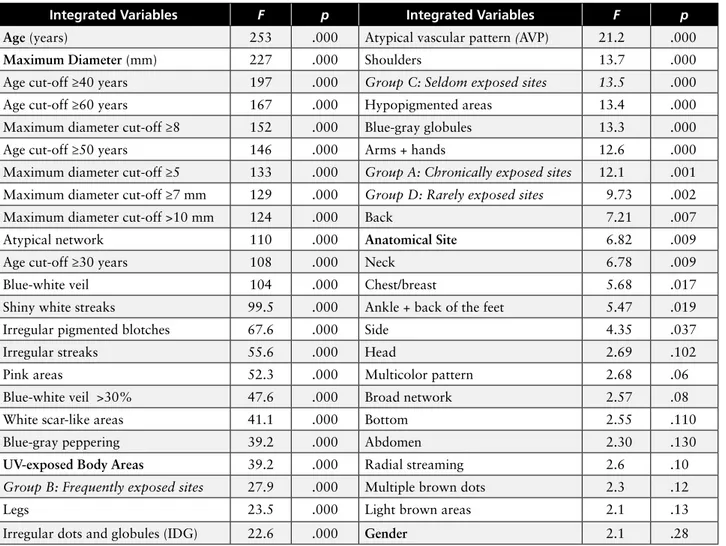
Univariate discriminant analysis of all 47 integrated variables, shown in Table 1, was performed taking the histo-pathological diagnosis as outcome. Uni-variate power to discriminate between Overall interobserver agreement was
estimated by Fleiss’ κ and its 95% con-fidence interval (CI). In a second phase, we retrospectively collected 2 clini-cal data (diameter and body site) and 2 personal data (age and sex) sets for each of the 450 MSLs, obtaining an integrated database of 450 images asso-ciated with 18 subjective (ie, dermo-scopic data) and 4 objective variables (ie, clinical-personal data). In order to be tested for risk factors for malignancy, they were evaluated both in their origi-nal form as 5 whole variables and in their binary-coded form as 42 simple variables. Age and maximum diameter were dichotomized to account for some interesting cut-off values. The lesion site was described according to
ana-TABLE 1. Discriminant analysis showing F-statistics (F) and P-value (P) of all
der-moscopic, clinical, and personal variables (47) coded into 38 simple
vari-ables, 5 whole variables (bold), and 4 grouped variables (italics)
Integrated Variables F p Integrated Variables F p
Age (years) 253 .000 Atypical vascular pattern (AVP) 21.2 .000
Maximum Diameter (mm) 227 .000 Shoulders 13.7 .000
Age cut-off ≥40 years 197 .000 Group C: Seldom exposed sites 13.5 .000
Age cut-off ≥60 years 167 .000 Hypopigmented areas 13.4 .000
Maximum diameter cut-off ≥8 152 .000 Blue-gray globules 13.3 .000
Age cut-off ≥50 years 146 .000 Arms + hands 12.6 .000
Maximum diameter cut-off ≥5 133 .000 Group A: Chronically exposed sites 12.1 .001
Maximum diameter cut-off ≥7 mm 129 .000 Group D: Rarely exposed sites 9.73 .002
Maximum diameter cut-off >10 mm 124 .000 Back 7.21 .007
Atypical network 110 .000 Anatomical Site 6.82 .009
Age cut-off ≥30 years 108 .000 Neck 6.78 .009
Blue-white veil 104 .000 Chest/breast 5.68 .017
Shiny white streaks 99.5 .000 Ankle + back of the feet 5.47 .019
Irregular pigmented blotches 67.6 .000 Side 4.35 .037
Irregular streaks 55.6 .000 Head 2.69 .102
Pink areas 52.3 .000 Multicolor pattern 2.68 .06
Blue-white veil >30% 47.6 .000 Broad network 2.57 .08
White scar-like areas 41.1 .000 Bottom 2.55 .110
Blue-gray peppering 39.2 .000 Abdomen 2.30 .130
UV-exposed Body Areas 39.2 .000 Radial streaming 2.6 .10
Group B: Frequently exposed sites 27.9 .000 Multiple brown dots 2.3 .12
Legs 23.5 .000 Light brown areas 2.1 .13
Irregular dots and globules (IDG) 22.6 .000 Gender 2.1 .28
eMM and aN was quantified by means of F-statistics. Statistical significance (P<0.05) was obtained by 37/47 vari-ables. Taken together, the results of this analysis showed that: 1) age and diameter exhibited the highest discrim-inant power for eMM when consid-ered as whole or simple variables; 2) the classification of anatomical sites into 4 body area groups according to UV exposure resulted in association with malignancy (eg, body site “head” obtained P>0.05 and F<2.69 as simple variable, but P<0.05 and F=12.1 when as part of Group A, chronically exposed body areas); 3) none of the dermoscopic features reached F>110, demonstrating moderate impact; and 4) as reported in Table 2, agreement between experts
Research | Dermatol Pract Concept 2018;8(4):16
327
that develop due to UV exposure [5-7].In conclusion, despite the contempo-rary presence of an atypical network, blue-white veil, and shiny white streaks within an equivocal MSLs, which may indicate malignancy, the objective clini-cal and personal data collected could supply a fundamental contribution in the correct diagnosis of equivocal MSLs and should be included in diagnostic algorithms.
References
1. Argenziano G, Cerroni L, Zalaudek I, et al. Accuracy in melanoma detection: a 10-year multicenter survey. J Am Acad
Dermatol. 2012;67(1):54-59.
2. Carrera C, Marchetti MA, Dusza SW, et al. Validity and reliability of dermoscopic criteria used to differentiate nevi from melanoma: a web-based international der-moscopy society study. JAMA Dermatol. 2016;152(7):798-806.
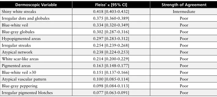
3. Abbasi NR, Yancovitz M, Gutkowicz-Krusin D, et al. Utility of lesion diameter in the clinical diagnosis of cutaneous mela-was generally poor, with the exception
of white shiny streaks (κ =0.418, 95% CI 0.403-0.432) albeit of intermediate level, which was probably due to the clear-cut appearance of this pattern.
Conclusions
The method of combining clinical and personal data with dermoscopic vari-ables proved to be highly useful diag-nostically in differentiating regressing MM from regressing nevi [8]. Here in this dataset of eMM and aN, the rela-tive impact of dermoscopic structures was moderate and their recognition was confirmed to be a rather subjective and equivocal method with unsatisfactory agreement [1,2]. Our findings are in line with recent epidemiological data in that eMM shows a trend to be increas-ing in prevalence in the elderly with no gender predominance [5,6], a strong correlation with lesion maximum diam-eter [7,8], and a moderate correlation with UV exposure (F=39.4). This prob-ably reflects only a fraction of eMM
TABLE 2. Concordance levels of experts (D
1-D
5) in recognition of
dermo-scopic structures (only variables that obtained P > 0.05 are shown)
Dermoscopic Variable Fleiss’ κ [95% CI] Strength of Agreement
Shiny white streaks 0.418 [0.403-0.432] Intermediate
Irregular dots and globules 0.375 [0.360-0.389] Poor
Blue-white veil 0.334 [0.320-0.349] Poor
Blue-gray globules 0.302 [0.287-0.316] Poor
Hypopigmented areas 0.297 [0.283-0.312] Poor
Irregular streaks 0.254 [0.239-0.268] Poor
Atypical network 0.238 [0.224-0.253] Poor
White scar-like areas 0.214 [0.200-0.229] Poor
Pigmented areas 0.163 [0.148-0.177] Poor
Blue-white veil >30 0.151 [0.137-0.166] Poor
Atypical vascular pattern 0.100 [0.085-0.114] Poor
Blue-gray peppering 0.098 [0.084-0.113] Poor
Irregular pigmented blotches 0.077 [0.063-0.091] Poor
noma. Arch Dermatol. 2008;144(4):469-474.
4. Moreno-Ramírez D, Ojeda-Vila T, Ríos-Martín JJ, et al. Association between tumor size and Breslow’s thickness in malignant melanoma: a cross-section-al, multicenter study. Melanoma Res. 2015;25(5): 450-452.
5. Haenssle HA, Mograby N, Ngassa A, et al. Association of patient risk fac-tors and frequency of nevus-associated cutaneous melanomas. JAMA
Derma-tol. 2016;152(3):291-298.
6. Whiteman DC, Green AC, Olsen CM. The growing burden of invasive melanoma: projections of incidence rates and numbers of new cases in six susceptible popula-tions through 2031. J Invest Dermatol. 2016;136(6):1161-1171.
7. Liu F, Bessonova L, Taylor TH, et al. A unique gender difference in early onset melanoma implies that in addition to ul-traviolet light exposure other causative factors are important. Pigment Cell
Mela-noma Res. 2013; 26(1):128–135.
8. Rubegni P, Tognetti L, Argenziano G, et al. A risk scoring system for the differentia-tion between melanoma with regression and regressing nevi. J Dermatol Sci. 2016; 83(2):138-144.