Clinical Study
Cultivated Oral Mucosa Epithelium in Ocular Surface
Reconstruction in Aniridia Patients
Dariusz Dobrowolski,
1Boguslawa Orzechowska-Wylegala,
2Bogumil Wowra,
1Ewa Wroblewska-Czajka,
1Maria Grolik,
1Krzysztof Szczubialka,
3Maria Nowakowska,
3Domenico Puzzolo,
4Edward A. Wylegala,
1Antonio Micali,
4and Pasquale Aragona
51Department of Ophthalmology, Ophthalmology Clinic, II School of Medicine with the Division of Dentistry in Zabrze,
Medical University of Silesia, District Railway Hospital, Panewnicka 65 street, 40760 Katowice, Poland
2Clinic of Maxillo-Cranio-Facial Surgery, Clinical Hospital, Medical University of Silesia, Francuska 20-24 street,
40027 Katowice, Poland
3Department of Nanotechnology of Polymers and Biomaterials, Faculty of Chemistry, Jagiellonian University, Ingardena 3 street,
30060 Cracow, Poland
4Department of Biomedical Sciences and Morphofunctional Imaging, University of Messina, 98 125 Messina, Italy 5Department of Surgical Specialties, Section of Ophthalmology, University of Messina, Policlinico “G. Martino”,
Gazzi, 98125 Messina, Italy
Correspondence should be addressed to Dariusz Dobrowolski; [email protected] Received 27 March 2015; Accepted 29 July 2015
Academic Editor: Flavio Mantelli
Copyright © 2015 Dariusz Dobrowolski et al. This is an open access article distributed under the Creative Commons Attribution License, which permits unrestricted use, distribution, and reproduction in any medium, provided the original work is properly cited.
Purpose. Efficacy of cultivated oral mucosa epithelial transplantation (COMET) procedure in corneal epithelium restoration of
aniridia patients. Methods. Study subjects were aniridia patients (13 patients; 17 eyes) with irregular, vascular conjunctival pannus involving visual axis who underwent autologous transplantation of cultivated epithelium. For the procedure oral mucosa epithelial cells were obtained from buccal mucosa with further enzymatic treatment. Suspension of single cells was seeded on previously prepared denuded amniotic membrane. Cultures were carried on culture dishes inserts in the presence of the inactivated with Mitomycin C monolayer of 3T3 fibroblasts. Cultures were carried for seven days. Stratified oral mucosa epithelium with its amniotic membrane carrier was transplanted on the surgically denuded corneal surface of aniridia patients with total or subtotal limbal stem cell deficiency. Outcome Measures. Corneal surface, epithelial regularity, and visual acuity improvement were evaluated. Results. At the end of the observation period, 76.4% of the eyes had regular transparent epithelium and 23.5% had developed epithelial defects or central corneal haze; in 88.2% of cases visual acuity had increased. VA range was from HM 0.05 before the surgery to HM up to 0.1 after surgery. Conclusion. Application of cultivated oral mucosa epithelium restores regular epithelium on the corneal surface with moderate improvement in quality of vision.
1. Introduction
During second decade of life, aniridic patients begin devel-oping pathologic cellular exchange. Epithelial cells covering the cornea are removed by the surrounding conjunctival
epithelium with its vascularized matrix [1]. This leads to a
local disorder called limbal stem-cell deficiency (LSCD) [2].
Its stages lead to superficial conjunctival ingrowth, corneal haze, discomfort, and deep loss of visual acuity. Since
the 1980s, the concept of limbal disease management has focused on limbal transplantation and removal of pathologic tissue followed by transplantation of autologous or allogeneic
limbal tissue carried on the donor’s sclera [3, 4]. Many
studies have confirmed that delivery of healthy limbal tissue
improves vision and decreases ocular discomfort [5–7].
Development of new culture techniques has changed
the treatment of LSCD [8]. Transplantation became limited
to cultured autologous cells, without additional tissues of
Volume 2015, Article ID 281870, 7 pages http://dx.doi.org/10.1155/2015/281870
the donor’s sclera or conjunctiva [9]. Application of oral mucosa epithelium was proposed by Shigeru Kinoshita and
was turned into clinical practice by Nakamura et al. [10].
Another new concept was use of oral mucosa epithelium in
reconstruction of the ocular surface [11]. With a phenotype
similar to the cornea, it allowed the possibility of autologous
treatment in patients with bilateral ocular involvement [12].
Congenital aniridia is a rare disorder of autosomal-dominant or sporadic occurrence. It occurs with an incidence of slightly less than 1 : 100,000 to 1 : 60,000 in the general population. It manifests with low visual acuity and ambly-opia early. Later it usually is accompanied by cataract and glaucoma. From the second decade of life, an additional factor influences visual quality: corneal haze caused by the
conjunctivalization of the cornea surface [13].
In aniridia patients, signs of LSCD are ocular discom-fort, superficial vascular conjunctival pannus, and decreased visual acuity. Severity is mild; therefore, for several years those patients were disqualified from any surgery. Cultivated oral mucosa epithelium transplantation (COMET) offers minimal and effective surgical approach for this group of patients.
2. Materials and Methods
The experimental procedure was performed under the tenets of the Declaration of Helsinki. Signed written informed consent was obtained from all patients before the procedure began.
Oral mucosa epithelium specimens (3–5 mm2) were
collected under local anesthesia and decontaminated with 5% povidone-iodine solution. Biopsies were taken from the inferior buccal mucosa; no sutures were applied. The tissue was transferred immediately to corneal storage medium at
4∘C and then taken to the laboratory.
The tissue specimen was treated with Dispase II for 1 hour, after which it was trypsinized with 1% trypsin/0.01% EDTA
mixture for 10 min in 37∘C to obtain a single cell suspension.
Epithelial cells were prepared for seeding with a density of
4× 10⁴cells per 1 mL (Cell counter, Coulter Z1, Miami, USA).
Culture media and chemicals were purchased from Sigma (Germany). The reagents for immunostaining were obtained from Santa Cruz Biotechnology Inc. (USA). The epithelial culture was carried in the presence of 3T3 fibroblasts, a source of growth factors. One week prior to specimen collection, six-well culture dishes (Becton Dickinson, USA) were prepared. Bottoms of the plates were covered with a monolayer of 3T3 fibroblasts (ATCC, USA). The 3T3 cells were cultivated in Dulbecco Modified Eagle’s Medium (DMEM) with 10%
bovine serum and a 100𝜇g/mL penicillin and streptomycin
mixture. From the fifth to seventh day of culture, when the monolayer was reached, fibroblasts were inactivated by
incubation in DMEM containing 2𝜇g/mL of Mitomycin C for
2 h.
The carrier for the oral mucosa epithelial sheet was the amniotic membrane located on the dish insert over the monolayer of fibroblasts. Amniotic membrane carriers were obtained from the Homograft Tissue Bank in Zabrze,
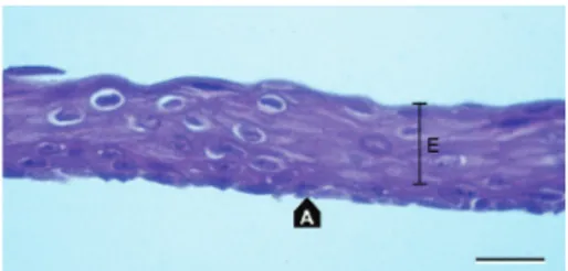
Figure 1: Cultivated oral epithelium (E) on the amniotic membrane
(A) prior to transplantation. H&E stain. Scale bar: 20𝜇m.
Poland. Amniotic membrane samples were washed from the cryoprotective medium with Phosphate Buffered Saline (PBS), and the amniotic epithelium was removed gently with
a culture scraper. Denuded amniotic membrane slides (15×
15 mm) on nitrocellulose paper were put over the layer of fibroblasts in the culture plates.
Single oral mucosa cells were seeded on denuded amni-otic membrane spread on nitrocellulose paper lying on the inserts over the fibroblast monolayer. Cultures were
carried out in standard conditions of 37∘C and a humidified
atmosphere of 5% CO2 and 95% air. The medium was a
supplemented Dulbecco Modified Eagle’s Medium (DMEM) with HAM F12 mixture with 10% of serum (bovine serum in first 12 experiments, autologous serum in the next 5 cultures), 0.5% dimethyl sulfoxide (DMSO), 10 ng/mL mouse
Epidermal Growth Factor (EGF), 5𝜇g/mL bovine insulin,
0.1 nM cholera toxin, 0.18 mM adenine, 2 nM triiodothyro-nine, 4 mM L-glutamine, 0.4 mg/mL hydrocortisone, and
100𝜇g/mL penicillin and streptomycin mixture. The culture
medium was changed every 48 h. Cultures were carried out on at least 2 amniotic membranes for each patient. On the seventh day of culture, the epithelial growth stage was evaluated under light microscope and small specimens of
each membrane (Figure 1) were stained with
hematoxylin-eosin (H-E). Amniotic membranes (A inFigure 1) covered
completely with multilayer epithelium (E inFigure 1) were
suitable for a graft. Cultivation over 7 days caused loss of superficial cells of the proliferating epithelium to the medium. At the time of microscopic qualification, airlifting was done. Cells were airlifted with a minimal amount of culture medium for 1 h.
One day before the transplantation, immunostaining for cytokeratin 4, cytokeratin 13, protein p63, and connexin 43 was performed to confirm the origin of the epithelium and identify the presence of low differentiated cells. Both cytok-eratins 4 and 13 were described by Ang et al. as characteristic for oral mucosa epithelial cells, their presence confirming expected cellular structure of the graft just before application
[14]. Protein p63 (transcription factor) plays an important
role in epithelial cells proliferation and differentiation, in cultured cells it shows cellular potential for epithelial renewal
[15]. Gap-junction protein connexin 43 is present in the
basal epithelial layer, and positive staining confirms proper
architecture of cultured epithelial layers [16].
Hematoxylin-eosin (H-E) staining was used in histological analysis of epithelial layer regularity and thickness. Regular structure
of intact epithelium on amniotic membrane presented on
Figure 1was expected by researchers. Only such grafts were qualified in surgery.
Study subjects were 13 aniridia patients (17 eyes) suffering from LSCD with central corneal involvement. Vascular con-junctival pannus involved the entire limbal area (total LSCD: 14 eyes) or at least 9 clock-hours of the limbus with central involvement (subtotal LSCD: 3 eyes). The main indication for the study was presence of semitransparent conjunctival pan-nus covering the cornea over the pupil. It caused decreased quality of vision even in cases of subtotal limbal involvement. The patients were both oral mucosa epithelium donors and recipients of the cultured epithelium. There were 3 men and
10 women. Ages ranged from 16 to 54 with an average of31.1±
11.5 years. In 4 cases, surgery was bilateral and sequential (3–6 months between each surgery). Visual acuity before intervention ranged from HM to 0.05. Inclusion criteria were congenital aniridia, conjunctivalization of the corneal surface with neovascularization (with or without corneal scarring) and at least 6 months from last eye surgery. In addition, the study required healthy oral mucosa, no smoking for at least 7 days before taking a sample of epithelium, and regular teeth brushing (twice a day). Exclusion criteria were acute systemic infections, acute eye infection during the last 6 months, neoplasm history, pregnancy, and mental disorders. In addition patients with dense cataract coexisting corneal disorders were excluded.
Surgery was performed under topical anesthesia (Alcaine, Alcon, USA). A 360-degree peritomy was done, followed by conjunctival suturing at 1-2 mm behind the limbal area with single 10-0 nylon sutures. The conjunctival pannus was removed gently with a spatula or crescent knife. If necessary, fibrotic tissue spread under the conjunctival pannus was removed with gentle keratectomy to prepare a smooth cor-neal surface. Then, round, 14 mm diameter trephined amni-otic carriers with oral mucosa epithelial cells were trans-planted onto the denuded corneas. Peripheral, continuous 10-0 nylon suture was used to stabilize the graft. The entire cornea was covered, and suture was placed in the corneolim-bal interface. Then, the graft was covered by a bandage contact lens.
After the procedure, levofloxacin (Santen, Finland) and preservative-free dexamethasone eye drops (Thea Laborato-ries, Paris, France) were administered 5 times a day for 2 weeks. Then, dexamethasone alone was applied 3 times a day for 6 weeks. All subjects received preservative-free lubricants every 2 hours, and in cases of epithelial defects or erosions, autologous serum drops were administered. On each con-trol visit, stabilization of the corneal surface was evaluated with topical 10% fluorescein staining. Lack of epithelial defects (negative fluorescein staining) with regular, stratified epithelium or no more than 3 clock-hours of peripheral conjunctival neovascularization was classified as a successful case (coded as +++); partial success consisted of punctate epithelial defects or peripheral conjunctival neovasculariza-tion up to 9 clock-hours without pupil area involvement (coded as ++); and failure consisted of vascularization of the pupil area, persistent nonhealing epithelial defects or deep stromal vascularization (coded as +). Whether LSCD
0.96 C u m u la ti ve gra ft s u rv iv al 0.94 0.92 0.90 0.88 0.86 0.84 0.82 0.80 2 4 6 8 10 12 Months Complete censored 14 16 18 20
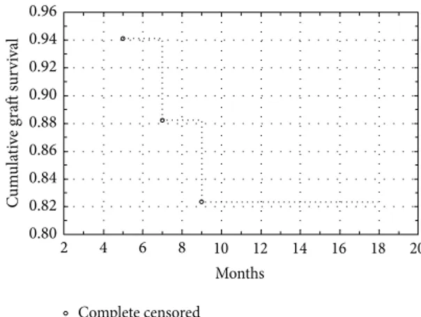
Figure 2: Cumulative graft survival: Kaplan-Meier curve for COMET in aniridia.
occurrence was total or subtotal made no differences in surgical management or postoperative treatment regarding 360-degree peritomy and removal of all fibrotic tissue or epithelia from the corneal surface.
3. Results
Local complications were not seen after oral mucosa dona-tion. Transplanted specimens showed regular, stratified epithelium of 4–9 layers of cells. Epithelium origin was confirmed by previous immunostaining, as described in the culture procedure section.
Over the observation period of 12–18 months, 13 operated eyes showed stable epithelium; however, peripheral vascular
irregularities were present in all of them. Table 1 shows
detailed patient data. These corneas were not stained by fluorescein solution (qualified as +++); 4 eyes suffered various epithelial defects, from single local defects (++) to large areas of denuded cornea (+) with peripheral conjunctival ingrowth. At the end of the observation period, 3 eyes developed stromal scarring, conjunctival vascularization, or stromal vascularization. All 3 of these eyes were qualified as graft failure. In successful cases, the epithelium was smooth and
regular (qualified as +++ and ++).Figure 2presents the graft
survival rate on a Kaplan-Meier curve. The first 9 postop-erative months were critical to the final results. Occurrence of epithelial defect was the indication for autologous serum eye drops. Despite careful treatment, 3 failed cases developed after 6 months of progressive invasion of the central cornea by vascular conjunctival tissue. In other cases, the periph-eral area was characterized by irregularities in the vascular network, with peripheral ingrowth of new vessels without central area involvement. Comparison of surfaces before and
after surgery (Figures3(a)and3(b)) shows improvement of
corneal transparency in the central area and irregular distri-bution of vessels in the limbal area. Inflammatory response, defined as hyperemia and neovascularization at the donor-host interface, was moderate and easily controlled with a steroid agent. Limited vascular ingrowth in eyes with stable autologous oral epithelium confirms that COMET with its autologous origin limits inflammatory response. There is also
Table 1: Overview of aniridia patients data.
Case Age Gender Eye BCVA
pre
BCVA
post Epithelial regularity Follow-up (m.) Treatment
1 28 F R CF 0.05 +++ 18 Routine
2 28 F L 0.05 0.1 +++ 12 Routine
3 27 F R 0.02 0.1 +++ 14 Routine
4 54 F R HM CF + 13 Routine, PSED, DEX
5 26 F L 0.02 0.05 +++ 18 Routine
6 29 F R 0.05 0.05 +++ 18 Routine
7 27 F L 0.01 0.03 + 17 Routine, PSED, DEX
8 18 F L HM 0.1 +++ 18 Routine 9 18 F R HM 0.05 +++ 14 Routine 10 49 M L CF 0.1 +++ 18 Routine 11 40 F R CF 0.05 +++ 15 Routine, TSED 12 23 M L HM CF +++ 18 Routine, TSED 13 38 F L 0.01 0.02 +++ 18 Routine 14 38 F R HM 0.05 ++ 15 Routine, PSED 15 49 M L CF 0.1 +++ 12 Routine 16 16 F L HM HM +++ 18 Routine, TSED
17 22 F R HM 0.05 + 16 Routine, PSED, DEX
Fluorescein staining: absent (+++), local defects (++), and present (+). CF: counting fingers and HM: hand movements.
Routine treatment: Levofloxacin and dexamethasone eye drops 5 times a day for a 2-week period and then only dexamethasone 3 times a day for the next 6 weeks.
Additional treatment: PSED: permanent serum eye drops, TSED: temporary serum eye drops, and DEX: additional dexamethasone administration over 2nd month from surgery.
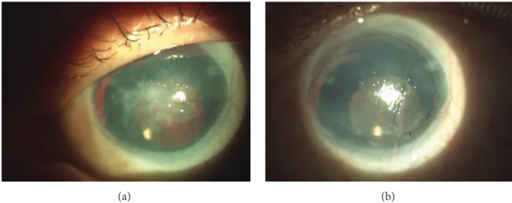
(a) (b)
Figure 3: (a) and (b) Aniridia patient, 49-year-old man, who underwent cataract surgery 5 years before COMET. Preoperative view with central fibrovascular scar on the left; on the right, 6 months after surgery, VA improvement from CF to 0.1 with central cornea covered by regular and smooth epithelium.
no immune reaction characteristic for allotransplantation in, for example, keratolimbal allografts (KLAL). Excluding the failed grafts, patients noted improvement in their quality of vision, as well as decreased ocular discomfort. Postoperative occurrence of epithelial defects did not interfere with visual acuity and were treated with autologous serum drops 7 times a day. Four eyes with epithelial defects received persistent therapy; in three others (graded as +++) therapy was tem-porary for up to four weeks. Visual acuity improvement was
noted in 88.2% of cases. It ranged from HM up to 0.1. As
shown in Figure 3 main benefit of COMET was restoring
of transparent epithelium without pathologic vessels on the corneal surface. This 49-year-old, single-eyed man developed conjunctival pannus after cataract surgery performed 5 years ago. Improvement to 0.1 restored his ability to read; he pointed it as a main benefit of surgery. Despite moderate improvement of VA the majority of patients were satisfied after surgery.
4. Discussion
LSCD is a difficult therapeutic challenge in patients with con-genital aniridia. This condition is mild in severity and is usually accompanied by amblyopia, cataracts, and corneal haze caused by conjunctival pannus. It is believed that pro-gressive conjunctivalization with age is connected to congen-ital impairment of pluripotent cells located in the hem.
In 2003, Nakamura and Kinoshita proposed a cultivation method of oral epithelium to restore ocular epithelial surface
based on experiments on rabbits [17]. Several benefits made
the procedure very interesting. Damaged corneas covered by autologous epithelium have a lower risk of immunologic reaction. Oral epithelium transplanted onto the new locus can maintain its proliferative potential without developing keratinization. In addition, low differentiation and quick cellular turnover result in regular stratified epithelium.
An important factor of this method is the cellular phe-notype of oral epithelium. The phephe-notype is similar to the corneal epithelium phenotype, making this procedure safe and easy to apply. Allografts often require systemic or local immunosuppressive agents administered for long periods postoperatively. Autografts do not have such requirements, as the induction of neovascularization is lower if the tissue
comes from the same patient [18].
As described by Hata et al., oral epithelium culture is very effective for recipients, as these cells have a low stage of
dif-ferentiation combined with fast cellular turnover [19]. To
obtain a regular epithelial layer, only a few days of culturing are needed. In addition, these features are prolonged for the postoperative period. In long-term observation, it improves stability of the transplanted epithelium. In our patients, the surface epithelium in successful cases remained stable with temporary epithelial disorders. In local defects, autologous serum drops were administered to improve epithelial growth. For large defects autologous serum or bandage contact lenses were ineffective. Such patients in our study developed secondary conjunctival ingrowth leading to failure of the procedure.
COMET is widely proposed in Stevens-Johnson Syn-drome (SJS), ocular cicatricial pemphigoid (OCP) and ocular burns. However, in those cases many patients require 2-step surgical approach to restore vision due to accompanied stromal damage. Sotozono et al. describes such cases showing that long-term visual improvement is possible even with
high rate of postoperative epithelial defects (40%) [20]. In
acute cases of SJS or OCP damage of the ocular surface and inflammation grade is high. Authors have only one case of aniridia and a few cases with slight ocular surface damage. In those cases visual improvement is significantly high. In aniridic patients ocular surface disease is limited to the epithelial surface and subepithelial fibrosis. There is no severe inflammation or destruction of surrounding tissues. In our group there was also no need of stromal procedures of lamellar or penetrating keratoplasty. Final vision remained low; however, main benefit was stable epithelial surface with only 3 failed grafts and 4 patients with epithelial defects (3 of them were temporary). If compared to severe cases of
ocular surface disease in aniridia there was no indication to administration of systemic steroids.
Historically, therapeutic treatment has included Scheffer Tseng’s approach of removing the corneal pannus com-bined with lamellar keratoplasty or penetrating keratoplasty combined with limbal allotransplantation. However, allo-transplants require subsequent systemic immunosuppres-sion, which can interfere with the patient’s general health. Complicity and difficulty of the technique resulted in the need to transplant only those epithelial cells involved in the pathologic process. COMET offered the opportunity to focus on the affected area of the corneal surface.
Tsubota et al. describing keratolimbal allografts shows high risk of immune adverse reactions (graft rejection), infections, and systemic disorders associated with immuno-suppressive agents administered for many postoperative
months [21]. In his study stable ocular surface in achieved
in 51% of the cases. If we compare improvements in vision,
allografts limit the quality of vision [22]. Risk of rejection
and revascularization result in loss of visual acuity in long-term observations. The Kaplan-Meier curves for the allograft approach show continuous reduction of success rate in the long term. Daya et al. reports results of allogeneic cultured corneal epithelium transplantation with success rate of 70%
[23]. It indicates that limitation of surgical approach to
epithelial surface only increases success rate. Nishida et al. report 100% success rate after COMET; however, in other studies authors achieve stabilization of corneal surface in
66.7% or even 57.5% [24–26]. The autologous approach
with oral epithelium transplantation seems to cause less immunization, less inflammatory response, and less vascular ingrowth. These benefits should increase the success rate in bilateral limbal stem-cell insufficiency present in aniridia.
There were not many differences between culture pro-cedures for the oral and corneal epithelium transplantation techniques. Amniotic membrane substrate as a cellular car-rier and the source of basement membrane is widely applied
[27, 28]. Its efficacy is confirmed in papers on cultured
epithelial transplantation. Amniotic membrane makes the application of oral epithelial sheets safe and easy. In mild cases with partial limbal insufficiency in aniridia, transplantation limited to the application of amniotic membrane can support the corneal epithelium for a long period of time. Coculture with 3T3 fibroblasts results in fast proliferation and maintains low differentiation of the oral epithelium. This parameter is crucial for graft survival, despite fast cellular turnover. Another advantage is the almost unlimited source of tissue, which can be collected easily. The regenerative potential of oral epithelium is high. During cultivation, loss of superficial epithelial cells to the media was observed after seven days. In our opinion, a culture period of one week is sufficient to reach stable oral epithelial multilayers.
In 2003, Nakamura et al. described the first oral epithe-lium transplantations in humans with corneas severely dam-aged by Stevens-Johnson Syndrome (SJS) and severe ocular burns. Results were promising, and oral epithelium became a substitute for corneal epithelium with satisfactory corneal transparency and reduction of ocular discomfort. Nakamura et al. later described 17 patients treated with the COMET
procedure with a mean follow-up of 55 months [29]. All eyes manifested revascularization of the surface with various stages of severity. The most stable epithelia were observed within six months after surgery. Visual acuity improvement was observed in 95% of patients after surgery. After 36 months, only 53% of eyes noted improved visual acuity. The authors concluded that despite the decrease in vision quality, the reconjunctivalization was less severe than that before surgery. In failed cases presented in the paper, conjunctival invasion was not severe; however, involvement of visual axis caused failure of the entire treatment.
Another paper on COMET by Ang et al. described stabi-lization of the ocular surface without major complications in
all investigated eyes for at least 12 months after surgery [14].
Graft survival was limited in COMET; however, scarification of the ocular surface was much less severe compared with untreated patients. In addition, combining COMET with penetrating keratoplasty led to improved ocular surface and
decreased risk of PK failure [30].
Satake presented the results of 40 eyes after COMET. Pro-cedure were performed on SJS, cicatricial pemphigoid, and
ocular burn patients [26]. Results showed the fast decline of
graft survival in the first six months after the procedures with subsequent stabilization. Outcomes depended on the severity of ocular involvement; patients with severe vascularization and damage to the cornea surface were at greater risk of early graft failure than those with less severe symptoms. Similarly, poor primary visual acuity resulted in a worse outcome for those in the aniridia group. However, analysis has not been performed on patients with congenital aniridia. According to our study, graft survival and epithelium regularity are much better in aniridia patients.
Visual acuity in patients with aniridia depends on the optical transparency of both the cornea and the lens. In 75% of patients, cataracts occurred next to the corneal pannus, which greatly affected visual acuity. Therefore, in our opinion, visual acuity is a poor success parameter of transplant-cultured oral mucosal epithelium. In the success group graded +++, there were eyes with no visual acuity improve-ment despite a stable surface, showing that other factors, such as foveal hypoplasia, optic nerve lesions, or cataracts, were disturbing vision. In our study, the criterion for success was restoration of the corneal epithelium, although it is known that the most important criterion for patients is visual acuity. Increased corneal vascularization with patient age is associated with epithelial cell dysfunction, but the exact cause has not been documented clearly yet. Some authors make effective use of anti-VEGF, but the literature does not describe the use of anti-VEGF in corneal neovascularization.
Application of COMET was successful in aniridia patients. The visual acuity improvement rate for aniridia cases was lower than that for SJS, ocular cicatricial pemphigoid, or ocular burn cases. While actual visual acuity improvement was lower, the main benefit was improvement of epithelial regularity. Chen et al. investigated cell survival in COMET
patients [31]. The majority of recipients in the follow-up had
oral mucosa-derived cells The present study has confirmed the usefulness of the procedure in cases of mild limbal insufficiencies, such as in aniridia patients. In conclusion,
COMET is a very useful surgical procedure for patients with congenital aniridia, as it delivers benefits with minimal harm. Further studies are needed to compare epithelium restoring procedures including etiology of limbal stem deficiency.
Summary Statement
COMET procedure is a minimal invasive treatment, which enable exchanging irregular corneal epithelium for regular stratified oral mucosa epithelium. Aniridia causes huge dis-comfort; COMET not only restores stable ocular surface but also improves quality of life. Another advantage is simple surgical approach, autologous transplantation, and nonag-gressive postoperative treatment. Visual acuity improvement is more efficient for near than for far distance; the majority of patients were able to read again.
Ethical Approval
Study design, patient’s brochure, and consent form were approved by The Bioethics Committee at the Silesian Regional Medical Chamber, Agreement no. 24/2012, Katow-ice, Poland.
Disclosure
This paper was a poster presentation: American Academy of Ophthalmology Annual Meeting, November 2011, Best Poster Award.
Conflict of Interests
None of the authors has conflict of interests with the submis-sion.
References
[1] S. C. G. Tseng, “Concept and application of limbal stem cells,”
Eye, vol. 3, no. 2, pp. 141–157, 1989.
[2] J. J. Y. Chen and S. C. G. Tseng, “Abnormal corneal epithelial wound healing in partial-thickness removal of limbal epithe-lium,” Investigative Ophthalmology and Visual Science, vol. 32, no. 8, pp. 2219–2233, 1991.
[3] K. R. Kenyon and S. C. G. Tseng, “Limbal autograft transplan-tation for ocular surface disorders,” Ophthalmology, vol. 96, no. 5, pp. 709–723, 1989.
[4] S. C. G. Tseng, P. Prabhasawat, K. Barton et al., “Amniotic mem-brane transplantation with or without limbal allografts for severe ocular surface disorders,” Ophthalmology, vol. 102, pp. 1486–1496, 1995.
[5] S. C. G. Tseng and K. Tsubota, “Important concepts for treat-ing ocular surface and tear disorders,” American Journal of
Oph-thalmology, vol. 124, no. 6, pp. 825–835, 1997.
[6] D. J. Coster, R. K. Aggarwal, and K. A. Williams, “Surgical man-agement of ocular surface disorders using conjunctival and stem cell allografts,” British Journal of Ophthalmology, vol. 79, no. 11, pp. 977–982, 1995.
[7] E. J. Holland and G. S. Schwartz, “The evolution of epithelial transplantation for severe ocular surface disease and a proposed classification system,” Cornea, vol. 15, no. 6, pp. 549–556, 1996.
[8] R. J.-F. Tsai, L.-M. Li, and J.-K. Chen, “Reconstruction of dam-aged corneas by transplantation of autologous limbal epithelial cells,” The New England Journal of Medicine, vol. 343, no. 2, pp. 86–93, 2000.
[9] G. Pellegrini, R. Ranno, G. Stracuzzi et al., “The control of epi-dermal stem cells (holoclones) in the treatment of massive full-thickness burns with autologous keratinocytes cultured on fibrin,” Transplantation, vol. 68, no. 6, pp. 868–879, 1999. [10] T. Nakamura, K.-I. Endo, L. J. Cooper et al., “The successful
cul-ture and autologous transplantation of rabbit oral mucosal epithelial cells on amniotic membrane,” Investigative
Ophthal-mology and Visual Science, vol. 44, no. 1, pp. 106–116, 2003.
[11] S. Kinoshita, N. Koizumi, and T. Nakamura, “Transplantable cultivated mucosal epithelial sheet for ocular surface recon-struction,” Experimental Eye Research, vol. 78, no. 3, pp. 483– 491, 2004.
[12] T. Inatomi, T. Nakamura, N. Koizumi, C. Sotozono, and S. Kinoshita, “Current concepts and challenges in ocular surface reconstruction using cultivated mucosal epithelial transplanta-tion,” Cornea, vol. 24, no. 8, supplement, pp. S32–S38, 2005. [13] K. Nishida, S. Kinoshita, Y. Ohashi, Y. Kuwayama, and S.
Yamamoto, “Ocular surface abnormalities in aniridia,”
Ameri-can Journal of Ophthalmology, vol. 120, no. 3, pp. 368–375, 1995.
[14] L. P. K. Ang, T. Nakamura, T. Inatomi et al., “Autologous serum-derived cultivated oral epithelial transplants for severe ocular surface disease,” Archives of Ophthalmology, vol. 124, no. 11, pp. 1543–1551, 2006.
[15] G. Pellegrini, E. Dellambra, O. Golisano et al., “p63 identifies keratinocyte stem cells,” Proceedings of the National Academy of
Sciences of the United States of America, vol. 98, no. 6, pp. 3156–
3161, 2001.
[16] M. Grueterich, E. Espana, and S. C. G. Tseng, “Connexin 43 expression and proliferation of human limbal epithelium on intact and denuded amniotic membrane,” Investigative
Ophthal-mology and Visual Science, vol. 43, no. 1, pp. 63–71, 2002.
[17] T. Nakamura and S. Kinoshita, “Ocular surface reconstruction using cultivated mucosal epithelial stem cells,” Cornea, vol. 22, no. 7, supplement, pp. S75–S80, 2003.
[18] G. Pellegrini, C. E. Traverso, A. T. Franzi et al., “Long term resto-ration of damaged corneal surface with autologous cultivated corneal epithelium,” The Lancet, vol. 349, pp. 990–993, 1997. [19] K.-I. Hata, H. Kagami, M. Ueda, S. Torii, and M. Matsuyama,
“The characteristics of cultured mucosal cell sheet as a material for grafting; comparison with cultured epidermal cell sheet,”
Annals of Plastic Surgery, vol. 34, no. 5, pp. 530–538, 1995.
[20] C. Sotozono, T. Inatomi, T. Nakamura et al., “Visual improve-ment after cultivated oral mucosal epithelial transplantation,”
Ophthalmology, vol. 120, no. 1, pp. 193–200, 2013.
[21] K. Tsubota, Y. Satake, M. Kaido et al., “Treatment of severe ocular-surface disorders with corneal epithelial stem-cell trans-plantation,” The New England Journal of Medicine, vol. 340, no. 22, pp. 1697–1703, 1999.
[22] H. S. Dua and A. Azuara-Blanco, “Allo-limbal transplantation in patients with limbal stem cell deficiency,” British Journal of
Ophthalmology, vol. 83, no. 4, pp. 414–419, 1999.
[23] S. M. Daya, A. Watson, J. R. Sharpe et al., “Outcomes and DNA analysis of ex vivo expanded stem cell allograft for ocular sur-face reconstruction,” Ophthalmology, vol. 112, no. 3, pp. 470–477, 2005.
[24] K. Nishida, M. Yamato, Y. Hayashida et al., “Corneal reconstruc-tion with tissue-engineered cell sheets composed of autologous
oral mucosal epithelium,” The New England Journal of Medicine, vol. 351, no. 12, pp. 1187–1196, 2004.
[25] T. Inatomi, T. Nakamura, N. Koizumi, C. Sotozono, N. Yokoi, and S. Kinoshita, “Midterm results on ocular surface recon-struction using cultivated autologous oral mucosal epithelial transplantation,” American Journal of Ophthalmology, vol. 141, no. 2, pp. 267–e1, 2006.
[26] Y. Satake, K. Higa, K. Tsubota, and J. Shimazaki, “Long-term outcome of cultivated oral mucosal epithelial sheet trans-plantation in treatment of total limbal stem cell deficiency,”
Ophthalmology, vol. 118, no. 8, pp. 1524–1530, 2011.
[27] N. Koizumi, N. J. Fullwood, G. Bairaktaris, T. Inatomi, S. Kinoshita, and A. J. Quantock, “Cultivation of corneal epithelial cells on intact and denuded human amniotic membrane,”
Inves-tigative Ophthalmology and Visual Science, vol. 41, no. 9, pp.
2506–2513, 2000.
[28] B. Seitz, M. D. Resch, U. Schl¨otzer-Schrehardt, C. Hofmann-Rummelt, R. Sauer, and F. E. Kruse, “Histopathology and ultrastructure of human corneas after amniotic membrane transplantation,” Archives of Ophthalmology, vol. 124, no. 10, pp. 1487–1490, 2006.
[29] T. Nakamura, K. Takeda, T. Inatomi, C. Sotozono, and S. Kinoshita, “Long-term results of autologous cultivated oral mucosal epithelial transplantation in the scar phase of severe ocular surface disorders,” British Journal of Ophthalmology, vol. 95, no. 7, pp. 942–946, 2011.
[30] T. Inatomi, T. Nakamura, M. Kojyo, N. Koizumi, C. Soto-zono, and S. Kinoshita, “Ocular surface reconstruction with combination of cultivated autologous oral mucosal epithelial transplantation and penetrating keratoplasty,” American Journal
of Ophthalmology, vol. 142, no. 5, pp. 757.e1–764.e1, 2006.
[31] H. J. Chen, H. L. Chen, J. Y. La et al., “Persistence of transplanted oral mucosal epithelial cells in human cornea,” Investigative
Submit your manuscripts at
http://www.hindawi.com
Stem Cells
International
Hindawi Publishing Corporationhttp://www.hindawi.com Volume 2014
Hindawi Publishing Corporation
http://www.hindawi.com Volume 2014
INFLAMMATION
Hindawi Publishing Corporation
http://www.hindawi.com Volume 2014
Behavioural
Neurology
Endocrinology
International Journal ofHindawi Publishing Corporation
http://www.hindawi.com Volume 2014
Hindawi Publishing Corporation
http://www.hindawi.com Volume 2014
Disease Markers
Hindawi Publishing Corporation
http://www.hindawi.com Volume 2014
BioMed
Research International
Oncology
Journal ofHindawi Publishing Corporation
http://www.hindawi.com Volume 2014
Hindawi Publishing Corporation
http://www.hindawi.com Volume 2014 Oxidative Medicine and Cellular Longevity Hindawi Publishing Corporation
http://www.hindawi.com Volume 2014
PPAR Research
The Scientific
World Journal
Hindawi Publishing Corporation
http://www.hindawi.com Volume 2014
Immunology Research
Hindawi Publishing Corporation
http://www.hindawi.com Volume 2014
Journal of
Obesity
Journal ofHindawi Publishing Corporation
http://www.hindawi.com Volume 2014
Hindawi Publishing Corporation
http://www.hindawi.com Volume 2014 Computational and Mathematical Methods in Medicine
Ophthalmology
Journal ofHindawi Publishing Corporation
http://www.hindawi.com Volume 2014
Diabetes Research
Journal ofHindawi Publishing Corporation
http://www.hindawi.com Volume 2014
Hindawi Publishing Corporation
http://www.hindawi.com Volume 2014 Research and Treatment
AIDS
Hindawi Publishing Corporation
http://www.hindawi.com Volume 2014
Gastroenterology Research and Practice
Hindawi Publishing Corporation
http://www.hindawi.com Volume 2014