The past is always present, as the context from which the present has emerged; thus, there is theoretical continuity between material changes in the past, present, and future.
University of Catania
Department of Biological, Geological and Environmental Sciences Ph.D. in Earth Science XXVIII cycle (2013-2015)
Complex Pore Geometries in Natural Building Stones:
an experimental and theoretical approach for the modeling of porosity changes
in natural, degraded and treated calcarenites
by
Simona Raneri
Under the supervision of Prof. Germana Barone
i
ABSTRACT
Calcarenites are the most widely used natural stones in Sicily as building materials. The comprehension of the degradation style of these natural building stone is of great interest in order to preserve the ancient masonry, especially in the case of relevant Cultural Heritages. A large number of Sicilian monuments are included in UNESCO Heritage List as their special cultural or physical significance. Considering the cultural importance of these ones, smart solutions are required in the perspective of their conservation.
In the framework of restoration actions, the complete knowledge of the internal structure of building stones is fundamental in order to estimate durability and prevent degradation processes. Salt growth is a significant cause of damage for natural stones. The effects of the salt weathering are, in addition to the properties of the salt itself and the climatic conditions, strongly related to the structural and textural features of materials. For this reason, an integrate study on petrographic, physical, mechanical and porosimetric proprieties changes due to salts crystallization could be useful to highlight correlation among textural, structural and engineering parameters. Moreover, one of the main problems in conservation field is the use of suitable consolidant and protective treatments able to preserve the masonry, being compatible with the stone substrate and not affecting its aesthetical proprieties. There are numerous tests able to verify the efficiency of consolidant or protective products, mainly devoted to verify their ability to not modify the appearance of stone and its physical proprieties, especially against water; however, particularly in the case of consolidant, one of the most important feature is related to its penetration depth, often difficult to evaluate by using the standard test routines. For aforementioned, this research work is devoted to apply a multi-methodological approach for characterizing and modeling pore structure modifications due to weathering processes in a coarse grained Sicilian calcarenite used as building and replace stones in Sicilian Cultural Heritages (Sabucina Stone), also through fractal models and innovative and non invasive methods. The obtained data on Sabucina stone have been employed for planning conservative treatments devoted to coarse grained calcarenite substrates. Therefore, innovative nanostructured and hybrid consolidant and protective products have been experimented and the results of efficiency tests along with the determination of surface and sub-surface changes due to the treatments are proposed.
In conclusion, the applied theoretical and empirical approach has allowed to: highlight the potential of classical and innovative complementary methods in quantifying the structural changes in porous materials due to degradation processes and conservative treatments, explain how they help to facilitate the understanding of stone weathering and validate the use of geometrical models in describing complex pore systems.
ii
RIASSUNTO
Nel panorama dei materiali lapidei naturali utilizzati come pietre da costruzione nell‘edilizia storica siciliana, le calcareniti rappresentano uno dei litotipi maggiormente impiegati; testimonianza di ciò è la particolare diffusione di tali materiali nella maggior parte dei Beni di interesse storico e culturale, tra i quali numerosi sono quelli inclusi nella Lista del patrimonio UNESCO. In ragione di ciò, è di prioritaria importanza l‘adozione di soluzioni idonee e specifiche per la loro protezione e conservazione.
Al fine di programmare e pianificare adeguati interventi di conservazione e restauro è di fondamentale importanza una corretta caratterizzazione delle proprietà fisiche e strutturali dei materiali, avendo queste un‘influenza diretta sulla durabilità degli stessi e sull‘evoluzione dei processi di alterazione e degrado. La cristallizzazione dei sali rappresenta uno degli agenti maggiormente dannosi nei confronti dei materiali lapidei naturali; nel caso del degrado da sali solubili, l'entità del danno causato è influenzata non solo dalle proprietà chimico-mineralogiche dei sali, ma anche dalle condizioni climatico-ambientali, nonché dalle modificazioni indotte nella struttura e tessitura dei materiali interessati dal processo.
Da quanto detto appare chiaro che la valutazione dello stato di conservazione e delle modificazioni indotte dal degrado da sali solubili nei materiali lapidei naturali non può che prevedere l‘applicazione di un approccio integrato e multidisciplinare, capace di indagare tutte le proprietà soggette all‘azione della cristallizzazione salina, sia quelle prettamente materiche (petrografiche, mineralogiche, chimiche e fisiche), che quelle tecniche (ingegneristiche, meccaniche e micro-strutturali).
Altro aspetto di interesse nella conservazione dei materiali lapidei naturali riguarda l‘intervento diretto di restauro; deprecabile è l‘utilizzo di consolidanti e protettivi senza verificarne la loro compatibilità materica ed estetica con il substrato. A tal proposito, diverse metodologie permettono di valutare l‘efficienza e la capacità di questi di non interferire con le proprietà chimico-fisiche del materiale; tuttavia, per i consolidanti, aspetti quali la loro profondità di penetrazione, sono spesso di difficile valutazione attraverso l‘esecuzione di test standard. Alla luce di quanto detto, il presente lavoro di tesi espone lo sviluppo di un approccio multi-metodologico basato sull‘applicazione integrata di metodi classici e innovativi, finalizzato a indagare le modificazioni della struttura porosa indotte dalla cristallizzazione salina nella Pietra di Sabucina, calcarenite grossolana utilizzata come materiale da restauro e da costruzione in Sicilia centro-orientale. Ciò ha consentito l‘elaborazione di un modello interpretativo del processo di degrado, descrittivo dell‘evoluzione micro-strutturale e tessiturale della roccia e dell‘influenza dello stesso sulle sue caratteristiche fisico-meccaniche, nonché funzionale alla scelta delle più idonee modalità di intervento conservativo. La particolare diffusione, nel panorama attuale, di prodotti nanostrutturati, ha indirizzato alla scelta di sistemi ibridi,
iii
innovativi, ecocompatibili e ad alta efficienza; in riferimento a ciò, sono state verificate le caratteristiche di compatibilità chimico-fisica ed estetica con il litotipo oggetto di studio e le eventuali modificazioni micro-strutturali e micro-tessiturali indotte nello stesso.
L‘insieme dei risultati ottenuti ha consentito quindi di verificare le potenzialità dell‘applicazione di metodologie complementari, tradizionali e innovative, atte a quantificare e visualizzare le modificazioni strutturali e tessiturali superficiali e sub-superficiali indotte dal degrado e dai trattamenti conservativi nei materiali lapidei naturali, indicando quindi un modus operandi capace di descrivere geometrie complesse attraverso modelli teorici ed empirici.
iv
RESUMÈ
Les calcarénites sont les pierres les plus utilisés en Sicile comme matériaux dans constructions. La compréhension des mécanismes de dégradation de ce matériel naturel a un grand intérêt pour la préservation de l‘ancienne maçonnerie, surtout lorsqu'il s'agit de patrimoine culturels reconnu. Un grand nombre des monuments siciliens sont inclus dans la Liste du Patrimoine de l‘Unesco grâce à leur portée culturelle et diversification des matériaux composants. Pour cette raison il faut identifier des solutions appropriées afin de les conserver.
Dans le cadre des activités de restauration, la connaissance complète de la structure interne des pierres de construction est essentielle pour estimer la durabilité et prévenir la dégradation du matériau. La croissance des sels est l‘une des premières causes des altérations des pierres naturelles. En addition à les propriétés du sel lui-même et des conditions climatiques, les effets d'altération de la pierre sont fortement associés aux caractéristiques structurelles et à la texture des matériaux. Pour cette raison il est nécessaire un étude intégré sur le changement des propriétés pétrographiques, physiques, mécaniques et des pores lié à la présence des sels pour mettre en évidence les corrélations entre les paramètres de la texture, de la structure et de la mécanique. En outre, la science de la conservation a le devoir de considérer le problème des traitements, de la consolidation et de la protection qui doivent être compatibles avec les caractéristiques de la pierre et ne pas altérer l'esthétique du monument. Différents test sont disponibles pour vérifier l‘efficacité d‘un produit de consolidation ou de protection, la plus part ont l‘objectif de prouver la capacité des produits de ne pas altéré l‘apparence des pierres et ses propriétés physiques, en particulier vers l‘eau; cependant, pour les consolidants, en outre un de plus grand problème est lié à la profondeur de la pénétration de ces produits, souvent difficile à déterminer par le test de routine.
En conséquence, ce travail de recherche a l‘objectif d'appliquer une approche multi-méthodologique avec des techniques innovatrices et très avant-gardistes pour le regroupement d'informations qui nous permettrons de créer un modèle d'interprétation du processus d'altération de la structure de pore sur une calcarénite sicilienne à grain grossier (Sabucina Stone). Cette pierre est très utilisée pour la construction et la restauration de monuments siciliens. Les données obtenues ont été utilisés pour la planification des traitements conservateurs spécifiques aux calcarenites. Pour cette raison des consolidants et protecteurs, hybrides, innovants et nanostructurés, ont été testés sur le matériau; cette expérience fais surgir l'efficience des produits employés et leur interaction avec la superficie et l'intérieure de la pierre en question.
L'ensemble des résultats met en évidence la potentialité de l‘utilisation des méthodes complémentaires, traditionnelles et innovantes, pour quantifier le changement structurel des matériaux poreux causé par la détérioration et par l'utilisation des traitements conservateurs.
v
L'approche utilisée explique clairement comme interpréter les mécanismes de dégradation des pierres en utilisant des modèles empiriques and théoriques pour décrire des géométries complexes des pores.
vi
Acknowledgments
This Thesis work is the result of several scientific collaborations, internship activities and international projects. Therefore, I would like to thank all supervisors and collaborators who support my researches.
Firstly, my supervisors Germana Barone and Paolo Mazzoleni; they introduced me in the field of conservation studies, especially in geomaterial researches concerning degradation processes in building and decorative stones. In addition, during my Ph.D. studies, they involved me in several projects about archeaometry and applied petrography and mineralogy, allowing me to acquire expertise and skills in several research fields; they also encouraged me to participate to numerous International Schools, Conferences and Congresses, during which I had not only the opportunity to share our researches to the International Scientific Community, but also enrich my personal and professional profile. They revised with great care this thesis work, significantly improving the research. Thanks for all these opportunities, for the scientific criticism, for the educational support, for the advises and the suggestions, and, last but not least, thanks for all the signs of respect and affection for me.
I‘d like to thank Giovanni Predieri, Pier Paolo Lottici, Danilo Bersani and their research teams for letting me work in their group at the Departments of Chemistry and Physic at the Parma University (Italy) during several internship activities in 2013 and 2014. Thank to Giovanni Predieri for introducing me in the field of nano-technologies and nanostructured materials for the conservation and protection of building stones, as well as for training in Chemistry laboratories and for supplying the innovative products tested in the framework of this research. Thank to Pier Paolo Lottici and Danilo Bersani for their charming guest at the Physic laboratories and for endorsing my knowledge about spectroscopic methods and applications. I would like to greatly thank Veerle Cnudde for hosting me during an internship activity in 2014 at the University of Ghent (Belgium) and supervising my researches at UGCT Facility. Thanks for the warming welcome and the absolutely indispensible educational training in high-resolution X-ray techniques, without which an important section of this research cannot be realized. Thank also to all her great research team for supporting me in my researches; thanks to Tim De Kock, Hannelore Derluyn, Jeroen Van Stappen, Tom Bultreys, Wesley De Boever and Marijn Boone.
I‘m also grateful to Donatella Capitani, Noemi Proietti and Valeria di Tullio who let me visit their NMR laboratory at CNR in Rome (Italy) during an internship activity in 2015. Thank for the training in the field of nuclear magnetic resonance methods and applications and for having
vii
supervised me in the acquisition and processing of portable-NMR data about my research, allowing the development of a relevant part of this work.
I want also thank Josè Teixiera and Eva Rabot for supporting me in the acquisition and processing of neutron imaging data during an experimental session performed in 2015 at IMAGE beamline in LLB, Saclay (France). Thank for all contributes in the correct interpretation of data and also for ―working remotely‖ together.
I would like to greatly thank Marco Giamello and Jacopo Crezzini for their contribute in the surface metrology studies included in this work. Thank for the measurements performed at the University of Siena (Italy) and for the fruitfully discussions about data acquisition and processing.
I want also thank all the researchers of the Department of Biological, Geological and Environmental Sciences of the University of Catania who collaborated with me in the development of this research. Thank to Professors, technicians and colleagues for all their support.
Furthermore, I want thank all the colleagues and researches met during these years in the framework of Schools and Conferences for the really productive scientific discussions in multifarious research topics. Special thanks to the students of GDSISN 2014; I‘d like to wish to all of them a brilliant academic career. Thank also to all the students met during seminars and thesis works which I had co-supervised; thank for the incentives, scientific questions and didactic issues inspected together.
Moreover, thank to Silvia and Chiara for the linguistic support in the developing of the ―resumé‖ of this thesis work.
Finally, I would greatly thank my parents Rosario e Pina, my sister Cinzia, all my family (especially who be no more here and should be really proud of me) and my lovely partner Federico, for having support me during this demanding and laborious Ph.D. studies. Thanks for their patient, enthusiasm, advises, suggestions and anxiety during my numerous abroad trips. A special thank for having encouraged me to persevere, in spite of difficulties, in the field of the research and academic career. Thank for their unconditional love, fondness and consideration.
November 2015 Simona Raneri
viii
List of Figures and Tables
Figure 1.1 - The iterative structure of Menger sponge 7
Figure 1.2 - From Kracek FC, International Critical Tables 3, 1928, Na2SO4: p. 371 10
Figure 1.3 - (a) Geological sketch map and localization of the quarried area of Sabucina Stone; (b) Aerial view of the Sabucina quarry (digital elevation model and orthophotos); (c) Photograph of the
quarry. 12
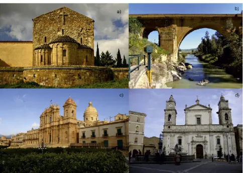
Figure 1.4 - The use of Sabucina Stone in some representative monuments of southern Italy. (a) S. Spirito Church; Caltanissetta (1151); (b) Capodarso bridge (1556); (c) Noto Cathedral (1703-2007); (d)
Caltanissetta Cathedral. 13
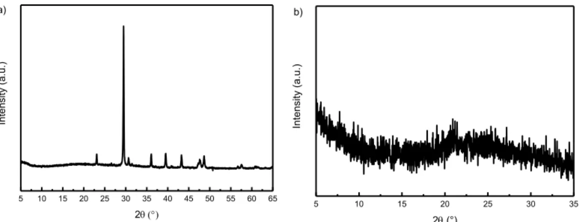
Figure 1.5 - Microphotographs of Sabucina Stone. All, allochems; Sp, sparite; Mc, micrite; Inter_v, inter-particle voids; Intra_v: intra inter-particle voids. Intra_v, intrainter-particle voids 14 Table 1.1 - Semi-quantitative mineralogical composition of the bulk rock and insoluble residue. Cc = calcite; Dol= dolomite; Qz = quartz; Hm = hematite; Goeth = goethite; C. M. = clay minerals. xxx = abundant; xx = present; x = scarce; tr = traces; – = absent. 14 Figure 1.6 - X-ray diffraction patterns acquired on (a) randomly oriented powders and (b) on insoluble
residue of bulk rock oriented slides 15
Figure 1.7 - SEM images of Sabucina Stone. Textural features analyzed are shown. Mc = micrite; Sp = sparite; Dol = dolomite; FeTiO3 = iron titanium oxide; FeS2 = iron sulfide oxide. 15
Table 1.2 - Average values and standard deviations of the chemical composition of the principal textural and petrographic component of the studied stone are reported. Values are assessed on almost three point
analyses. 16
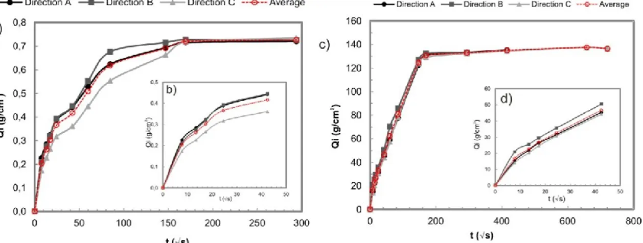
Figure 1.8 - Water absorption by capillarity: (a) curves related to the three investigated directions and average curve in 4 cm cubic samples and (b) absorption in the first 30 minutes. (c) Curves related to the three investigated directions and average curve in 7 cm cubic samples and (d) absorption in the first 30
minutes. 18
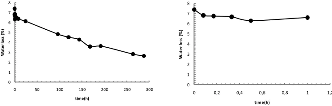
Figure 1.9 - Water absorption by total immersion: averages curve referred to the whole duration of the test and water absorption in the first 30 minutes for cubic samples of 4 cm (a, b) and 7 cm (c, d). 19 Figure 1.10 - Curves of the amount of water loss Qi plotted as a function of time (in hours) 21 Figure 1.11 - Average curve of weight loss percentage during crystallization cycles and picture of sample at the end of the test in 7 cm (a, b) and 4 cm (c, d) cubic samples, respectively. 22 Table 1.5 - Physical and mechanical proprieties determined through laboratory tests on fresh samples of Sabucina Stone with indication of samples ID, average values and standard deviation (St. Dev). 25
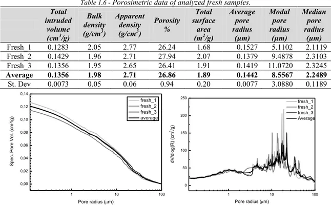
Table 1.6 - Porosimetric data of analyzed fresh samples. 26
Figure 1.12 - (a) cumulative pore volume (cm3/g) vs. pore radius (µ) and (b) pore size distribution
dV/dlog(R) (cm3/g) vs. pore radius (µ) curves of fresh samples. Measurements have been performed on three samples (gray scale curves); the average curve is shown in black. 26 Figure 2.1 - Schematic model of intrusion process for a non-wetting liquid 33 Figure 2.2 - Schematic representation of nuclei in an external magnetic field B0 38
Figure 2.3 - Schematic representation of the magnetization vector M0 39
ix
Figure 2.5 - Schematic representation of a NMR set-up 40
Figure 2.6 - Magnetization of nuclei in external magnetic field B0 41
Figure 2.7 - Inversion recovery sequence 42
Figure 2.8 - Saturation recovery sequence 42
Figure 2.9 - Hahn spin echoes sequence 43
Figure 2.10 - CPMG pulse sequence 43
Figure 2.11 - Multi-exponential fit analysis finalized to transform the echo signal in T2 distribution in
function of time 44
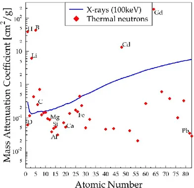
Figure 2.12 - Relation between T2 distribution and pore size in saturated porous media 45 Figure 2.13 - Schematic representation of portable NMR Mouse 46 Figure 2.14 - Dependence between mass attenuation coefficient and atomic number for neutrons and
X-rays 47
Figure 2.15 - The principle of the radiography system: a surface detector fixed behind the sample records, the radiation emitted by the source, thus revealing the weakening effect within the sample; from
50 48
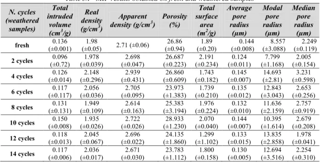
Figure 2.16 - The two images on the left are radiographs of the reinforced concrete sample photographed on the right. The blue and green images are tomograms; from51 48 Figure 2.17 - Representation of some profile geometrical elements 52 Figure 3.1 - (a) Cumulative pore volume vs. pore size and (b) pore size distribution curves (dV/dlog(R) vs. pore size) collected for fresh (in red) and weathered samples (gray scale) 61 Table 3.1 - MIP results obtained on fresh and weathered samples 62 Figure 3.2 - Weathering mechanism due to salts crystallization and porosimetric curves modified from
Angeli et al., 20071 62
Figure 3.3- (a)Cumulative pore volume vs. pore size and (b) pore size distribution curves (dV/dlog(R) vs. pore size) collected for fresh (in red) and cleaned samples (gray scale) 63 Table 3.2 - MIP results obtained on fresh and cleaned samples 63 Figure 3.4 - Comparison between fresh (red), weathered (gray) and cleaned (black) samples. 64 Figure 3.5 - Single-sided NMR sensor by RWTH Aachen University 65 Figure 3.6 - The equation shows the relation between pore distribution and transverse relaxation time, where T2 is the transverse relaxation time resulted from surface interactions, 2 is a constant
representing the transverse relaxation strength and S/V refers to the surface to volume ratio of the
analyzed solids. 66
Table 3.3 - T2 values and populations density Wi for each relaxation time components for fresh sample at
the different depths investigated 67
Figure 3.7 - T2 relaxation time distributions obtained at the different depths (400, 700 and 1000 µm) for
unweathered sample 68
Table 3.4 - T2 values and populations density Wi for each relaxation time components for sample subject
to VIII salt crystallization cycles at the different depths investigated 68 Figure 3.8 - T2 relaxation time distributions obtained at the different depths (400, 700 and 1000 µm) for
x
Table 3.5 - T2 values and populations density Wi for each relaxation time components for subject to XIV
salt crystallization cycles at the different depths investigated 69 Figure 3.9 - T2 relaxation time distributions obtained at the different depths (400, 700 and 1000 µm) for
sample subjected to XIV crystallization cycles. 70
Table 3.6 - T2 values and populations density W for each relaxation time components for fresh and
weathered samples. In bold the most significant variation attributable to weathering action. 70 Figure 3.10 - Average T2 relaxation time distributions for fresh and artificially weathered samples 71
Figure 3.11- Comparison between NMR and MIP data obtained on fresh and artificially weathered samples subjected to eight and fourteen salt crystallization cycles according to UNI EN 12370. 72 Table 3.7 - Free water absorption at atmospheric pressure and under vacuum, interconnection between the pores and saturation parameters obtained for fresh and weathered samples 73 Figure 3.12 - Variation of average values of (a) real density (g/cm3); (b) porosity (%); (c) ultrasonic velocity (m/s) and (d) UCS (MPa) parameters in function of weathering cycles. 75 Table 3.8 - Average values of the main physical-mechanical parameters of Sabucina Stone at different
salt weathering cycles. 75
Figure 3.13 - (a)Drawing of fractures on 2D-reconstructed slices by using NeuronJ; cracks have been indicated with red lines (b) Rose diagram for analyzed samples, representing the frequency of fractures in
a given orientation. 78
Table 3.9 - Features of fractures in analyzed samples. 78
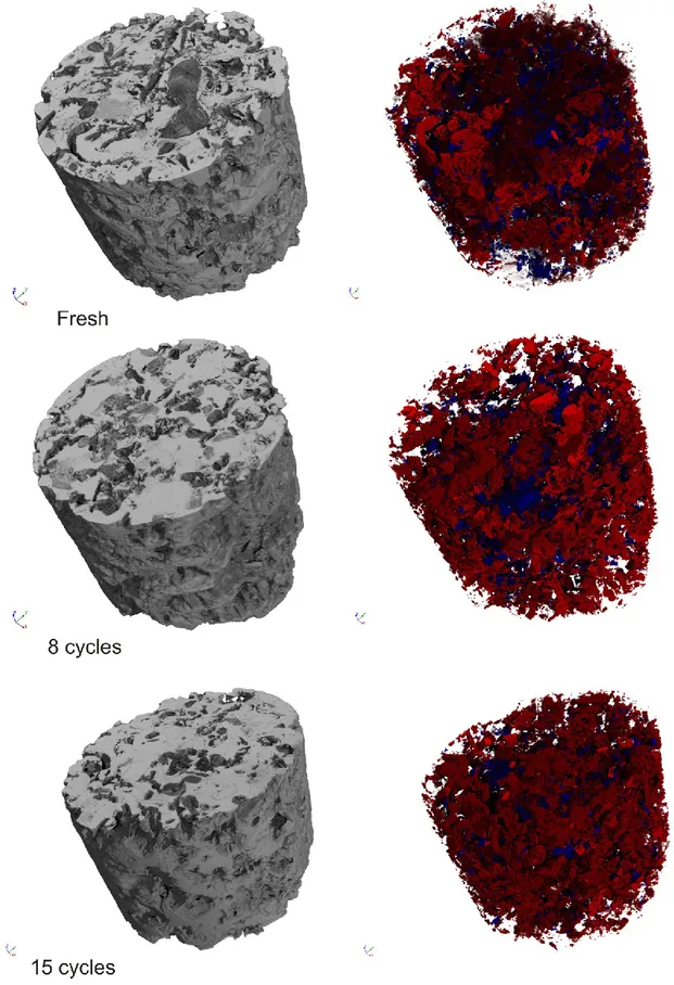
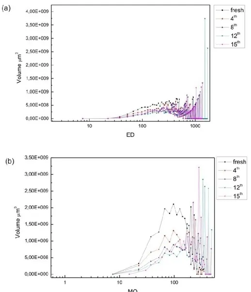
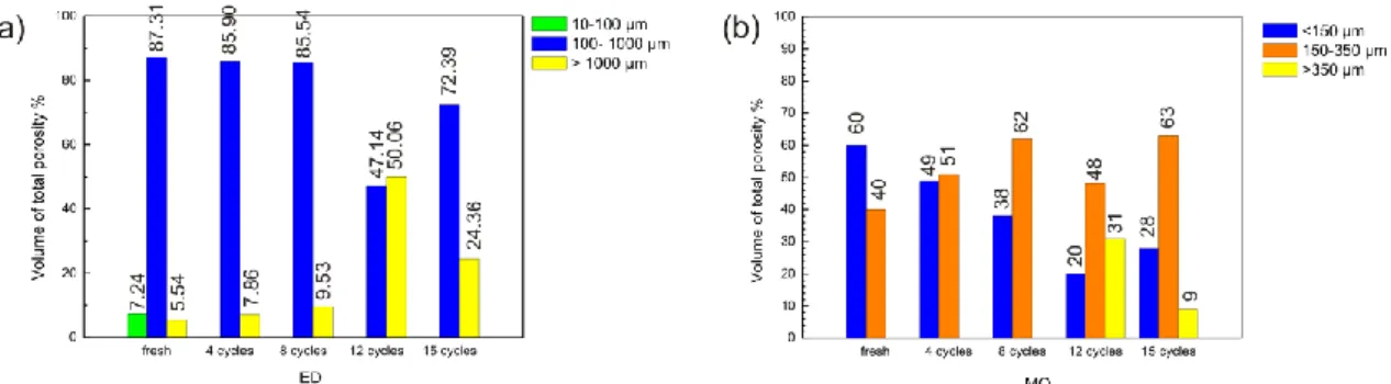
Figure 3.14 - Changes in open, closed and total porosity for fresh and weathered samples. 79 Figure 3.15 - 3D reconstruction images of the bulk (left) and pore structure (right). Closed porosity (blue) and open porosity (red) network are shown. Diameter sample size is about 7 mm. 80 Figure 3.16 - Pore volume vs. equivalent diameter ED (a) and maximum opening OM (b) curves as a function of the number of salt weathering cycles. Step intervals 2 voxels (i.e. 15 µm). 81 Figure 3.17 - Volume fraction (%) of ranges in equivalent diameter (a) and in maximum opening (b) with
respect to the total open porosity. 82
Figure 3.18 3D volume reconstruction of Sabucina Stone. Equivalent diameter and maximum diameter are visualized in function of porosity ranges and crystallization cycles. (a) Green, blue and yellow colors represent pores with ED values of 10–100 µm, 100–1000 µm, and 1000 µm, respectively. (b) Blue, orange and yellow colors represents pores with MO <150 µm, 150–350 µm, and > 350 µm, respectively.
83 Figure 3.19 - Variation of sphericity as a function of the number of salt-weathering cycles. 84
Table 3.10 - Neutron imaging experimental parameters 86
Figure 3.20 - From Kim et al., 201272, as example of image processing 87
Figure 3.21 - Water movement inside samples as examples of un-weathered and weathered Sabucina
stone. 89
Table 3.11 - Penetration coefficients calculated for representative samples subjected to different
weathering degree 90
Figure 3.22 - Position of the wetting front as a function of the square root of time for representative
xi
Figure 3.23 - Water content distribution (WC%) obtained from neutron radiography images in function
of time and degradation degree. 92
Figure 3.24 Amount of water absorbed per area in function of time during the first 30 minutes of water absorption by capillarity for unweathered and weathered samples at different degradation degree 94
Table 3.12 - Report of NORMAL 11/88 recommendation test 94
Figure 3.25 - 3D reconstruction images of the bulk (gray), pore structure (red) and water flow
(blue).Diameter sample size about 2 cm. 96
Figure 3.26 - Roughness maps and surface plots of unweathered and weathered stone surfaces 100 Table 3.13 - Roughness parameters (µm ) obtained by applying a Gaussian filter with SurfCharJ plug-in
in ImageJ 101
Figure 3.27 - Correlation between roughness parameters and weathering cycles 101 Figure 3.28 - Waviness maps and surface plots of unweathered and weathered stone surfaces 102 Table 3.14 - Waviness parameters (µm )obtained by applying a Gaussian filter with SurfCharJ plug-in in
ImageJ 103
Figure 3.29 - Correlation between waviness parameters and weathering cycles 103 Figure 3.30 - Surface roughness and waviness profiles in function of weathering cycles 104 Table 4.1 - Slopes and fractal dimension calculated for fresh, weathered and cleaned samples 114 Figure 4.1 - Logarithmic derivative intrusion volume vs. logarithmic pressure plot for fresh weathered
and cleaned samples. 114
Figure 4.2 - Correlation between fractal dimension as obtained by mercury intrusion porosimetry and
weathering cycles. 115
Table 5.1 - Chromatic coordinates a*, b* and L* collected on untreated and treated samples 121
Table 5.2 - Color difference ΔE values for each treatment 122
Figure 5.1 - Normalized chromatic changes ΔC (chrome) of the stained at 0.01 M as a function of the irradiation time on Sabucina stone samples of (a) MeO with the UV source, (b) MeO with Daylight source, (c) MB with the UV source and (d) MB with Daylight source 123 Table 5.3 - Water absorption coefficient (AC) by capillarity estimated for untreated and treated samples
124 Figure 5.2 - Capillarity water absorption curves (in g/cm2 units) of Sabucina stone samples before and
after the application of TiO2 coatings. 124
Table 5.4 - Water absorption coefficient (CI%) by total immersion estimated for untreated and treated
samples 124
Figure 5.3 - Water absorption curves by total immersion of Sabucina stone samples before and after the
application of TiO2 coatings. 124
Table 5.5 - Percentage mass difference during the 15 crystallization cycles of coated and uncoated Sabucina stone samples. Average values on fifteen samples are reported. 125 Figure 5.4 - Percentage mass difference curves during the 15 crystallization cycles of coated and uncoated Sabucina stone samples. Average curves on fifteen samples are reported. 125 Figure 5.5 - SEM images taken on (a) TiAcN and (b) TiMaA coated Sabucina Stone sample surfaces 125 Figure 5.6 - Picture of water drops on WS3 coated Sabucina stone sample 126
xii
Table 5.6 - Color difference ΔE values for each treatment (PAASi and PAASi+WS3) 127 Table 5.7 - Data report of the water absorption by capillarity test carried out on PAASi treated and
untreated Sabucina Stone samples 128
Figure 5.8 - Water absorption by capillarity average curves (a) referred to the whole duration of test and (b) in the first 30 minutes for untreated and treated Sabucina stone samples with PAASi and PAASi+WS3
129 Table 5.8 - Water absorption coefficient (CI%) estimated for untreated and treated samples (PAASi and
PAASi+WS3) after 72h of total immersion 129
Figure 5.9 - Water absorption curves by total immersion of Sabucina stone samples before and after the
application of PAASi and PAASi+WS3 treatments 129
Table 5.9 - Data report of dry index measurement test on untreated and PAASi treated Sabucina samples 130 Figure 5.10 - Curves of the amount of water loss Qi plotted as a function of time (in hours) for untreated
and PAASi treated samples of Sabucina stone 130
Table 5.10 - Percentage mass difference during the 15 crystallization cycles of PAASi treated and untreated Sabucina stone samples. Average values on fifteen samples are reported. 131 Figure 5.11 - Percentage mass difference curves during the 15 crystallization cycles of PAASi treated and untreated Sabucina stone samples. Average curves on fifteen samples are reported. 131 Figure 5.12 - Pictorial sketch is reported showing the main reactions involved in the sol formation. 131 Table 5.11 - Color difference ΔE values for each treatment (AlSiX and AlSiX+WS3) 132 Table 5.12 - Data report of the water absorption by capillarity test carried out on AlSiX treated and
untreated Sabucina Stone samples 132
Figure 5.13 - Water absorption by capillarity average curves (a) referred to the whole duration of test and (b) in the first 30 minutes for untreated and treated Sabucina stone samples with AlSiX and
AlSiX+WS3 133
Table 5.13 - Water absorption coefficient (CI%) estimated for untreated and treated samples after 72 h of
total immersion 133
Figure 5.14 - Water absorption curves by total immersion of Sabucina stone samples before and after the
application of AlSiX and AlSiX +WS3 treatments 133
Table 5.14 - Data report of dry index measurement test on untreated and AlSiX treated Sabucina samples 134 Figure 5.15 Curves of the amount of water loss Qi plotted as a function of time (in hours) for untreated
and AlSiX treated samples of Sabucina stone 134
Table 5.15 - Percentage mass difference during the 15 crystallization cycles of AlSiX treated and untreated Sabucina stone samples. Average values on fifteen samples are reported. 135 Figure 5.16 - Percentage mass difference curves during the 15 crystallization cycles of AlSiX treated and untreated Sabucina stone samples. Average curves on fifteen samples are reported. 135 Figure 5.17 Final report of the comparative water absorption by capillarity and total immersion, drying and durability tests performed on untreated and treated samples with both consolidants and hydrophobic.
xiii
Table 6.1 - Porosimetric parameters obtained on untreated (NT) and treated samples with AlSiX product 140 Table 6.2 - Porosimetric parameters obtained on untreated (NT) and treated samples with PAASi product
141 Figure 6.1 - Variation of porosimetric parameters in function of treatment applied 141 Figure 6.2 - (a)Cumulative pore volume vs. pore size and (b) pore size distribution curves (dV/dlog(R) vs. pore size) collected for untreated (in black) and treated AlSiX (gray) samples. 142 Figure 6.3 - (a)Cumulative pore volume vs. pore size and (b) pore size distribution curves (dV/dlog(R) vs. pore size) collected for untreated (in black) and treated PAASi (gray) samples. 142 Figure 6.4 - Changes in open, closed and total porosity for fresh and treated samples (with AlSiX
consolidant). 144
Figure 6.5 - Pore volume vs. equivalent diameter ED (a) and maximum opening OM (b) curves as a function of the AlSiX treatment. Step intervals 2 voxels (i.e. 15 µm). 144 Figure 6.6 - 3D reconstruction images of the bulk (in gray) and AlSiX product distribution (red).
Diameter sample size about 7 mm. 145
Figure 6.7 - Changes in open, closed and total porosity for fresh and treated samples (with PAASi
consolidant). 146
Figure 6.8 - Pore volume vs. equivalent diameter ED (a) and maximum opening OM (b) curves as a function of the PAASi treatment. Step intervals 2 voxels (i.e. 15 µm). 146 Figure 6.9 - 3D reconstruction images of the bulk (in gray) and PAASi product distribution (green).
Diameter sample size about 7 mm. 147
Figure 6.10 - Comparison between ED and MO parameter ranges in treated and untreated samples. 148 Figure 6.11 - Radiographs of samples representative of each treatment applied 150 Figure 6.12 - Sequence of radiographs taken at different time intervals for samples treated with
consolidant and protective products 151
Figure 6.13 - Position of the wetting front as a function of the square root of time for representative
samples treated with consolidant and protective products 152
Table 6.3 - Penetration coefficients calculated for untreated and treated samples 152 Figure 6.14 - Water content percentage (WC%) contour plots for untreated and treated samples over the
monitoring time 154
Table 6.4 - Report of NORMAL 11/88 recommendation test 155
Figure 6.15 - Amount of water absorbed per area in function of time during the first 30 minutes of water absorption by capillarity for untreated and treated samples with consolidant AlSiX and PAASi. 155
Table 6.5 - Report of NORMAL 11/88 recommendation test 156
Figure 6.16 - Amount of water absorbed per area in function of time during the first 30 minutes of water absorption by capillarity for untreated and treated samples with the hydrophobic product WS3 156 Table 6.6 - Roughness parameters (µm )obtained by applying a Gaussian filter with SurfCharJ plug-in in
ImageJ 158
Table 6.7 - Waviness parameters (µm) obtained by applying a Gaussian filter with SurfCharJ plug-in in
ImageJ 158
xiv
Figure 6.18 - Waviness maps and surface plots of untreated and treated stone surfaces 160 Figure 6.19 - Comparison between Ra/Wa, Rq/Wq and Rt/Wt values for uncoated and coated surfaces and
xv SUMMARY ABSTRACT I RIASSUNTO II RESUMÈ IV ACKNOWLEDGMENTS VI
LIST OF FIGURES AND TABLES VIII
INTRODUCTION 1
Research highlights 1
Outlines 2
Bibliography 3
CHAPTER 1. CHARACTERIZATION OF POROUS STRUCTURE AND SURFACE FEATURES OF NATURAL BUILDING STONES: THE CASE OF SABUCINA STONE
(SICILY, ITALY) 5
1.1 Natural Building Stones 5
1.1.1 Limestone and sandstone as building stones: the case of Noto Valley (Sicily) 5
1.1.2 Complex porous structure in building stones 6
1.1.3 Fractal geometry in natural building stones 6
1.1.4 Weathering process: the role of salts crystallization in masonry decay 8
1.2 Sabucina Stone 11
1.2.1 Geological framework, quarries and use of Sabucina Stone as building material 11 1.2.2 Petrographic, mineralogical and chemical proprieties 13
1.2.2.1 Optical microscopy 13
1.2.2.2 X-Ray diffraction on insoluble residue 14
1.2.2.3 Single phases chemical composition 15
1.2.2.4 Non-destructive chemical analyses by using portable X-Ray fluorescence 16
1.2.3 Physical proprieties 17
1.2.3.1 Water absorption by capillarity 17
1.2.3.2 Water absorption by total immersion 18
xvi
1.2.3.4 Resistance to salts crystallization 21
1.2.4 Ultrasonic test, mechanical proprieties and density 22
1.2.4.1 Ultrasonic test 22
1.2.4.2 Compressive strength 23
1.2.4.3 Flexural strength 23
1.2.4.4 Apparent and real density and porosity 23
1.2.5 Mercury intrusion porosimetry (MIP) test 25
1.3 Discussion and conclusions 26
1.4 Bibliography 27
CHAPTER 2. METHODS AND MODELS FOR INVESTIGATING THE
IMPACT OF WEATHERING IN THE POROUS SYSTEM OF BUILDING STONES 31
2.1 Introduction 31
2.2 Mercury Intrusion Porosimetry 32
2.2.1 Theoretical background 33
2.2.2 MIP measurement 34
1.2.1 Pore volume and pore size distribution 34
1.2.2 Density 35 1.2.3 Surface area 35 1.2.4 Compressibility 35 1.2.5 Tortuosity 36 1.2.6 Permeability 36 1.2.7 Fractal dimension 36
2.3 Nuclear Magnetic Resonance (NMR) 37
2.3.1 Theoretical background 37
2.3.1.1 Longitudinal relaxation time T1 40
2.3.1.2 Transversal relaxation time T2 42
2.3.1.3 NMR in porous media 44
2.3.1.4 Unilateral portable devices for material science studies 45
2.4 X-ray and neutron imaging 46
2.5 Surface analysis 50
xvii
CHAPTER 3. QUANTIFICATION AND VISUALIZATION OF PORE
STRUCTURE, SURFACE TEXTURE AND PHYSICAL-MECHANICAL FUTURES
CHANGES DUE TO SALT WEATHERING 60
3.1 Pore structure modification due to salt weathering 60
3.2 Pore network changes observed by Mercury Intrusion Porosimetry 61
3.2.1 Weathered samples: degradation mechanism 61
3.2.2 Cleaned samples: location of salts 63
3.2.3 Final remarks 64
3.3 Nuclear Magnetic Resonance (NMR) application 64
3.3.1 Transverse relaxation times and distributions 66
3.3.2 Final remarks 71
3.4 Changes in physical and mechanical proprieties 72
3.4.1 Physical proprieties changes after salt weathering 72 3.4.2 Mechanical proprieties changes after salt weathering 73
3.4.3 Final remarks 75
3.5 Pore structure changes quantified by using X-ray computed µ-CT28 76
3.5.1 2D image analysis 77
3.5.2 Open, closed and total porosity 78
3.5.3 Equivalent diameter and maximum opening 81
3.5.4 Micro-cracks estimation by using sphericity 83
3.5.5 Final remarks 84
3.6 Visualization and quantification of weathering effects and water uptake process by using
neutron imaging 84
3.6.1 Neutron images of water capillary absorption: the effects of weathering on the behavior of the
stone against water 88
3.6.2 Quantification of water content: the fluid-flow patterns inside the degraded stone 91 3.6.3 3D visualization of water movement by neutron tomography 95
3.6.4 Final remarks 96
3.7 Surface modifications due to salt weathering 97
3.7.1 Roughness and waviness analysis 98
3.7.2 Final remarks 103
3.8 Discussion and Conclusions 104
xviii
CHAPTER 4. USE OF FRACTAL MODELS TO DESCRIBE PORE STRUCTURE
OF BUILDING STONES 113
4.1 Fractal geometry of stone pore surface 113
4.2 Fractal dimension as weathering descriptor of stone pore surface 113
4.3 Bibliography 115
CHAPTER 5. INNOVATIVE CONSOLIDANT AND PROTECTIVE
TREATMENTS FOR CALCARENITE SUBSTRATE: EFFICIENCY TESTS 117
5.1 Protective and consolidant treatment for protection and conservation of natural building stones 117
5.1.1 Sol-gel process 117
5.1.2 Titanium nanoparticles based products 118
5.1.3 Hybrids organic-inorganic products 120
5.2 Protective treatments 120
5.2.1 Self-cleaning photocatalytic products based on TiO2 nanoparticles 120
5.2.1.1 Synthesis via sol-gel and application of TiO2 nanoparticles 120
5.2.1.2 Efficiency test 121
5.2.2 Colloidal silicon-based hydrophobic treatment (WS3) 126
5.2.2.1 Synthesis via sol-gel and application of WS3 126
5.2.2.2 Efficiency tests 126
5.3 Hybrid organic-inorganic consolidant treatments 126
5.3.1 Polyammidoammine with silicon alkoxide functions (PAASi) 126
5.3.1.1 Synthesis and application of PAASi 127
5.3.1.2 Efficiency test 127
5.3.2 Silicon alkoxide with epoxidic functional group and aluminium oxide (AlSiX) 131
5.3.2.1 Synthesis and application 131
5.3.2.2 Efficiency test 132
5.4 Discussion and Conclusions 135
5.5 Bibliography 137
CHAPTER 6. QUANTIFICATION OF PORE STRUCTURE AND SURFACE
TEXTURE MODIFICATIONS DUE TO PROTECTIVE AND CONSOLIDANT
TREATMENTS 139
xix
6.2 Pore structure modifications due to consolidant treatments 140
6.2.1 Porosimetric test by Mercury Intrusion porosimetry (MIP) 140
6.2.1.1 Results 140
6.2.1.2 Final remarks 142
6.2.2 Determination of the impregnation depth of consolidants by X-ray µ-CT 143
6.2.2.1 AlSiX product 143
6.2.2.2 PAASi product 145
6.2.2.3 Final remarks 147
6.2.3 Visualization of water repellents and consolidants in building stones and monitor water uptake
by neutron radiography 148
6.2.3.1 Visualization of treatments and water capillary uptake monitoring 149 6.2.3.2 Quantification of changes in capillary absorption proprieties 152
6.2.4 Final remarks 156
6.3 Surface modification due to protective treatments 157
6.4 Discussion and conclusions 161
6.5 Bibliography 163
CHAPTER 7. FINAL REMARKS 165
CHAPTER 8. BIOGRAPHIC NOTES 171
8.1 Scientific production 171
8.1.1 Journal 171
8.1.2 Books 173
8.1.3 Extended abstracts 173
1
Introduction
Research highlights
The durability of natural building stones when they are subjected to weathering processes is a current issue in geological, engineering and conservation fields in the perspective of preserving Cultural Heritage. The esteem of this parameter is strongly related to several features of rocks, namely mineralogical, petrographic, chemical, textural and physical-mechanical proprieties, including pore structure1.
Among the previous ones, the description of the pore structure is one of the more difficult to obtain, because of the variability of the size distribution of voids, ranging from nanometers to millimeters, which is generally not accessible by a single methodology. For this reason, a multi-technique approach is often required for obtaining a complete model of internal structure of building stones that usually exhibits complex pore geometries2.
Moreover, as the great influence of porosity on durability, the quantification of the eventually changes in pore structure due to degradation processes represents a key element in the correct interpretation of stone behavior as well as in the correct use of conservative treatments in the framework of restoration works3. Finally, as degradation processes greatly affect not only the
inner structure of building stones but also their surface, the analysis of surface texture changes due to weathering represents an additional tool in understanding these processes.
In the recent literature, durability of natural stones is usually esteemed by using parameters based on physical, mechanical and porosimetric data4; additionally, useful information can be
obtained by accelerated aging tests such as frost resistance (EN 12371), resistance to ageing by SO2 action in the presence of humidity (EN 13919), resistance to ageing by thermal shock (EN
14066), resistance to ageing by salt mist (EN 14147) and by sodium sulphate solutions (UNI EN 12370). However, during accelerated aging tests, the evaluation of behavior of stones is mainly based on visual inspection and only some quantitative analysis on inner micro-structural modification are available in a non-destructive way, for example by using sound propagation tests5. Furthermore, although the origin of the extensive damage caused by some weathering
agents is widely inspected (for example in the case of salt crystallization6), the comprehension
of how the complex porous network of a stone is modified from these processes is actually incomplete. Usually, in order to characterize pore structure of stone materials, classical approach by using intrusion methods (e.g., mercury intrusion porosimetric) are used; these methods allow to investigate a great number of features such as pore size distribution, total pore volume or porosity, skeletal and apparent density, specific surface of samples7-8. However,
intrusion methods exhibits numerous limits due to assumption made in the process.
In recent years, the use of innovative, non-invasive and non-destructive technique able to characterize both sub-surface and surface features of porous materials has largely increased,
2
especially in stone conservation studies. Among them, neutron and X-Ray imaging methods have been proved to be really valuable procedure for the characterization of archaeological, geological and industrial materials9-11. The results of the imaging analyses are, in fact, not
limited to a simple 2D or 3D representation of the studied objects, as a large amount of quantitative information can be also obtained; in the field of geomaterial, for example, by analyzing and processing radiographies and tomographies aspects as porosity, water movement in porous structure, degradation effects, amount and distribution of degradation products, penetration depth of protective and consolidant products can be extensively inspected. In the framework of non-destructive and non-invasive techniques able to characterize the pore structure of stones, NMR (i.e. Nuclear Magnetic Resonance) applications are widely used to study the petrophysical features of rock materials, especially porosity and permeability 12-13.Beside the investigation of sub-surface modifications, the understanding of the weathering on
exposed surfaces is a key element in building stone conservation studies, as surface represents the direct interface with atmosphere and, therefore, with the weathering agents. In this context, the recent development of non-destructive digital image techniques provides a new tool for measuring changes in stone surfaces in response to weathering, assuring a high resolution and the possibility to monitor the measured parameters over the time14-15.
On the basis of the aforementioned, the main goal of this project consists in applying a multi-methodological approach for the modeling of porosity and surface changes in natural, degraded and treated building stones, for a complete understanding of degradation process due to salt crystallization and the effectiveness of nanostructured and innovative hybrids organic-inorganic products for their preservation and conservation. The research aims also to validate the use of different methods and models in studying porous materials, especially in the framework of innovative, non-invasive and non-destructive techniques.
In detail, this empirical approach has been applied on a Sicilian coarse grained calcarenite known as Pietra di Sabucina, widely used as building and replacement stone16 and particularly
suitable for this study, as characterized by a wide range of pores and a complex porous structure.
Outlines
This dissertation consists in seven parts in which are proposed four main research topics, i.e. a) characterization of porous structure and surface features of natural building stones; b) 2d and 3d models and methods for complex porosity and pore geometry; c) innovative consolidant and protective treatment for calcarenite substrate; d) quantification of pore structure, surface texture, physical-mechanical proprieties changes due to degradation processes and conservative treatments, by using a multi-methodological approach.
3
First of all, an introduction section devoted to coarse grained calcarenite used as natural building stones and a focus on Sabucina Stone is proposed (Chapter 1); in particular, the main features that affect the behavior of the stones when they are used in masonry are investigated in order to understand the influence of microtextural, microstructural, physical-mechanical and especially porosimetric proprieties in degradation processes. Finally, a complete characterization of studied stone is supplied. Classical destructive (i.e. mercury intrusion porosimetry) and innovative non invasive methods (i.e. X-ray tomography, neutron radiography and tomography, nuclear magnetic resonance, digital surface analysis) for the 2D and 3D characterization of complex porosity and pore geometry are summarized in Chapter 2. In Chapter 3 the results obtained by comparative standard tests, mercury intrusion porosimetry (MIP), X-Ray and neutron imaging, nuclear magnetic resonance (NMR) and 3D digital microscope analyses carried out on fresh and artificially weathered samples are discussed, with the aim to quantify changes in physical and mechanical proprieties as well as surface and sub-surface modification of the studied stone due to salt crystallization action. On the basis of experimental numerical data, geometrical models (i.e. fractal models) are applied in order to investigate the potential of fractal dimension in studying and describing complex pore networks (Chapter 4). Finally, the obtained results on Sabucina stone are employed for planning conservative treatments devoted to coarse grained calcarenite substrate. For aforementioned, a complete overview on innovative protective and consolidant products based on sol-gel process is proposed in Chapter 5. In detail, the efficiency of self-cleaning photocatalytic nanoparticles TiO2-based surface coatings and hybrids organic-inorganic products are evaluated by standard
test on treated samples of Sabucina Stone. Moreover, pore structure and surface modifications of studied stone due to treatments are investigated in Chapter 6 by using porosimetric, X-ray and neutron imaging and 3D digital microscope analysis techniques. All data acquired are discussed in Chapter 7, highlighting the complementary of the applied methodologies and models in studying complex pore geometries in natural building stones.
Bibliography
[1] Anania L, Badalà A, Barone G, Belfiore C, Calabrò C, Mazzoleni P, Pezzino A. The stones in monumental masonry buildings of the ―Val di Noto‖ area: New data on the relationships between petrographic characters and physical-mechanical properties. Construction and Building Materials, 2012. 33:122-132.
[2] Barone G, Crupi V, Longo F, Majolino D, Mazzoleni P, Raneri S, Teixeira J, Venuti V. A multi-technique approach for the determination of the porous structure of building stone. European Journal of Mineralogy, 2014. 26:189-198.
4
[3] Bergamonti L, Alfieri I, Lorenzi L, Montenero A, Predieri G, Barone G, Mazzoleni P, Pasquale S, Lottici PP. Nanocrystalline TiO2 by sol-gel: characterization and photocatalytic activity on Modica and
Comiso stones. Appl. Surf. Sci., 2013. 282: 165-173.
[4] Yu S, Oguchi CT. Role of pore size distribution in salt uptake, damage, and predicting salt susceptibility of eight types of Japanese building stones. Engineering Geology, 2010. 115:236-226. [5] Barbera G, Barone G, Mazzoleni P, Scandurra A. Laboratory measurement of ultrasound velocity during accelerated aging tests: Implication for the determination of limestone durability. Construction and Building Materials, 2012. 36:977–983
[6]Scherer GW. Stress from crystallization of salt. Cement and Concrete Research, 2004. 34:1613–1624. [7]Giesche H. Mercury Porosimetry: A General (Practical) Overview. Part. Part. Syst. Charact. 2006. 23:9–19
[8] Leòn y Leòn CA. New perspectives in mercury porosimetry. Advances in Colloid and Interface Science, 1998. 76-77: 341-372
[9] Cnudde V, Boone M. High-resolution X-ray computed tomography in geosciences: a review of the current technology and applications Earth-Science Reviews, 2013. 123:1-17.
[10] Derluyn H, Griffa M, Mannes D, Jerjen I, Dewanckel J, Vontobel P, Sheppard A, Derome D, Cnudde V, Lehmann E, Carmeliet J. Characterizing saline uptake and salt distributions in porous limestone with neutron radiography and X-ray micro-tomography. Journal of Building Physics, 2013. 36:353-374.
[11] Perfect E, Cheng CL, Kanga M, Bilheux MZ, Lamanna JM, Gragg MJ, Wright DM. Neutron imaging of hydrogen-rich fluids in geomaterials and engineered porous media: A review. Earth-Science Reviews, 2014. 129:120:135.
[12] Capitani D, Di Tullio V, Proietti N. Nuclear magnetic resonance to characterize and monitor cultural heritage. Progress in nuclear magnetic resonance spectroscopy, 2012. 64: 29-69
[13] Proietti N, Capitani D, Di Tullio V. Applications of Nuclear Magnetic Resonance Sensors to Cultural Heritage. Sensors, 2014. 14:6977-6997
[14] Turkington AV, Paradise TR. Sandstone weathering: a century of research and innovation. Geomorphology, 2005. 67: 229–253.
[15] Moses C, Robinson D, Barlow J. Methods for measuring rock surface weathering and erosion: A critical review. Earth-Science Reviews, 2014. 135: 141–161.
[16] Bellanca A, Curcuruto E, Lo Bue S, Neri R. Petrografia, geochimica e riferimenti all‘impiego storico delle calcareniti plioceniche di Sabucina, Sicilia Centrale. Mineralogica et petrographica acta, 1999. 42:210-193.
5
Chapter 1. Characterization of porous structure and surface features
of natural building stones: the case of Sabucina Stone (Sicily, Italy)
1.1 Natural Building Stones
Much of the history of the world‘s civilizations is recorded in stones. Stones can be considered the primary building materials taken from the crust of the Earth; in fact, they have been employed since the earliest times of human history for convenience, endurance, and visualimpact1. Among them, worthy of note are decorative stones, referring with this term to a
group of polishable rocks, widely used in modern buildings and in archeological and monumental artifacts. This definition includes metamorphic, magmatic and sedimentary rocks with particular esthetic qualities, generally indicated in the trade contexts as marbles. Of course, the use of decorative stone in building has to be adapted to the mineralogical, physical and mechanical properties of them, as well as to the different architectural and sculptural issues.
1.1.1 Limestone and sandstone as building stones: the case of Noto Valley (Sicily)
Among sedimentary rocks, calcarenites are the most used building stone in Sicily, especially in the South-East area of the island, in view of their suitable proprieties, as availability, color, easy extractability and good workability. In particular, various typologies of Oligocene-Miocene limestone cropping out in different segments of the Hyblean Plateau and Pliocene sandstones outcropping in Central Sicily have been used over the time in the local masonry, especially in the historical town centers. A large use of these local stones, mainly characterized by white-cream and ochre colors, has be attested during the reconstruction of the Noto Valley cities after the well-known earthquake occurred in 1693. In this framework, the Baroque architectural style flourished and the majority of the new built monuments exhibited standards of beauty devoted to emphasize volumes and voids. The requirements of the new established architectural rules found a good agreement with the easy workability and bright color of these limestones and sandstones, so that all the nowadays appearance of the South-East Sicilian historical town center is characterized by Baroque civil and religious monuments, bestowing the characteristic frame of the Noto Valley. Considering the special cultural and physical significance of Baroque buildings, several monuments are nowadays included to the UNESCO Heritage List. Therefore, in view of preserving these Cultural Heritages, the complete knowledge of the features and the behavior of the building stones employed in local masonry is fundamental for planning appropriate conservation actions.
6
1.1.2 Complex porous structure in building stones
Porosity and pore structure represent relevant features for the conservation of stones materials, as they influence the permeability of liquids transport through them; water and moisture are, in fact, the most relevant agents in buildings and monuments deterioration processes.
Porosity of a rock is defined as the volume of void respect to the total volume. In rocks, porosity generally exhibits a ―dual‖ behavior, as it is possible discriminate a open (or effective) porosity, i.e. accessible to fluid flow, and a closed porosity, i.e. in which fluids or gases are present but in which fluid flow cannot effectively take place. In general, porosity (both accessible and inaccessible) strong affects physical and mechanical behavior of stones; however, for weathering processes, only the open/accessible porosity is of relevant interest as it is able to vehicle the water moisture responsible of frost and salt weathering2.
The effective porosity is usually investigated by means of microscopic observations combined with image analysis and porosimetric techniques based on gas adsorption and mercury intrusion. Nevertheless, these methods appear inadequate for the complete modeling of the microstructure of the rock, since they are destructive, measure only the open porosity and explore a limited dimensional range. Moreover, most of these techniques are based on assumptions about pores shape and dimension, representing an ideal geometric model of them. On the contrary, natural pores exhibit a complex geometrical form. Therefore, the determination of pore size by using these methods often gives back arbitrary results.
For aforementioned, a unique quantitative description of rock micro-architecture is generally very difficult, considering the variability in the dimensional distribution of the voids (ranging over length scales ranging four to five orders of magnitude, i.e. from nanometers to centimeters) and their real complex geometry.
In this framework, the application of mathematical models able to describe complex chaotic systems with an high degree of heterogeneity by using a single numerical value can be considered a useful tool for studying porosity in building stones. Among them, fractal geometry represents one of the most powerful methods.
1.1.3 Fractal geometry in natural building stones
Fractals are real and mathematics objects characterized by non-integer value of dimension. For fractal surfaces, fractal dimension can assume any value between 2 and 3; more it approach to 3, more complex is the surface. In this sense, the fractal dimension (D) describes “how” objects occupy the space.
Assuming a non-planar surface, we want measure the area of this surface determining how many squares of ϵedge are needed. The number of squares N (𝜖) needed to cover the surface are described by the following equation:
𝑁 𝜖 = 𝑘 1 𝜖
2
7
With k = 1, ½, π/4, …, depending on the shape of the non-planar surface. The exponent 2 represents the dimension of the surface, as following:
𝑁 𝜖 = 𝑘 1 𝜖 2 → ln 𝑁 𝜖 = ln 𝑘 + 2 ln1 𝜖 → 2 = 𝑙𝑛𝑁 (𝜖) ln1𝜖 − ln 𝑘 ln1𝜖 (2)
if 𝜖 → 0 , so that the edge of the planar squares tend to zero, ln 𝑘 ln1𝜖 → 0 the equation is:
2 = 𝑙𝑛𝑁 (𝜖)
ln1𝜖 (3)
In general, the previous can be written as:
𝐷 = 𝑙𝑛𝑁 (𝜖)
ln1𝜖 (4)
with D representing the fractal dimension of the surface.
For aforementioned, a fractal surface is characterized by a non integer fractal dimension D that is invariant at any scale used to examine it (i.e., self-similarity).
In conclusion, a fractal object is characterized by non-integer dimension and self-similarity proprieties at any scale of observation.
Stones contain pores with a large radius interval, so they can be associate to a fractal model represented by the Menger sponge3(Figure 1.1), even if a real system exhibits pores only in a
specific dimensional range and fractal behavior is defined in a specific interval between rmax and
rmin pore value.
Figure 1.1 - The iterative structure of Menger sponge
However, fractal geometry has been successfully applied to a great variety of porous solid materials4-7. Among the application of fractal geometry on complex pore systems, several
researches demonstrate the fractal behavior of pores in stones8-10, also for the prediction of
physical proprieties as permeability11. Additionally, recently application of fractal geometry on
weathered building stones demonstrates the powerfully of this method in describing the modification of pore system related to degradation processes12,13.
8
1.1.4 Weathering process: the role of salts crystallization in masonry decay
Even if rocks are considered really durable materials, they suffer of degradation processes that affect their physical, mechanical and aesthetical proprieties. Among them, weathering is a common process that interests natural stones and consists in structural and compositional modification of the material due to external factors, implying a worsening of its characteristics from a conservation point of view. The comprehension of weathering processes, in term of external agents, interaction between them, response of the rocks, scale and rate of action, represents therefore an element of great interest not only in conservation studies but also in geomorphology and rocks stability fields. Rock weathering is considered, in fact, one of the most relevant agents able to modify natural and artificial landscapes. For aforementioned, throughout the last centuries, several researchers have been focused on weathering studies, getting to numerous theories and models (e.g. traditional structural theory, climatic theory, single process model, reductionism, etc.14).
Generally speaking, weathering is considered the results of several external agents that can operate independently or simultaneously, determining modifications in morphology, structure, chemical and mineralogical composition and physic features under specific environmental conditions. In this sense, weathering can be expressed by the following general formula that takes in account all the previously in a certain time lap15:
𝐹 = 𝑓 𝐸, 𝑀, 𝑃 𝑑𝑡 (5)
with: F, morphological elements; P, processes operating; E, environmental conditions controlling the processes; M, materials upon which the processes operate over periods of time t. It is quite clear that each of these elements has to be in-depth analyzed and therefore correlated with the other ones in order to have a complete overview on weathering process.
Referring to processes (P), they can occur by different mechanisms and can be distinguished mainly in mechanical and chemical processes. Mechanical damage results when stones are subjected to a stress major than their mechanical resistance; sources of mechanical decay are ascribable to growth of vegetation, thermal stress, hydric swelling and salts growth. Chemical damage processes refer to the dissolution or alteration of the mineral constituents of a stone material by chemical reactions and they mainly consist in mineral dissolution reactions, surface recession, and action of salts. These processes produce in rocks different weathering patterns often due to the interaction of several external agents; therefore, in order to assess the relative importance of different processes and their rates, a detailed understanding of the underlying mechanisms and the interaction between processes is indispensable. In this framework, the interactions among processes can act in three different ways: in synergy, in mutually exclusive way and independently. The comprehension of how these factors affect the weathering process can therefore supply additional information, also considering the different scale at which processes take place (i.e. granular and outcrop/building scale).