Antioxidants 2021, 10, 486. https://doi.org/10.3390/antiox10030486 www.mdpi.com/journal/antioxidants Review
Endothelium as a Source and Target of H
2S to Improve Its
Trophism and Function
Valerio Ciccone, Shirley Genah and Lucia Morbidelli * Department of Life Sciences, University of Siena, Via A. Moro 2, 53100 Siena, Italy; [email protected] (V.C.); [email protected] (S.G.) * Correspondence: [email protected]; Tel.: +39‐0577‐235381 Abstract: The vascular endothelium consists of a single layer of squamous endothelial cells (ECs) lining the inner surface of blood vessels. Nowadays, it is no longer considered as a simple barrier between the blood and vessel wall, but a central hub to control blood flow homeostasis and fulfill tissue metabolic demands by furnishing oxygen and nutrients. The endothelium regulates the proper functioning of vessels and microcirculation, in terms of tone control, blood fluidity, and fine tuning of inflammatory and redox reactions within the vessel wall and in surrounding tissues. This multiplicity of effects is due to the ability of ECs to produce, process, and release key modulators. Among these, gasotransmitters such as nitric oxide (NO) and hydrogen sulfide (H2S) are very active molecules constitutively produced by endotheliocytes for the maintenance and control of vascular physiological functions, while their impairment is responsible for endothelial dysfunction and car‐ diovascular disorders such as hypertension, atherosclerosis, and impaired wound healing and vas‐ cularization due to diabetes, infections, and ischemia. Upregulation of H2S producing enzymes and administration of H2S donors can be considered as innovative therapeutic approaches to improve EC biology and function, to revert endothelial dysfunction or to prevent cardiovascular disease pro‐ gression. This review will focus on the beneficial autocrine/paracrine properties of H2S on ECs and the state of the art on H2S potentiating drugs and tools. Keywords: vascular endothelium; hydrogen sulfide; endothelial dysfunction; hypertension; atherosclerosis; diabetes; angiogenesis; wound healing; H2S donors 1. Vascular Endothelium The vascular endothelium is the tissue that lines the inside of the circulatory system (blood vessels, lymphatic vessels and heart). The cells, arranged in a single layer oriented on the longitudinal axis of the vessel, assume a flattened shape and lay side by side with each other to form a complete monolayer. Structurally, the endothelial cell (EC) apical domain is in direct contact with blood or lymph, while the basolateral domain anchors to the basal lamina, which connects EC to the underlying tissues, such as the medial or mus‐ cular layer and the adventitia, rich in fibrous tissue.The vascular endothelium acts as a selectively permeable barrier between extravas‐ cular and intravascular compartments and provides a non‐thrombogenic surface for the cardiovascular system [1]. Nowadays, the endothelium can no longer be considered a passive barrier. Indeed, its anatomical position allows it to integrate the physical and neu‐ rohumoral signals from the blood and surrounding tissues for regulating vascular tone and permeability, cell adhesion, inflammation, smooth muscle phenotype and prolifera‐ tion, as well as thromboresistance and blood fluidity [2,3]. The endothelial lining represents a wide area for the exchanges between blood and tissues (about 350 m2 in humans) [3]. Electron microscopy observations reveal the contin‐ uous nature of arterial endothelium, characterized by tight junctions among adjacent cells Citation: Ciccone, V.; Genah, S.; Morbidelli, L. Endothelium as a Source and Target of H2S to Improve Its Trophism and Function. Antioxidants 2021, 10, 486. https:// doi.org/10.3390/antiox10030486 Academic Editor: Thomas C. Resta Received: 16 February 2021 Accepted: 16 March 2021 Published: 19 March 2021 Publisher’s Note: MDPI stays neu‐ tral with regard to jurisdictional claims in published maps and insti‐ tutional affiliations. Copyright: © 2021 by the authors. Li‐ censee MDPI, Basel, Switzerland. This article is an open access article distributed under the terms and con‐ ditions of the Creative Commons At‐ tribution (CC BY) license (http://crea‐ tivecommons.org/licenses/by/4.0/).
in order to limit macromolecule exchange, and by a complex micro‐vesicular system in‐ volved in macromolecular transport. In spite of its apparent morphological lack of com‐ plexity, the endothelium is characterized by heterogeneity, with differences in permeabil‐ ity, reactivity, and biosynthesis in relation to the type of vascular district and organ con‐ sidered [1–3]. 2. Role of Endothelium in Physiology
The endothelium’s role as a semipermeable barrier is one of its fundamental and basic functions: it regulates macromolecule transport between the lumen and vascular smooth muscle tissue [4]. Several mechanisms control the passage of macromolecules across the endothelial barrier: (i) through ECs themselves (transcellular flux); (ii) through the cell–cell junctions (paracellular flux); and (iii) via vesicular transport.
Most biological transmitters consist of large molecules with anionic and hydrophilic features, unable to diffuse across the membrane bilayer. The majority of those transmitters are believed to move through intracellular junctions between cells, or via vesicular transport, thanks to the formation of transient channels resulting from vesicle fusion. The reorganization of the intercellular junctions, which involves actin and myosin reconfigu‐ ration or direct collapse of junctional connections, appears to be the main process by which ECs increase their permeability to solutes and water [1]. The function of the endothelium is not limited to the internal surface lining of vessels or to constitute the vascular wall in the microcirculation, but it produces and releases vas‐ oactive factors such as nitric oxide (NO), prostacyclin (PGI2), hydrogen sulfide (H₂S) and endothelin (ET) which, in the appropriate concentration and balance, maintain adequate vascular tone and blood fluidity, giving the endothelium itself an antithrombotic pheno‐ type [5,6]. The synthesis of NO by ECs is constitutive, but it can be augmented by a wide variety of compounds, including acetylcholine, angiotensin II (AngII), bradykinin, histamine, ar‐ achidonic acid. NO has a very short half‐life and is synthesized from L‐arginine and oxy‐ gen by the NO synthase enzyme (NOS). The endothelial isoform of this enzyme, eNOS, constitutively expressed, appears to be Ca2+/calmodulin‐dependent. Once synthesized,
NO rapidly spreads to vascular smooth muscle cells where it stimulates soluble guanylate cyclase (sGC), with an increase in cGMP formation and consequent vascular relaxation, while its autocrine function is related to the control of EC trophism and angiogenesis [7]. NO is not the only endothelial‐dependent vasodilator. The endothelium also consti‐ tutively generates PGI2, which relaxes the underlying smooth muscle cells by activating adenylate cyclase and increasing cAMP. PGI2 is released in high quantities following the binding of transmitters, such as thrombin, histamine, serotonin, on cell surface receptors. The endothelium also produces and releases a hyperpolarizing factor (EDHF) whose chemical nature is still debated. EDHF’s function is to hyperpolarize vascular smooth muscle cells, causing these cells to relax and allowing dilation of blood vessels [6]. In addition to NO and carbon monoxide (CO), H2S is an endogenous gasotransmitter involved in the regulation of the cardiovascular, nervous, gastrointestinal, and renal sys‐ tems, with a great impact on inflammatory and immune responses [8]. Recently, it has been proposed as one of the EDHFs. H₂S exerts a multitude of physiological effects on the wall of the vessels, acting in an autocrine/paracrine manner. It is produced by vascular cells and exhibits antioxidant, anti‐apoptotic, anti‐inflammatory and vasoactive proper‐ ties. Indeed, it reduces arterial blood pressure, limits the formation of atheromatous plaques, and promotes the vascularization of ischemic/injured tissues [8].
In some pathophysiological circumstances including hypoxia, tissue hypoperfusion or arterial hypertension, some vasoconstrictor factors may be released from the endothe‐ lium as endothelins (ETs), thromboxane A2, and prostaglandin H2. Moreover, vascular endothelium participates at the regulation of vessel tone and trophism and blood flow
through the processing of angiotensin, via the expression of angiotensin converting en‐ zyme isoforms ACE‐1 and ACE‐2, responsible for the balance between AngII/AT‐1 recep‐ tor and Ang(1–7)/Mas receptor [9].
ECs respond to the increase in blood flow through the release of NO and PGI2 by the same cells. Indeed, shear stress causes rapid activation of eNOS and increases its gene transcription; it also endorses ECs to release factors that inhibit coagulation, leukocyte migration, and smooth muscle cell proliferation, simultaneously promoting EC survival. Conversely, low shear stress and turbulent blood flow promote a pathological feature in the endothelium responsible for atherosclerosis ignition, documenting the pivotal role of ECs in finely controlling vascular functions [2,6]. A healthy functioning endothelium also provides protection against radical species of oxygen and nitrogen (ROS/RNS). It is now clear that increased levels of ROS and RNS are harmful to cells and tissues and are involved in a wide range of cardiovascular dis‐ eases having endothelial dysfunction as an underlying phenomenon. This boosted the concept of oxidation as synonymous with cell damage and senescence. The post‐transla‐ tional modifications involving RNS share a common ancestor—high NO concentrations mainly synthetized by inducible NOS (iNOS), upregulated in response to various endo‐ toxin or cytokine signals. Several pathological states are linked to the deregulation of NO levels, indicating that aberrant production of NO and its products can have deleterious consequences on cells [10]. Again, one of the functions related to a healthy endothelium is the scavenging activity of reactive species through the production of antioxidant prod‐ ucts as H2S or protective enzymatic pathways. The intimate surface of a healthy endothelium is both anticoagulant and antithrom‐ botic: ECs secrete a wide range of molecules relevant to the regulation of blood clotting and platelet functions, as PGI2 and NO. Damage to the vessels or exposure to certain cy‐ tokines and proinflammatory stimuli overturns the equilibrium towards a procoagulant and prothrombotic EC phenotype, through the exposure of basal membrane components and/or tissue factor, and reduced presence of glycosaminoglycans or tissue factor inhibi‐ tor [5]. Endothelial trophism is guaranteed by the response to vasoactive and growth factors produced by surrounding tissues or autocrinally by the same ECs. Among the various examples, we and others have contributed to characterizing the beneficial effects on vas‐ cular endothelium by NO derived from eNOS, bradykinin, substance P, vascular endo‐ thelial growth factor (VEGF), fibroblast growth factor‐2 (FGF‐2), prostaglandin E2, H2S
[11–15]. The molecular mechanisms responsible for cell survival, proliferation, migration and functioning include eNOS/NO/cGMP/protein kinase G (PKG), PI‐3K/Akt, MAPK/ERK1/2 and gene transcription of autocrine factors as FGF‐2 [7].
Epigenetics is an emergent mechanism involved in the regulation of vascular biology and endothelial trophism. Through chromatin structure modification, epigenetics can modify endothelial functions with an impact on cardiovascular disease, being the regula‐ tory functions of epigenetics also active on endothelial precursor cells and circulating an‐ giogenic cells [16,17]. DNA methylation, variants, histone post‐translational modifica‐ tions, and recently discovered RNA‐based mechanisms represent the major pathways in‐ volved in the molecular basis of epigenetics. VEGF‐A and NOS are the key players in regulating and maintaining cardiovascular functions. Their expression can be controlled by epigenetic mechanisms. In particular, VEGF‐A epigenetic control can occur mainly through changes in histone code by RNAs. VEGF‐A acts through VEGFR2, which in turn is regulated by promoter DNA methylation [18]. Furthermore, accumulating evidence in‐ dicates that epigenetic pathways play an important role in eNOS gene regulation [19]. These findings suggest the importance to deeply understand the epigenetic mecha‐ nisms involved in the regulation of vascular functions both in physiology and in patho‐ logical conditions.
3. Endothelial Dysfunction
Endothelial dysfunction refers to a systemic condition in which the endothelium loses its physiological properties, including the control of vasodilation, fibrinolysis and platelet aggregation. Key features of the endothelial dysfunction are: (1) the reduced local production of NO due to impaired activity (uncoupling) of eNOS, and of other vasodilat‐ ing mediators as H₂S; (2) the decrease in anticoagulant factors such as heparin; (3) the increase in the secretion of reactive species, von Willebrand factor, and tissue factor; (4) the overexpression of adhesion molecules for leukocytes and platelets [20,21]. All these factors concur to compromise the physiological vascular homeostasis. Due to the impair‐ ment of the main protective transmitters NO and H₂S, the resulting endothelial dysfunc‐ tion is associated with increased ROS and RNS levels and vascular oxidative stress [22,23]. The perpetuation of this condition then leads to retraction and death of the endothelium with increased permeability and exposure of the components of the basement membrane, that further amplifies the picture of vascular inflammation [4,20,22]. Endothelial dysfunction risk factors are represented by pathological states such as hypertension, diabetes, and hyperlipidemia, and improper lifestyles such as high‐fat di‐ ets, tobacco and alcohol consumption, and physical inactivity [24]. Most cardiovascular diseases share endothelial dysfunction as a hallmark: atherosclerosis, diabetes complica‐ tions, thrombosis, and hypercoagulation [20]. Moreover, physiological ageing through the phenomenon of mild chronic inflammation (“inflammaging”) is accompanied by endo‐ thelial dysfunction [25]. Inflammatory factors such as tumor necrosis factor‐α (TNF‐α), inteleukin‐6 (IL‐6), intercellular adhesion molecule 1 (ICAM‐1), and loss of the antioxidant mechanism are among the most influential promoters of vascular impairment [21]. Mounting evidence suggests that epigenetic mechanisms may contribute to vascular complications in many pathological conditions, such as diabetes or atherosclerosis, linked to altered endothelial trophism and functions [17,26]. The inflammatory phenotype in ECs induces the transcription of many cytokines and adhesion molecules, in a nuclear factor‐ kappa B (NF‐κB)‐dependent manner. Epigenetic modifications in the NF‐κB promoter re‐ gion produce an increased expression of p65 subunit of NF‐κB, and a hyper‐activation of the NF‐κB pathway [27]. Another mechanism could involve histone deacetylase 2 (HDAC2), which interacts and deacetylates Nrf2. Oxidized low‐density lipoproteins (ox‐ LDLs) are able to downregulate HDAC2 expression, resulting in increased production of eNOS‐dependent reactive species [16]. Furthermore, shear stress represented by blood flow alteration modifies EC gene expression and function. Dunn et al. demonstrated that disturbed blood flow stimulates DNA methyltransferase‐1 (DNMT‐1) expression in endo‐ thelial cells with aberrant DNA methylation at the promoter of flow‐inducible genes, con‐ tributing to atherosclerosis [28]. Finally, recent data showed the role of non‐coding RNA in regulating the expression of endothelial adhesion molecule [29]. Acute or chronic infections both by bacteria and viruses have cardiovascular conse‐ quences for their direct or indirect effects on vascular endothelium, through bacterial products or cytokines released by tissue and immune system cells [30,31], and through epigenetic regulation [32]. The recent pandemic due to SARS‐CoV‐2 supports this concept [33,34].
Furthermore, our studies and those of others have revealed that endothelial dysfunc‐ tion is associated to impaired EC survival and physiological angiogenic outcomes with subsequent rearrangement of the microcirculation that contributes to the emergence of various pathological conditions and healing disorders [7,35]. ECs play a key role in the adaptation of tissues to damage, revealing their plasticity. A change in endothelial func‐ tions following ischemia can induce the transition to a mesenchymal phenotype charac‐ terized by functional, metabolic and gene expression signatures. Indeed, the mesenchy‐ mal phenotype, with increased cell migration and clonal expansion, participates in regen‐ erating a functioning vascular network [36].
Considering endothelial function as a “barometer for cardiovascular risk”, it is cru‐ cial to identify the molecular determinants underlying endothelial integrity and function‐ ality. Seeing endothelium as an exchange regulator between the vascular wall and sur‐ rounding tissues, it is expected that dysfunctional ECs can determine damage to other tissues [37]. Indeed, a detailed assessment of the cellular and molecular mechanisms at the base of vascular function, and, particularly, of endothelial dysfunction, will help the diagnosis and treatment choice for a broad array of human disorders, including cardio‐ vascular and neurodegenerative diseases [21,24,38–40]. 4. Biochemistry of H₂S Production H2S is a gas physiologically produced by tissue and vascular cells. The enzymes re‐ sponsible for the synthesis of H2S are cystathionine β‐synthase (CBS), cystathionine‐γ‐ly‐ ase (CSE), and 3‐mercaptopyruvate sulfurtransferase (3‐MST). The first two enzymes use L‐cysteine as a substrate and are dependent on pyridoxal‐5′‐phosphate. 3‐MST, on the other hand, works in association with cysteine aminotransferase which, starting from L‐ cysteine and α‐ketoglutarate, produces 3‐mercaptopyruvate. These enzymes are differen‐ tially expressed in the various tissues. In particular, the expression of CSE and 3‐MST pre‐ dominates in the cardiovascular system [8,41]. CSE is only present in the cytoplasm, while CBS and 3‐MST both have a cytosolic and mitochondrial form, with the latter predomi‐ nating. CSE is the principal enzyme responsible for H2S biosynthesis, located in vascular smooth muscle cells and found mainly in ECs [42,43]. This can explain why the concen‐ tration of H2S in the vascular tissues is around 100 times greater than in other tissues [44], suggesting a crucial role in vascular homeostasis, endothelial function, and trophism [45]. In addition, H2S can be also generated via a reduction in thiols and thiol‐containing mol‐ ecules, in a non‐enzymatic manner [8]. Compared to CBS, knockout mice for CSE have no severe phenotype and normal lifespan. Their phenotype is mainly cardiovascular, with hypertension and endothelial dysfunction [46]. Information has become available about the regulation of the expression and activity of these enzymes (Figure 1). It has been reported that NADPHox4 derived ROS (via heme‐ regulated inhibitor kinase/eIF2/activating transcription factor 4 (ATF4) signaling) enhance the expression of CSE [47]. Blood flow has been reported to exert divergent effect of H2S producing enzymes, depending on the type of endothelium and flow stress. While lami‐ nar flow was initially demonstrated to enhance the expression of CSE and 3‐MST in ECs [47,48], recent data are more complex. In particular, laminar flow (high shear stress) has been demonstrated to inhibit CSE expression via KLF2 regulated miRNA‐27b [49], while turbulent flow (low shear stress) seems to upregulate CSE [50,51]. In rat aortic ECs, cal‐ cium‐sensing receptors increase CSE expression in a phospho‐calmodulin kinases II‐de‐ pendent manner to inhibit platelet activation [52]. Transcription factors specifically con‐ trolling CSE expression are among the others NF‐κB in lipopolysaccharide (LPS)‐stimu‐ lated macrophages [53], specificity protein 1 (Sp1) in smooth muscle cells [54] and Elk1 in beta pancreatic cells [55]. Nuclear factor of activated T cell (NFAT) binding sites have been identified in the CSE promoter. Intermittent hypoxia exposure reduces Ca2+‐dependent
activation of calcineurin/NFAT to lower CSE expression and impair vasodilation, while NFAT activation lowers CSE expression at the cell and microvascular levels [56].
Figure 1. Summary of the molecular mechanisms controlling CSE expression and function in endothelial cells (up‐stream
signaling) and of the multiple downstream signaling activated or inhibited by H2S in ECs. Red target or lines means inhi‐ bition. Note that some signals are both up‐ and downstream, strengthening the central role of CSE/H2S in controlling vascular trophism and functions. ACE‐2, angiotensin converting enzyme 2; Akt, protein kinase B; AngII, angiotensin II; ARE, antioxidant responsive elements; ATF4, activating transcription factor 4; BMP4, bone morphogenetic protein 4; cGMP, cyclic guanosine monophosphate; COX‐2, cyclooxygenase‐2; CSE, cystathionine γ‐lyase; Elk1, ETS Like‐1 protein; eNOS, endothelial NO synthase; ERK1/2, extracellular signaling regulated kinase ½; ET‐1, endothelin‐1; HIF‐1a, hypoxia inducible factor‐1a; HuR, human antigen R; ICAM, intercellular adhesion molecule; IL‐1, interleukin 1; IP3, inositol‐3‐ phosphate; IP3R, inositol‐3‐phosphate receptor; KATP, ATP‐sensitive K+ channels; Keap1, Kelch‐like ECH associated pro‐ tein 1, KLF2, Krüppel‐like Factor 2; MAPK, mitogen‐activated protein kinase; MEK1, MAP kinase kinase 1; NFAT, nuclear factor of activated T‐cells; NF‐κB, nuclear factor‐kappa B; NLRP3, nucleotide‐binding oligomerization domain, leucine rich repeat, and pyrin domain containing protein 3; NO, nitric oxide; Nox4, NADPH oxidase 4; Nrf2, nuclear factor erythroid 2‐related factor 2; p38, p38 mitogen‐activated protein kinases; PDE, phosphodiesterase; PI‐3K, phosphoinositide 3‐kinase; PKG, protein kinase G; PLCβ, phospholipase Cβ; PPARδ, peroxisome proliferators‐activated receptor δ; Prx6, thioredoxin‐dependent peroxiredoxin; PTEN, phosphatase and tensin homolog; sGC, soluble guanylate cyclase; SOD, su‐ peroxide dismutase; Sp1, specificity protein 1 transcription factor; STAT3, signal transducer and activator of transcription 3; VCAM, vascular cell adhesion molecule; VEGF, vascular endothelial growth factor; VEGFR2, vascular endothelial growth factor receptor 2.
OxLDL in one of the major stimuli to cause the endothelial damage that leads to ath‐ erosclerosis. One of the epigenetic mechanisms underlying CSE reduced expression at both mRNA and protein levels by oxLDL is increased histone deacetylase 6 (HDAC6) [57]. Recently this finding has been extended to blood pressure control, by assessing the role of tubastain A in AngII induce hypertension, and documenting that upregulation of CSE and H2S through HDAC6 inhibition can be a valid therapeutic strategy [58]. Additional information on CSE gene and enzyme control is provided in the chapters below and is summarized in Figure 1. H2S plasma levels are kept at appropriate concentrations by three elimination sys‐ tems. The first pathway involves mitochondrial oxidative metabolism which converts H2S into thiosulfate, followed by further conversion into sulfate and then sulfite. The second metabolic pathway is cytosolic methylation to dimethyl sulfide via thiol S‐methyltrans‐ ferase. Finally, the binding of H2S to hemoglobin leads to the formation of sulfhemoglobin [45].
5. Molecular Signaling Activated by H₂S into ECs
H2S in ECs performs a protective action on vessels in an autocrine/paracrine manner.
It plays a role in the regulation of vasodilation, angiogenesis, inflammation, oxidative stress and apoptosis [59]. Three are the main mechanisms through which H2S exerts it
biological effect: (i) reactive oxygen species/nitrogen species scavenging; (ii) interaction with metal centers; (iii) persulfidation (called also S‐sulfhydration).
H2S acts through a post‐translational modification—the S‐sulfhydration of cysteine
residues, which modifies the structure and activity of the target proteins [60]. The mech‐ anism is persulfidation on reactive cysteine residues (‐SH) of target proteins to form a persulfide group (‐SSH). An example is the persulfidation of the ATP‐dependent K chan‐ nel (KATP) in ECs and smooth muscle cells, responsible for fast hyperpolarization and vas‐
orelaxation [61]. Indeed, evidence has been provided in support of H₂S function as an EDHF, exerting more remarkable vasorelaxation in the peripheral resistance arteries [62]. H2S also reduces ROS levels through their direct inactivation and by enhancing anti‐ oxidant defense mechanisms. One of the mechanisms underlying oxidative protection is given by the H₂S regulation of the Keap1/Nrf2 pathway. Normally, the transcription factor Nrf2 is inhibited by its binding to Keap1 in the cytoplasm. In conditions of oxidative stress, H2S promotes the translocation of Nrf2 into the nucleus by means of the S‐sulfhydration of the Keap1 inhibitor, causing the dissociation of the Keap1/Nrf2 complex. Nrf2 in the nucleus activates the antioxidant responsive element. Consequently, the transcription of many antioxidant genes such as superoxide dismutase, catalase, glutathione peroxidase and glutathione‐S‐transferase is induced, requiring hours or days to produce a biological effect [63]. Among other targets, H2S decreases inflammation by inhibiting transcription factors such as NF‐κB through persulfidation [64], thus decreasing the expression of pro‐inflam‐ matory mediators. Most persulfidation reactions lead to target inhibition as phosphatase and tensin homolog (PTEN) [65], except for MEK1 activity, which in HUVEC leads to in‐ creased activity with DNA damage repair and senescence impairment [66]. Recently, Prx6 has been identified as a further target of sulfhydration on Cys47, which controls its decamerization and peroxidase activity [49], while the “S‐sulphydrome” was identified among the many target proteins, and β3 integrin was identified as the key element of en‐ dothelial mechanotransduction [67]. The interaction of H₂S with the NO/NOS pathway involves different modalities: as inhibition of PDE in smooth muscle cells, PI‐3K/Akt‐dependent phosphorylation of eNOS in Ser1177 [68] and stabilization of eNOS in the dimeric state through enzyme persulfida‐ tion [69]. Additionally, heme reduction in sGC enzyme with facilitated response to NO [70] and activation of protein kinase G Iα (PKG Iα) through disulfide bond formation [71] have been reported to potentiate the NO/cGMP pathway. H2S also stimulates endothelial proliferation and migration, aiding the process of an‐ giogenesis and wound repair. H2S acts at several levels on the mechanisms responsible for angiogenesis, including the control of VEGF expression, through upregulation of the transcription factor hypoxia inducible factor‐1α (HIF‐1α) or direct modulation of the PI‐ 3K and Akt pathways in ECs (signaling pathways also activated by VEGF) [41,72,73]. In angiogenic ECs, H2S has also been reported to activate signal transducer and activator of transcription 3 (STAT3) [74], mammalian target of rapamycin (mTOR) and the VEGFR2 pathway [75]. It has been reported that CBS silencing in ECs reduces VEGF signaling through VEGFR2 and neuropilin‐1 downregulation [76].
A schematic summary of the molecular mechanisms activated or inhibited by H2S in
ECs is reported in Figure 1. As a note, the effects of H2S are context and tissue‐dependent,
sometimes producing divergent functional effects. It is plausible that this depends on en‐ dothelial heterogeneity, tissue microenvironment, and physio‐pathological conditions where there is the influence of epigenetic mechanisms.
6. Cardiovascular Diseases Associated with Altered Levels of H₂S
6.1. Hypertension
Altered levels of H₂S have been reported in both experimental models and clinical studies on patients with severe hypertension, where lower plasma H₂S levels are de‐ scribed along with reduced content of CBS and CSE (see [77,78] for recent reviews). A human cohort study demonstrated a reduced H2S plasma level in hypertensive patients, suggesting H₂S as a potential therapeutic target and diagnostic marker [79]. The intimate relationship between H₂S‐associated endothelial dysfunction and hy‐ pertension comes from the observation that CSE KO mice develop hypertension particu‐ larly with impaired endothelium‐dependent relaxation in resistance mesenteric arteries [46]. The levels of H₂S‐producing enzymes are reduced in the vessel wall of spontaneous or drug induced hypertensive animals [80–82]. Exposure of cultured endothelial cells to AngII and, similarly, to H2O2, downregulated the expression and activity of CSE with in‐ duction of endoplasmic reticulum stress [83]. In a mouse model of Ang II‐induced hyper‐ tension, H₂S reversed the aortic endothelial dysfunction and reduced NO bioavailability, while blockade of endogenous H₂S exacerbated these alterations [84]. Other studies demonstrated that administration of H₂S donors decreases blood pressure and reverses vascular remodeling through the suppression of smooth muscle cell proliferation and col‐ lagen deposition in the vessel wall [61,85–89]. H₂S treatment noticeably reestablishes eNOS function and NO bioavailability in Nω‐nitro‐l‐arginine methyl ester (L‐NAME)‐in‐
duced hypertensive rats [90]. From a mechanistic point of view, H₂S improves endothelial function through the inhibition of oxidative stress, suppression of renin angiotensin sys‐ tem, downregulation of bone morphogenic protein 4/cyclooxygenase‐2 (BMP4/COX‐2) pathway, or activation of the PPARδ/PI‐3K/Akt/AMPK/eNOS cascade, thus contributing to the antihypertensive mechanism of H2S in renovascular hypertensive rats [91–93]. In
SHR, administration of H₂S significantly decreases blood pressure and abrogates endo‐ thelial dysfunction through inactivation of NLRP3 inflammasome and oxidative stress [94]. In another disease model, lead‐induced hypertension in rats, H₂S treatment normal‐ izes blood pressure and ameliorates endothelial dysfunction with an inhibition of oxida‐ tive stress [95].
Recently, it has been proposed that H2S can regulate EC pathological behavior
through epigenetic mechanisms. H2S induces miR‐129, which inhibits DNA methyltrans‐
ferase‐3 (DNMT3) and IL‐17, found to be overexpressed in hypertension [96].
Summarizing all results, it can be concluded that the CSE/H₂S signaling pathway may represent a potential therapeutic target for hypertension. 6.2. Diabetes The relation between diabetes‐induced endothelial dysfunction and H₂S impairment is now well established (for review see [77,97]). H₂S levels have been observed to be re‐ duced in rats with diabetes induced by streptozotocin (STZ) and in subjects with type 2 diabetes mellitus [98–102]. In line with these findings, a high fat diet downregulates and dietary restriction induces (via ATF4) CSE expression [103,104]. Hyperglycemia lowers H₂S levels due to the high H₂S catabolism favored by the extremely oxidizing environment or the reduced gasotransmitter production due to a lower expression of the generating enzymes or their inhibition such as oxidative inactivation of the 3‐MST at endothelial level [105]. The lack of H₂S bioavailability supports the accumulation of intracellular ROS, which are not completely scavenged by H₂S due to its consumption in high‐glucose‐ treated ECs [106]. The consequent oxidative status favors mitochondrial dysfunction and mitophagy, cell damage, and apoptosis [107–110].
Endothelial dysfunction in diabetes correlates with angiogenesis impairment. CSE expression and H₂S levels are strongly diminished in wound granulation tissues of obese diabetic mice [111], thus explaining the angiogenesis impairment described in wounds
and critical limb ischemia in diabetes [112–114]. The availability of H₂S donors or CSE upregulators could be an innovative therapeutic strategy to promote endothelial function and proper neovascularization of wounds. However, despite the protective effect of H₂S on endothelial function and wellness, the stimulation of angiogenesis in atherosclerosis plaques by high CSE expression could have a negative outcome, resulting in plaque vul‐ nerability and rupture [115]. The choice of the proper strategy and best control of H₂S at tissue level is still a critical point to be resolved at the experimental and clinical level. 7. Molecular Mechanisms Regulated by H₂S in Support of EC Function and Trophism 7.1. Antioxidant and Anti‐Inflammatory Properties Several studies document that H₂S limits vascular permeability, directly or indirectly through antioxidant and anti‐inflammatory actions. The multiplicity of the mechanism downstream H2S production is schematically reported in Figure 1. Vascular hyperperme‐
ability was inhibited in mice undergoing cardiac arrest and blood–brain barrier disruption following H₂S inhalation [116]. This protective effect was linked to reduced expression of VEGF and metalloproteinase‐9 and increased angiopoietin‐1. Another study documented scavenging of ROS and activation of Akt [117]. Data, however, are not all in the same direction, documenting that the final effect is context dependent. The protective effect of H₂S in conditions such as hypertension, atherosclerosis, and vascular diabetic complications may be related to multiple actions by the gasotransmitter: H₂S inhibition of ROS production, blunting of ROS by direct scavenging, upregulation of glutathione, and antioxidant enzymes [101,118]. H₂S reduces ROS levels in ECs exposed to high glucose, preventing their apoptosis and damage [108,119]. Gene transfer of CSE or administration of exogenous H₂S in diabetes models reduced ROS levels and improved endothelial dependent vasorelaxation, while CSE KO was responsible for a greater im‐ pairment of endothelial function [106]. Many studies support the inhibitory effect of H₂S on endothelial inflammation [120]. The autocrine/paracrine action of endothelial‐derived H₂S has been documented by endothelial specific deletion of CSE, which predisposes to vascular inflammation and atherosclerosis [50].
In ECs exposed to high glucose, a suppression of NF‐κB activity and reduction in ICAM‐1 levels were found upon NaHS pretreatment [121]. Moreover, stimulation of ECs with high glucose significantly promotes ET‐1 secretion, which was reduced by admin‐ istration of H₂S [122]. Recently, inhibition of necroptosis together with ROS downregulation have been de‐ scribed in ECs exposed to hyperglycemia [123]. Inhibition of adhesion molecules such as ICAM‐1 in ECs has been described in response to NaHS through NF‐κB inhibition [124], while CSE inhibition increased leukocyte adherence to the endothelium [125]. The anti‐ inflammatory activity of H₂S is not only related to the impairment of adhesion molecules as vascular cell adhesion molecule (VCAM) and ICAM, but also to the inhibition of in‐ flammatory mediator production, such as IL1‐β, TNF‐α, IL‐6 and monocyte chemoattract‐ ant protein‐1 by ECs and monocytes/macrophages [126,127]. IL‐1β, in turn, was found to be increased in atherosclerotic plaques, and induces the phosphorylation of Ser377 and inactivation of CSE [127]. In cultured ECs, the stability of eNOS regulated by miR‐455‐3p and NO production is induced by H₂S. Moreover, H₂S levels and miR‐455‐3p are incremented in human ath‐ erosclerosis plaques, implying that H2S could be involved in the miR‐455‐3p/eNOS/NO pathway controlling atherosclerosis development [128]. Sirtuin‐1 has been reported to prevent premature senescence of ECs, protecting from dysfunction [129]. Exogenous H₂S directly induces sirtuin‐1 sulfhydration and stability, reducing aortic inflammation and formation of atherosclerosis plaques [130].
Further studies demonstrated that H₂S reduces the severity of atherosclerosis in a mouse model of disturbed blood‐flow, through the upregulation of ACE‐2 and increase
in Ang(1–7) levels [131]. At cellular level, in LPS‐activated ECs, H₂S promotes the upreg‐ ulation of the beneficial side of the renin‐angiotensin system [131], documenting a multi‐ targeting effect of H₂S. An original mechanism of action of H₂S in controlling endothelial dysfunction in ath‐ erosclerosis was recently proposed [50]. In both cultured ECs and in mice, endogenous CSE‐derived H₂S leads to sulfhydration and dimerization of the RNA‐binding protein hu‐ man antigen R (HuR), described to be inhibited in atherosclerosis [50]. The administration of SG1002, a slow polysulfide donor, in ECs isolated from CSE knockout mice, re‐estab‐ lished HuR sulfhydration with subsequent inflammatory marker (CD62E) downregula‐ tion. Moreover, SG1002, administered to ApoE−/− CSE knockout mice exposed to partial carotid ligation, limited plaque formation, demonstrating an H₂S‐induced antiatherogenic effect [50]. Based on the above results, it appears that H₂S donors may be a potential promise for the treatment of endothelial inflammation related disorders [132]. 7.2. Proangiogenic Effect Several studies report the effects of H2S, derived from endogenous biosynthesis or released by exogenous donors, on the process of angiogenesis and in the wound healing context, mainly at low micromolar concentration range, mimicking the physiological con‐ centration of the gasotransmitter [15,68,105]. Additionally, CSE overexpression promotes in vitro angiogenesis [68,133], while CSE silencing, KO or pharmacological inhibition blocks in vitro and in vivo neovascularization responses [133,134]. In addition to the acti‐ vation of the autocrine eNOS pathway [7], the exposure of ECs to VEGF produces an in‐ crease in CSE‐dependent H2S [134].
Recently, a role of 3‐MTS participation in angiogenesis occurrence has been demon‐ strated in vitro [135]. A connection between 3‐MTS‐derived H2S and EC metabolism has
been demonstrated: 3‐MTS downregulation decreased mitochondrial respiration and ATP production, increased glucose uptake, and perturbed the whole EC metabolome [135].
Pro‐angiogenic effects of H2S are evident as increased EC proliferation, migration,
and tube formation in vitro. Exogenous H2S has been shown also to promote in vivo an‐
giogenesis in models of chicken chorioallantoic membrane and to induce neovasculariza‐ tion in mouse subcutaneous Matrigel plugs [15,134]. In a model of cutaneous burn injury and wound healing, topical administration of a H2S‐saturated physiological solution has
been demonstrated even to significantly increase the wound closure [134].
Therefore, various studies have investigated the cellular signaling pathways in‐ volved in the pro‐angiogenic effect of H2S to discover its molecular targets (Figure 1). Hy‐
drogen sulfide has been shown to activate multiple signaling pathways with a key role in the contribution of EC migration during angiogenesis. Exposure of ECs to H2S donors
induced increased phosphorylation of Akt, ERK1/2, and p38 MAPK, resulting in their ac‐ tivation [15,134]. There is also evidence about the effect of H2S on the activity of eNOS, promoting its phosphorylation on Ser1177 and consequent NO production inside ECs [133,136,137]. Ul‐ timately, H2S and NO emerged as being mutually dependent in inducing angiogenesis of ECs and vasorelaxation [68]. In addition, a reverse mechanism appeared to also be effective in controlling EC via‐ bility: NO, produced by eNOS, is able to induce CSE activation, resulting in further pro‐ duction of H2S in ECs [138].
However, the direct molecular target of H2S on angiogenesis remains to be eluci‐
dated. From studies of mass spectrometry and additional investigations, it emerged that a disulfide bond between Cys1045 and Cys1024 in the intracellular kinase core of VEGFR‐ 2 serves as a molecular switch for H2S to regulate the function of VEGFR‐2 [139]. In par‐
ticular, data revealed that HS− (in aqueous solution, H2S is a mixture of H2S and HS−)
breaks an inhibitory disulfide bond, bringing VEGFR‐2 in an active conformation, proba‐ bly promoting the activation of downstream signaling [139,140].
An alternative theory about H2S interaction with its target molecules is associated with S‐sulfhydration, a post‐translational modification of cysteine residues, induced by H2S on target proteins, involved in signaling pathways [141]. An example of this mecha‐ nism is S‐sulfhydration of eNOS on Cys443 by NaHS, resulting in increased activity and stability of eNOS and promotion of its phosphorylation, with higher NO bioavailability in ECs to promote their survival and trophism [69]. 7.3. Wound Healing Promotion
H₂S has been reported to accelerate the healing of gastric ulcers and skin burn wounds [15,134,142]. Topical application of H₂S improved recovery from burns in wild‐ type rats, while genetic ablation of CSE delayed healing in mice [134]. H₂S improves an‐ giogenesis and wound healing in db/db mice by promoting transcription of VEGF, epi‐ dermal growth factor (EGF), HIF‐1α and eNOS, by upregulating VEGF and platelet‐de‐ rived growth factor (PDGF) proteins and receptor phosphorylation [143,144]. H₂S acceler‐ ates wound healing in STZ‐induced diabetic mice with the formation of granulation tissue and increased levels of anti‐inflammatory factors and VEGF [59]. Additionally, attenua‐ tion of inflammation has been attributed to H₂S, thus improving diabetic wound healing in ob/ob mice [111]. Accordingly, H₂S facilitates wound closure through the inhibition of neutrophil extracellular traps (NET) release‐coupled neutrophil death (NETosis) in db/db mice [114]. Interestingly, H₂S improved wound healing via restoration of endothelial pro‐ genitor cell functions and activation of angiopoietin‐1 in db/db mice [113]. Recent epigenetic data document that treatment of ECs with H₂S or upregulation of CSE rescued migration impairment due to high glucose, through a pathway involving miR126‐3p upregulation and DNA methyl trasferase‐1 downregulation [145]. 8. Therapeutic Strategies to Improve H₂S Concentration at Endothelial Level The use of H₂S donor compounds or gene therapy to increase the expression of en‐ zymes responsible for the endogenous synthesis of H₂S has the aim of restoring endothe‐ lial function and preventing the onset of pathologies associated with endothelial damage. Several efforts have been made to synthetize effective H₂S donors showing different H₂S releasing kinetics and site of action. The main objective has been to control blood pressure and correct endothelial dysfunction, vascular inflammation and redox state and to im‐ prove neovascularization and healing of wounds (Figure 2). Furthermore, H₂S donor drugs have been evaluated as hypoglycemic agents in type 2 diabetes. H₂S has been shown not only to protect cells from damage induced by hyperglycemia, but to prevent the onset of type 2 diabetes, preserving the functionality of β‐pancreatic cells and regulat‐ ing the sensitivity of target organs to insulin [146]. Figure 2. H2S exerts autocrine/paracrine actions in vascular endothelial cells in order to maintain their trophism and physiological functions.
Due to the divergent responses induced by different concentrations of H₂S, it is im‐ portant for H₂S donor drugs to maintain the plasma concentration of H₂S at physiological levels, in the nanomolar order. Therefore, an ideal H₂S donor should possess two qualities: slow and gradual production and intracellular release of H₂S. Indeed, NaHS, widely used for experimental purposes as H₂S donor, is unsuitable for clinical use, due to its fast kinetics of H₂S release, difficulties to titer the dosage, and its toxic effects [147,148]. A slow‐releasing H₂S donor was developed, GYY4137, demonstrating its vasodilat‐ ing property in aortic, renal, and cardiac arteries in an L‐NAME‐induced hypertension model [85]. The antiatherogenic and endothelium‐dependent vasodilating effects of GYY4137 were reported in ApoE−/− mice, through decreasing vascular inflammation
(lower ICAM‐1, IL‐6 and TNF‐α expression) and oxidative stress [149]. An additional an‐ tithrombotic action was demonstrated in mice [150]. Among the others, GYY4137 was proposed in post‐ischemia remodeling. The beneficial effects on cardiac functions were correlated to greater vessel density in the infarcted area [151]. The mitochondria‐targeted H₂S donors AP123 and AP39 have been demonstrated to prevent hyperglycemia induced oxidative stress and metabolic alteration in microvascu‐ lar ECs, suggesting their use in vascular complications of diabetes [119]. The slow releas‐ ing H₂S donor AP39 remarkably reduced systemic blood pressure, heart rate and arterial stiffness in L‐NAME treated rats [88]. The positive interaction and synergistic action between NO and H₂S [152] lead to the development of H₂S‐NO hybrid donor as ZYZ‐803, recently reported to promote angio‐ genesis with a crosstalk between STAT‐3 and CAMKII [153]. The authors reported an in‐ creased blood flow and vascular density in the hind limbs of mice exposed to femoral artery ligation. The orally available prodrug SG1002 is an inorganic mixture (sodium polysulthio‐ nate) which in vivo was demonstrated to increase both H₂S and NO levels [154]. Its pro‐ tective effects have been demonstrated in animal models of atherosclerosis and acute limb ischemia, and patients with heart failure [50,132,155,156]. Among the established drugs, ACE inhibitors bearing a SH group, such as captopril, can promote blood pressure reduction though the sulfhydryl moiety beside the primary pharmacological target. Zofenopril demonstrates vasorelaxant and proangiogenic prop‐ erties in addition to its ACE inhibitory activity. Indeed, we have contributed to demon‐ strating that its active moiety, zofenoprilat, can be considered an H₂S donor and an up‐ regulator of CSE expression at the EC level [80,157–159]. A further therapeutic option is represented by H₂S‐releasing derivatives of a number of drugs, such as non‐steroidal anti‐inflammatory drugs [160]. Alongside its antithrom‐ botic properties, H₂S releasing‐aspirin was recently demonstrated to exert pro‐prolifera‐ tive and anti‐apoptotic actions on cultured ECs together with anti‐inflammatory and anti‐ oxidative features [161]. The use of orally active compounds able to endogenously produce H2S, such as N‐
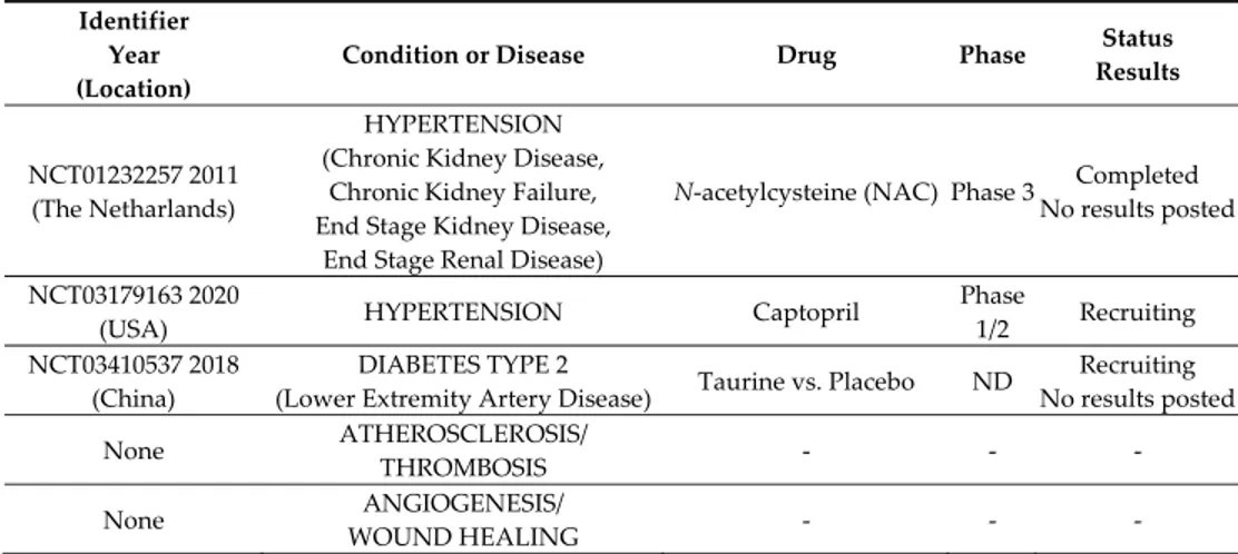
acetylcysteine (NAC) and taurine, has been proposed, but clinical trials unfortunately were not followed up with published data (Table 1). NAC, a well‐tolerated compound, clinically employed to enhance cellular levels of glutathione, is rapidly cleaved in vivo to yield cysteine. On the other hand, in vivo and ex vivo studies demonstrate that the sulfur amino acid taurine markedly and dose‐dependently increased the expression of both CSE and CBS, with a higher effect on CSE upregulation [162]. A reduction in blood pressure in patients with prehypertension has been described [163]. In the complex, only very few clinical studies on H2S donors or enhancers adminis‐ tered in endothelial dysfunction‐related disorders are currently registered in NIH Clini‐ calTrials.gov as listed in Table 1, but no results have been posted or are available on Pub‐ Med.
Table 1. Clinical trials on endothelial dysfunction‐related diseases with H2S donors/enhancers. Identifier
Year (Location)
Condition or Disease Drug Phase Status
Results NCT01232257 2011 (The Netharlands) HYPERTENSION (Chronic Kidney Disease, Chronic Kidney Failure, End Stage Kidney Disease, End Stage Renal Disease)
N‐acetylcysteine (NAC) Phase 3 Completed No results posted
NCT03179163 2020
(USA) HYPERTENSION Captopril
Phase 1/2 Recruiting NCT03410537 2018 (China) DIABETES TYPE 2 (Lower Extremity Artery Disease) Taurine vs. Placebo ND Recruiting No results posted None ATHEROSCLEROSIS/ THROMBOSIS ‐ ‐ ‐ None ANGIOGENESIS/ WOUND HEALING ‐ ‐ ‐ The search of clinical trials listed in ClinicalTrials.gov was performed combining the keywords: H2S, hydrogen sulfide, endothelial dysfunction and the disease listed in the second column. Other not recent studies performed on patients are reported in the text (see [154]). In a manner similar to NAC, cysteine/cysteine‐rich undenatured whey protein sup‐ plement improved pressure ulcer recovery in a small group of diabetic patients [164]. The development of natural compounds, present in the diet, as a H2S source is inter‐ esting, such as polysulfides. Diallyl trisulfide (DATS), diallyl disulfide (DADS) and diallyl sulfide (DAS) are the active principles of the Alliaceae family, such as garlic, which is recognized worldwide as a popular remedy of hypertension. These polysulfides have been demonstrated to exert vasodilating properties in relation to H₂S release [165], behav‐ ing as anti‐hypertensives in L‐NAME‐treated rats [166]. In animal experiments, DATS im‐ proved cardiac function in aortic constricted mice, via an upregulation of VEGF, reduced angiostatin and increased myocardial vascular density [167]. Systemic administration of DATS or local transplantation of DATS‐treated or CSE‐overexpressing bone marrow cells improved capillary density, cell survival and blood perfusion in ischemic hindlimb of db/db mice [168]. Administration of DATS improved neovascularization in STZ‐induced diabetic mice through increased NO availability [169]. Erucin [4‐(methylthio) butyl isothiocyanate] is a natural isothiocyanate particularly abundant in Eruca sativa Mill. (rocket salad), an edible cruciferous plant belonging to the family of Brassicaceae. Isothiocyanates (ITCs) in general represent a source of different beneficial biological effects on human health, and most are investigated in relation to their chemo‐preventive and anti‐cancer properties [170–172]. Numerous studies demonstrated a general anti‐inflammatory and antioxidant activity [173], together with protective prop‐ erties for the cardiovascular system, where ITCs exhibit vasorelaxing and antihyperten‐ sive activity and a protective effect against endothelial dysfunction [174–176]. Several bi‐ ological effects of ITCs may be associated with their ability to release H₂S inside cells in a slow and long‐lasting manner, leading to the definition of “smart H2S‐donors” [175,177]. H2S release from ITCs occurs in a specific manner, depending on the presence of thiols, and it is particularly facilitated in the cell cytosol, where high concentrations of organic thiols, glutathione (in 1–10 mM range), and cysteine (in 30–200 μM range) are present [175,176]. Natural isothiocyanates, including erucin, may therefore represent a possible exogenous source of H2S, which, if gradually released, could mimic the physiological con‐ centrations of the endogenous gasotransmitters. On the other hand, they can be the base for the design of synthetic H2S donor hybrids with antioxidant property and interesting pharmacological development [178]. Considering the requirement of dressings able to protect ulcers with high exudate levels and to promote wound healing (i.e., in diabetic patients), medicated dressings have been designed and developed. In particular, a functional sodium alginate dressing with
H₂S‐releasing properties (SA/JK‐1) was fabricated incorporating a pH‐dependent donor, JK‐1 molecule, into a sodium alginate sponge [179]. The resulting construct provided a moist healing protection able to continuously release H₂S under acidic pH and absorbing exudate at the wound interface. In vitro, the construct was demonstrated to be biocom‐ patible and effective in improving fibroblast migration and proliferation. When tested in animal model of full thickness dermal defect, SA/JK‐1 promoted granulation tissue for‐ mation, angiogenesis, collagen deposition, and re‐epithelization [179]. Overlapping re‐ sults were demonstrated by the same group with hyaluronic acid hydrogels doped with H₂S which was shown to induce M2 macrophage polarization [180]. Another example is represented by silk fibroin porous scaffold loaded with GYY4137, reported to facilitate in vitro bone cell trophism and angiogenesis [181]. These data demonstrate that H₂S‐medi‐ cated wound dressing/biomaterial may represent promising strategies for non‐healing wounds or bone healing and regeneration. Extensive animal and clinical studies are, how‐ ever, necessary for assessing their safety and validation.
9. Concluding Remarks
H₂S is nowadays considered an important transmitter able to maintain vascular ho‐ meostasis. Most of its activities are due to autocrine/paracrine actions by ECs, with a fine control of its plasma concentrations. The availability of H₂S depends on the activity of endothelial and other cells that express the key enzymes involved in the gasotransmitter release, the reactivity of H₂S and its inactivation by redox systems and the efficacy of elim‐ ination reactions. On top of these, the production of H₂S by the gut microbiota and intes‐ tinal epithelium is important to consider, due to the increasing recognition of circulating molecules coming from this source and finely controlling the cardiovascular system per‐ formance [96,182]. There is still demand for the availability of safe and effective synthetic H₂S donors or enhancers, and natural products or nutraceuticals are helping to fulfil this demand. In‐ deed, few clinical trials based on H₂S exogenous sources have been interrupted or have not published their results for unknown reasons. Since endothelial dysfunction and inflammation continue to be the main causes of morbidity and mortality all over the world, knowledge of the molecular and biochemical mechanisms underlying cardiovascular pathologies and their complications is still re‐ quired, as well as the definition of new treatment options to prevent endothelial dysfunc‐ tion or revert cardiovascular disorders [183,184]. The recent pandemic evidenced this un‐ resolved medical need [185] What it is expected from novel molecules in order to be druggable is the exhibition of H₂S levels near the physiological ones, and many compounds are actually druggable H2S‐donors, but it seems that no clinical trials are currently running against endothelial dysfunction (Table 1). Sulfur compounds with natural origin represent helpful pharma‐ ceutical/nutraceutical tools to be used in therapy or as a template for the ideation of ad‐ vanced H₂S‐donor molecules with improved pharmacodynamic and/or pharmacokinetic properties [132,178].
Although experimental data clearly document a protective effect of H₂S donors against endothelial dysfunction, further clinical studies are needed. To the best of our knowledge, there are no clearly active clinical trials on patients affected by pathologies due to endothelial dysfunction and treated with H₂S donors. Even a modest improvement in endothelial function and viability would be a therapeutic success due to the lack of drugs against this diffuse condition predisposing to cardiovascular pathologies.
Author Contributions: Conceptualization, methodology, investigation, resouces, writing‐original draft preparation, writing—review and editing, V.C., S.G. and L.M.; supervision and funding ac‐ quisition, L.M. All authors have read and agreed to the published version of the manuscript. Funding: This research was funded by MIUR‐PRIN project, grant number 2017XP72RF. Conflicts of Interest: The authors declare no conflict of interest. References
1. Cahill, P.A.; Redmond, E.M. Vascular endothelium—Gatekeeper of vessel health. Atherosclerosis 2016, 248, 97–109, doi:10.1016/j.atherosclerosis.2016.03.007. 2. Boulanger, C.M. Endothelium. Arterioscler. Thromb. Vasc. Biol. 2016, 36, e26–e31, doi:10.1161/ATVBAHA.116.306940. 3. Michiels, C. Endothelial Cell Functions. J. Cell Physiol. 2003, 196, 430–443. 4. Wagner, D.D.; Frenette, P.S. The vessel wall and its interactions. Blood 2008, 111, 5271–5281. 5. Boyce, S.; Lwaleed, B.; Kazmi, R. Homeostasis of hemostasis: The role of endothelium. Semin. Thromb. Hemost. 2015, 41, 549–555, doi:10.1055/s‐0035‐155658. 6. Good, S.; Shimokawa, H. Endothelial Functions. Arterioscler. Thromb. Vasc. Biol. 2017, 37, e108–e114. 7. Morbidelli, L.; Donnini, S.; Ziche, M. Therapeutic implications of nitric oxide pathway in angiogenesis of tumors and inflam‐ matory related disorders. In Therapeutic Application of Nitric Oxide in Cancer and Inflammatory Disorders; Morbidelli, L., Bonavida, B., Eds.; Elsevier: Amsterdam, The Netherlands, 2019; pp. 65–91. 8. Bełtowski, J. Synthesis, Metabolism, and Signaling Mechanisms of Hydrogen Sulfide: An Overview. Methods Mol. Biol. 2019, 2007, 1–8, doi:10.1007/978‐1‐4939‐9528‐8_1. 9. Kangussu, L.M.; Marzano, L.A.S.; Souza, C.F.; Dantas, C.C.; Miranda, A.S.; Simões E Silva, A.C. The Renin‐Angiotensin System and the Cerebrovascular Diseases: Experimental and Clinical Evidence. Protein Pept. Lett. 2020, 27, 463–475, doi:10.2174/0929866527666191218091823. 10. Adams, L.; Franco, M.C.; Estevez, A.G. Reactive nitrogen species in cellular signaling. Exp. Biol. Med. 2015, 240, 711–717. 11. Ziche, M.; Morbidelli, L.; Masini, E.; Amerini, S.; Granger, H.J.; Maggi, C.; Geppetti, P.; Ledda, F. Nitric oxide mediates angiogenesis in vivo and endothelial cell growth and migration in vitro promoted by substance P. J. Clin. Investig. 1994, 94, 2036–2044. 12. Parenti, A.; Morbidelli, L.; Ledda, F.; Granger, H.J.; Ziche, M. The bradykinin/B1 receptor promotes angiogenesis by upregula‐ tion of endogenous FGF‐2 in endothelium via the nitric oxide synthase pathway. FASEB J. 2001, 15, 1487–1489. 13. Finetti, F.; Solito, R.; Morbidelli, L.; Giachetti, A.; Ziche, M.; Donnini, S. Prostaglandin E2 regulates angiogenesis via activation of fibroblast growth factor receptor‐1. J. Biol. Chem. 2008, 283, 2139–2146. 14. Ziche, M.; Morbidelli, L.; Choudhuri, R.; Zhang, H.‐T.; Donnini, S.; Granger, H.J.; Bicknell, R. Nitric oxide synthase lies down‐ stream from vascular endothelial growth factor‐induced but not basic fibroblast growth factor‐induced angiogenesis. J. Clin. Investig. 1997, 99, 2625–2664. 15. Cai, W.J.; Wang, M.J.; Moore, P.K.; Jin, H.M.; Yao, T.; Zhu, Y.C. The novel proangiogenic effect of hydrogen sulfide is dependent on Akt phosphorylation. Cardiovasc. Res. 2007, 76, 29–40. 16. Yan, M.S.; Marsden, P.A. Epigenetics in the Vascular Endothelium: Looking From a Different Perspective in the Epigenomics Era. Arterioscler. Thromb. Vasc. Biol. 2015, 35, 2297–2306, doi:10.1161/ATVBAHA.115.305043. 17. Recchioni, R.; Marcheselli, F.; Antonicelli, R.; Mensà, E.; Lazzarini, R.; Procopio, A.D.; Olivieri, F. Epigenetic effects of physical activity in elderly patients with cardiovascular disease. Exp. Gerontol. 2017, 100, 17–27, doi:10.1016/j.exger.2017.10.016. 18. Turunen, M.P.; Ylä‐Herttuala, S. Epigenetic regulation of key vascular genes and growth factors. Cardiovasc. Res. 2011, 90, 441– 446, doi:10.1093/cvr/cvr109. 19. Russell‐Hallinan, A.; Watson, C.J.; O’Dwyer, D.; Grieve, D.J.; O’Neill, K.M. Epigenetic Regulation of Endothelial Cell Function by Nucleic Acid Methylation in Cardiac Homeostasis and Disease. Cardiovasc. Drugs Ther. 2020, doi:10.1007/s10557‐020‐07019‐4. 20. Rajendran, P.; Rengarajan, T.; Thangavel, J.; Nishigaki, Y.; Sakthisekaran, D.; Sethi, G.; Nishigaki, I. The vascular endothelium and human diseases. Int. J. Biol. Sci. 2013, 9, 1057–1069, doi:10.7150/ijbs.7502. 21. Haybar, H.; Shahrabi, S.; Rezaeeyan, H.; Shirzad, R.; Saki, N. Endothelial Cells: From Dysfunction Mechanism to Pharmacolog‐ ical Effect in Cardiovascular Disease. Cardiovasc. Toxicol. 2019, 19, 13–22, doi:10.1007/s12012‐018‐9493‐8. 22. Loscalzo, J. Oxidative stress in endothelial cell dysfunction and thrombosis. Pathophysiol. Haemost. Thromb. 2002, 32, 359–360. 23. Daiber, A.; Oelze, M.; Wenzel, P.; Wickramanayake, J.M.; Schuhmacher, S.; Jansen, T.; Lackner, K.J.; Torzewski, M.; Münzel, T. Nitrate tolerance as a model of vascular dysfunction: Roles for mitochondrial aldehyde dehydrogenase and mitochondrial oxi‐ dative stress. Pharmacol. Rep. 2009, 61, 33–48. 24. Donato, A.J.; Machin, D.R.; Lesniewski, L.A. Mechanisms of dysfunction in the ageing vasculature and role in age‐related dis‐ ease. Circ. Res. 2018, 123, 825–848, doi:10.1161/CIRCRESAHA.118.312563. 25. Trott, D.W.; Fadel, P.J. Inflammation as a mediator of arterial ageing. Exp. Physiol. 2019, 104, 1455–1471, doi:10.1113/EP087499. 26. Khyzha, N.; Alizada, A.; Wilson, M.D.; Fish, J.E. Epigenetics of Atherosclerosis: Emerging Mechanisms and Methods. Trends Mol. Med. 2017, 23, 332–347, doi:10.1016/j.molmed.2017.02.004.