Università degli Studi di Ferrara
DOTTORATO DI RICERCA IN
"Farmacologia e Oncologia Molecolare"
CICLO XXI
COORDINATORE Prof. Pier Andrea Borea
ALTERED EXPRESSION AND FUNCTIONALITY OF
A
2AADENOSINE RECEPTORS IN HUNTINGTON’S
DISEASE AND OTHER POLYGLUTAMINE DISORDERS
Settore Scientifico Disciplinare BIO/14
Dottorando Tutore
Dott. Vincenzi Fabrizio Chiar.mo Prof. Borea Pier Andrea
Contents
Pag.
General introduction 005
Aim of the thesis 070
CHAPTER 1 073
Aberrant amplification of A2A receptor signaling
in striatal cells expressing mutant huntingtin.
Introduction 075
Materials and Methods 076
Results 079
Discussion 091
CHAPTER 2 095
Early and transient alteration of A2AAR signaling
in a mouse model of Huntington disease.
Introduction 097
Materials and Methods 098
Results 103
Discussion 113
CHAPTER 3 117
Aberrant A2AAR expression and function in
peripheral blood cells of patients with Huntington's disease.
Introduction 119
Materials and Methods 120
Results 125
Discussion 133
CHAPTER 4 137
Biological abnormalities of peripheral A2AARs in a large representation of
polyglutamine disorders and Huntington's disease stages.
Introduction 139
Materials and Methods 140
Results 144 Discussion 155 General conclusions 157 References 161 Curriculum Vitae 181 List of publications 183 Meetings 185 Acknowledgements 187
ADENOSINE
Adenosine is a nucleoside composed of a molecule of adenine attached to a ribose sugar molecule (ribofuranose) via a β-N9-glycosidic bond (Figure 1).
Figure 1 – Chemical structure of Adenosine
Adenosine is an endogenous nucleoside-signalling molecule, which, by acting on specific membrane receptors produces a number of physiological and pathophysiological effects in both the central nervous system and peripheral organs. Under normal conditions, adenosine is continuously formed intracellularly as well as extracellularly. The intracellular production is mediated either by an intracellular 5'-nucleotidase, which dephosphorylates or by hydrolysis of S-adenosyl-homocysteine (Fredhoml et al., 2001). Adenosine generated intracellularly is transported into the extracellular space mainly via specific bi-directional transporters through facilitated diffusion that efficiently evens out the intra- and extracellular levels of adenosine. The dephosphorylation of extracellular AMP to adenosine, mediated by ecto-5'-nucleotidase, is the last step in the enzymatic chain that catalyzes the breakdown of extracellular adenine nucleotides, such as ATP, to adenosine. Ectonucleotidases include
N N N N NH2 O OH OH H H H H HO
ectonucleoside triphosphate diphosphohydrolase which can hydrolyze ATP or ADP, ectonucleotide pyrophosphatase/phosphodiesterases, alkaline phosphatases and 5'-nucleotidases (Zimmermann, 2000). When adenosine levels in the extracellular space are high, adenosine is transported into cells by means of transporters. It is then phosphorylated to AMP by adenosine kinase or degraded to inosine by adenosine deaminase. Adenosine deaminase, but not adenosine kinase, is also present in the extracellular space (Fredhoml et al., 2001). Another potential source of extracellular adenosine is cAMP, which can be released from neurons and converted by extracellular phosphodiesterases into AMP and thereafter by an ecto-5'-nucleotidase to adenosine. The transport of adenosine by facilitated diffusion is equilibrative and bidirectional, meaning that the net transport of adenosine either into or out of the cell depends upon the adenosine concentration gradient in both sides of the membrane. Inhibition of adenosine transport can, therefore, inhibit either adenosine release or adenosine uptake, depending upon the intra- and extracellular levels of adenosine (Gu et al., 1995). However, since the extracellular formation of adenosine from released adenine nucleotides constitutes a second source of adenosine, which is not affected by transport inhibition, the transport inhibitors usually cause an increase in the extracellular adenosine levels. Under hypoxic and ischemic conditions there is a marked increase in cytoplasmatic adenosine leading to an intense release of adenosine, which is inhibited by adenosine uptake inhibitors (Parkinson et al., 2002).
Excitatory amino acid-mediated release of adenosine is certainly involved; however, of greater importance is probably the fact that whenever intracellular levels of adenine nucleotides fall as a result of excessive energy use, the intracellular levels of adenosine will rise dramatically (Fredhoml et al., 2001). For example, following hypoxia there is a decrease of intracellular ATP, accompanied by an accumulation of 5'-AMP and subsequently adenosine. The nucleoside is thereafter transported into the extracellular
space via the transporters. Furthermore, when the intracellular level of adenosine is very high, adenosine simply diffuses out of cells. Direct release of intracellular adenine nucleotides, such as ATP, that is thereafter converted extracellularly by ecto-ATPase and ecto-ATP-diphosphohydrolase (ecto-apyrase) to AMP and dephosphorylated by ecto-5'-nucleotidase to adenosine, should also be considered (Zimmermann et al., 2000). Adenosine is neither stored nor released as a classical neurotransmitter since it does not accumulate in synaptic vesicles, being released from the cytoplasm into the extracellular space through a nucleoside transporter. The adenosine transporters also mediate adenosine reuptake, the direction of the transport being dependant upon the concentration gradient at both sides of the membrane (Fredhoml et al., 2001). Since it is not exocytotically released, adenosine behaves as an extracellular signal molecule influencing synaptic transmission without itself being a neurotransmitter, i.e. modulates the activity of the nervous system at cellular level presynaptically by inhibiting or facilitating transmitter release, postsynaptically by hyperpolarising or depolarising neurones and/or exerting non-synaptic effects. Adenosine, therefore, belongs to the group of neuromodulators.
Adenosine receptors
Four adenosine receptor (AR) subtypes (A1, A2A, A2B, and A3) have been cloned and
pharmacologically characterized, all of which are G protein-coupled receptors (GPCRs). Adenosine receptors can be distinguished according to their preferred mechanism of signal transduction: A1 and A3 receptors interact with pertussis toxin-sensitive G
proteins of the Gi and Go family; the canonical signaling mechanism of the A2A and of
coupling to adenylyl cyclase, all four subtypes may positively couple to phospholipase C via different G protein subunits (Fredholm et al, 2001).
Considering the overall protein structure, ARs display the topology typical of GPCRs. Sequence comparison between the different GPCRs revealed the existence of different receptor families sharing no sequence similarity even if specific fingerprints exist in all GPCR classes. However, all these receptors have in common a central core domain consisting of seven transmembrane helices (TM1-7), with each TM composed of 20–27 amino acids, connected by three intracellular (IL1, IL2, and IL3) and three extracellular (EL1, EL2, and EL3) loops. Two cysteine residues (one in TM3 and one in EL2), which are conserved in most GPCRs, form a disulfide link which is possibly crucial for the packing and for the stabilization of a restricted number of conformations of these seven TMs. Aside from sequence variations, GPCRs differ in the length and function of their N-terminal extracellular domain, their C-terminal intracellular domain, and their intracellular loops. Each of these domains provides very specific properties to these receptor proteins. Particularly, consensus sites for N-linked glycosylation exist on the extracellular regions of ARs, although the precise location of the sites for this post-translational modification varies amongst the AR subtypes. The carboxyl-terminal tails of the A1AR, A2BAR, and A3AR, but not A2AAR, possess a conserved cysteine residue
that may putatively serve as a site for receptor palmitoylation and permit the formation of a fourth intracellular loop (Moro et al., 2005).
The A1AR, A2BAR, and A3AR are very similar in regard to the number of amino acids
composing their primary structure, and in general, these AR subtypes are among the smaller members of the GPCR family. For example, the human homologs of the A1AR,
A2BAR, and A3AR consist of 326, 328, and 318 amino acid residues, respectively.
Conversely, the human A2AAR is composed of 409 amino acids. It should be noted that
consistent with the mass estimated by polyacrylamide gel electrophoresis of the expressed proteins. The post-translational glycosylation of ARs, which may vary in a cell type-dependent fashion, likely accounts for these discrepancies. The human A1AR
and human A3AR display 49% overall sequence identity at the amino acid level, while
the human A2AAR and human A2BAR are 45% identical (Fredholm et al, 2001).
A1 adenosine receptors
The A1 receptor is widely expressed throughout the body, having its highest expression
in the brain, spinal cord, atria and adipose tissue (Baraldi et al., 2000). Via adenosine A1ARs, adenosine reduces heart rate, glomerular filtration rate, and renin release in the
kidney; it induces bronchoconstriction and inhibits lipolysis (Elzein and Zablocki, 2008). Adenosine A1Rs can be coupled to different pertussis toxin-sensitive G proteins,
which mediate inhibition of adenylate cyclase and regulate calcium and potassium channels, as well as inositol phosphate metabolism (Fredholm et al., 2001). A1ARs and
A2AARs are primarily responsible for the central effects of adenosine (Dunwiddie and
Masino, 2001). In addition to their postsynaptic locations in different brain regions, A1ARs can be found presynaptically and modulate neurotransmitter release. Presynaptic
A1ARs are the prototype of GPCRs, the stimulation of which decreases the probability
of neurotransmitter release. The main mechanism of A1AR-mediated inhibition of
exocytosis is a direct inhibitory effect on voltage-dependent Ca2+ channels (Moore et al., 2003). A1AR displays two different affinities for agonist, which have classically
been attributed to a different coupling to heterotrimeric G proteins. According to this two independent site model, coupled receptor–G protein complexes display high affinity for agonists and uncoupled receptors display low affinity. The reported cluster-arranged cooperative model predicts that the high- and low-affinity sites are a consequence of the negative cooperativity of agonist binding and do not seem to be related to the content of
G protein-coupled or –uncoupled receptors (Franco et al., 1996). Like other GPCR members, A1AR expression is regulated in response to agonist or antagonist stimulation.
Desensitization of A1ARs has been described in intact animals and in cell cultures.
Prolonged administration of A1AR agonists to animals leads to functional
desensitization of A1ARs in guinea pig heart, rat adipocytes, rat atrial muscle, and rat
brain (Moro et al., 2006). The reduced functional response is attributable to a net loss of A1ARs or down-regulation, a decrease in the proportion of A1ARs displaying the
high-affinity state for agonists, and a decrease in the content of Gi proteins. The loss of binding sites on the cell membrane owing to internalization of A1ARs is a slower event.
Ser/Thr phosphorylation seems to be related to short-term clustering and desensitization, as well as long-term internalization of A1ARs (Ciruela et al., 1997).
A2A adenosine receptors
The A2AAR exists in a wide variety of organs including major peripheral tissues (e.g.,
liver, heart, lung, and the immune system) and the central nervous system (CNS) (Lee et al., 2003). In the developing rat brain, expression of the A2AAR is transiently regulated
in various areas (e.g., the striatum, cortex, and hippocampus), perhaps implying a role of adenosine in neuronal development. Soon after neurogenesis, the A2AAR is highly
expressed by striatal neurons and co-localizes with the D2 dopamine receptor in
GABAergic striatopallidal neurons (Ferrè et al., 2008). In addition to the intense expression in the striatum, low levels of A2AAR are found in many brain regions (e.g.,
the cortex and hippocampus) and it has been suggested that adenosine acting at the A2AAR regulates important neuronal functions including neuronal protection and
synaptic transmission (Ferrè et al., 2008). Regulation of A2AAR gene expression is
therefore likely to play an important role in neuronal development, basal ganglia activity, and many other peripheral functions. In the CNS, l-DOPA enhanced the gene
expression of the striatal A2AAR in 6-OHDA-lesioned rats (Tomiyama et al., 2004).
Treatment with an antagonist of the NMDA receptor (memantine) was also reported to elevate the transcript level of striatal A2AARs (Marvanova and Wong, 2004). The
adenosine A2AAR couples primarily to members of the Gs family. Like other GPCRs it
can also interact with other G proteins if the receptor is very over-expressed, but the evidence for such coupling in vivo is not compelling. In striatum the A2AAR interacts
with Golf proteins (Corvol et al., 2001). It is not known if there are significant differences in receptor affinity or in signaling dependent on which of the two partners (or which variant of Gs) the receptor interacts with. There are instances where other G protein pathways have been implicated, and it will be important to determine if this alternate coupling is a regulated process, for example via phosphorylation. There is no compelling reason to assume that this GPCR coupling to members of the Gs family would signal in anything but a canonical way. Thus, most effects are probably due to activation of adenylyl cyclase and generation of cAMP. The A2AAR can recruit
β-arrestin via a GRK-2 dependent mechanism (Khoa et al., 2006). This is influenced by activation of cytokine receptors, which cause reduced desensitization of the A2AAR
(Khoa et al., 2006).
One key target of PKA is the cAMP responsive element-binding protein (CREB) which is critical for many forms of neuronal plasticity as well as other neuronal functions (Josselyn and Nguyen, 2005). Phosphorylation of CREB at Ser133 by PKA activates CREB and turns on genes with cAMP responsive elements (CRE sites) in their promoters. One important feature of CREB is that it is a point of convergence for the cAMP/PKA and MAPK pathways. Stimulation of the A2AARs counteracts the
inhibition of neurite outgrowth due to MAPK blockade (Cheng et al., 2002). Stimulation of the A2AAR alone also activates the Ras/Raf-1/MEK/ERK signaling
mechanisms, respectively (Schulte and Fredholm, 2003). Interestingly, phosphorylation/activation of CREB has been shown to compete with nuclear factor-κB (NFκB) p65 for an important co-factor, CBP. Phosphorylated CREB was therefore proposed to mediate the anti-inflammatory effect of the A2AAR receptor and inhibition
of NFκB by A2AAR activation during acute inflammation in vivo was demonstrated
(Fredholm et al., 2007).
An interesting observation is that activation of A2AAR receptor facilitates activities of
adenosine transporters via a PKC-dependent pathway in the hippocampus, and thus reduces the level of extracellular adenosine available for A1AR activation (Pinto-Duarte
et al., 2005). In addition, PKC was shown to play a key role in mediating the enhancement of noradrenaline release by the A2AAR in rat tail artery (Fresco et al.,
2004). Activation of multiple signaling pathways by the A2AAR appears to contribute to
its diverse and complex functions in various tissues.
A2B adenosine receptors
A2BAR mRNA was originally detected in a limited number of rat tissues by Northern
blot analysis, with the highest levels found in cecum, bowel, and bladder, followed by brain, spinal cord, lung, epididymis, vas deferens, and pituitary. The use of more sensitive reverse transcriptase-polymerase chain reaction techniques revealed a ubiquitous distribution of A2BAR (Spicuzza et al., 2006). mRNA encoding A2BAR was
detected at various levels in all rat tissues studied, with the highest levels in the proximal colon and lowest in the liver. In situ hybridization of A2BARs showed
widespread and uniform distribution of A2BAR mRNA throughout the brain (Dixon et
al., 1996).
Pharmacological identification of A2BARs, based on their low affinity and characteristic
brain, functional A2BARs are found in neurons and glial cells. Although there is no
evidence that A2BAR are present in microglia, there is ample data that show that they
are expressed in astrocytes and in different glioma cell lines (Fiebich et al., 1996). The expression of A2BARs in glial cells, which represent a majority of the brain cell
population, can explain the original observation that slices from all brain areas examined showed an adenosine-stimulated cAMP response.
Functional A2BARs have been found in fibroblasts and various vascular beds,
hematopoietic cells, mast cells, myocardial cells, intestinal epithelial and muscle cells, retinal pigment epithelium, endothelium, and neurosecretory (Gessi et al., 2005). Although activation of adenyl cyclase is arguably an important signaling mechanism for A2AARs, this is not necessarily the case for A2BARs, as other intracellular signaling
pathways have been found to be functionally coupled to these receptors in addition to adenyl cyclase. In fact activation of adenosine A2BARs can increase phospholipase C in
human mast cells and in mouse bone marrow-derived mast cells. A2BAR activation also
elevates inositol triphosphate (IP3) levels, indicating this receptor can couple also to Gq-proteins. A2BARs have been implicated in the regulation of mast cell secretion and,
gene expression, intestinal function, neurosecretion, vascular tone and in particular asthma (Varani et al., 2005).
A3 adenosine receptors
The A3AR has widely distributed its mRNA being expressed in testis, lung, kidneys,
placenta, heart, brain, spleen, liver, uterus, bladder, jejunum, proximal colon and eye of rat, sheep and humans. However, marked differences exist in expression levels within and among species. In particular rat testis and mast cells express high concentrations of A3AR mRNA, while low levels have been detected in most other rat tissues (Gessi et
A3AR mRNA in human, while low levels have been found in aorta and brain. Lung,
spleen, pars tuberalis and pineal gland expressed the highest levels of A3AR mRNA in
sheep.
The presence of A3AR protein has been evaluated through radioligand binding,
immunoassay or functional assay in a variety of primary cells, tissues and cell lines (Gessi et al., 2008). In the mouse brain a widespread, relatively low level of A3AR
binding sites was found (Jacobson et al., 1993). Similar data were obtained in the rat and in gerbil and rabbit brain. Electrophysiological and biochemical evidence suggested the presence of A3ARs in the rat hippocampus and cortex, and functional studies also
indicated its presence in the brain. In cardiomyocytes, there was no direct evidence of the presence of A3ARs but several studies reported that it was responsible for
cardioprotection in a variety of species and models, including isolated cardiomyocytes and isolated myocardial muscle preparations (Peart and Headrick, 2007). In lung parenchyma and in human lung type 2 alveolar-like cells (A549), the A3AR was
detected through radioligand binding and immunohistochemical assays (Varani et al., 2006).
The classical pathways associated with A3AR activation are the inhibition of adenylyl
cyclase activity, through the coupling with Gi proteins, and the stimulation of phospholipase C (PLC), inositol triphosphate (IP3) and intracellular calcium (Ca2+), via Gq proteins (Fredholm et al., 2001). However, more recently additional intracellular pathways have been described as relevant for A3AR signaling. For example, in the
heart, A3AR mediates cardioprotective effects through ATP-sensitive potassium
(KATP) channel activation. Moreover, it is coupled to activation of RhoA and a subsequent stimulation of phospholipase D (PLD), which in turn mediates protection of cardiac myocytes from ischemia (Mozzicato et al., 2004). In addition, in different recombinant and native cell lines, A3AR is involved, like the other adenosine subtypes,
in the modulation of mitogen-activated protein kinase (MAPK) activity (Schulte and Fredholm, 2003). A3AR signaling in Chinese Hamster Ovary cells transfected with
human A3AR (CHO-hA3) leads to stimulation of extracellular signal-regulated kinases
(ERK1/2). In particular, A3AR signaling to ERK1/2 depends on βγ release from
pertussis toxin (PTX)-sensitive G proteins, phosphoinositide 3-kinase (PI3K), Ras and mitogen-activated protein kinase kinase (Schulte and Fredholm, 2003). It has been reported that A3AR activation is able to decrease the levels of PKA, a downstream
effector of cAMP, and of the phosphorylated form of PKB/Akt in melanoma cells. This implies the deregulation of the Wnt signaling pathway, generally active during embryogenesis and tumorigenesis to increase cell cycle progression and cell proliferation (Fishman et al., 2002). Involvement of the PI3K/PKB pathway has been linked with preconditioning effects induced by A3AR activation in cardiomyocytes from
newborn rats (Germack and Dickenson, 2005). An elegant study has recently documented a role of A3AR in cell survival signaling in resveratrol preconditioning of
the heart. This study provides evidence that resveratrol preconditions the heart through the activation of adenosine A1 and A3AR, transmitting a survival signal through both
the PI3K-Akt-Bcl2 and, only in the case of A3AR, cAMP response element-binding
protein (CREB)-Bcl2 pathways (Das et al., 2005). Subsequently it has been demonstrated that CREB phosphorylation occurs through both Aktdependent and -independent signaling. Activation of PI3K-Akt-pBAD by A3AR has been observed
recently in glioblastoma cells leading to cell survival in hypoxic conditions (Merighi et al., 2007). Further studies indicate that A3AR activation by interfering with PKB/Akt
pathways can decrease interleukin-12 (IL-12) production in human monocytes (la Sala et al., 2005). Collectively, these findings demonstrate that several intracellular mechanisms are involved following A3AR stimulation, the understanding of which may
be essential and crucial for explaining the different aspect of its activation.
Therapeutic potential
Cardiovascular system
A1AR is the most extensively studied and well characterized of the adenosine receptor
subtypes in relation to cardiac protection. The A1AR is best understood insofar as its
effects on injury and in terms of receptor-coupled kinase/protein signaling. For the most part, transduction cascades induced by A1AR agonism follow those of several other
protective GPCR systems (Hausenloy and Yellon, 2004). However, there remain some controversies regarding both signal cascades and A1AR-mediated responses in the heart.
Originally, adenosine (via A1AR activation) was thought to induce myocardial
protection through preservation of ATP (and improved nucleotide repletion on reperfusion), stimulation of glycolysis, and normalization of the hearts so-called “oxygen supply/demand ratio” (Ely and Berne, 1992). Subsequent investigations have identified essential protein kinase signaling cascades together with putative end-effectors (including the mitochondrial KATP channel), in the protective and preconditioning actions of A1ARs.
Adenosine enhances tolerance to ischemia via metabolic substrate effects (Headrick et al., 2003). Adenosinergic cardioprotection in ischemic-reperfused hearts involves reductions in oncotic and apoptotic death, and improved functional outcomes (Willems et al., 2005). Recent work supports differential effects of acute adenosine vs. transient adenosinergic preconditioning, consistent with multiple pathways of protection (Peart and Headrick, 2003). In terms of cellular targets, adenosine appears to directly protect cardiomyocytes or myocardial tissue (likely via A1 and A3ARs), and additionally
cells and vascular and myocardial tissue. The different cardioprotective effects of AR agonism have been verified in animal and human tissue (Willems et al., 2005). However, few studies have addressed the possibility that altered AR-mediated protection might underlie specific cardiovascular disorders, though there is evidence to support this. Hypertrophic hearts, for example, display abnormal adenosinergic signaling, and dysregulated adenosine formation. Interestingly, ARs impact on many processes implicated in cardiovascular "aging", regulating Ca2+ influx and oxidant injury, substrate metabolism, angiogenesis, myocardial fibrosis, and apoptotic processes (Willems et al., 2005). Given evidence of a role for ARs in intrinsic cardioprotection, mediation of preconditioning, and modifying the above-mentioned processes, alterations in AR signalling could contribute both to ischemic intolerance and emergence of other features of aged myocardium. All ARs are considered to be expressed within cardiovascular cells. Studies in different species verify endogenous adenosine contributes to intrinsic ischemic tolerance, and support cardioprotective roles for A1ARs in vitro and in vivo, and for A2AARs in vivo (Willems et al., 2005).
Anti-ischemic effects of A1ARs appear direct (at cardiomyocytes), since similar protection is
observed in isolated hearts, cardiomyocytes, and in vivo (Roscoe et al., 2000). Protective A2AAR effects involve modulation of vascular function, platelet adhesion and
neutrophil activation. There is currently no direct evidence for acute A2BAR mediated
cardioprotection, partially due to lack of selective A2BAR agonists/antagonists.
In contrast to A1 and A2AARs, there is little evidence that intrinsically activated A3ARs
mediate protection. A3AR antagonists have no effect on ischemic outcomes in myocytes
Airways
A role for adenosine in pulmonary disease was first suggested when it was found that adenosine and related synthetic analogues were potent enhancers of IgE-dependent mediator release from isolated rodent mast cells (Holgate et al., 1980). A few years later, adenosine administered by inhalation was shown to be a powerful bronchoconstrictor of asthmatic but, importantly, not of normal airways (Cushley et al., 1983). Further work showed that both allergic and non-allergic asthmatics responded in a similar way and that the effect was also seen with adenosine 5′-monophosphate (AMP), ADP and ATP (Basoglu et al., 2005). Elevated levels of adenosine are present in chronically inflamed airways; they have been observed both in the bronchoalveolar lavage fluid and the exhaled breath condensate of patients with asthma (Caruso et al., 2006). Adenosine levels are also increased after allergen exposure and during exercises in atopic individuals. The observed increase in tissue levels of adenosine suggests that adenosine signaling could regulate important features of chronic inflammatory disorders of the airways, including asthma and chronic obstructive pulmonary disease (COPD). Consistent with the hypothesis of adenosine playing an important role in the pathogenesis of chronic inflammatory disorders of the airways, mice deficient in adenosine deaminase (ADA) develop severe pulmonary inflammation and airway remodeling in association with elevated adenosine concentrations in the lung (Blackburn et al., 2000). The pulmonary phenotype in ADA-deficient mice consists of airway accumulation of eosinophils and activated macrophages, mast cell degranulation, mucus metaplasia in the bronchial airways, and emphysema-like devastation of the lung parenchyma. Although these histological traits do not completely resemble those of human asthma, the ADA-deficient mouse model is a useful tool to study the pathogenic role of adenosine in chronic airway inflammation.
The central role of adenosine in chronic lung inflammation is also supported by studies carried out in mice that have increased levels of interleukin IL-13 in the lung. These mice develop inflammation, fibrosis and alveolar destruction in association with elevated adenosine concentrations in the lung (Caruso et al., 2006). Treatment with ADA to prevent the increase in adenosine concentrations resulted in a marked decrease in the severity of the pulmonary phenotype, suggesting that adenosine mediates IL-13-induced inflammation and tissue remodeling. Blockade of adenosine re-uptake by administration of dipyridamole has been used in humans to test the hypothesis that the accumulation of extracellular adenosine functionally modulates important features of the asthmatic response. In addition, it has been shown that a rapid increase in sputum eosinophilia occurs when asthmatics are exposed to adenosine by means of a provocation test with AMP (van der Berge et al., 2003). Taken together, these observations indicate that adenosine is likely to play an important role in asthma and COPD through interaction with specific cell-surface receptors. Expression of the four identified adenosine receptors has been shown in a large number of proinflammatory and structural cells and recently in the peripheral lung parenchyma of patients with COPD. The affinity of A1, A2A, and A3ARs, studied by means of saturation binding
assays, was substantially decreased in patients with COPD, whereas their level of expression appears to be increased. Conversely, the affinity of A2BARs was not altered,
but the density was significantly decreased in patients with COPD (Varani et al., 2006). This suggests that adenosine signalling play an important but rather complex role in COPD. Hence, adenosine responses are not only dictated by the bioavailability of the nucleoside but also by the pattern of adenosine receptor expression, which is known to be finely modulated by physiological and/or pathological tissue environments. Stimulation of A1ARs promotes activation of human neutrophils and enhances
this receptor. However, in ADA/A1ARs double knockout mice, the lack of A1ARs
results in enhanced pulmonary inflammation, mucus metaplasia, alveolar destruction and earlier death from respiratory distress, indicating a protective function. Activation of A2AARs on activated immune cells by adenosine appears to largely suppress the
inflammatory response. In human neutrophils, stimulation of A2AARs reduces
neutrophil adherence to the endothelium, inhibits formyl-Met-Leu-Phe (fMLP)-induced oxidative burst and inhibits superoxide anion generation (Fredholm et al., 1996). In monocytes and macrophages, activation of A2AARs inhibits lipopolysaccharide-induced
tumour necrosis factor-α expression. Therefore, A2AAR agonists might have
anti-inflammatory effects in diseases such as COPD, where neutrophil- and monocyte-mediated tissue injury is implicated (Caruso et al., 2006).
Initial evidence for the role of A2BARs in asthma and COPD came from
pharmacological studies of enprofylline, a methylxanthine structurally related to theophylline (Feoktistov and Biaggioni, 1995). It was proposed that the A2BAR might
be the therapeutic target in the long-term clinical benefit achieved with relatively low doses of theophylline and enprofylline. Recently, A2BARs have been shown to mediate
several pro-inflammatory effects of adenosine in the large majority of inflammatory and structural cells of the lung. For example, functional human A2BARs have been identified
in mast cells, bronchial smooth muscle cells and lung fibroblasts. In these cells, adenosine, via activation of A2BARs, increases the release of various inflammatory
cytokines, which induce IgE synthesis from human B lymphocytes and promote differentiation of lung fibroblasts into myofibroblasts. Such findings provide support for the view that activation of A2BARs could enhance the inflammatory response associated
with asthma and that selective blockade of these receptors would be potentially beneficial in the treatment of asthma and other pulmonary inflammatory diseases. The functional significance of the A3AR in the pathogenesis of chronic inflammatory airway
diseases remains controversial largely owing to major species differences (Caruso et al., 2006).
Adenosine and cancer
One of the difficulties in treating most of the common cancers (colon, lung, breast, prostate, etc.) is that they form solid tumors. The individual cancer cells, being different from normal cells, form a tissue mass that behaves in a radically different way from normal tissues in the body. This is because the major cell population (the cancer cells) has grown in a way that is out of step with all of the other cells that would normally form a supportive network. In particular, the growth of the cancer is not coordinated with the development of a proper blood supply. The vascular network of a tumor is usually inadequate, the blood vessels are often too few in number, the network is improperly branched, and their calibre is not well controlled. This means that the blood supply is inadequate. Consequently, most solid tumors do not receive sufficient oxygen and the cells are hypoxic. Specifically, hypoxia is conducive to adenine nucleotide breakdown, which is responsible for the adenosine release (Vaupel et al., 2001). As a consequence, adenosine accumulates to high levels in hypoxic tissues. In particular, it is recognized that significant levels of adenosine are found in the extracellular fluid of solid tumors, suggesting a role of adenosine in tumor growth (Merighi et al., 2003). Adenosine, released from hypoxic tissue, is thought to be an angiogenic factor that links altered cellular metabolism, caused by oxygen deprivation, to compensatory angiogenesis. Angiogenesis (or neovascularization) begins with the migration of endothelial cells, originating from capillaries, into the tissue being vascularized. Adenosine has been reported to stimulate or inhibit the release of angiogenic factors
depending on the cell type examined (Burnstock, 2006). On one hand, adenosine is known to cause the synthesis of vascular endothelial growth factor (VEGF) and increase the proliferation of endothelial cells obtained from the aorta, coronary vessels, and retina (human retinal endothelial cells, HREC). In particular, adenosine has been shown to induce the DNA synthesis in cultures of human umbilical vein endothelial cells (HUVEC) (Burnstock, 2006).
In the human leukemia HL60, human melanoma A375, and human astrocytoma cells, adenosine at millimolar concentrations caused apoptosis. It seems likely that apoptosis is mediated by the intracellular actions of adenosine rather than through surface receptors (Merighi et al., 2002). It has been argued that the effect of high adenosine concentration might be subsequent to uptake of adenosine by the cell and intracellular accumulation of AMP, leading to caspase activation (Merighi et al., 2003).
In many cases, tumor-induced immune suppression is mediated by soluble inhibition factors or cytokines elaborated by the tumor cells. Extracellular fluid of solid carcinomas contains immunosuppressive concentrations of adenosine, suggesting that this autacoid constitutes an important local immunosuppressant within the microenvironment of solid tumors.
Antigen-presenting cells such as dendritic cells and macrophages are specialized to activate naïve T-lymphocytes and initiate primary immune responses. Adenosine inhibits interleukin-12 (IL-12) and tumor necrosis factor-α (TNF-α) production in dendritic cells and in macrophages impairing T-cell priming and suppressing the anticancer immune response. Furthermore, adenosine impairs the induction and expansion of cytotoxic T-lymphocytes and the antitumor activity of natural killer (Hoskin et al., 2002).
Differential effects of adenosine on normal and cancer cells have been previously reported, showing that the proliferation of lymphocytes derived from patients with
chronic lymphocytic leukemia was inhibited by adenosine, whereas the proliferation of lymphocytes from healthy people was inhibited to a lesser extent.
In vivo studies have shown that adenosine exerts a profound inhibitory effect on the induction of mouse cytotoxic T-cells, without substantially affecting T-cell viability (Hoskin et al., 2002).
Adenosine sustains a complex role in the immune system activity, because when given to mice pretreated with cyclophosphamide it demonstrated a myeloprotective effect by restoring the number of white blood cells and the percentage of neutrophils as compared with normal values. Furthermore, it has been demonstrated that the elevation of the extracellular adenosine concentrations induced a radioprotective effect in mice by the stimulation of hematopoiesis in the bone marrow and the spleen. In support of this myelostimulatory role, it has been demonstrated that adenosine enhances cycling of the hematopoietic progenitor cells (Pospísil et al., 2001).
The ability of adenosine to specifically inhibit tumor cell growth in vitro and in vivo suggests that the activation and/or blockade of the pathways downstream of adenosine receptors may contribute to tumor development. Furthermore, the extracellular adenosine concentration may be a crucial factor in determining the cell progression pathway, either in the apoptotic or in the cytostatic state (Merighi et al., 2003).
Adenosine and central nervous system
Adenosine levels in the brain extracellular space increase dramatically during metabolically stressful conditions, such as ischemia, seizures, or trauma. Adenosine, acting via its receptors,modulates excitability in the central nervous system (CNS)and has a role in mechanisms of seizure susceptibility, sleep induction, basal ganglia function,pain perception,cerebral blood flow, and respiration (Benarroch, 2008).
Adenosine functions as a natural sleep-promoting agent accumulating during periods of sustained wakefulness and decreasing during sleep. It was suggested that adenosine participates in resetting of the circadian clock by manipulations of behavioural state. Indeed, A1ARs of the suprachiasmatic nucleus regulate the response of the circadian
clock to light (Elliott et al., 2001). The sleep inducing properties of adenosine is in line with its A1AR-mediated inhibitory action and may involve multiple neuronal
populations in the central nervous system; however, the actions upon the basal forebrain nuclei involved in sleep and arousal appear to be particularly important (Ribeiro et al., 2002). In healthy humans, caffeine inhibits psychomotor vigilance deficits from sleep inertia, a ubiquitous phenomenon of cognitive performance impairment, grogginess and tendency to return to sleep immediately after abrupt awakening from intermittent and short sleep periods (van Dongen et al., 2001). It thus emerges that there exists a potential role of adenosine-related compounds and of A1AR agonists as sleep promoters
and adenosine receptor antagonists as arousal stimulators. Adenosine A1AR agonists
have anxiolytic activity in rodent models of anxiety, whereas caffeine and the adenosine A1AR selective antagonist, cyclopentyltheophylline, have anxiogenic properties. The
involvement of adenosine A1ARs in anxiety was confirmed by the finding that the mice
knocked out for this receptor showed increased anxiety-related behaviour. Interestingly, patients suffering from panic disorder, a serious form of anxiety disorder, appear to be particularly sensitive to small amounts of caffeine (Ribeiro et al., 2002). These observations, taken together, suggest that drugs that facilitate adenosine A1AR-mediated
actions may be effective for the treatment of anxiety. Endogenous adenosine, through A1AR activation, modulates long-term synaptic plasticity phenomena, such as long-term
potentiation (LTP), long-term depression (LTD), and depotentiation. In accordance with the notion that synaptic plasticity is the basis for learning and memory in different brain areas, adenosine correspondingly modulates behaviour in various learning and memory
paradigms, and adenosine A1AR antagonists have been proposed for the treatment of
memory disorders (Ribeiro et al., 2002). Cognitive effects of caffeine are mostly due to its ability to antagonise A1ARs in the hippocampus and cortex, the brain areas mostly
involved in cognition, positive actions of caffeine on information processing and performance might also be attributed to improvement of behavioural routines, arousal enhancement and sensorimotor gating (Fredholm et al., 1999).
One of the first pathophysiological roles proposed for adenosine was as an endogenous anti-convulsant (Dragunow et al., 1986). Limitations of the use of adenosine receptor agonists as anti-convulsant drugs are due to their pronounced peripheral side effects as well as central side effects like sedation. To circumvent this limitation, the possibility of using compounds to increase the concentrations of endogenous adenosine was proposed, in particular inhibitors of adenosine kinase (McGaraughty et al., 2001). These compounds would facilitate the increase in extracellular adenosine caused by the seizure, having limited effects in other brain areas where the levels of adenosine are low. The anti-epileptic properties of adenosine are mostly due to the well-known inhibitory actions of A1ARs upon synaptic transmission in the hippocampus, but not
only presynaptic actions upon glutamate release are involved (Khan et al., 2000). In the spinal cord, adenosine A1 AR activation produces anti-noceptive properties in
acute nociceptive, inflammatory and neuropathic pain tests. Therefore, there was an increasing interest in the development of drugs that, by influencing extracellular adenosine levels, could have analgesic actions. Probably due to the anti-inflammatory actions of adenosine, adenosine kinase inhibitors administered orally are even more effective to reduce inflammatory pain than neuropatic or acute pain (Jarvis et al., 2002). By comparing the anti-nociceptive and anti-inflammatory properties of adenosine kinase inhibitors administered at the ipsilateral or contralateral side of the injury, it was concluded that much of the anti-inflammatory action is locally mediated, whereas the
anti-nociceptive action is systemically mediated, exerted predominantly at the spinal dorsal horn level. Indeed, adenosine kinase inhibitors are able to reduce the increase in
c-fos expression in the spinal dorsal horn, induced by peripheral injection of an inflammatory (carrageenan) substance (Poon and Sawynok, 1999). Modulation of spinal chord-mediated pain by A1ARs is probably related to its ability to presynaptically
inhibit excitatory transmission to substantia gelatinosa neurones in the spinal cord (Ribeiro et al., 2002).
Anti-depressants are widely used in the treatment of neuropatic pain, but their analgesic efficacy seem to occur irrespective of mood altering effects. Recent studies showed that increases in extracellular adenosine levels and subsequent receptor activation are involved in the peripheral anti-nociceptive effect of amitriptyline in nerve injury-induced neuropatic pain in rats, since co-administration of modest doses of caffeine reduces the action of acutely and chronically administered amitriptyline. Similarly, endogenous adenosine seems to be involved in the anti-allodynic action of amitriptyline in a rat model of painful diabetic neuropathy. Tricyclic anti-depressants, including the active metabolite of amitriptyline, nortriptyline, are potent inhibitors of neuronal uptake of adenosine and this is most probably the mechanism by which amitriptyline interacts with endogenous adenosine. The manipulation of endogenous adenosine by amitriptyline, while important, is unlikely to be the sole mechanism underlying its anti-hyperalgesic action, but the attenuation by modest doses of caffeine (within those easily attained in humans after two cups of strong coffee) raise the possibility that dietary caffeine consumption might influence the efficacy of amitriptyline in alleviating neuropatic pain in humans (Esser and Sawynok, 2000).
Morphine induces the release of adenosine and this also contributes to analgesic action of opioids. In neuropathic rats the release of adenosine induced by morphine is reduced, which might explain a decreased efficacy and potency of opioids in the treatment of
neuropathic pain (Sandner-Kiesling et al., 2001). An increase in the expression of kappa opioid receptors and a decrease in the expression of delta receptors, together with corresponding alterations in delta- and kappa-mediated anti-nociception, were recently detected in mice lacking the A2AAR gene, suggestive of a functional interplay between
A2AAR and opioid receptors in the control of pain pathways (Bailey, 2002).
Phase I clinical safety studies in healthy volunteers showed that intrathecal adenosine administration attenuated several types of experimental pain without causing significant side effects. Allosteric modulation of adenosine receptors, namely of A1ARs, has been
attempted with success with the objective of developing drugs that by sinergising with endogenous adenosine action could have minimal side effects in the absence of adenosine. A further advantage of allosteric modulators is that they usually possess some degree of tissue selectivity. Allosteric modulation of adenosine A1ARs reduces
allodynia, and, more interesting, the allosteric modulator T62 was effective not only after intrathecal injection but also after systemic administration, which reinforces the interest of adenosine-related compounds as putative drugs for the treatment of chronic pain associated with hyperalgesia and allodynia (Pan et al., 2001).
A1AR agonists were conclusively shown to attenuate ischemic or excitotoxic neuronal
damage both in vitro (cell cultures, brain slices) and in vivo in different models of ischemia/hypoxia (Wardas, 2002). Using primary cortical or hippocampal cell cultures subjected to hypoxia or glucose deprivation, it was demonstrated that both adenosine and the selective A1AR agonist CHA reduced the neuronal damage (Lynch et al., 1998).
It has been demonstrated that local administration of CADO, an adenosine analogue, can attenuate cell loss in the CA1 region of the rat hippocampus in a model of global forebrain ischemia (temporary occlusion of carotid arteries) (Lynch et al., 1998). Moreover, acute systemic or intracerebroventricular injection of CHA attenuates the
neuronal loss in the hippocampus and improves neurological deficits in gerbils or rats subjected to global forebrain ischemia (Wardas et al., 2002).
Studies in knockout mice or by using pharmacologic blockade indicate that A2AARs in
the striatum modulate locomotor activity. The A2AARs are highly expressed in medium
spiny GABA/enkephalin neurons projecting to the glubus pallidus and may form heterodimers with D2 dopamine receptors and functionally antagonize the effects of
dopamine on striatopallidal neurons (Benarroch 2008). Activation of A2AARs results in
increased GABA release in the globus pallidus, which would lead to disinhibition of the subthalamic nucleus (Ribeiro et al., 2002). The A2AARs are also coexpressed with
A1ARs in glutamatergic corticostriate terminals and antagonize the presynaptic
inhibitory effect of the A1ARs on glutamate release in the striatum. It has been proposed
that increased release of glutamate from the subthalamic nucleus projections to the substantia nigra pars compacta would predispose to excitotoxic injury of these dopaminergic cells whereas presynaptic A2AAR mediated increase in glutamate release
from corticostriate synapses may predispose to excitotoxic injury of medium spiny neurons (Bara-Jimenez et al., 2003). Epidemiologic and laboratory data suggest that caffeine may reduce the risk of Parkinson’s disease by preventing degeneration of nigrostriatal dopaminergic neurons. Furthermore, caffeine and selective A2AAR
antagonists protect against dopaminergic cell loss in several toxin models of Parkinson’s disease (Simon et al., 2008). After the first studies demonstrating that the administration of methylxanthines, which have been later recognized as adenosine receptor antagonists, can exert the same behavioural effects as dopamine receptor agonists, the discovery of the colocalization of D2 and A2AARs has provided an
anatomical basis to the functional antagonism between adenosine and dopamine in the basal ganglia. In particular, A2AARs and D2 dopamine receptors colocalize on the
the excitatory actions of glutamatergic transmission (Hettinger et al., 2001). The final functional result of this interaction is that the activation of the A2AARs induces
hypolocomotion, whereas the opposite is observed after D2 receptor activation. One
explanation of the antagonistic interaction between A2AAR and D2 receptor stimulation
came from the observation that they are both coupled to adenylyl cyclase, but with opposite effects, namely stimulation and inhibition, respectively (Kull et al., 1999). Subsequent studies have provided more in-depth molecular explanations by demonstrating that under physiological conditions, activation of the A2AARs is
responsible for a tonic increase in basal cAMP levels on which dopamine can exert its inhibitory effects (Svenningsson et al., 1999). Furthermore, A2AARs have been also
demonstrated to regulate the phosphorylation status of dopamine- and cAMP-regulated phosphoprotein 32 kDa (DARRP-32), and the PKA/CREB cascade, thereby promoting immediate early genes and enkephalin expression (Svenningsson et al., 2000). More recently, the existence of a physical protein–protein interaction between the two receptor subtypes has been demonstrated, which involves peculiar peptide residues (Canals et al., 2003). This interaction leads to the formation of receptor heteromers in the plasma membrane, which contributes to explain the early observation of agonist affinity loss at the D2 receptor after activation of the A2AARs. The adenosine/dopamine
interaction could have very important implications for basal ganglia functioning. It has provided the molecular and biochemical basis to postulate the possible therapeutic use of A2AAR antagonists in Parkinson's disease, and has prompted chemists to synthesize
more selective and clinically useful molecules (Pinna et al., 2005). Besides caffeine and theophylline, which represent the first A2AAR antagonists, the synthetic compounds
SCH58261 and ZM241385 have proven extremely potent and selective toward A2AARs,
and have represented useful in vitro and in vivo tools to characterize the pathophysiological role of this receptor subtype in the CNS. Other compounds, such as
KW-6002 (istradefylline) and V2006, are currently undergoing clinical trials as novel approaches in the therapy of Parkinson's disease (Jacobson and Gao, 2006).
POLYGLUTAMINE DISEASES
The group of inherited neurodegenerative diseases designated the polyglutamine diseases share many seminal features with the other families of neurodegenerative diseases (Zoghbi and Orr, 2000). They typically manifest with a late age of onset and, at least during their initial stages, these disorders are characterized by a specific set of clinical signs with pathology limited to a distinct subset of neurons. Like many other neurodegenerative diseases, the polyglutamine diseases have, as a hallmark of pathology, the accumulation of insoluble material within neurons, adding further to the concept of a common pathogenic theme, the generation and accumulation of misfolded proteins.
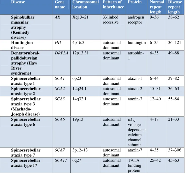
Nine neurodegenerative diseases have as their disease-causing mutation the expansion of a polyglutamine tract (Table 1). This involves the unstable expansion of a CAG sequence within the coding region of each gene. Thus, these diseases fall within a broader class of disorders, i.e. diseases that involve the expansion of an unstable repetitive element, usually triplet sequences (Gatchel and Zoghbi, 2005). Interestingly, expansions of an unstable nucleotide repeat is a mutational mechanism that appears to be unique to the human genome. Furthermore, although genes that are highly homologous to the polyglutamine genes are present in the genomes of other mammals, the polyglutamine tract is not, suggesting that the polyglutamine stretches are not necessary for normal function.
The polyglutamine disorders include spinobulbar muscular atrophy, Huntington's disease (HD), the spinocerebellar ataxias (SCA1, SCA2, SCA3/MJD, SCA6, SCA7 and SCA17), and dentatorubral-pallidoluysian atrophy (DRPLA). All of these disorders are progressive, typically striking at midlife and having a course that consists of an extended period of neuronal dysfunction followed by neuronal loss and eventually death 10–20 years after onset. Other features that characterize this group of neurodegenerative disorders include a direct relationship between the length of the polyglutamine tract and the severity of the disease, and between the number of glutamines and the age of onset and the severity of the disease (Gatchel and Zoghbi, 2005). Mutant CAG repeats show both germline and somatic instability. Germline instability along with the relationship between repeat length and disease course are the basis for the phenomena of genetic anticipation. As the repeat grows upon passage from generation to generation, affected members in successive generations have an earlier age of onset and more rapidly progressing form of the disease.
The autosomal dominant spinocerebellar ataxias (SCAs) are a complex group of neurodegenerative disorders characterized by progressive cerebellar ataxia of gait and limbs variably associated with ophthalmoplegia, pyramidal and extrapyramidal signs, dementia, pigmentary retinopathy and peripheral neuropathy (Zoghbi et al., 2000). Disease onset is usually between 30 and 50 years of age, although early onset in childhood and onset in later decades after 60 years have been reported. The prognosis is variable depending on the underlying cause of the spinocerebellar ataxia subtype. Epidemiological data indicate that SCAs might be more common than that previously estimated with prevalences of up to 5–7 in 100 000 in some populations (Craig et al., 2004).
The clinical features are caused by a combination of degeneration of the cerebellum, basal ganglia, cerebral cortex, optic nerve, pontomedullary systems, spinal tracts or
peripheral nerves. Although the aetiology of SCAs is still poorly understood, genetic analyses, epidemiological data, neuropathological investigations and new experimental models are providing important new insights into the pathogenic mechanisms. At least 28 distinct loci are responsible for rare Mendelian forms of SCA. Interestingly, a few SCA subtypes, including SCAs 1, 2, 3, 6, 7, 17 and dentatorubral pallidoluysian atrophy (DRPLA), are caused by the expansion of a CAG (DNA sequence coding for glutamine) repeat sequence located within the coding region of specific genes, leading to an abnormally long polyglutamine (polyQ) tract in the encoded proteins named ataxins 1, 2 and 3, alpha 1A-voltage-dependent calcium channel, ataxin 7, TATA box binding protein (TBP) and atrophin 1, respectively. These SCAs show, as common features, the progressive neurodegeneration of neuronal subsets in distinct brain areas and the formation of polyQ-containing protein aggregates forming characteristic nuclear or cytoplasmic inclusions (Zoghbi and Orr, 2000). The age at onset and severity of disease symptoms inversely correlate with the length of the glutamine repeat. A second group of SCAs, including SCAs 8, 10 and 12, are caused by a repeat expansion located outside of the coding region of the disease genes leading to dysregulation of gene expression (Zoghbi and Orr, 2000). While the molecular mechanisms underlying SCAs 8 and 10 are unclear, SCA12 appears to be caused by dysregulation of the activity of the crucial enzyme protein phosphatase 2 (PP2, formerly named PP2A) in cerebellar Purkinje cells. Different mechanisms cause cerebellar ataxia and neurodegeneration in SCAs 5, 13, 14 and 27, where alterations in amino acid composition in beta-III spectrin (SPTBN2) (Ikeda et al., 2006), potassium channel KCNC3 (Waters et al., 2006), protein kinase C (PRKCG) and fibroblast growth factor 14 (FGF14), respectively, elicit disease symptoms in these four SCA subtypes (Ikeda et al., 2006). In the rest of SCAs, the genes and, therefore, the mutations remain to be identified and characterized.
SCA1 is one of several inherited forms autosomal dominant ataxia. Typical of most ataxias, SCA1 consists clinically of gait ataxia dysarthria and bulbar dysfunction, with death usually ocurring between 10 and 15 years after the onset of symptoms. Despite the protein, ataxin-1, being widely expressed in the CNS, the most frequently seen and most severe pathological alterations are restricted to loss of Purkinje cells in the cerebellar cortex, as well as loss of neurons in the inferior olivary nuclei, the cerebellar dentate nuclei and the red nuclei (Mascalchi, 2008).
With the identification of an expanded polyglutamine tract as the mutational basis for several neurodegenerative disorders, a pathogenic mechanism largely dependent on the biochemical property of the polyglutamine tract itself gained quick favor. In contrast, two experiments utilizing SCA1 transgenic mice showed that amino acid residues outside of the polyglutamine had a crucial role in pathogenesis. In the first example an amino acid substitution was made in the nuclear localization signal of ataxin-1, such that the protein could no longer be translocated into the nucleus. When this substitution was placed into a mutant allele of ataxin-1 with an expanded polyglutamine tract that was then used to generate transgenic mice, the mice failed to develop disease (Klement et al., 1998). Thus by restricting the subcellular distribution of mutant ataxin-1 to the cytoplasm of susceptible neurons the protein was no longer pathogenic. Perhaps a more dramatic illustration of the importance of 'host' protein sequence for pathogenesis was shown recently when a site of phosphorylation of ataxin-1 was identified, the serine at position 776 (Emamian et al., 2003). Replacing this serine with an alanine yielded a protein that still was transported to the nucleus, but when transported in a mutant ataxin-1 with 82 glutamines failed to cause disease.
TABLE 1. Characteristics of Polyglutamine diseases Disease Gene name Chromosomal location Pattern of inheritance Protein Normal repeat length Disease repeat length Spinobulbar muscular atrophy (Kennedy disease) AR Xq13–21 X-linked recessive androgen receptor 9–36 38–62 Huntington disease HD 4p16.3 autosomal dominant huntingtin 6–35 36–121 Dentatorubral-pallidoluysian atrophy (Haw River syndrome) DRPLA 12p13.31 autosomal dominant atrophin-1 6–35 49–88 Spinocerebellar ataxia type 1 SCA1 6p23 autosomal dominant ataxin-1 6–44 39–82 Spinocerebellar ataxia type 2 SCA2 12q24.1 autosomal dominant ataxin-2 15–31 36–63 Spinocerebellar ataxia type 3 (Machado-Joseph disease) SCA3 14q32.1 autosomal dominant ataxin-3 12–40 55–84 Spinocerebellar ataxia type 6 SCA6 19p13 autosomal dominant α1A - voltage-dependent calcium channel subunit 4–18 21–33 Spinocerebellar ataxia type 7 SCA7 3p12–13 autosomal dominant ataxin-7 4–35 37–306 Spinocerebellar ataxia type 17 SCA17 6q27 autosomal dominant TATA binding protein 25–42 45–63
HUNTINGTON’S DISEASE
Huntington’s disease (HD) is the most common and well-studied polyglutamine neurodegenerative disorder. It has a prevalence of 3–10 affected subjects per 100.000 individuals in Western Europe and North America (Gil and Rego, 2008). The disorder was first described in the 19th century by George Huntington, who identified both its clinical features and its pattern of familial transmission. However, it was not until 1993 that a multicenter consortium, organized by the Hereditary Disease Foundation, discovered the actual HD gene mutation. This is an unstable expansion of CAG repeats within the coding region of the HD gene, which is located on the short arm of chromosome 4 (4p63) and encodes the protein huntingtin. The mutation results in a stretch of glutamine residues located in the NH2-terminal of huntingtin (The Huntington’s Disease Collaborative Research Group, 1993). Although the abnormal protein is ubiquitously expressed throughout the organism, cell degeneration occurs mainly in the brain (Vonsattel and DiFiglia, 1998).
CLINICAL MANIFESTATIONS
Classically described as Huntington's Chorea (the Greek word for dance), the first signs of the disease are subtle: clumsiness, difficulties with smooth eye pursuit, and slight uncontrolled and awkward movements. These motor disturbances, associated with the loss of voluntary movement coordination, progress slowly. The involuntary movements of the proximal and distal muscles become more severe and the patients gradually lose their capacity to move and eventually communicate. Bradykinesia and rigidity are also common symptoms in Huntington's patients, especially in late stages of the disease. Death generally occurs as a consequence of heart failure or aspiration pneumonia (Sánchez-Pernaute et al., 2000). In juvenile patients the symptomatology is considerably different, being characterized by bradykinesia, tremors, rigidity and dystonia, and
chorea may be completely absent. Affected children may also suffer epileptic seizures. The majority of patients suffer also from inexplicable muscle wasting and weight loss, despite constant caloric intake (Aziz et al., 2008). Although the cause of these peripheral symptoms is still unclear, a recent study has demonstrated the existence of a positive correlation between the levels of branched chain amino acids (valine, leucine and isoleucine), weight loss and disease progression in HD patients, reinforcing the importance of a systemic metabolic defect in HD pathology (Mochel et al., 2007). A number of endocrine abnormalities have also been reported in HD patients, including increased levels of corticosteroids and reduced levels of testosterone (Markianos et al., 2005). Furthermore, 10–25% of HD patients exhibit diabetes mellitus. The cognitive capacities are also severely affected during the course of HD. The slowing of intellectual processes is the first sign of cognitive impairment in HD patients and deficits in some cognitive functions can in some cases be detected decades before the onset of motor symptoms. These cognitive impairments get worse over time and late-stage HD patients show profound dementia. Manic–depressive behavior and personality changes (irritability, apathy and sexual disturbances) are often part of the psychiatric syndrome. Based on the above features, the criteria used for the diagnosis of HD include: (i) a family history of HD; (ii) progressive motor disability with chorea or rigidity with no other cause; and (iii) psychiatric disturbances with progressive dementia with no other cause (Vonsattel and DiFiglia, 1998). Currently, individuals showing these symptoms are submitted to genetic testing in order to screen for the HD mutation and confirm the diagnosis.
GENETICS
The mutation responsible for HD constitutes a stretch of uninterrupted CAG trinucleotide repeats located near the 5'-end in exon 1 of the HD gene coding sequence, which comprises 67 exons. Consequently, mutant huntingtin bears a tract of consecutive glutamine residues in its NH2-terminal, 17 amino acids downstream of the initiator methionine (The Huntington's Disease Collaborative Research Group, 1993). The disease is inherited in an autosomal dominant manner. The normal allele is transmitted from generation to generation in a Mendelian fashion. The mutant allele is unstable during meiosis, changing in length in the majority of intergenerational transmissions, with either slight increases of 1–4 units or decreases of 1–2 units. In rare occasions, larger-sized increases occur in parental transmissions, reflecting a particularly high mutation rate during spermatogenesis (The Huntington's Disease Collaborative Research Group, 1993). The number of CAG repeats (i.e. glutamine residues) is the primary and major determinant of disease severity, accounting for 30–60% of the
variance in the age of onset of the first symptoms. The remaining variance is attributable to other genetic features and environmental factors (Arning et al., 2005). Interestingly, in a recent study that involved 167 HD patients, the length of the polyglutamine tail (which ranged from 41 to 45 units) accounted for only 30.8% of the variance in the age of onset, whereas 12.3% additional variance could be attributed to genotype variation in the gene coding for the N-methyl-D-aspartate (NMDA) receptor subunit NR2B and 4.5% to genotype variation in the gene coding for the NMDA receptor subunit NR2A (Arning et al., 2005). Normally, asymptomatic individuals have >35 CAG repeats whereas HD is manifested when the number of repeats exceeds this threshold. Alleles with 35–39 repeats are associated with later onset of the disease, although incomplete penetrance has been observed in some individuals who have shown no symptoms or neuropathological signs. Alleles of 40–50 units give rise to the most


![Figure 1.2 - Saturation curves and Scatchard plot of [ 3 H]-SCH 58261 specific binding to ST14A ( • ), N548 mu (■) (A) and ST14A ( • ), FL mu ( ▲ ) (B)](https://thumb-eu.123doks.com/thumbv2/123dokorg/4708309.45164/88.892.129.752.252.1002/figure-saturation-curves-scatchard-plot-sch-specific-binding.webp)