SAPIENZA Università di Roma Facoltà di Farmacia e Medicina
Ph.D. in MORPHOGENESIS AND TISSUE ENGINEERING
XXXII Ciclo A.A. 2018/2019
Evaluation of the histopathological modifications induced by the positioning of
autologous platelets-rich plasma vs. biosynthetic, absorbable mesh as reinforcement of the cruroplasty:
a pilot study on an animal model Cristian Eugeniu Boru, MD, Ph.D. Student
Tutor Coordinator
Evaluation of the histopathological modifications induced by the positioning of autologous platelet-rich plasma vs. biosynthetic, absorbable mesh as reinforcement of the cruroplasty: a pilot study on an animal model
Dr. Cristian Eugeniu Boru MD, PhD student
PhD candidate in Morphogenesis & Tissue Engineering
Department of Anatomical Sciences, Histology, Forensics and of the Locomotor Apparatus (SAIMLAL)
Histology and Medical Embryology Section University La Sapienza of Rome
Prof. Gianfranco Silecchia, MD, PhD Full Professor of Surgery
Director of UOC & Bariatric Centre of Excellence-IFSO EC Department of Medico-Surgical Sciences and Biotechnologies UOC General Surgery & Bariatric Centre of Excellence-IFSO EC University La Sapienza of Rome, Polo Integrato AUSL LT-ICOT
Prof. Dr. Nicolae Manolescu, PhD, Professor Emeritus
Member of Romanian Academy
Member of The French National Academy of Medicine Department of Morphopathology and Comparative Oncology Faculty of Veterinary Medicine
Spiru Haret University, Bucharest, Romania
[email protected] Prof. Catalin Copaescu, MD, PhD
Associated Professor of Surgery
Head of Minimal Invasive and Metabolic Surgery Dept., Director of the Bariatric & Metabolic Surgery Program Bariatric Centre of Excellence IFSO EAC-EC
Ponderas Hospital, Bucharest, Romania
[email protected] Prof. Dan Ulmeanu, MD, PhD,
Associated Professor of Surgery
Faculty of Medicine, Titu Maiorescu University, Bucharest Head of General and Thoracic Surgery Department
Regina Maria Baneasa Hospital, Bucharest, Romania
INDEX
1. Index pag. 5
2. Summary pag. 7
3. Introduction pag. 9
4. Aims of the study pag. 17
5. Materials and methods pag. 19
5.1 type of study pag. 19
5.2 PRP preparation pag. 20
5.3 surgical procedures pag. 21 5.4 histopathological examination pag. 25 5.5 statistical considerations pag. 29
6. Results pag. 31
6.1 histopathological results pag. 31 6.2 statistical analysis results pag. 34
7. Discussions pag. 37
8. Conclusions pag. 47
9. Figures pag. 49
10. Tables pag. 71
11. List of publications pag. 77
1. SUMMARY
Introduction: Symptomatic hiatal hernia’s (HH) incidence, associated with gastroesophageal reflux disease (GERD), ranges between 10-20% in the general population. Laparoscopic suture of the diaphragmatic crura (posterior cruroplasty PC) during the HH’s surgical treatment determines good long-term results in the majority of cases, without cancelling the risks of recurrences or surgical revisions. Based on anatomical, physiological, morphological and evolution of certain diseases, the use of porcine model became one of the most important research fields, including laparoscopic implant of prosthetic materials. Cruroplasty’s reinforcement is widely used in large hiatal defects for recurrences’ reduction with no consensus on type or shape of mesh. The use of autologous platelet-rich plasma (PRP) concentrate, simple and cheap prepared, might be an alternative option. Aims: finding new solutions for the laparoscopic treatment of symptomatic HH, in particular comparing a biosynthetic, absorbable mesh, vs. autologous PRP in a porcine model, in order to determine local histopathological (HP) changes and complications. Rationale: the literature lacks on comparative studies on the use of PRP for the reinforcement of hiatal defects repair. Materials and methods: Prospective, randomized study on 12 pigs. The laparoscopic procedure consisted in creation of a hiatal defect, then sutured PC, reinforced with absorbable mesh (group A) or PRP positioning (group B). Blood withdrawal was made prior to surgery for autologous concentrate’s preparation in group B. The animals survived the procedure and professional care was taken for the next 7 months. Second procedure was performed afterwards, consisting in euthanasia, laparoscopy, and laparotomy for specimen retrieval (whole hiatal area). HP examination was performed, evaluating 4 main components: proliferative fibroblast – fibrocyte – collagen neoformation; vascular; inflammatory; and dystrophic. A semiquantative HP scoring of tissue reaction was elaborated.
management of the HH/GERD, definition of the technical indications, avoiding costly materials loss. Results: animals survived 7 months of postoperative care, with normal growth and without complications related to the surgical procedures. There were no complications, local or general, in both groups, with more intense adherential process in group A at the gross examination. Complete resorption of both products was demonstrated on gross and HP examination. Both reinforcements determined inflammatory infiltrates predominantly mononuclear, with induced local collagen production and tissue neo-vascularization. Animals in the mesh group had increased mean chronic inflammation score (2.66 vs. 1.83, p = 0.908) compared to the PRP group. Vascular network did not differ between groups (p = 0.3061), although there was an increased trend toward mean collagen fibers in the mesh group (score 2.83±0.4) compared to PRP (2.33±0.81), p=0.248.
Conclusions: Our laparoscopic, animal model of hiatal hernia repair was reliable, feasible and reproductible. The comparative study of both materials, autologous and biosynthetic, did not show any specific complications, with no residual material found after 7 months. The mesh group showed a more important score of sclerotic collagenising process. PRP was shown to be an autologous source of pro-regenerative growth factors and chemokines suited to hiatal area wound healing, enhanced angiogenesis, myofibroblast recruitment and tissue ingrowth and less severe peritoneal adhesions. The novelty of the study and the histopathological results suggest the use of combined products, so autologous PRP could find a clinical application in the surgical treatment of the hiatal defects, as a promising co-adjuvant to local remodeling and healing in this animal model. Next studies should verify that the use of PRP could improve the crura’s weakness.
Keywords: Hiatal hernia, platelets-rich plasma, biosynthetic absorbable mesh, laparoscopy, pigs.
2. INTRODUCTION
Hiatal hernia’s (HH) prevalence is estimated up to 50% of the general population, while the incidence of the symptomatic HH, associated with gastroesophageal reflux disease (GERD) ranges between 10-20%. Symptomatic GERD represents the most prevalent gastrointestinal disturb in the USA, with a relevant risk of severe chronic esophagitis, Barrett esophagus and even esophageal adenocarcinoma development1.
Laparoscopic treatment of symptomatic hiatal hernia is already established as first choice option, and laparoscopic Nissen fundoplication (LFN), associated with posterior cruroplasty (PC), represents the gold standard2. The fundoplication remakes an
insufficient lower esophageal sphincter (LES) using the gastric fundus, in order to create a barrier at the level of the gastroesophageal junction (GEJ)3, while reinforcement of the PC is
advised in case of large hiatal defects and/or weakness of the crura4.
Simple suture of the hiatus has been associated with good long-term results, but recurrences and increased rate of surgical revision may be seen even in high volume, specialized centers5. Reparation’s
susceptibility of disruption in patients with HH was demonstrated to be caused by the ultrastructural abnormalities at the muscular tissue of the crura and the outcome of the anti-reflux surgery could depend not only on the surgical technique, but also on the quality of the diaphragmatic crura6.
Reinforcement of the sutured PC in HH repair, in the presence of large hiatal defects and / or weakness of the diaphragmatic pillars, has gained worldwide interest due to the availability of new materials.
Mesh reinforcement has been proposed as a method of recurrence prevention by Frantzides7. The indication for mesh use is
directly determined by the defect size and quality of the pillars. There is no consensus on the size, type, shape, and position of mesh on the hiatus (SAGES2, EAES4). The concerns regarding permanent
and other nearby vital organs, local infections, and technical difficulties, dysphagia, fibrosis, stenosis of the esophago-gastric junction, and complete transmural gastric migration8, 9. The
European Association of Endoscopic Surgeons (EAES) recommended in 2014 the use of mesh limited to large hiatal defect and weak crura4 while SAGES recognized the controversies on the
use of mesh on the hiatus2. A tailored surgical approach was
suggested by different Authors10, 11: simple suture of the crura,
reinforced suture or tension-free reinforced suture; the choice should be made based on pre- or intraoperative measurement of Hiatal Surface Area. This area is determined by the two-dimensional expanse of the hiatal orifice, measuring the length of the crura in centimeters beginning at the crural commissure up to the edge where the pars flaccida begins and the circuit between the both crural lateral edges.
Mesh materials in hiatal hernia repair
There are 3 categories of mesh available on the market:
1. Biological scaffolds origin from porcine dermis or intestine,
bovine dermis or pericardium or even human cadaveric dermis or
fascia lata. It was suggested that biological, absorbable meshes
might be used even in clean-contaminated or contaminated situations, due to a better clearance of the bacteria, whereas biosynthetic meshes are not indicated. In the same time bacterial colonization may lead to accelerated enzymatic degradation of the mesh, with less long-term satisfactory results. Oelschlager reported use of biologic mesh for laparoscopic repair of large, complicated hiatal hernias that appeared safe, with no major complications related to the mesh, and overall satisfaction with the operation was very good12.
Over these advantages, the costs of these prostheses are high, handling and fixation might be difficult, while the possibility of immunological reactions are also reasons of concern. There is insufficient data to establish the optimal use in different surgical procedures, as well as on the crura13, 14.
2. Non-absorbable biosynthetic meshes raise major concerns
about the long-term risk of mesh erosion. Popular use for repair of incisional hernias might advocate for their use in reinforced cruroplasty15, because they incorporate rapidly into host tissue and
have lower recurrence rate7, 16. On the other hand, anecdotal reports
contra-indicated the use of these prostheses in the reinforcement of the HH’s repair; permanent mesh might be associated with risks of organ resections at revisions for failed HH’s surgery9, 17. Specially
designed, V-shaped, non-absorbable, covered mesh was specially created for use on the hiatal area16,with large-scale use, but recently
it was withdrawn from the market for not-reported adverse effects18. 3. Absorbable biosynthetic scaffolds (on short and
long-term) have different components like polyglycolic acid (PGA) in Dexon® (Covidien, Mansfield, MA); copolymer of glycolide and
lactide in Vicryl® (Ethicon, Inc., Somerville, NJ); copolymer of
polyglycolide: thrimethylene carbonate in Gore® Bio-A® (W.L.
Gore & Associates, Inc., Flagstaff, AZ); or different combinations, like macroporous multifilament absorbable biosynthetic mesh in Tigr Matrix® (Novus Scientific, Uppsala, Sweden). The use of
biosynthetic meshes reduced the incidence of recurrences 8,19,20.
This type of mesh requires the presence of a substrate where fibrous tissue can be formed, so that the new structure remains after mesh absorption. Rapid absorption, like in case of Vicryl® or
Dexon®, determine weakening of the mechanical strength and make
them not suitable for the use in hernia repairs that requires long-term support. Medium or long-term absorbable scaffolds like Gore®
Bio-A®, Tigr Matrix® and PhasixTM Mesh (C.R. Bard, Inc./Davol Inc.,
Warwick, RI) are fully absorbable scaffolds due to hydrolysis and/or enzymatic reactions, providing long-term support (6-18 months), bacteria cleaning, with use in different abdominal wall defects21.
There is a shortness of studies to establish the safety and effectiveness of absorbable mesh reinforcement in hiatal hernia repair; it was shown that the use of resorbable biosynthetic meshes in the prevention or treatment of abdominal wall defects in animal experiments with small defects in clean setting seems safe, with no
serious complications related to the device during short-term follow-up22. Although a certain degree of remodeling occurs, it remains
completely unclear whether this can lead to strong fascial tissue of good quality in the long term.
Currently the only biosynthetic, absorbable mesh with CE mark for hiatal hernia use is Gore® Bio-A® Tissue Reinforcement
since 2008, as well as Food and Drug Administration’s (FDA, USA) approval for soft tissue reinforcement since 201723.
There is no clear experimental evidence currently available that can support the advantages of absorbable biosynthetic meshes over the use of biosynthetic non-absorbable or biological meshes in human settings, mostly due to lack of good data. More experimental studies were shown to be needed, using standardized models and parameters, followed by randomized controlled trials and prospective registries in humans with a sufficiently long follow-up period to reveal the potential advantages in clinical practice21. The characteristics of ideal reinforcement on the hiatus are:
- to ensure durable and perfect reparation; - perfect integration in the area’s biodynamic;
- completely absorbable (fully disappearing from the body, in order to avoid long-term presence of foreign body);
- tolerance of infection, or positioning’s possibility in a contaminated environment;
- no premature absorption in presence of bacteria; - simple positioning;
- stable (no migration);
- possibility of use as bridge situations; - cost-effective (accessible price);
- prevention of visceral complications (avoidance of adherences, fistulas, occlusions);
- resist shrinkage; - promote minimal pain; - non-carcinogenic.
Ideal mesh does not exist yet24. Biological meshes, even proving
good short-term results, haven’t proved long term efficacy, while non-absorbable meshes might cause serious complications like erosion or migration. Reduction of HH’s recurrence and reinterventions became a priority due to more complicated situations in these cases, with a higher incidence of complications. That’s why it became necessary to study and evaluate biocompatible materials and techniques that could improve the results of hiatal hernia repair.
Platelet-rich plasma and its use in surgery
Recent studies on platelet-rich plasma (PRP) showed a positive effect on tissue remodeling and neovascularization, due to platelets’ capacity of different proteins’ liberation (growth factors, chemokine, cytokines etc.) that stimulates the tissue reparation25.
Independently of the role in thrombus formation during hemorrhage, the platelets attract cells that stimulate the tissue reparation (neutrophils, macrophages, stem cells etc.) through growth factors liberation, with consecutive activation of the intracellular cascade that generates migration, proliferation and diversification26. The
degranulation of the platelets’ alpha-granules releases an increased number of growth factors, like platelet-derived growth factor (PDGF aa/bb/ab), transforming growth factor b1/2 (TGF-b1/2), vascular endothelial growth factor (VEGF), basic fibroblast growth factor (bFGF), platelet-derived epidermal growth factor (PDEGF), and insulin-like growth factor-I/II (IGF-I/II) 25, 26, 27,28. Not only this, but
platelets have a role in the immunomodulation and antiseptic proprieties. All these characteristics suggested the possibility of platelets’ use in various surgical fields as adjuvant therapy.
The platelet-rich plasma PRP is a hemocomponent for local use, autologous or allogenic, obtained from the aggregation of a platelet concentrate put in contact with calcium and pro-aggregation biological (thrombin) or pharmacological factors25, 29. The PRP is,
basically, a super-concentrated form of platelets positioned in situ, on the lesion, in various surgical branches: oral and maxillo-facial, orthopedics and bone reconstructions, cutaneous ulcers, chronic
wounds, esthetic and plastic-reconstructive surgery, and ophthalmology30, 31, 32, 33, 34, 35, 36, 37, 38, 39, 40.
The local use of PRP is favored by its characteristics like plasticity and possibility of modelling on the application spot. After its activation and platelets’ degranulation, growth factors (GFs), chemokine and other bioactive metabolites are released, similar to the physiological process. These components could contribute to rapid reparation of abdominal wall defects due to the anabolic and chemotactic activity. There are reports on the PRP’s efficacy on improvement of tissue response, on reduction of the incisional hernia’s incidence and in reinforcement of tissue resistance41, 42. Use
of PRP in ventral hernia repair with acellular dermal matrices (biological mesh) as an autologous source of pro-regenerative growth-factors and chemokines was advocated in a rat model, due to the enhanced local angiogenesis, myofibroblast recruitment and tissue ingrowth. Further favorable effects as less severe peritoneal adhesions and diminished hernia recurrence might stimulate the use of PRP as an augmentation of the ventral hernia repair, with less recurrences and preserved mesh integrity43.
Use of PRP in HH repair is anecdotal and only on animal models. Altieri reported the use of a filtered platelet concentrated on the HH’s repair in pigs, after the surgical cruroplasty reinforced with the positioning of a biological mesh. Practically, the mesh was impregnated with the filtered platelet concentrate and this reparation was compared with 2 groups (control as simple surgical suture and reinforced cruroplasty without PRP). The hiatal area was histological analyzed and tensile strength testing was performed. A trend toward increased collagen deposition and vascularity in the filtered plasma concentrate group was shown, with increase tensile strength and yield force44.
Improvement of the incidence of postoperative complications and recurrences after hiatal defects repair has a crucial importance. Our study proposes to confront a biosynthetic, absorbable mesh (Gore® Bio-A® Tissue Reinforcement) used for the reinforcement of
laparoscopic PC vs. the surgical repair reinforced by the application of an autologous platelet concentrate PRP in a porcine model of HH repair.
Tissue renewal and repair, as a pathological process, consists in regeneration (restitution of tissue components identical to those removed) and healing, a fibroproliferative respond involving a number of processes: inflammatory reaction to the initial injury, with removal of damaged and dead tissue; proliferation and migration of parenchymal and connective tissue; angiogenesis and granulation tissue; collagen deposition; tissue remodeling; wound contraction and acquisition of wound strength. It was shown that Gore® Bio-A® tissue reinforcement scaffold was designed to break
down primarily by hydrolysis. Within one to two weeks after positioning, the patient’s cells migrate into the scaffold and begin generating vascularized soft tissue. Gradually over approximately 6 to seven months, the material is absorbed by the body and replaced 1:1 with the patient’s own favorable type 1 collagen.
In the meantime, autologous PRP, having all the characteristics of a self-product, contains an array of growth factors and chemokines that can boost tissue healing25-27,44.
3. AIMS OF THE STUDY
1. To establish a laparoscopic model of a hiatal defect in pigs 2. to analyze the hiatal area’s HP modifications induced by the
biosynthetic, absorbable mesh positioning as reinforcement of the laparoscopic posterior cruroplasty and its full absorption;
3. to analyze the hiatal area’s histopathological HP modifications induced by the autologous platelet-rich plasma PRP positioning as reinforcement of the laparoscopic posterior cruroplasty and its absorption;
4. to evaluate the safety profile of the reinforcements (adhesions, infections, periesophageal fibrosis, distortion of the local anatomy, inflammatory pattern)
Rationale of the study: The rationale behind this experimental
study is to evaluate the anatomical and HP pattern of the hiatal area after mesh and PRP absorption, because the use of meshes on the hiatus keeps on being controversial due to the severe complications related to their use and the recurrence’s rate. The study proposes to evaluate if the PRP’s application could improve the crural repair and the tissue remodeling in a porcine model of hiatal hernia repair. Proposed objectives of the reinforcement are:
- No recurrence;
- Increase of the security of the positioning near the
esophagus;
- Tissue remodeling improvement, with stronger reparation. The particularity of the study (justification): evaluation of a
model, outcomes, pitfalls, and applicability to the clinical setting of an autologous hematologic product, adjuvant for soft tissue repair in abdominal or inguinal wall but applied in the HH repair. The PRP is compared with a mesh already used for this pathology. The literature is lacking in studies on the use of PRP in hiatal defects repair, with only one published study on a porcine model and one report of its
use in humans, with no comparative studies between autologous and biosynthetic reinforcement.
Justification of the animal model
The necessity of choosing the porcine model is based on the fact that the surgical procedure must reproduce faithfully human intraoperative conditions that surgeons should face during laparoscopic hiatal defects reparations. The porcine model is one of the most used between superior mammals, due to the genetically vicinity, being widely recognized as the most relevant pre-clinical model, based on the organs, physiology and digestive similarities45.
That’s why it is hypothesized that the experimental surgery’s results are transferable on human species and that is why the information gained cannot be obtained in different ways. Histological evaluations, gained on medium term, will offer original data useful for the medical industry’s efforts on more and more affordable, efficient biomaterials production and use on esophago-gastric pathology.
4. MATHERIALS AND METHODS: 4.1. TYPE OF STUDY
This is a pilot study, designed as a prospective, open, randomized study, performed on fourteen (n=14) young female Yorkshire pigs, aged 4-6 months, weighting 20-25 kg, obtained from an institutionally approved vendor. University “La Sapienza” of Rome, Faculty of Medicine and Pharmacy, Department of Medico-Surgical Sciences and Biotechnologies Board’s Committee approved the study’s experimental protocol, and ulterior it was approved even by the Minister of Health of Italy.
Pigs were acclimated after acquisition and operated in an advanced experimental center, Surgical Training Institute, located in Ponderas Academic Hospital, Bucharest Romania. Logistics, operating rooms fully equipped for laparoscopic approach and oro-tracheal intubations machines, and authorized animal manipulation by the National Health Minister of Romania were provided in this quality-controlled center. The center has over 18 years of experience in experimental surgery on animals, simulators and training.
Study groups:
Group B: Hiatal surgical suture (PC) + Bio-A® mesh: n=6 animals;
Group 2. Hiatal surgical suture (PC) + PRP: n=6 animals.
A biosynthetic, absorbable prosthetic mesh was considered for the study: a copolymer of polyglycolic acid (67%)- trimethylene carbonate (33%), GORE® Bio-A® Tissue reinforcement (W. L. Gore
& Associates, Newark, NJ, USA) – was placed after posterior crural closure as reinforcement of the surgical procedure. The use of Bio-A® mesh in the reinforcement of the cruroplasty has already been
reported (see table 1), with good results46, with no local or general
complications and tendency in reducing postoperative recurrence20.
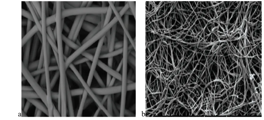
The mesh is composed of a 3-dimensional web of polymers (figure 1), gradually absorbed over 6 months and replaced by vascularized soft tissue, due to the open, highly interconnected pores that facilitates cell infiltration and vascularization to generate quality tissue fast, leaving no permanent material behind. This
biodegradable mesh provides a temporary scaffold for ingrowth of native tissue and collagen deposition with no risk of contamination and infection. It does not require any preparation, such as soaking or stretching, and no special storage conditions. It was suggested that fully absorbable meshes with longer-term absorption profiles might provide improved mechanical and histological properties compared to biologically derived scaffolds47.
4.2. PRP PREPARATION
The autologous PRP concentrate is prepared in concomitancy with the surgical procedure through a method that permits to obtain 2 fundamental elements: the platelets concentrate and the thrombin, the last one necessary for the platelets’ activation and degranulation. The preparation procedure consists in 4 phases 38:
4.2.1 Autologous, full blood withdrawal
A 20 ml venous blood withdrawal was performed from the pigs randomized in the PRP reinforcement group, using jugular or femoral vein access, with a 20 Gauge needle and a 60 ml syringe preloaded with a sodium citrate, citric acid and dextrose anticoagulant solution (ACD-A, Salf SpA, Canate Sotto, Bergamo, Italia), in a 1:9 ratio. The ACD-A solution contains: 22.0 g/L bihydrate sodium citrate, 8.0 g/L mono hydrated citric acid, 24.5 g/L monohydrated glucose (dextrose) in i.v. sterile solution. A 10 ml additional quantity of autologous venous blood is withdrawal and distributed in 2 tubes of 5 ml each, containing sodium citrate as anticoagulant (Terumo Medical Corporation, Elton, MD, USA), in order to obtain the autologous thrombin.
4.2.2 Platelet rich plasma preparation
The 20 ml blood sample is divided in 2 tubes (Sterilin Ltd, Newport, UK). Both tubes are centrifuged (Rotina 46R, Hettich, Milano, Italia), in order to obtain various blood components’ separation, based on their density. The plasma and buffy coat will be centrifuged again in order to obtain the platelet concentrate sedimentation (CP or platelet pellet) on the base of the tube and the platelet-poor plasma (PPP) above. Some of the PPP will be
discarded, leaving an 8 ml volume of PRP, obtained from the re-suspension of the CP in the PPP. Small quantities (0,5 ml) of blood and of final product are used in order to confront the platelets’ concentrations (Cell Dyn 3500R, Abbott, Wiesbaden, Germania) and confirm the adequate platelets’ therapeutically concentration40.
4.2.3 Autologous thrombin preparation
Autologous blood contained in the 5 ml tubes with sodium citrate was centrifuged for 10 minutes. At this plasma fraction 10% calcium gluconate is added (446 mEq/l di Ca++) (Monico SpA,
Venezia-Mestre, Italia), in a 5:1 ratio. The plasma – calcium gluconate mixture is incubated at 37°C in CO2 incubators a (NuAire DH
Autoflow, Plymouth, MN, USA). The obtained clot is pressed, and the thrombin rich solution is collected for the next phase, the PRP activation.
4.2.4 The PRP activation
The PRP gel is obtained putting together the PRP, the thrombin rich solution and the calcium gluconate in an 8:1:0.5 ratio in about 10 min. The entire lab procedure is performed with sterile, single use materials, in asepsis conditions, under a hood of laminar flow (Bicasa, Barnareggio, MB, Italy), respecting the lab’s good practice.
4.3 SURGICAL PROCEDURES
4.3.1 Phase one: first surgical intervention March 2018
Animals were fasted for at least 12 hours prior to surgery. An antibiotic (amoxicillin 15 mg/kg IM every 24 h) and anti-inflammatory therapy (flunixin meglumin 2,2 mg/kg IM every 24 h) was initialized in the day of surgery and followed for the next 5 days.
A clinical examination was performed, necessary for the clinical status confirmation (ASA I, American Society of Anesthesiologists classification48), followed by i.m. premedication
with a mixture of ketamine (5 mg/kg), xilazin (2.5 mg/kg) e butorfanolo (0.2 mg/kg), part of the multimodal anesthesiologic plan.
reduction of the psycho-motor response to manipulations) a central venous line was positioned near the auricular vein. General anesthesia was induced with thiopental sodium (6 mg/kg) and maintained on inhalator way through oro-tracheal intubation and mechanical ventilation, with a mixture of isoflurane in oxygen. These procedures guarantee a stable surgical anesthesia during all procedures, with the main aim to limit / cancel the consequent nociceptive stimuli. In case of anesthetic plan reduction (like cardiac frequency increasing, respiratory frequency increasing, blood pressure increment, muscular rigidity, voluntary movements), seen on clinical and instrumental monitoring, as routine during veterinary anesthesia, an adaptation of the treatment was done with supplements of analgesics and anesthetics.
A blood withdrawal was performed in the Group B before surgical procedure started, necessary for the PRP and thrombin production that followed the steps described above.
Animals were placed in dorsal recumbency, in anti-Trendelenburg position, and the ventral abdomen was prepared for aseptic survival surgery by shaving the entire abdominal region, cleaning the operative area with three alternating scrubs of povidone-iodine solution and 70% isopropyl alcohol solution, and applying sterilized surgical drapes over the entire field.
Following preparation of the abdomen, laparoscopy was performed using standard laparoscopic trocars and instruments: usually one optical trocar placed supra-umbilical, two 5 mm trocars laterally for the operating surgeon and one 5 mm in the epigastrium for liver/organs retraction. After pneumoperitoneum induction and exploration of the abdominal cavity, the hiatal area was exposed. Diaphragm’s left crus was exposed after upper gastric greater curvature and gastro-phrenic ligament dissection, with mobilization of the gastric fundus. Short gastric vessels were partially resected in case of necessity of better gastric mobility using advanced electrosurgery (Ultracision, Ethicon Johnson & Johnson). Afterwards right crus was exposed and proper dissection of the esophagus was made in order to fully exposed the intra-abdominal
esophagus, eso-gastric junction and the retroesophageal window. Periesophageal dissection was completed in case of necessity.
In order to create an artificial hiatal defect, which can simulate real defects, a cut or partial excision of right crus was executed, with further dissection. The resulting defect was measured based on a simplified calculation of the hiatal surface area (HSA), with the area of a rhombus formula (d1X d2/2), and a surface up to 4 cm2 was obtained (see figure 2).
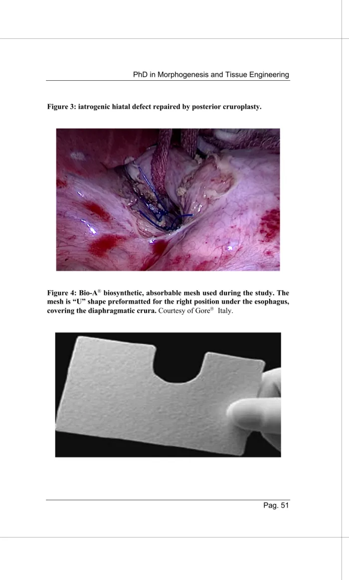
Crura’s suture: Repair of the iatrogenic hiatal defect was
performed by laparoscopic suture of the hiatus (posterior cruroplasty PC, figure 3) with interrupted, non-absorbable stiches (2–0 polypropylene sutures, Ethicon, Cincinnati, OH, USA).
Reinforcement of crura suture: randomization between the
two types of available reinforcement was computer generated (Excel, Microsoft): Bio-A® mesh (Group A) vs. PRP (Group B).
The absorbable, biosynthetic mesh was tailored for local use. Due to standard dimension (rectangle of 7X10 cm), its length was be reduced to 5/8 cm, cutting the left part of the mesh, creating a U-shape hole in the upper part of the mesh, in order to obtain an asymmetrical ‘‘U’’, leaving the right part longer than the left to obtain adequate right pillar reinforcement (figure 4) The ‘‘U’’ concavity was slightly enlarged downward to reduce the risk of possible contact with the esophagus. Then it was rolled like a cigarette and inserted through the 10-mm trocar, superimposed on the cruroplasty and fixed with Prolen 2/0 stiches. The incised part of the mesh (the so-called U) was positioned under the esophagus. Due to the total absorbable characteristics of the mesh and of the fixing devices, some titan clips were used for marking the covering area. These clips could have been useful for radiological identification as well.
preparation of the hiatal area. The autologous PRP was prepared concomitant with the surgical preparation of the hiatal area and PC in about 40 min. It was presented as a thick gel (figure 5), and it was positioned over the PC pillars, using a laparoscopic applier and fixed with absorbable stitches. Final image after reinforcement positioning over the PC is shown in figure 6.
4.3.2 Second phase: postoperative animals monitoring 03. 2018 – 09. 2018
After operation and 2 days recovery and strict monitoring, pigs were transferred, cured, raised and followed for the next 7 months in the stalls of a conventional animal farm, where they were routinely monitored, daily feed and routinely cleaned. Wounds were checked daily for the first 10 days, till stiches removal and full healing. Assistance was provided daily, according to the national regulations for animal testing (Allegato III del Decreto Legislativo 26/2014 regarding porcine housing), by a collaborating veterinary doctor, using the auxiliary models, see Table 2: Clinical card of daily monitoring. Grimace scale49 (0 = not present, 1 = moderately present, 2 = obviously present), and Table 3: Body Condition Scoring50. In case of significant variations of the monitored
parameters, an increment of the monitoring and required therapy was applied. In case of medical issues that could became too painful for the animals or incompatible with life, euthanasia would have been performed before the ending of the study.
The humanitarian endpoints will consist in the following:
1. Grimace scale >15, or < 3 in the Body Condition Scoring; 2. External physical aspect: dehydration, necrosis and ulcers,
edema, or surgical wound dehiscence;
3. Behavioral modifications: abatement and severe depression of the sensory, voluntary fasting.
4.3.2 Third phase: second surgical intervention Oct 2018
Pigs were returned to the operating rooms 12 weeks after primary operation, in the same Surgical Training Institute, located
in Ponderas Academic Hospital, Bucharest Romania. The animals were prepared to surgery in the same way like in phase 1, with oro-tracheal intubation and field preparation. Afterwards, laparoscopy was performed on the anesthesiated pigs. After inspection of the abdominal cavity and dissection of the eventual adherences, the hiatal area was exposed and video registration was performed, in order to demonstrate any modification of the hiatal area.
After video recording, the animals were sacrificed, being under general anesthesia. Euthanasia was obtained accordingly to the
American Veterinary Medical Association (AVMA)51. Afterwards,
laparotomy was performed, and all animals were visually checked for local effects like adherences, stenosis, mesh presence, cruroplasty’s previous sutures. In order to obtain a bigger specimen, like en-bloc resection of the hiatal area (thoracic and abdominal esophagus, diaphragm, periesophageal fat, hiatal crura containing the studied area), with full removal of the mesh-reinforced area, conversion to laparotomy was done for an easier and faster access. In this way we could demonstrate the full or partial absorption of the mesh and of the PRP. After inspection of the abdominal cavity and dissection of the eventual adherences, the hiatal area was exposed and surgically removed (figure 7). The entire specimen was preserved in formalin solution 10% and prepared for transportation in refrigerated containers.
4.4 HISTOPATHOLOGICAL EXAMINATION
Specimens were transported to the Department of Morphology and Morphopathology, Faculty of Veterinary Medicine, University of Agronomic Sciences and Veterinary Medicine, Bucharest Romania, for the definitive histopathological diagnosis. The specimens were placed in a liquid fixing agent (fixative) such as formaldehyde solution (formalin). Following fixation, the specimens were dissected to select appropriate areas for examination. Processed specimens were placed in suitable labelled cassettes (small perforated baskets) to segregate them from other
specimens. Because melted paraffin wax is hydrophobic (immiscible with water), most of the water in a specimen must be removed before it can be infiltrated with wax. This process is commonly carried out by immersing specimens in a series of ethanol (alcohol) solutions of increasing concentration until pure, water-free alcohol is reached. The next step is clearing with xylene. This solvent will displace the ethanol in the tissue, then this in turn will be displaced by molten paraffin wax. Then the tissue was infiltrated with a suitable histological wax. After the specimen is thoroughly infiltrated with wax it must be formed into a “block” which can be clamped into a microtome for section cutting. This step was carried out using an “embedding center” where a mold was filled with molten wax and the specimen placed into it. After paraffin embedding, samples were sectioned at 3µm width using the microtome (Leika®, Germany), mounted onto electrostatically
treated glass slides (Bio-Optica®, Milano, Italy), and stained with
morphological stain hematoxylin and eosin (H&E) and special histochemical stains:
- Trichrome of Masson: this is a three-color staining protocol
used for distinguishing cells from surrounding connective tissue. Most recipes produce red keratin and muscle fibers, blue or green collagen and bone, light red or pink cytoplasm, and dark brown to black cell nuclei.
- Alcian blue/Periodic Acid-Schiff (AB/PAS): is a staining method used to detect polysaccharides such
as glycogen, and muco-substances such as
glycoproteins, glycolipids and mucins in tissues. The reaction of periodic acid oxidizes the vicinal diols in these sugars, usually breaking up the bond between two adjacent carbons not involved in the glycosidic linkage or ring closure in the ring of the monosaccharide units that are parts of the long polysaccharides, and creating a pair of aldehydes at the two free tips of each broken monosaccharide ring. The oxidation condition has to be sufficiently regulated so as to not oxidize the aldehydes
further. These aldehydes then react with the Schiff reagent to give a purple-magenta color.
Alcian blue was used in combination with the PAS staining procedure, so that both acid and neutral mucins can be demonstrated in the same tissue sample. Alcian blue will stain acidic mucins blue and PAS will stain neutral mucins rose red.
All histological assessments were conducted by a board-certified, experienced veterinary pathologist in the Department of Morphopathology and Comparative Oncology, Faculty of Veterinary Medicine, Spiru Haret University, Bucharest and in the Department of Morphology and Morphopathology, Faculty of Veterinary Medicine, University of Agronomic Sciences and Veterinary Medicine, Bucharest, Romania. The following outcome measurements were assessed:
- resorption (disappearance of the reinforcement method from
its initial place due to physical or chemical phenomena);
- degradation (loss of the performance or of the characteristics
of device, regardless of the mechanism)
- organization of connective tissue, inflammatory response,
tensile strength, and amount of adhesion formation.
In order to characterize the host inflammatory/fibrotic response, collagen deposition or remodeling and absorption proprieties associated with each specimen, based on a standardized scoring system, specimens were scored (proposal)21 using appropriately
selected antibody panels, using the following parameters:
• Inflammatory infiltrated presence (leucocytes, neutrophils, eosinophils, macrophages, giant cells, lymphocytes) and its quantifications using the standard, International scoring
system standard;
• Infiltration and organization pattern of inflammation (diffuse inflammation, granulomatous inflammation, etc.);
• Neo-angiogenesis (evaluated using the neoformation of an elaborate network of small vessels on the superficial unit,
after marking with VEGF, FVIII e CD31);
• Fibrosis (evaluated using the collagen characterization and of the organization factors – maturation of the same one: Collagen I, Collagen III, Decorin, Aggrecano, Metalloproteasis);
• Presence of necrosis phenomena, dystrophic calcification, chondroid metaplasia of the fibrotic areas and capillary microthrombosis.
For any parameter a score was attributed, obtained from the validated scoring systems, valued from 0 to 3, based on the response:
- absent (0);
- minimally or barely detectable (1); - mild or easily detectable (2); - marked / very evident (3).
In order to study the collagen networks (quantitative-qualitative analyze), slides were stained with H&E, Trichrome of Masson, AB/PAS: the quantity of the produced collagen and visualization of the organized grade of collagen fibers, both under microscope and under polarized light, whereas collagen bundles appear green, red or yellow, and are easily differentiated from the black background, thus allowing for quantitative morphometric analysis. Collagen was analyzed using mucopolysaccharides coloration, both neutral (PAS will stain neutral mucins rose red) and acid (AB will stain acidic mucins blue).
a) Vascularization—Degree of vascularization was assessed histologically at 40X magnification by averaging the total number of blood vessels present on five non-overlapping fields of fibroconnective/ granulation tissue from site of the hiatal closure.
b) Collagen deposition - A comparative analysis of collagen organization, abundance and myocyte degeneration based on H&E, Trichrome Mason and AB/PAS staining was performed. Based on existing literature, specimens were scored using a semiquantitative scale of 0–3, where a 0
reflected a complete absence of fibrosis and a 3 reflected broad bands of collagen deposition.
c) Histologic examination of mesh or PRP hiatal interface zone: the degree of inflammation, manifested by infiltration of lymphocytes, macrophages, and plasma cells per high power field, was semiquantitative estimated by cell density and scored based on a scale of 0–3.
4.5 STATISTICAL CONSIDERATIONS Statistical analysis
A minimal number of 12 animals is necessary (6 for each group) in order to obtain solid data for a significant statistical analyze. These numbers correspond with similar reports from the literature 19, where a study group of 4 subjects resulted as a
limitation for the study (small sample size, with lack of calculation power) 21,26, 44, 47, 52. The minimal number of subjects for each group
was considered 6, due to the fact that our project confronts the histopathological modifications induced by two types of reinforcement. Due to the risks of intra- and postoperative incidents (like iatrogenic injuries, spontaneous deaths, postoperative medical conditions independent of the surgical procedure, etc.), with possible loss of subjects, 14 animals were considered the minimally indispensable number included in the study, in order to obtain valuable statistical data.
Sample Size
The purpose of this study was to assess the performance of the autologous PRP in covering the hiatal hernia repair and determine a durable local tissue ingrowth, in order to avoid recurrence, compared to a biosynthetic, absorbable Bio-A® mesh. The proposed
sample size of 12 subjects, with multiple measurements taken per subject, are deemed sufficient to assess the safety and performance of the mesh with respect to the data collected for the primary endpoint.
Data Analysis for the criteria presented in the primary endpoint.
Study data were summarized mainly with descriptive statistics, where continuous data were summarized by a mean standard deviation, minimum, median and maximum. Discrete data will be presented as a count and percentage. Continuous measurements were compared with the Wilcoxon rank test. The level of statistical tests’ significance will be considered achieved with P≤0.05.
The cumulative incidence (and 95% CI) of device-related complications and adverse events (AEs) observed intraoperative and throughout the follow-up period, are presented in a tabular format. A detailed list of all adverse events will be presented, if recorded. The adverse event rate will be compiled with respect to frequency, nature, severity of the event, and relationship to the study device. In addition, listings of all safety measures will be produced.
5. RESULTS
Twelve animals survived after the laparoscopic surgery and were sacrificed 7 months later (28 weeks), 6 for each study group. Two animals died during the laparoscopic procedure, one suffered a tension bilateral pneumothorax, and one did not recover spontaneous respirations following extubating. The autopsy of the two animals confirmed the suspected diagnosis by the anesthesiologist. The long-term survival animals had a normal postoperative evolution, with normal wounds healing, stable weight gain and no pathology under veterinary surveillance in a proper recovery animal farm.
The estimated time of the laparoscopic surgery was 60 minutes, with no difference between the 2 groups. Time spent with mesh tailoring, insertion in the abdominal cavity through the trocar, position and fixation over the posterior cruroplasty in group A was comparable with the time consumed by PRP clot intraabdominal insertion, position and fixation in group B (mean operative time 15 min for each group). The mean concentration of therapeutic PRP was 106 platelets in 1 µl of plasma.
All surviving animals completed laparoscopically all the planned steps of the procedure. The second operation was performed as scheduled 7 months afterwards in all animals. At macroscopic examination of the abdominal cavity and hiatal surface area and palpation, no animal had local complications or possible side effects (obstructions, esophageal stenosis or kinking, intense adherences or hiatal hypertrophy, anatomic distortion of the hiatal surface area). No failure of the posterior cruroplasty was registered for both groups. In particular, in the group A no residual mesh on the hiatus was observed at gross examination, but more adhesions vs group B (see figure 8) were evident. Thereafter, mild reactive ascites and hepatic hilum lymph nodes enlargement were evident. In group B there was no residual PRP on the hiatus as well (figure 8).
5.1 Histopathological results
A full resorption of both Bio-Aâ mesh and autologous PRP
was observed at HP examination at 7 months. Results of the HP interpretation were classified based on following components (see table 4):
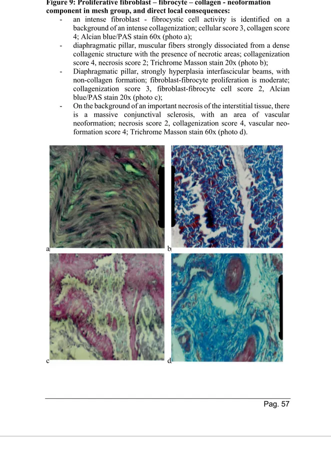
- Proliferative fibroblast – fibrocyte – collagen -
neoformation component: both groups presented cellular
(fibroblasts and fibrocytes) and neo-fibril-conjunctively parts (collagen PAS +, alcian blue +, Trichrome Masson+), with predominance in the Bio-A group, shown and interpreted in figures 9 and 10. Neo-collagen fibers, disposed parallel, are presented in increased number in group A in the connective tissue, with consequent muscular fibers’ dissociation and creating the premises for an intense sclerosis area.
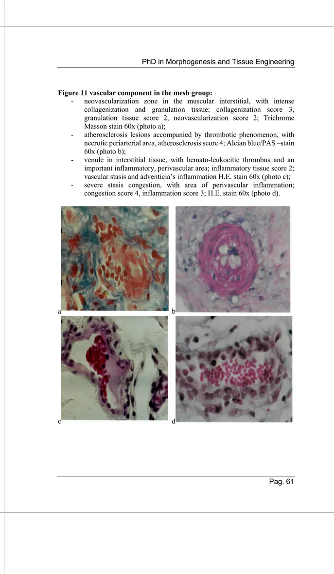
- Vascular component:
Group A (figure 11): Endothelial cells of the neo-formed vessels are plump, presented large nuclei and showed numerous pycnotic vesicles that were also observed in the subendothelial pericytes, (different calibers), intense and diffuse active hyperemia, both arterioles and veins ± perivascular hematic extravasation; endothelithis and sclerogenic thrombo-arteriopathy, with collagen fibers proliferations mainly PAS positive and alcian blue weak positivity.
Group B (figure 12): Only neovascularization phenomenon, with relatively frequent aspects of active hyperemia.
- Inflammatory component (chronic nodular aspect mainly,
with rare acute or sub-acute zones)
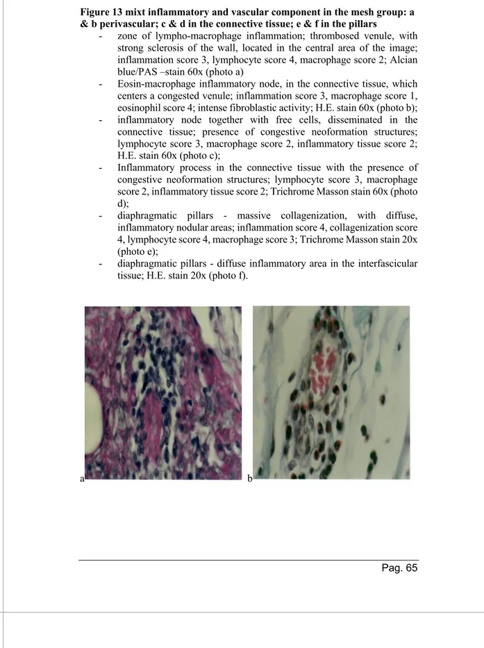
Group A (figure 13): Granulocytes, neutrophils and eosinophils, mononuclear cells like lymphocytes, plasmocytes, macrophages and histiocytes.
organized as nodular chronic inflammation, with lympho-plasmocytes and mono-histiocytes cells.
- Dystrophic component similar for both groups (shown and interpreted in figure 15): The most intense and evident,
localized on the crura, with skeletal muscle degeneration, vascular lesions and of adipose metaplasia.
A semiquantitative histological scoring of tissue reaction was elaborated based on the above results (see table 5), calculated based on:
- inflammation 0 = absent, 1 = mild detectable, 2 = present, 3 = marked/very evident;
- fibrosis (collagen, fibroblast, fibrocytes): 0= absent, 1 slightly detectable, 2 mild,
3 marked;
- vascularization: 0 = absent, 1= rare, 2 = some vessels (3-5), 3 = over 5 vessels.
Based on this semiquantative HP scores, animals in the mesh group showed an increased pattern of chronic inflammation (2.66 vs. 1.83, p = 0.04) compared to PRP group. Vascular and collagen network did not differ between groups (p = 0.306), although there was an increased trend toward mean collagen fibers in the mesh group(2.83±0.4) compared to PRP (2.33±0.81).
As a summary of the observed pattern, in group A there was an evident inflammatory reaction, composed from mononuclear inflammatory cells located between young collagen fibers, disposed parallel, and edged by hyperemic capillaries with rare hematic extravasations (granulation tissue). The adjacent muscles are marked by fibroblast proliferations, with interfibrillar connective tissue’s accumulation. From the point of view of the phenomenon of sclerogenic collagenization of interstitial connective tissue, meshed pigs recognized a larger process, respectively 2.83 compared to 2.33 in those without mesh.
In group B the inflammatory reaction was characterized by the active fibroblasts in the connective tissue, with young collagen fibers, with neovascularization phenomenon (different calibers), with relatively frequent aspects of active hyperemia. This group was characterized by less inflammatory response compared with group A, but an important fibrosis phenomenon (collagen, fibroblast, fibrocytes) and neovascularization were seen as well, creating the premises of tissue remodeling and improved healing.
5.2 Statistical analysis results
Results of the semiquantitive histological scoring of tissue reaction statistical analyze between the 2 groups are reported in Table 5 and Figure 16 for each variable examined. There was no statistically significant difference between the 2 groups. For each variable, there is a minimum, 1st quartile, median, mean, 3rd quartile and maxim reported. Wilcoxon non-parametric test was used, but chi quadro test as well and they are reported in table 5.
1. For the “inflammatory response” variable Mesh group
Min. 1st Qu. Median Mean 3rd
Qu. Max.
2.000 2.250 3.000 2.667 3.000 3.000
PRP group`
Min. 1st Qu. Median Mean 3rd
Qu. Max.
0.000 1.250 2.000 1.833 2.750
3.000
Mesh group PRP group 0 0 1
1 0 1 2 2 2 3 4 2
2. For the “Vascularization” variable Mesh group`
Min. 1st Qu. Median Mean 3rd
Qu. Max.
1.000 2.000 2.000 2.167 2.750 3.000
PRP group`
Min. 1st Qu. Median Mean 3rd
Qu. Max.
1.000 1.000 1.500 1.667 2.000
3.000
Mesh group PRP group 1 1 3
2 3 2 3 2 1
3. For the “Fibrosis” variable Mesh group
Min. 1st Qu. Median Mean 3rd
Qu. Max.
2.000 3.000 3.000 2.833 3.000 3.000
PRP group
Min. 1st Qu. Median Mean 3rd
Qu. Max.
1.000 2.000 2.500 2.333 3.000
3.000
Mesh group PRP group 1 0 1
2 1 2 3 5 3
6. DISCUSSION
The results of the present experimental study demonstrate that a reliable big animal model for the study of hiatal surface area pathology can be set up also using a minimally invasive approach. The highlight of the study from the technical point of view are:
1. A devoted anesthesiologist is mandatory to obtain a high survival rate considering the specific physiopathology related to laparoscopy and induced pneumoperitoneum; 2. The laparoscopic approach to the hiatal area can be
standardized in the pig, obtaining a reliable defect of the hiatus mimicking the human hiatal hernia;
3. A standard cruroplasty can be carried out in the pig with a proper reconstruction of the hiatal surface area diameter;
4. The coverage of the cruroplasty and the hiatal area by reinforcement material is feasible and safe in this animal model;
5. This experimental animal model can be used for further study of the hiatal area using different autologous or heterologous material.
6. The tissue harvesting technique was standardized allowing proper histopathological examination.
Coming back to the rationale of this pilot study, a fundamental question remains regarding the etiology of HH: is it caused by the mechanical stress to which the gastroesophageal junction (GEJ) is exposed and/or is related to a biological change in the structures of this region, such as the crura of the diaphragm and as the fixation structures around of the GEJ (gastrohepatic ligament, gastrophrenic ligament, and periesophageal ligament)? Fei et al showed that patients with hiatal hernia have ultrastructural abnormalities at the crura’s muscular tissue that are not present in patients with a normal GEJ: dilation of intermyofibrillar spaces, swelling of sarcotubular structures, focal degeneration of
myofibrils, and extended disruption and degeneration of the muscle’s architecture6. All these findings suggested signs of
structural weakness in the crura of the patients with HH, that might be the cause of recurrence after primary surgical repair.
Regarding the periesophageal ligament, Fei didn’t find any difference in the microscopic damage at theconnective tissue of the phrenoesophageal membrane surrounding the esophagus in normal subjects and HH patients. But von Diemen showed on biopsies obtained from the phrenoesophageal membrane in patients undergoing HH surgical treatment that the collagen content is reduced when compared with healthy subjects53. Levels of total,
type-I, and type-III collagens are reduced by about 60% at this level in patients with GERD and HH compared to cadavers without HH. Thus, the quality of the periesophageal ligament may be an etiological factor in the development of HH and/or could facilitate the occurrence of GERD in patients with GERD but without an associated HH.
Thus, the outcomesof the anti-reflux surgery could depend not only on the adopted surgical technique but also on the underlying status of the diaphragmatic crura. Surgical treatment of the hiatal hernia consists of dissection of the hernial sac from the mediastinum into the abdomen, followed by cruroplasty with sutures and fundoplication, with demonstrated acceptable morbidity and low symptomatic recurrence rates3,4. A higher than
expected radiologic, asymptomatic recurrent hiatal hernia was reported, with rates up to 30–42%, while 5-13% of these patients had symptomatic recurrence, that can require surgical revision in 5-10% of the cases19,54. Even an asymptomatic recurrence may
become symptomatic over time and can lead to severe complications. Currently, non-resorbable biosynthetic meshes are not routinely used anymore for HH’s repair due to possible severe local complications determined by a chronic inflammatory response. This response can cause symptoms like chronic postoperative pain, discomfort, and mesh erosion, sometimes underestimated54. Under
for the most complex cases, especially in patients at risk for complications of infection. Unfortunately, these biological meshes seem to provide insufficient strength for adequate repair, and in addition, the costs of this variety of meshes are extremely high21,22.
Due to the disadvantages of both biosynthetic and biological meshes, strategies focused on the development of new type of mesh, like slowly resorbable biosynthetic meshes, were gradually introduced. This new generation of meshes aims to combine the advantages of both a biosynthetic mesh (no degradation shortly after implantation) and a biological mesh (the “remodeling” aspects and the better tolerance in case of contamination).
Some reports doubt about the efficacy of mesh use during hiatal hernia’s repair. It was shown in a cohort of 189 cases that radiologic recurrences, symptomatic recurrences and reoperation rates were equal after laparoscopic hiatal hernia repair with or without non-absorbable mesh reinforcement, irrespective of hernia’s size and type55. Quality of life, dysphagia and patient
satisfaction were comparable, with no serious mesh-related complications occurred. More favorable conclusions in term of mesh-use were highlighted by a recent review and meta-analysis conducted56 on 942 patients, demonstrating that mesh hiatoplasty
and sutured hiatoplasty produced comparable results with regards to reoperation rate and complications following the repair of paraoesophageal hernias (POH), with significant recurrence’s reduction following use of mesh. All kinds of mesh were included in this study, from non-absorbable to biologic. Authors concluded that in the future a number of issues need to be addressed to determine the clinical outcomes, safety, and effectiveness of these 2 methods for elective surgical treatment of large hiatal hernias. These include:
- standardized definition of hiatal hernia’s dimension; our
group recently introduced the Hiatal Surface Area CT-scan measurement as a useful, minimally invasive tool in hiatal hernia’s treatment of obese patients undergoing anti-reflux
surgery10. Nevertheless, the suggested cut-off HSA > 4 cm2
was proved to be effective on the choice of when to apply the reinforced cruroplasty20;
- standardized techniques of suture and prosthetic repair; - type of prosthesis used: biologic/nonabsorbable/biosynthetic
absorbable;
- standardized method of securing the mesh such as use of
sutures, tacks, or biologic glues;
- standardized classification of recurrent hiatal hernia post
repair;
- long term postoperative data collection of at least 5 years to
detect the true incidence of hiatal hernia recurrence, reoperation and mesh migration or erosion between suture cruroplasty and prosthetic hiatal herniorrhaphy.
Still, the presence of a foreign body, the costs of these devices, the surgical skills needed for positioning, and nevertheless, patient’s choice, made us look for other available solutions for the augmented, reinforced surgical treatment of the HH in order to obtain a reproductible technique. The use of an autologous, simple and cheap product like PRP might be an important adjuvant of the surgical cure. In the last years it was highly demonstrated the recurrences’ reduction of hiatal hernia after cruroplasty’s reinforcement. An autologous product, with similar proprieties like a certified mesh for local tissue remodeling, totally absorbable and with minimal impact on the nearby organs, it is for long time waited for the guarantee of a minor physical and psychological impact, faster recovery and reduction of the social burden. This is the first study that compares in an animal model the autologous PRP clot, like a self-standing procedure, with an already established surgical solution, the biosynthetic, absorbable Goreâ Bio-Aâ mesh implant
for hiatal hernia’s repair. The study proposed to offer experimental answers with impact on the public health for the management of this pathology, definition of the technical indications and reduction of the expensive prosthetic materials waste. Long term benefits are easily understood because the use of new products, like autologous
PRP, laparoscopic dispenser, and of a new surgical technique, like laparoscopic application of PRP on the cruroplasty, might improve the results of surgical cure of symptomatic HH and GERD, pathologies that represents the most prevalent gastrointestinal disturb worldwide.
Studies published recently concentrated on the use of PRP in ventral (VHR) or inguinal hernia’s reparation, with promising results. PRP was used as reinforcement of mesh repair of abdominal wall’s iatrogenic defects in 12 dogs and compared with mesh alone repair group of 12 animals57. Hernia’s recurrence was not recorded
in PRP-treated dogs, that also displayed significantly more neovascularization and less severe adhesion to the underlying structures in comparison to control group. Histological and molecular evaluation confirmed the gross findings that collagen deposition, new vessel formation, and overexpression of angiogenic and myofibroblastic genes were observed more frequently in the PRP group. In conclusion, the Authors found that addition of allogenic PRP in mesh enhanced neo-vessels formation, and increased tissue deposition and incorporation, with subsequent reduction of the peritoneal adhesion and of the recurrence rate.
Fernandez-Moure et co studied the effect of PRP on a porcine non-crosslinked acellular dermal matrix (ADM) in an in
vivo model of VHR, hypothesizing that PRP would enhance
ADM-tissue incorporation in a rat model58. Enhanced neo-vascularization
was associated with earlier and greater tissue deposition on the ADM. This suggested for the Authors that PRP could be used as an adjunct to VHR in clinical scenarios where poor wound healing is anticipated, and enhanced neovascularization and early tissue deposition is desired. Same Authors hypothesized that recurrence after VHR remains a multifactorial problem due to mechanical strain, poor tissue-mesh integration, and degradation of matrices43.
The high recurrence rate witnessed with the use of ADM for definitive hernia repair has reduced their use largely to bridging repair and breast reconstruction. Use of PRP might improve classic
cellular metrics of successful VHR, resulting in improved rates of hernia’s recurrence. The study consisted in reparation with StratticeÔ (Allergan, USA) mesh alone or impregnated with PRP in
32 Lewis rats with surgically induced, chronic ventral hernias. Samples were harvested at 3 and 6 months postoperatively and compared for gross, histologic, and molecular outcomes of: neovascularization, tissue incorporation, peritoneal adhesions, hernia recurrence, and residual mesh thickness. PRP was shown to be an autologous source of pro-regenerative growth factors and chemokines uniquely suited to soft tissue wound healing, enhanced angiogenesis, myofibroblast recruitment and tissue ingrowth, ADM preservation, less severe peritoneal adhesions, and diminished hernia recurrence. Authors advocated further investigation regarding PRP augmentation of human VHR.
The effect of PRP-gel coating of polypropylene mesh on inflammation, production of collagen, and on smooth muscle was analyzed in the rabbit vagina by Parizzi et al59. They analyzed 3
groups (sham, mesh only and mesh coated by PRP). The inflammatory infiltrate was evaluated using H&E staining, the Sirius Red stain to examine deposition of collagen I and III, and Masson’s trichrome staining was used to visualize the smooth muscle. The group with PRP-coated meshes had a lower inflammatory infiltrate count at 30 days, deposition of collagen III increased with the use of PRP-coating at 90 days. The inflammatory component was significantly increased in the group without the PRP-coated mesh at 30 days but not in the group with the PRP-coated mesh, indicating a less intense inflammatory response.
There’s only one study in a porcine model on the use of PRP in hiatal hernia’s repair. Altieri analyzed recently41 on 3 groups of
young pigs on which a iatrogenic defect of the hiatal area was created laparoscopically and then repair consisted on: hiatus repair alone (HR; n = 7 animals); hiatus repair and acellular dermal matrix (HRM; n = 8); hiatus repair and acellular dermal matrix and filtered plasma concentrate (fPC) (n = 9). Histopathological and