PhD in Biochemistry
XXXI Cycle (2015-2018)
Impact of non synonymous single nucleotide
variants on protein fitness: experimental
analysis for a comparative study
Candidate
Maria Petrosino
Supervisor Coordinators
Prof.ssa Roberta Chiaraluce Prof. Stefano Gianni
Prof. Francesco Malatesta
PhD in Biochemistry
XXXI Cycle (2015-2018)
Impact of non synonymous single nucleotide
variants on protein fitness: experimental
analysis for a comparative study
Candidate
Maria Petrosino
Supervisor Coordinators
Prof.ssa Roberta Chiaraluce Prof. Stefano Gianni
Prof. Francesco Malatesta
A tutti coloro che hanno creduto in me.
It always seems impossibile until it‟s done.
(Nelson Mandela)
Acknowledgements
Though the following PhD thesis is an individual work, I could never have reached it without the help, support, guidance and efforts of a lot of people.
Firstly, I would like to express my sincere gratitude to my supervisors Prof.ssa Roberta Chiaraluce and Prof. Valerio Consalvi for believing in me and for instilling in me the qualities of being a good scientist. Your help and guidance over the years has been unmeasurable and without it I would not be where I am today. It has been a pleasure for me work with you.
I am deeply grateful to Dr.ssa Alessandra Pasquo for the knowledge you have passed on. You are my primary re source for getting my science questions answered and I will always be grateful for having the opportunity to learn under your guidance. You have made me grow so much both from a personal and professional point of view. I will treasure your advices forever.
Thanks also go out to Prof. Francesco Malatesta for taking care about my professional growth, for his encouragement and helpful advice.
Thank to Dr.ssa Francoise Dantzer (CNRS, University of Strasbourg) for the opportunity to work in her respectable laboratory. You made me feel as a component of your group from the first day, making me feel at home and very welcome. Thanks to her research group, in particular I would like to thank Jean-Christophe for all the days that we spent working and smiling in front of the column to manage the purification, Josè for his patient and precious help and above all Leonel and Kathline. These two friends formed the core of my research time in the Dantzer‟s group. You are wonderful and generous friends who I admire a lot for your ability to smile despite the situation. I‟ll never forget the help you gave me in research activities and the wonderful lunches and fun activities we‟ve done together during my period in Strasbourg.
A very special thank you to my friends Leonore, Laura, Giusy, Marialaura, Giammarco, Ilaria, Elena, Claudia, Daniele, Flavio, Regine, Jo-Ann, Lais and to my friends and collegues Flavia, Francesca, Serena. A good support
system is important to surviving and staying sane during PhD. I was lucky to have all of you. You contribute to the following thesis in different ways: giving advices, working hard for helping me, making me smiling.
Thanks to my new collegues for helping and supporting me in this caotic period. A special thank to Giovanni for believing in me and for give me the opportunità to work in his research group and thanks to Silvia e Annalisa, too.
A special thank to my family for all their love and encouragement. Thanks becouse you supported me in all my choises being always with me and giving me the possibility to reach what I have now. For my parents that teach me what it means believe in something and work hard to reach your goal, and for my sister and her unmeasurable patient and love.
Index
Chapter 1 ... 1
1.1 General introduction ... ...1
1.2 Single nucleotide variants ... 2
1.3 Overview of possible effects induced by single amino acid substitution... 9
1.4 How can you study the effect of point mutations on protein? ... 9
1.5 nsSNVs and cancer ... ………..14
1.5.1 nsSNVs and cancer: the experimental system ... …..14
1.5.1i Phosphoglycerate kinase 1 ... 18
1.5.1ii Human Frataxin... ...23
1.5.1iii Poly (ADP-ribose) polymerase 3 (PARP3)... ...29
Chapter 2 ... 35
Aim of the study ... 35
Chapter 3 ... 39
Methodology ... 39
3.1 Protein expression and purification……….41
3.2 Spectroscopic measurements………..….43
3.3 Thermal denaturation experiments……….. 43
3.4 Urea-induced equilibrium unfolding………44
3.5 PGK1 Enzyme activity and kinetic studies….………45
3.6 PGK1 Protein crystallization, data collection, structures solution……….…….46
3.7 hFXN Stopped-flow measurements..………...46
3.8 Data analysis………..…..…………..…..47
3.8.1 Quantitative analysis of equilibrium transition……….……....47
3.9 Cell culture…..………..…...50
3.10 Western blot analysis……….………..…..50
3.11 Dose response……….………..….51
3.12 Clonogenic assay …………..………..….51
Chapter 4 ... .53
Results ... .53
4.1 Experimental analysis of PGK1 nsSNVs...56
4.1.1 Spectroscopic characterization of PGK1 wild-type and variants...57
4.1.2 Kinetic analysis...59
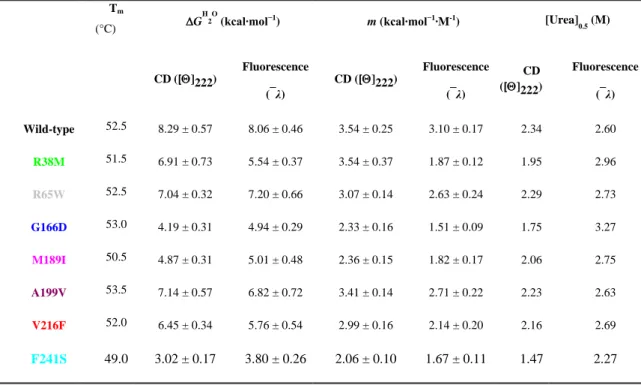
4.1.3 Thermal and thermodynamic analysis...62
4.1.4 Structural analysis...66
4.2 Experimental analysis of hFXN nsSNVs...72
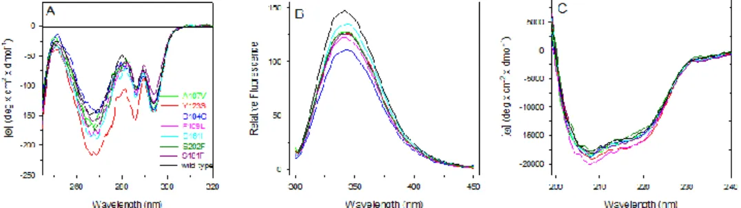
4.2.1 Spectroscopic characterization of hFXN wild-type and variants...74
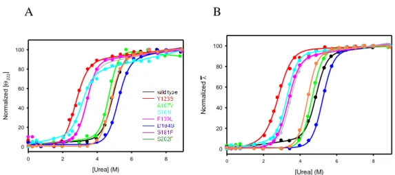
4.2.2 Thermal and thermodynamic analysis...76
4.2.3 Analysis of the folding pathway of hFXN wild-type and its variants ...80
4.3 Biochemical characterization of PARP3 and analysis of PARP3 nsSNVs...83
4.3.1 PARP3 in the tumorigenicity of glioblastoma ...85
4.3.2 PARP3 and nsSNVs ………...88
4.4 Analysis of the effects induced by single amino acid substitution...92
4.4.1 Effect on protein conformation in solution...93
4.4.2 Effect on protein structure...95
4.4.3 Effect on protein function...96
4.4.4 Effect on protein binding...98
4.4.5 Effect on protein stability...100
4.4.6 Effect of single amino acid substitution on protein production...102
Chapter 5... ...103
Discussion ... ...103
Chapter 6...109
Attachments ...109
Common acronyms and abbreviations...211
1
Chapter 1
1.1 General introduction
Proteins are large biological molecules that control most vital cellular functions. They consist of one or more chains of amino acids in an order determined by the base sequence of nucleotides in the DNA coding for the protein. Thanks to the information from the genetic code and according to the energy landscape, proteins fold into their correct three-dimensional structures and exert their specific function. The correct fold of large portion of the structure is generally related to specific protein functions and when any even small alterations occur, it is possible to observe a decrease, an increase or a drastic change in the protein function. In several cases alterations at the amino acid level can influence the conformational rearrangement, the function or the binding properties of a given protein. On this premise, knowledge on protein structure-function relationships can be crucial in finding the molecular basis for hereditary diseases and in predicting protein function from structure and vice versa. Therefore, the study of structure-function relationships is really important nowadays to better understand several diseases at their molecular level. In particular, this kind of approach seems to be relevant in cancer research considering that several somatic variants resulting from alterations at the amino acid level have been detected in cancer genome for several proteins. The analysis of this kind of alterations is key to understand the genetic bases of disease progression, patient survival
2
and also response to therapy. Since knowledge of protein function in health and disease is essential to identify new and more specific cures for different diseases and to design pharmacologically active and more selective drugs, the information resulting from the analysis of somatic mutations found in cancer tissues can improve the available therapies and create new and more specific ones suggesting that precision and personalized medicine is not anymore a daydream.
1.2 Single nucleotide variants
The human genome, which consists of over 3 billion base pairs, has remained well conserved throughout evolution, in fact it is at least 99.5 % identical between any two humans on the planet [Levy S et al, 2007]. Modern genomic sequence analysis have revealed that it is more complex, diverse, and dynamic than previously thought, with possible genetic variations in the range from 0.1 % to 0.4 % [Jorde LB et al, 2004]. Sequence variations at the amino acid level may influence the conformation, function or binding properties of a given protein [Bhattacharya R et al, 2017]. When sequence changes occur, even in non-protein coding regions of the DNA, the consequent alteration in the human genome may cause or just be linked to a specific disease.
In the last decades, several studies have focused their attention on protein variants and their relationship with rare, mendelian genetic diseases and some authors have recently hypothesized that the common variants may contribute significantly to genetic risk for common disease [Lander ES et al, 1996; Risch N et al, 1996; Collins FS et al, 1997].
3
The most common genetic differences in the human genome are polymorphisms, generally defined as single base pair variations in DNA sequence that occur in at least 1% of the population. These differences are called single nucleotide polymorphisms (SNPs) and they are considered the most common genetic variations observed in humans [Collins FS, 1998] (The International HapMap Consortium 2003) [Shastry, 2002], since they occur, on the average, once every 300–400 base pairs [Kruglyak and Nickerson, 2001] in contrast with other types of polymorphisms (for example, differences in copy number, insertions, deletions, duplications, and rearrangements) that also occur, but much less frequently. Some of them are located within the coding or regulatory regions of genes but, considering that only about 5% of the human genome codes for the production of proteins (The International Human Genome Sequencing Consortium 2001), most single nucleotide mutations may fall in the non-coding regions of genes or in the intergenic regions. Point mutations within a coding sequence are of particular interest because they can cause qualitative and quantitative changes in gene expression, RNA splicing, protein translation, or gene function. Coding SNPs, especially nonsynonymous coding SNPs (nsSNPs), induce changes at the amino acid level of the polypeptide sequence of the protein generating protein variants with single amino acid substitution (nsSNVs), also referred to as missense mutations, that may present structural and/or functional alterations [Zhou et al, 2010].
Only in the last years, millions of human SNVs have been identified, and these variants could be strongly correlated with phenotypic variations of traits/diseases [Cao et al, 2017]. It has been reported that nearly 30% of the nsSNVs are predicted to affect the protein function [Chasman and Adams, 2001; Ng and Henikoff, 2002; Ramensky et al, 2002]. Several nsSNVs have
4
been found neutral or with no effect on human health, but many nsSNVs have been linked to many diseases or may be responsible for individual susceptibility to contract diseases.
In the last years, several sequencing initiatives [Hudson et al, 2012; Stratton et al, 2012] have deeply contributed to the implementation of information about nsSNVs detected in humans and many databases are nowaday available, listing human SNVs and their effects, such as dbSNP (Single Nucleotide Polymorphism database) [Sherry et al, 2001], SAAPdb [Hurst et al, 2009] or SNPdbe [Schaefer et al, 2012]. Since genetic variation in the human genome is an emerging resource for studying cancer and other diseases [Mueller et al, 2015; Kunz et al, 2016, Zang et al, 2016, Didonna et al, 2015], in the last years databases like Online Mendelian Inheritance in Man (OMIM) [Hamosh a et al, 2005] and COSMIC [Forbes et al, 2010],that contribute to catalogue nsSNVs found in pathological tissues, have been created. Even if there is a huge amount of SNVs deposited in public platform, only a small proportion of them are functional polymorphisms that contribute to disease phenotypes [Tabor et al, 2002; Yuan et al, 2006]. Recently a new important database, named Pan-Cancer Atlas (https://www.cell.com/pb-assets/consortium/pancanceratlas/pancani3/index.html), is available and catalogues the nsSNVs found in cancer tissues in the Genomic Data Commons (https://portal.gdc.cancer.gov/).
Given the high potentiality of knowledge about the relationship between structure-function changes observed in nsSNVs and diseases, it is noteworthy that this field is extensively expanding in cancer research since several somatic mutations that have been identified in cancer tissues are nsSNVs. Until now a huge dataset has been generated and made available. A stumbling-block is represented by the fact that no tumor genome has been
5
completely characterized but the advances in high throughput technologies and the declining costs of high-throughput sequencing are transforming our understanding of cancer and will allow us to generate a comprehensive map of polymorphisms distributed over the entire genome that will be helpful to identify new and more appropriate therapies and to make effort in drug discovery.
1.3 Overview of possible effects induced by
single amino acid substitution
Single amino acid substitutions can be beneficial, adding new functionality and increasing the fitness of the cells or deleterious, damaging the protein, causing destabilization of the protein structure or reducing its functional activity because of the disruption of a site that is directly involved in the function of the protein and thereby contributing to disease phenotypes [Studer et al, 2013]. Usually, change in amino acids with similar size and physico-chemical properties (e.g. substitution from leucine to valine) has a mild effect. Similarly, if the point mutation disrupts secondary structure elements (e.g. substitution to proline in alpha helix region) such mutation usually may affect whole protein structure and function. Thus nsSNVs can potentially affect the function of the cell in a variety of ways.
The most important consequence of point mutations in different types of functional sites can be either the increase or the decrease of protein activity. The most dramatic effects have been detected when point mutations occur in the active sites of enzymes or in the binding pockets of receptors
6
[Stevanin et al, 2004; Yamada et al, 2006]. Considering that enzymes catalyze biochemical reactions not only thanks to catalytic residues, but also with several surrounding residues important for ensuring proper attachment of the substrates and cofactors to the active site cavity (binding sites residues), mutations that occur on the residues located in the neighborhood of the active site can influence the activity [Zhang et al, 2001; Takamiya et al, 2002; Zhang et al, 2010]. In the human population, 25% of the known nsSNVs significantly affect protein function in vivo [Yue and Moult, 2006]. It has been reported that in most cases nsSNVs lead to loss-of-function (LOF) generally due to perturbation of the active site of the enzyme or of its global structure [Kucukkal et al, 2015] or to a reduction of the thermodynamic stability of the protein leading to a shift of the folding equilibrium toward the nonfunctional unfolded state, possibly coupled to irreversible aggregation (thermal instability) and/or degradation by cellular quality control [Yue et al, 2005; Casadio et al, 2011; Shi et al, 2011; Stefl et al, 2013; Petukh et al, 2015]. Sometimes, but not commonly, nsSNVs can result in a protein that still functions, even if in a “non-canonical” way, as evident in the so-called gain-of-function (GOF) mutants that generally include oncogenic mutations in proteins such as the Ras GTPase and the epidermal growth factor receptor family of tyrosine kinases, which often drive cancer tumor cell development and proliferation [Schubbert et al, 2007; Arteaga et al, 2014].
The occurrence of a mutational event can generate a protein variant that perturbs conformational constraints of the native protein (e.g., substituting a small side chain residue to a large one and vice versa, resulting in backbone strain or overpacking) or have physicochemical effects (substitutions between hydrophilic residues and hydrophobic residues, burial of charged residues, the disruption of hydrogen bonds, loss of hydrogen
7
bonds, of S–S bonds) [Shirley et al, 1992], leading to considerable alterations of the stability of the native protein [de Cristofaro et al, 2006; Koukouritaki et al, 2007; Ode et al, 2007]. Protein stability is a key characteristic of a functional protein [Wang and Moult 2001; Zhang et al, 2001; Ramensky et al, 2002; Wang and Moult 2003; Capriotti et al, 2005; Karchin et al, 2005; Ye et al, 2006; Zhang et al, 2010]. It is known that a single base DNA substitution may result in alteration of stability of the corresponding native protein [Wang and Moult, 2001; Yue et al, 2005] and that about 80% of missense mutations associated with disease affects the stability of proteins by several kcal∙mol−1 [Wang and Moult, 2001]. Generally, a nsSNV can have destabilizing or stabilizing effects: most frequently, missense mutations lead to a destabilization of the protein stability by making it susceptible to proteolysis or by changing the thermal inactivation temperature. The impact of protein destabilization can be observed in several proteins involved in many neurodegenerative diseases, such as Parkinson's disease [Lin et al, 2008; Morais et al, 2009; Nuytemans et al, 2010] or in cancer, where destabilizing mutations in the core of the protein lead to an inactivation of many tumor suppressors [Stehr et al, 2011]. An amino acid substitution can occur at a position critical for the folding of the protein destabilizing it and/or stabilizing a misfolded state [Valastyan and Lindquist, 2014]. The importance of the correct folding of a protein is clear if we consider the hundreds of diseases associated with many misfolded proteins [Dobson, 2003; Thusberg and Vihinen, 2009; Groenendyk et al, 2010; Valastyan and Lindquist, 2014].
Several investigations have been addressed to better elucidate how missense variants can induce minimal or significant alterations on the protein flexibility [Young et al, 2001; Zhang et al, 2010; Karplus and Kuriyan, 2005]
8
considering that the conformational flexibility of a protein is crucial for its correct folding, for the binding with partners and for its function. The presence of a point mutation, by altering the protein flexibility, can affect protein aggregation propensity. This is considered a hallmark of some neurodegenerative diseases, like Alzheimer‟s disease (AD), Parkinson‟s disease (PD), Huntington‟s disease (HD), amyotrophic lateral sclerosis (ALS) and prion diseases [Guijarro et al, 1998; Chiti et al, 1999; Fandrich et al, 2001; Chiti et al, 2003; Bucciantini et al, 2004; Harris and True 2006; Keage et al, 2009; Khemtemourian et al, 2008; Robinson, 2008; Yankner and Lu 2009] and for this reason research on protein misfolding and aggregation is progressively gaining increasing attention. The general mechanism by which nsSNVs promote these diseases is the promotion of toxic protein aggregation [Calamini and Morimoto, 2012; Knowles et al, 2014; Valastyan and Lindquist, 2014] triggered by the destabilization and unfolding of the native protein structure, which exposes aggregation-prone regions previously buried inside the structure.
Approximately 60% of disease-associated nsSNVs induce a perturbation in the binding site of the protein, expecially if the point mutation affects residue located on protein-protein interface [Schuster-Bockler and Batemen, 2008; Teng et al, 2009; David et al, 2012], and may lead to changes in the interactions with partners (activators, repressors or substrates) and in the binding specificity [Kohler et al, 2008; Wu et al, 2008; Bauer-Mehren et al, 2009; Vanunu et al, 2010; Barabàsi et al, 2011].Many of these mutations induce significant perturbations that result in complete loss of interactions or complete abolishment of the function, generally leading to diseases [Wang et al, 2012; Sahni et al, 2015].
9
The impact of single amino acid substitutions on protein stability has been investigated by theoretical and experimental approaches [Bross et al, 1999; Wang and Moult, 2001; Ferrer-Costa et al, 2002; Steward et al, 2003; Yue et al, 2005; Scheraga et al, 2007; Dill et al, 2008; Fersht, 2008]. In general, the experimental studies are relatively limited due to the cost and time needed for the entire process which often includes mutagenesis, protein expression and purification, followed by thermal or chemical unfolding.
1.4 How can you study the impact of single
point mutations on the protein?
Single point mutations can occur between different individuals of the same species. These mutations can lead to a protein without any changes in its amino acid sequence (synonymous SNVs (sSNVs)), insert a stop codon or generate a protein variant with an amino acid change (nsSNVs). Even if most SNVs are neutral or have no effect on human health and embryonic development [Kimura M, 1983; Wang Z, Moult J, 2001], a large number of nsSNVs have been associated with diseases [Wang Z et al, 2001]. Due to the complexity of human biology, the association of a nsSNV with a complex phenotype is still a problem and methods that target understanding the effects of missense mutations on various sequence, structural and functional features are progressively implemented in the hope of deciphering the phenotype– genotype relations [Stanley et al, 2013; Yates and Sternberg 2013; Kucukkal et al, 2014].
Nowadays bioinformatics tools can be really helpful to filter large datasets of genetic variation and in the last decades good computational
10
methods have been developed to predict the effect of nsSNVs and their pathogenicity in order to predict the experimentally observed effects for disease-related variants [Hecht et al, 2013]. These methods are based on implemented computational algorithms that allow the evaluation and analysis of mutations as a function of their pathological consequence [Balu and Purohit, 2013; Kamaraj and Purohit, 2013; Kamaraj et al, 2013; Kumar et al, 2013] giving us the possibility to distinguish the non-significant SNVs from the ones that might produce major disease associated consequences. Computational methods such as SIFT [Sim et al, 2012], Polyphen-2 [Adzhubei I. et al, 2013], StSNP [Uzun et al, 2007], Bongo [Cheng et al, 2008], Condel [Ng and Henikoff, 2006; Gonzalez-Perez and Lopez-Bigas, 2011] and MAPP [Stone E.A. et al, 2005] classify SNVs according to neutral, negative or positive effects on the structure or function of a protein, while different computational methods, such as FoldX [Schymkowitz et al, 2005], PoPMuSiC [Dehouck et al, 2011], CUPSAT [Parthiban et al, 2006], Rosetta [Leaver-Fay et al, 2011] I-Mutant [Capriotti et al, 2005] or INPS (a predictor of the Impact of Non-synonymous-variations on Protein Stability) have also been developed to classify a single amino acid substitution according to its effect on the protein stability by comparing wild-type with mutated proteins, and predicting the variation of protein stability (∆∆G) of the mutation. However these methods are generally based on a single folded structure, but the native state of a protein is not unique, thus it is important to consider the entire ensemble of the protein conformers in dynamic equilibrium.
A deeper understanding of the effect of a point mutation on the protein structural, physical and chemical properties requires the utilization of several techniques: only a combination of predictive analysis and experimental procedures can give us a whole picture regarding the consequences of
11
missense mutations on protein. Since in the last years the accepted paradigm that proteins can tolerate nearly any amino acid substitution has been replaced by the awareness of deleterious effects of mutations, especially on the thermodynamic and kinetic stability of protein, several methods have been developed to catalogue the nsSNVs that affect the stability and function of the protein [Ng and Henikoff, 2006; Yue and Moult, 2006]. In order to combine the results of the various tools, consensus predictors have been developed to allow comparison between methods that use different analytical approaches [Vendruscolo et al, 2003; Bendl et al, 2014].
One useful method largely used to provide information about the conformational changes that a mutated protein may undergo under various conditions is the molecular dynamics simulation (MD) leading to a systematic evaluation of molecular properties in dynamic molecular systems. Using specific algorithms it is possible to assess the potential impact of variants within the protein revealing the possible various mechanisms by which variants may lead to functional alteration. Some of them can be revealed energetically, others structurally or dynamically analyzing the dynamic behaviour of proteins [Zimmermann MT et al, 2017].
The importance of a combination of experimental studies with molecular dynamics simulations allow us to highlight several experimental procedures that can be used to reveal the structural and functional effects of single amino acid changes in proteins owing nsSNVs. Of particular interest are experimental methods that provide 3D structures of genetic variant proteins revealing atomic-level structural details with which it‟s possible to analyze the structural differences caused by the variation at the DNA level. X-ray Diffraction, solution Nuclear Magnetic Resonance or Electron Microscopy contribute to implement the PDB archive [Rose PW et al, 2015]. Even if the
12
availability of the 3D structure of a gene product represents the most informative source of data that can explain what is able to cause a particular phenotype, most studies that analyze the relationship between point mutations and experimentally observed 3D protein structure published to date have been restricted to individual proteins or single diseases. There is a paucity of quantitative analyses of the consequences of SNVs on 3D protein structure going beyond the realm of prediction [Arodź and Płonka, 2012], even if tools that incorporate structural information when making predictions have been implemented [Brown DK Bishop ÖT, 2017]: an excellent communication between bioinformatics and biomolecular scientists is requested to increase the amount of data about genetic variants and phenotype.
Since several nsSNVs associated with different diseases can affect the stability of the native protein [Wang and Moult, 2001], protein folding analysis and stability measurements may be useful for the study of disease-associated processes [Adhikari et al, 2015]. With this purpose mutagenesis can be used to obtain mutated proteins and followed by protein expression and purification steps in order to obtain a pure protein product that can be analyzed by thermal or chemical unfolding to determine the difference of Gibbs free energy value (∆G) between the wild-type and the mutated proteins. Several forces contribute to the stabilization of the folding state of a protein, expecially hydrogen bonding, hydrophobic contacts between the buried side chains in the interior of the molecule out of contact with water, and the formation of intramolecular contacts between side chains [Eswar and Ramakrishnan 2000]. In their stable state, proteins are marginally stable, between −3 and −10 kcal∙mol−1 [Taverna and Goldstein, 2002]. They can tolerate some destabilization within a narrow range [Bershtein et al, 2006;
13
Goldstein, 2011] but when this range is overshot a pathological condition can occur. Point mutations may add energy (i.e. more than +2 kcal∙mol−1) to the folded state and destabilize the equilibrium between the native state and the unfolded one, making the protein more likely to aggregate in its unfolded form: this is a crucial event in some diseases characterized by unfolded aggregates of proteins [Yue et al, 2005]. However, in some cases a mutation removes energy from the folded state (i.e. less than +2 kcal∙mol−1) and stabilizes it making the protein too rigid and so non-functional. Most of the disease-causing mutations were found to be destabilizing [Pasquo et al, 2012; Grothe et al, 2013; Khan et al, 2013] and, in particular, the degree of destabilization was found to be elevated for mutations that introduce drastic changes such as charged to neutral, relatively rigid to relatively flexible, or aromatic to aliphatic mutation types.
Considering the plausible alterations in the folding process caused by point mutations, it is not surprising that experimental studies about protein folding and stability are needed.
Equilibrium studies allow to understand the cooperativity of the folding process and can be used to evaluate the conformational stability. Usually denaturation is studied monitoring changes in secondary or tertiary structural elements in protein, by means of circular dichroism or fluorescence spectroscopy, when different concentrations of a chemical denaturant agent are used to induce the denaturation process. Urea and guanidine hydrochloride are the most common chemical denaturant agents used, because it is well known that the interaction of urea or guanidine hydrochloride with the constituent groups of proteins is more favorable than the interaction of those groups with water [Tanford C, 1970]. Denaturants alter the equilibrium between the native (N) and the denatured (D) states and
14
the free energy of unfolding reaction is given by: G = RT ln ([D]/[N]), where [D] and [N] are the concentrations of protein in the denatured and native state, respectively.
Thermal denaturation experiments can also be performed due to the large amount of information that they can give about the denaturation process of a given protein. In particular, calculation of the Tm plotting the first derivative
of the molar ellipticity values at around 222 nm, where helices are known to give their spectral signal, as a function of temperature, it‟s possible to compare the transition curves of the wild-type and of the mutated proteins looking at the midpoint of the denaturation process.
Furthermore, the folding process of a given protein can be analyzed by following the kinetics of unfolding that allow to understand the protein dynamics and provide information about the folding pathway. Kinetic studies are performed by altering the equilibrium of the system introducing a perturbation, such as mixing two solutions with different urea concentrations or increasing rapidly the temperature of the sample, and evaluating how the system relaxes to a new equilibrium. The relaxation process can be estimated by following the variation of an optical probe with time, such as the fluorescence of the aromatic residue.
1.5 nsSNVs and cancer
Cancer is an important health problem worldwide, considered as one of the most frequent causes of death. It is a genetic disease caused by changes of genes that control the way in which cells function allowing them to grow out of control and become invasive. These kind of abnormalities have been
15
detected studying cancer genomes, however large-scale research studies are still needed to characterize some tumour types that have not been deeply characterized. Furthermore it is important to take into consideration that each cancer type has a unique combination of genetic changes in different patients that can be inherited from the parents or can be related to the adoption of unhealthy lifestyle, including smoking, drinking, physical inactivity and „„westernized‟‟ diets [Hua et al, 2014]. In fact, several types of cancer can be caused by environmental factor exposures that include substances, such as the chemicals in tobacco smoke, and radiation, such as ultraviolet rays from the sun, which can lead to cancer by inducing DNA damage [Levi et al, 1999]. In addition, in cancer cells genetic variation occurs with an increased frequency due to the uncontrolled proliferation and the compromised DNA integrity.
Since the mechanisms of cancer cells at their molecular level have not been identified, new technologies and the knowledge gained from previous genomic studies could be used to define the full set of alterations to DNA and RNA in many cancers. It can be really useful to set studies that compare genomic information from tumours and normal tissue from the same patient in order to allow researchers to discover genomic changes that may drive cancer. Another possibility is to expand the current use of genomic methods to investigate the molecular basis of clinical phenotypes in order to identify genetic changes that may distinguish aggressive cancers from indolent ones or to study the molecular basis of response to a given therapy, as well as mechanisms of resistance to treatment.
Nowadays the National Cancer Institute (NCI) is working to analyze the DNA and RNA of cancer cells using advanced technologies such as next-generation DNA sequencing. Using multiple genomic techniques the
16
landscape of the cancer genome is mapped in order to discover new changes linked to disease. In particular, a collaboration between NCI and the National Human Genome Research Institute (NHGRI), and Therapeutically Applicable Research to Generate Effective Treatments (TARGET), named The Cancer Genome Atlas (TCGA), have characterized thousands of genomes and matched normal samples making available a collection of cross-cancer analyses delving into overarching themes on cross-cancer, including cell-of-origin patterns, oncogenic processes and signaling pathways.
It is clear that in cancer research genetic susceptibility plays an important role: genome-wide association studies (GWAS) have been conducted in large cohorts of patients allowing us to say that cancer can be considered a genetic disease sometimes caused by a single or few catastrophic somatic mutations that are responsible for cellular transformation [Zhang CZ et al, 2015], and sometimes caused by a combination of several genomic alterations that work together to promote cancer. Many of the identified causal genetic changes are related to the SNV that derives from a substitution of one base pair in DNA and that can initiate the cascades of downstream signaling and eventually transforms the cellular phenotype from normal to malignant [Hyeonju S et al, 2017]. These mutations represent novel targets for therapeutic intervention in this otherwise incurable disease.
17
1.5.1 nsSNVs and cancer: the experimental
system
Although nsSNVs are theoretically expected in tumours, as it has been shown in the previous section, they can occur in mostly random manner across the genome or they can be significantly biased toward a specific codon transition [Hyeonju S et al, 2017]. Furthermore, it is not easy to characterize all the nsSNVs found in cancer tissues by an experimental point of view, due to the large amount of variants identified in several proteins involved in different pathways and to the cost and time required for the experiments. However, since structural and functional analysis of nsSNVs in the DNA sequences of humans may help to predict an individual‟s response to certain drugs, susceptibility to environmental factors, and risk of developing particular diseases [Johnson et al, 2008; Zhou et al, 2010], during the last years we focus our attention on the characterization of nsSNVs of different proteins involved in the regulation of cell metabolism and in the response of DNA damages.
In particular, during my PhD I carried on a biochemical characterization of nsSNVs that have been detected in different types of cancer of three proteins involved in the regulation of cell metabolism, such as the Phosphoglycerate kinase 1 (PGK1) and the human Frataxin (hFXN), or in the response of DNA damages, such as the Poly (ADP-ribose) polymerase 3 (PARP3). Using structural and functional analysis our aim was to investigate any possible correlations between alteration on protein structure, function or stability caused by single amino acid substitutions and cancer. Furthermore in order to present a detailed discussion about this issue, in this thesis I‟m going to
18
combine the results obtained during my PhD with those obtained for variants of other proteins studied by our group in the last years: Bromodomains (BRDs), Peroxisome Proliferator-Activated Receptor (PPAR), Serine/threonine-protein kinase pim-1 (Pim-1) and Protein tyrosine phosphatase (PTP) in order to present a systematic and comparative analysis of properties of protein variants caused by single amino acid substitutions.
1.5.1i Phosphoglycerate kinase 1
Tumors reprogram pathways of nutrient acquisition and metabolism to meet the bioenergetic, biosynthetic and redox demands of malignant cells: altered metabolic activity supports anabolic growth during nutrient replete conditions, catabolism increases to support cell survival during nutrient limitation, and fortification of redox homeostatic systems to counteract the metabolic effects of oncogene activation, tumor suppressor loss, and other stresses [Boroughs L.K. et al, 2015].
The greater example of a reprogrammed metabolic pathway in cancer is the Warburg effect or aerobic glycolysis [Lunt S.Y. et al, 2011]. Glycolysis is a physiological response to hypoxia in normal tissues, and Otto Warburg in the 1920s observed that cancer cells are able to adapt to survive in difficult conditions, like for example in O2 deficiency, through a reprogramming of
their metabolism according to their requirements [Bertout J.A. et al, 2008]. Phosphoglycerate kinase (PGK) (EC 2.7.2.3) is an essential glycolytic enzyme for all living organisms. It catalyzes the reversible phosphotransfer reaction from 1,bisphosphoglycerate (1,BPG) to MgADP to produce
3-19
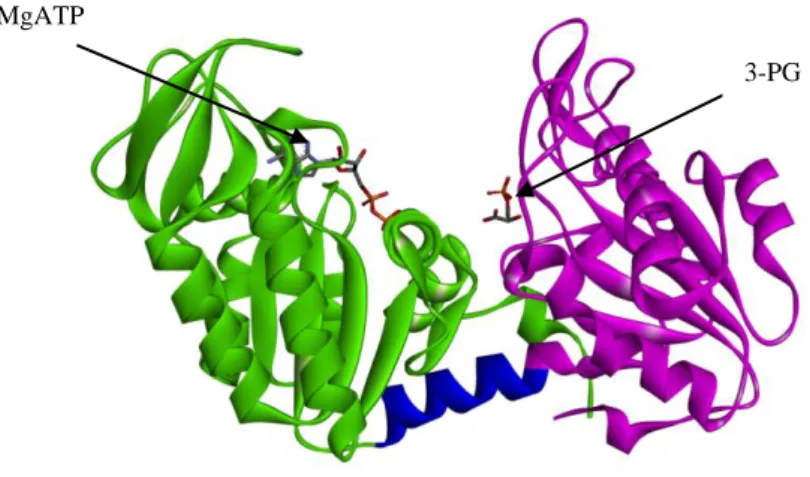
phosphoglycerate (3-PG) and MgATP, an important ATP-generating step in glycolysis [Beutler E., 2007]. By controlling ATP and 3-PG levels, PGK plays an important role in coordinating energy production with biosynthesis and redox balance. PGK is a typical hinge-bending monomeric enzyme of 417 amino acids with an apparent molecular mass of approximately 45 kDa. It is characterized of two domains of equal size, the N- and C-terminal domains. The N-terminal domain binds 3-PG or 1,3-BPG, whereas the C-terminal domain binds MgADP or MgATP. The two domains are separated by a deep cleft and linked by two alpha-helices (α-helix 7 and α-helix 14) [Vas et al, 2010; Palmai et al, 2009]. During the catalytic cycle this flexible hinge region allows the two domains to approach involving a large number of conformational rearrangements: in the open form the enzyme binds the substrates while in the closed form the enzyme performs the transfer of the phosphoryl group and it exerts its catalytic activity [Vas et al, 2010].
Fig. 1.1 Phosphoglycerate kinase 1 (PDB code: 2XE7, open conformation) PGK1 is
composed by two domains of equal size, the N- and C-terminal domains (violet and green,
MgATP
20
respectively), connected by a hinge region (blue). The N-terminal domain binds 3-PG or 1,3-BPG, whereas the C-terminal domain binds MgADP or MgATP.
Two human phosphoglycerate kinase isoenzymes have been identified: PGK1 and PGK2. They are characterized by distinctive tissue localization and encoded by two distinct genes [Willard et al, 1985; McCarrey et al, 1987]. PGK1 is ubiquitously expressed in all somatic cells while PGK2, also known as testis form, is unique to meiotic/postmeiotic spermatogenic cells. Interestingly, similar to many other glycolytic enzymes, human PGK1 was found implicated not only in the glycolytic pathway infact, in addition to its metabolic function, it may play different roles. In mammal cell nuclei, for example, this enzyme participates in the DNA replication and repair [Jindal et al, 1990]; it can also phosphorylate L-nucleoside analogues used in antiviral and anticancer therapies [Krishnan et al, 2003; Gallois-Montbrun et al, 2004; Gondeau et al, 2008].
Furthermore, this enzyme may be surprisingly secreted by tumors in the extracellular environment where it exhibits thiol reductase activity on plasmin that permits the cleavage of plasminogen in order to generate the vascular inhibitor angiostatin [Lay et al, 2000]. Thus the enzyme has been suggested to be an important negative regulator of the angiogenic process that is essential for tumour and metastatic growth [Shichijo et al, 2004; Wang et al, 2007]. It has been found that PGK1 regulates angiogenesis by generating angiostatin and by reducing the secretion of the proangiogenic cytokines VEGF and IL-8. CXCR4 signaling regulates PGK1 expression. At sites of high CXCL12 production, PGK1 secretion is inhibited by the CXCL12/CXCR4 axis. Thus, CXCL12 signaling through CXCR4 generates an „angiogenic switch‟ necessary for metastatic growth. Together, these data
21
further show that CXCL12/CXCR4 chemokine axis and PGK1 represent at least one of the critical events necessary for metastasis of prostate cancer as well as a mechanism for a proangiogenic switch to promote tumor growth [Wang et al, 2007].
In addition, under hypoxic conditions, PGK1 may translocate from cytoplasm to mitochondrion where it may act as a protein kinase and phosphorylate different protein substrates [Li et al., 2016]. In particular, under hypoxic stress, ERK activation-dependent mitochondrial translocation of a small portion of cytosolic PGK1 have been observed [Li X. et al, 2016]. Once activated, ERK1/2 phosphorylates PGK1 at Ser203 allowing the recruitment of the PIN1 prolyl isomerase and leading to isomerisation of the Ser203. Pro204 bond and the subsequent exposed PGK1 pre-sequence (38-QRIKAA-43) on its surface is recognized by the mitochondrial translocase of the outer membrane complex, leading to the translocation of PGK1 into the mitochondria. Here, PGK1 acts as a protein kinase, interacts with and directly phosphorylates pyruvate dehydrogenase kinase isozyme 1 (PDHK1) at Thr338 using ATP as a phosphate donor. This phosphorylation activates PDHK1 and enhances PDHK1- mediated pyruvate dehydrogenase E1 a phosphorylation at Ser293, which inactivates the pyruvate dehydrogenase complex preventing the conversion of pyruvate and coenzymeA (CoA) to acetyl-CoA and CO2 in the mitochondria. This phosphorylation inhibits
mitochondrial pyruvate metabolism and ROS production and enhances lactate production, thereby promoting tumour development [Xinjian et al, 2016]. Recently, the protein kinase activity of PGK1 has been related to initiation of autophagy [Qian et al., 2017].
22 Fig. 1.2 Mechanism of Mitochondrial PGK1-Coordinated Glycolysis and TCA Cycle in Tumorigenesis [Xinjian Li et al, 2006; doi:10.1016/j.molcel.2016.02.009].
PGK1 is regulated by hypoxia-induced factor-1ɑ (HIF-1ɑ), the most important factor involved in the cellular response to hypoxia (Wang et al, 2007). Several solid tumors exhibit an increased expression of glycolytic enzymes such as PGK1 to generate ATP in hypoxic conditions. Elevated levels of PGK1 protein have been detected in the serum of patients with pancreatic cancer (Hwang et al, 2006) and in breast cancer tissues compared with normal tissues (Kabbage et al, 2008) and this suggests a plausible activity of the enzyme as a biomarker for cancer.
A great number of mutations of the PGK1 gene have been so far identified: most of them are related to the X-linked human PGK1 Deficiency (OMIM entry 311800) [Chiarelli et al, 2012]. PGK1 Deficiency is a genetic
23
disease that has been found in ∼40 patients, and ∼20 different mutations have
been shown to be disease-causing mutations. Approximately 80% of the mutations cause single amino acid substitutions or small deletions [Fermo et al, 2012]. PGK1 deficient patients show heterogeneous phenotypes including chronic nonspherocytic hemolytic anemia, neurological dysfunctions, and myopathies, even though the presence of all three types of clinical signs in the same patient is rare.
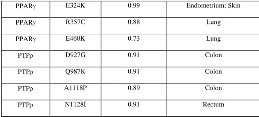
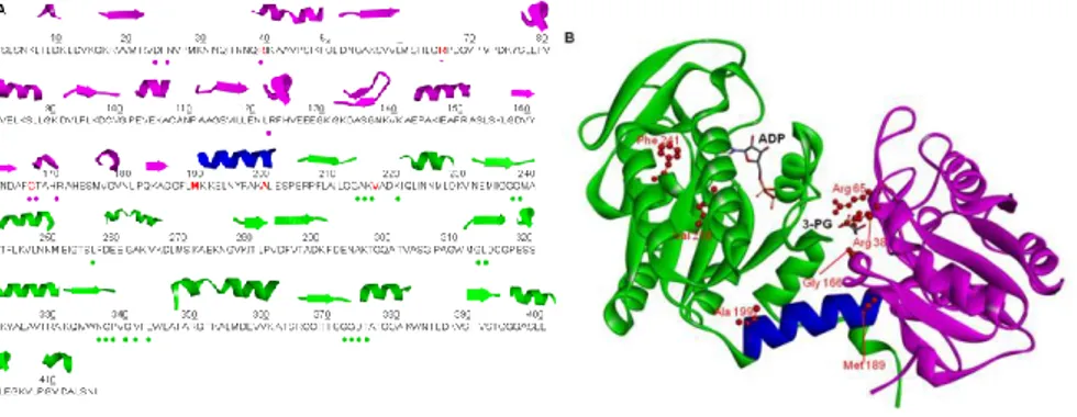
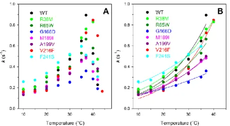
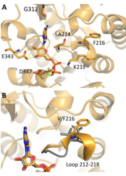
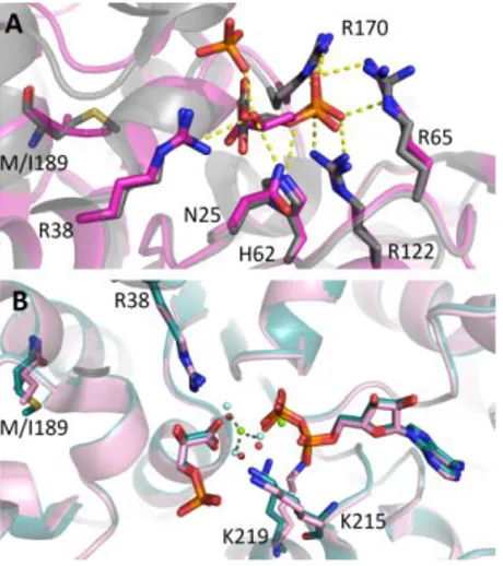
In addition, several somatic mutations of PGK1 have been exclusively identified in different cancer types, as reported in COSMIC (Catalogue of Somatic Mutations in Cancer) (http://cancer.sanger.ac.uk/cosmic) [Forbes et al., 2011]. The role of PGK1 variants in cancer is not still clear, so this study would give a biochemical characterization of some PGK1 variants (R38M, R65W, G166D, M189I, A199V, V216F and F241S) found in cancer tissues and annotated in the COSMIC database [Forbes et al, 2010].
1.5.1ii Human Frataxin (hFXN)
Several studies in the literature about cancer highlight the relationship between iron metabolism and carcinogenesis [Richardson D.R. et al. 2009; Torti S.V. 2011, 2013]. Iron is an inorganic element which is critical for cell proliferation and growth by incorporating into iron- or heme-containing enzymes. Since these enzymes are involved in respiratory complexes, DNA synthesis, cell cycle and detoxification processes, iron is essential in terms of cell replication, cellular metabolism and growth. Iron is not only fundamental for cell survival but it can be related to carcinogenesis because, beside the effects mentioned above, iron can create reactive oxygen species (ROS) by
24
participating in Fenton reaction where hydroxyl radical is produced: ROS can damage DNA and be mutagenic.
Frataxin (FXN) is a small highly conserved mitochondrial protein ubiquitously expressed in prokaryotes and eukaryotes [Adinolfi et al, 2002; Gibbson et al, 1996] that plays an important role in Fe-S cluster biosynthesis and iron and heme metabolism [Muhlenhoff et al, 2002; Che net al, 2002; Busi et al, 2006]. Sequence alignment of the frataxin family shows two distinct regions: an N-terminal region of 70-90 residues completely absent in prokaryotes and poorly conserved in eukaryotes with typical unfolded features [Huynen MA et al, 2001] and a highly conserved C-terminal region of 100-120 residues with a sequence identity of about 25% and similarity around 40-70% indicating that this part of the protein is functionally important [Pastore A et al, 2013]. Frataxin is nuclear encoded, expressed in the cytoplasm and imported in the mitochondrion through an import signal contained in the N-terminal region of the protein. In fact, human frataxin (hFXN) is synthesized as a precursor of 210 amino acids and imported into the mitochondrion where the mitochondrial processing peptidase operates several cleavage processes that remove the N-terminal region and allow the formation of the mature form of the protein [Koutnikova H et al, 1998; Condò I et al, 2007; Schmucker S et al, 2008].
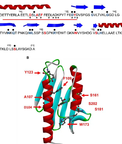
The mature protein consists of amino acids 81–210 and has an apparent molecular mass of about 14 kDa [Campuzano et al, 1996]. The human isoform shows an α- β sandwich structure composed of two α-helices, which form one plane over five antiparallel β-strands, which form the second plane. Furthermore, two others β-strands intersect the two planes in order to achieve the overall structure [Bencze et al, 2006].
25 Fig. 1.3 Human Frataxin (PDB code: 1EKG) Globular structure of hFXN with two
terminal α-helices which pack against a -sheet with 7 strands, giving a planar α- β sandwich structure motif.
Several conserved acidic residues (Asp, Glu) are located between the first helix and the edge of the β 1-sheet gene rating a semi-conserved acidic ridge that generates a negatively charged surface (Fig. 1.4) that seems to be involved in iron binding and in binding with partners [Dhe-Paganon et al, 2000]. Three tryptophans are also conserved even if the occurrence of this kind of residue is relatively low in proteins, suggesting that they could have an important structural and/or functional role.
26 Fig. 1.4 Molecular representation of the negatively charged surface of hFXN. This acidic
region is located on the interface between 1 and the first 1 strand and seems to be
involved in iron binding and in binding with partners.
In mammals, frataxin is mainly espressed in tissues with a high metabolic rate, like neurons, heart, kidney and liver and normally activates mitochondrial iron-sulfur-cluster protein assembly complex, which is composed of the Nfs1 enzyme and the scaffold protein Isu [Gerber J et al, 2003; Ramazzotti A et al, 2004]. These two proteins are, respectively, the cysteine desulfurase that converts cysteine to alanine and the transient scaffold protein on which the cluster assembles [Bandyopadhyay S et al, 2008; Py B et al, 2011].
The direct frataxin partner have been extensively studied: early genetic and biochemical studies were careful to generically indicate interaction between frataxin and the Nfs1/Isu complex [Gerber J et al. 2003; Ramazzotti A et al. 2004], however later it was reported that human frataxin interact with Isu with high affinity and in an iron-dependent way [Cook JD et al. 2006; Bencze
27
KZ et al. 2007]; finally, independent works confirmed that hFXN interacts with a preformed complex composed of Nfs1, Isu and Isd11 in an iron-independent fashion [Tsai and Barondeau 2010; and Schmucker S et al. 2011].
The notoriety of this protein is predominantly related to Friedreich ataxia (FRDA), an autosomal-recessively inherited disorder described for the first time in 1863 by Friedreich and with an incidence of 1:50000 in the European population. 95% of FRDA patients are homozygous for unstable guanine-adenine-adenine (GAA) expansions in the first intron of the frataxin gene (fxn) on the positive strand of chromosome 9q21.11 [Campuzano et al, 1996], however another 5% of FA patients are compound heterozygotes with an expansion on one allele and conventional mutations on the other. At least 15 missense point mutations have been reported [Cossée M et al, 1999; Labuda M et al, 1999; Musco G et al, 2000; Leidgens S et al, 2010; Schmucker S et al, 2011]. In vitro studies demonstrated that these variants (W155R, I154F, D122Y, G130V, N146K) retain a native fold under physiological conditions but have reduced thermodynamic stabilities and binding property of frataxin for its protein partners [Musco et al, 2000; Correira A et al, 2006, 2008]. The core syndrome is an early onset, slowly progressive ataxia associated with areflexia that appears during adolescence. Ataxia arises from combined afferent (peripheral sensory neuropathy plus spinal degeneration), cerebellar and sometimes also vestibular dysfunction. In addition to ataxia of stance and gait, patients develop appendicular and truncal ataxia. Dysarthria is another cerebellar feature present in 70% with abnormal pitch variation, loudness maintenance, breath support for speech, hypernasality and consonant imprecision due to laryngeal or velopharyngeal dysfunction [Folker et al, 2010]. Cardiac involvement is also possible, with
28
left ventricular hypertrophy, paroxysmal or permanent supraventricular tachycardias [Bourke et al, 2011]. In 8 to 49% of the cases it‟s possible to see diabetes [Dürr et al, 1996; Finocchiaro et al, 1988] with an usually late manifestation in the course of FRDA (mean 15 years after onset) [De Michele et al, 1996]. The onset is often acute, sometimes with ketoacidosis [Bird et al, 1978]. The main event in the etiology of diabetes is the loss of pancreatic islet ß cells with concomitant decline of insulin secretion. The process is further aggravated by increased insulin requirements due to insulin resistance. Scoliosis is considered typical for FRDA. Its prevalence varies between 33 and 100% depending on the individual study [Harding et al, 1981; Labelle et al, 1986]. Similarly, foot deformities (pes cavus, club foot, pes planus) may significantly interfere with mobility in 55 to 90% of patients. Severity does not depend on expansion size or age of onset, but is related to disease duration and age.
Despite several studies have been performed until now on frataxin, the exact function of this protein is still unclear: in addition to its certain implication in iron homeostasis [Richardson et al, 2010], iron–sulfur cluster (ISC) biosynthesis [Stemmler ey al, 2010] and protection from oxidative stress and apoptosis [Condo et al, 2006], it seems that frataxin can be involved in several other functions.
Frataxin is a protein required for cell survival since its complete knockout is lethal. This protein protects tumour cells against oxidative stress and apoptosis but also acts as a tumour suppressor [Guccini I et al, 2011] and, until now, the molecular bases of this apparent paradox are missing. Furthermore, conflicting results have been reported on the role of frataxin in cellular growth: both frataxin knockdown and frataxin overexpression were shown to impair cell growth [Stehling O et al, 2004; Thierbach R et al, 2005;
29
Schulz TJ et al, 2006]. Very little is known regarding the molecular regulation of frataxin expression: histone deacetylase inhibitors, erythropoietin, cisplatin, 3-nitropropionic acid and hemin increase levels of frataxin in vitro (Gottesfeld et al, 2007). Moreover, transcription factors peroxisome proliferator-activated receptor gamma (Marmolino et al, 2009) and hypoxia-inducible factor-2 alpha (HIF-2a) (Oktay et al, 2007) positively regulate frataxin expression. It has been reported that frataxin overexpression can protect tumour cells from apoptosis but can also act as a tumour suppressor participating, by regulating p53 activation, in tumour adaptation to hypoxia, a critical feature associated with tumour growth and progression, thus suggesting that frataxin levels can influence tumour cell fate [Guccini I et al, 2011].
Furthermore, in several databases such as COSMIC database or ATLAS, several FXN variants have been reported suggesting that abnormalities in this highly conserved essential protein can be implicated in cancer onset and/or progression. This study would give a biochemical characterization of some FXN variants (D104G, A107V, F109L, Y123S, S161I, W173C, S181F and S202F) found in cancer tissues and annotated in the COSMIC database [Forbes et al, 2010].
1.5.1iii Poly (ADP-ribose) polymerase 3 (PARP3)
In addition to the accepted role for cell metabolism in cancer, it is well established that DNA-repair/DNA-damage pathways are important in cancer progression because dysregulation leads to higher levels of genomic instability, increased mutation rate, and enhanced intra-tumor heterogeneity [Burrel RA et al, 2013; Chae YKet al, 2016; Gavande NS et al, 2016].
30
Mutations in DNA-repair genes can confer growth and survival advantages of cancer cells, therefore it can be interesting to improve knowledge about this important aspect of cancer biology because this might lead to the development of new targeted therapies in DNA-repair deficient cancers or even can improve the efficacy of existing therapies, such as PARP inhibitors.
The biological roles of Poly (ADP-ribose) polymerase 3 (PARP3) are currently under investigation; however, several key reports indicate the integral roles of PARP3 in DNA damage repair responses [D‟Amours et al, 1999; Ame et al, 2004], and thus it has been investigated as a novel target in oncology.
PARP3 is an important member of the PARP family, also referred to as diphtheria toxin-like ADP-ribosyltransferases (ARTD), composed of 17 members which all share a conserved ADP-ribosyl transferase (ART) fold and involved in a number of crucial cellular processes that are linked to genomic DNA integrity such as DNA repair, genome stability, and cellular stress responses [D‟Amours et al, 1999; Ame et al, 2004; Hottiger et al, 2010]. In the PARP family, only a subset of members is predicted to have the ability to produce Poly-ADP-ribose (PARP-1 to PARP-5a and PARP-5b) while two are inactive enzymes (PARP-9 and PARP-13) and the remaining members are able to produce a mono-ADP-ribose modification [Kleine et al, 2008]. The 17 members of the PARP family show very different structures and cellular functions but are all related by the presence of the PARP signature, a conserved PARP catalytic domain [Otto H et al, 2005].
The founding and most studied member PARP1 as well as PARP2 and PARP3 play important roles in the repair of DNA strand breaks and are known to be catalytically activated through interaction with DNA damaged and catalyze auto-ADP-ribosylation and ADP-ribosylation of other nuclear
31
acceptor proteins [Pascal JM et al, 2015; Martin-Hernandez K et al, 2016; Wei H et al, 2016]. PARP1, PARP2 and PARP3 show a characteristic fold that is characterized by conserved tryptophan-glycine-arginine (WGR) and C-terminal catalytic (CAT) domains and of an N-terminal region (NTR) that is really different between the three proteins [Oliver AW et al, 2004]. In fact, in contrast to PARP1, the NTRs of PARP-2 and PARP-3 are much shorter (78 and 40 residues, respectively), and they do not contain the N-terminal zinc finger domains essential for initial DNA binding of PARP1 [Langlier MF et al, 2012]. Short NTRs of PARP2 and PARP3 are not strictly required for DNA-dependent activation but rather contribute to the overall binding affinity and specificity for SSB due to the presence of basic residues in this region, as shown for PARP2 [Ame et al, 1999], and contribute to the allosteric regulatory mechanism of DNA-dependent catalytic activation via local destabilization of CAT through local rearrangements of the WGR domain [Dawicki-McKenna JM et al, 2015; Obaji E et al, 2016].
32 Fig. 1.5 Domain architecture of PARP-1, PARP-2 and PARP-3 The WGR and CAT
domains are conserved, while the N-terminal regions (NTRs) are distinct [John M. Pascal Nucleic Acids Research, 2014, Vol. 42, No. 12]
PARP3 has been found to associate with Polycomb group proteins involved in transcriptional silencing and with DNA repair networks, including base excision repair/single-strand break repair (BER/SSBR) and nonhomologous end-joining (NHEJ), suggesting an active role for PARP3 in the maintenance of genomic integrity [Rouleau M et al, 2007]. Furthermore, PARP3 has been described as a critical player in the stabilization of the mitotic spindle and in telomere integrity notably by associating and regulating the mitotic components NuMA and Tankyrase 1 suggesting PARP3 as a positive regulator of the mitotic network containing Tankyrase 1 and NuMA with important implications in spindle dynamics and telomere integrity during mitosis [Boehler C et al, 2011].
PARP3 serves TGFβ-dependent epithelial-to-mesenchymal transition (EMT), a transdifferentiation programme that is important for organogenesis in the developing embryo, tissue injury repair and cancer progression, by stimulating a TG2-Snail-E-cadherin axis in a manner involving its response to TGFβ-induced ROS [Karicheva O et al, 2016]. The fact that PARP3 is upregulated and responds to ROS produced from both exogenous (genotoxic agents) and endogenous (TGFβ) sources makes it a major driver of the ROS response that is likely to function in various other physiological and pathophysiological events. One interesting example is suggested by the prominent role of PARP3 in neural crest cell differentiation in the zebrafish [Rouleau M et al, 2011], a process in which the generation of ROS by the
33
NADPH oxidase NOX4 was documented to be required for efficient differentiation [Lee JE et al, 2014].
Since point mutations in DNA repair pathways can alter repair function and contribute to cancer risk [García-Closas et al. 2006a; Goode et al. 2002], in this study we selected some nsSNVs found in cancer tissues and annotated in the COSMIC database [Forbes et al, 2010] (F125L, P147T, G148D, L233F and F440L) in order to investigate their biophysical and biochemical properties respect to its wild-type counterpart.
The data presented in this thesis will provide a comprehensive analysis of three-dimensional structure and biophysics of wild-type and nsSNVs with the purpose to give a contribution to fill the gap between the collection of thermodynamic data and disease-related information on protein variants.
35
Chapter 2
Aim of the study
nsSNVs are caused by missense mutations, changes in one DNA base pair that result in the substitution of an amino acid in the protein encoded by a gene, and contribute to ̴ 80% of the total number of variants annotated as pathogenic. They are the most common type of variation among humans and pathogenic nsSNVs, or missense mutations, account for approximately half of the allelic variants causative of hereditary disease. During the last decade several databases were created collecting data associated to protein variations, above all focusing their attention on the impact of protein variants at the structural, functional and phenotypic levels. Some amino acid substitutions are functionally neutral, however most of them may have different effects on protein structure and function and can be related to several diseases. It was demonstrated that mutations can frequently affect several biophysical characteristics simultaneously and may or may not cause diseases [Gong et Blundell, 2010]. Even if the available data on single amino acid substitutions are expanding rapidly, the knowledge of the possible disease association of nsSNVs and the molecular mechanisms of genetic disease is lagging due to the limited integration between experimental data and predictions because of the laborious and time consuming nature of experimental studies. There is no clear threshold of how large the change of the wild-type characteristics should be in order to alter protein function and
36
result in disease. Predictions of damaging effects of mutations are further complicated by the observations that enhanced protein activity [Zhang et al, 2013], higher stability or binding affinity [Witham et al, 2011; Takano et al, 2012] are not necessarily advantageous for the cell and can be disease causing.
Nowadays several computational methods are available for predicting the possible pathogenicity of nsSNVs; they are predominantly based on evolutionary information and/or varying structural descriptors of the protein in question. These methods aim at automating the annotation process of nsSNV effects and therefore would be very useful for the mutation research community. In silico study can be useful for obtaining a statistical picture of the extent to which these SNVs influence phenotype and for prioritizing SNVs for experimental study [Hallali-Assani et al, 2009].
Computational analysis has predicted that around 30% of protein variants resulting from non synonymous single nucleotide substitutions are less stable than the wild-type. Experimental studies have been carried out on very few proteins due to the cost and time needed for the entire process that requires mutagenesis, protein expression and purification followed by thermal and chemical unfolding [Pasquo A et al, 2012; Lori C et al, 2013; Lori L et al, 2016; Gao et al, 2015; Petrosino M et al, 2017].
In this study I‟m going to present a systematic and qualitative analysis of structural and functional properties of protein variants caused by single amino acid substitutions leading to non synonymous variants found in cancer tissues, including changes in enzyme activity, aggregation propensity, structural stability and binding ability. The aim of this study is the investigation of plausible correlations, if any, between alteration on protein
37
structure, function or stability caused by single amino acid substitutions and cancer.
During my PhD I focused my attention on three case-reports analyzing the expression, the structural characterization and stability and binding properties of some variants of three different proteins related to several cancer types:
Phosphoglycerate kinase 1 (PGK1) is an essential enzyme that catalyzes the reversible phosphotransfer reaction from 1,bisphosphoglycerate (1,BPG) to MgADP to produce 3-phosphoglycerate (3-PG) and MgATP, an important ATP-generating step in glycolysis. Several solid tumors exhibit an increased expression of glycolytic enzymes such as PGK1 to generate ATP in hypoxic conditions [Hwang et al., 2006; Sun et al., 2015] suggesting a plausible role of PGK1 as a cancer biomarker.
Human Frataxin (hFXN) is a nuclear-encoded protein highly conserved implicated in Fe-S cluster biosynthesis and in iron and heme metabolism. Iron is an inorganic element which is critical for cell proliferation and growth and alterations in proteins involved in the regulation of iron levels in cells can be dangerous for living organisms and can be related to carcinogenesis.
Poly (ADP-ribose) polymerase 3 (PARP3) is a member of the PARP family, a group of enzymes that synthesize poly (ADP-ribose) on themselves or other acceptor proteins, that catalyses a post-translational modification of proteins involved in