Biological Tools to Study the Effects of
Environmental Contaminants at the
Feto–Maternal Interface
Chiara Mannelli
1,2, Francesca Ietta
1, Anna Maria Avanzati
1,
Dariusz Skarzynski
2, and Luana Paulesu
1Abstract
The identification of reproductive toxicants is a major scientific challenge for human health. Prenatal life is the most vulnerable and important time span of human development. For obvious ethical reasons, in vivo models cannot be used in human pregnancy, and animal models do not perfectly reflect human physiology. This review describes the in vitro test models representative of the human feto–maternal interface and the effects of environmental chemicals with estrogen-like activity, mainly bisphenol A and para-nonylphenol, with a particular emphasis on the effects at low, nontoxic doses similar to concentrations commonly detected in the population.
Keywords
in vitro models, human placenta, human endometrium, xenoestrogens, bisphenol A, para-nonylphenol
The Feto–Maternal Interface: A Complex
Network of Endocrine, Paracrine, and
Autocrine Factors
Human pregnancy is a complex and finely regulated process during which 2 genetically different organisms, the mother and the embryo/fetus, establish a very intimate contact.1-3A huge number of molecules are secreted at the feto–maternal interface by both the mother and the embryo/fetus and act on both sides.4 Thus, the mother and the embryo interact via specific tissues in a reciprocal exchange of molecules that act as communication signals.
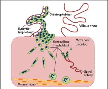
Placentation in human pregnancy involves invasion of the blastocyst in maternal endometrium up to the spiral arteries. The trophoblast cells, which surround the blastocyst, first con-tact the uterine epithelium, then dislodge the endometrial epithelial cells, and invade the maternal endometrium. During its journey, the trophoblast follows 2 differentiation path-ways—the villous and the extravillous ones (Figure 1).5In the villous pathway, the mononuclear cytotrophoblast cells form-ing the internal layer of villi fuse and form the multinucleated syncytiotrophoblast, which is the epithelial covering of the chorionic villi (floating villi). In the extravillous pathway, the cytotrophoblast cells move beyond the overlying syncytium and form multilayer cell columns of extravillous trophoblast that fix the villi to the maternal tissues (anchoring villi). These
cells then move deeper into the maternal tissues up to the proximal third of the myometrium and endometrial spiral arteries (Figure 1).
Human endometrium has a key role for reproductive effi-ciency, and its remodeling takes place under the control of steroid hormones.6,7 This tissue undergoes dramatic cyclic changes regarding cell proliferation, secretory functions, regression, and regeneration. Hormonal stimuli lead the mater-nal tissues to the formation of the decidua at the end of each cycle. If fertilization has occurred, the blastocyst reaches the uterine epithelium and implantation starts.
The correct signaling between the decidua and the fetal trophoblast is of paramount importance for blastocyst implan-tation and successful pregnancy (Figure 2).2,7
In the complex scenario of the feto–maternal interface, endocrine, paracrine, and autocrine factors must be taken into
1Department of Life Sciences, University of Siena, Siena, Italy 2
Department of Reproductive Immunology and Pathology, Institute of Animal Reproduction and Food Research, Polish Academy of Sciences, Olsztyn, Poland
Corresponding Author:
Luana Paulesu, Department of Life Sciences, University of Siena, via A. Moro, 2, Siena, Italy. Email: [email protected] An International Journal 2015:1-11 ªThe Author(s) 2015 DOI: 10.1177/1559325815611902 dos.sagepub.com
Creative Commons CC-BY-NC: This article is distributed under the terms of the Creative Commons Attribution-NonCommercial 3.0 License (http://www.creativecommons.org/licenses/by-nc/3.0/) which permits non-commercial use, reproduction and distribution of the work without further permission provided the original work is attributed as specified on the SAGE and Open Access page (https://us.sagepub.com/en-us/nam/open-access-at-sage).
consideration. Hormones, such as estrogens, progesterone, and human chorionic gonadotropin (hCG) are the major players of the endocrine regulation that take place at the feto–maternal interface. Human chorionic gonadotropin, which is the hor-mone produced by the trophoblast in the very early stages of pregnancy, plays a key role in making sure that the endome-trium is ready to receive the embryo implantation.8 Those molecules which have an autocrine/paracrine action are impor-tant factors in the establishment and advancement of preg-nancy.9Among these are cytokines including the interleukins (IL)s IL-1, IL-4, IL-5, IL-6, IL-8, and IL-10, the macrophage migration inhibitory factor (MIF), colony stimulating factors (CSFs) and the leukemia inhibiting factor, and different growth factors (GFs) such as the epidermal GF and the vascular endothelial GF. All these molecules are potent immunoregula-tors that play a key role in the feto–maternal tolerance to the
semiallogeneic embryo.10 These molecules are also media-tors of cell proliferation/differentiation and apoptosis con-tributing to fetal growth and expansion in the maternal tissues.11,12 Therefore, their presence at the feto–maternal interface may modulate the maternal immune response and contribute to the expansion of fetal tissues in the maternal uterus.
Prostaglandins (PGs) also play a pivotal role in angiogen-esis, mitogenangiogen-esis, cell proliferation, and differentiation and exert an important role during the early stages of pregnancy.7,13,14
Environmental Contaminants: A Threat for
Human Reproductive Health
There are increasing data that support the adverse effects on human reproductive health caused by environmental contami-nants, especially endocrine-disrupting chemicals. These chemi-cals are man-made or plant-derived compounds that can bind to the receptors of steroid hormones and thus impair hormone-driven physiological functions in female and male reproductive system.15,16Among these, diethylstilbestrol (DES), a nonster-oidal estrogenic compound, has been commercialized as ther-apeutic for reproductive disorders. Regrettably, only after its commercialization its deleterious effects became evident. Among industrial compounds is bisphenol A (BPA), a polymer that can be released by polycarbonated plastics and by the linings of metal cans that are used for food and beverages.17,18 Consequently, BPA can be easily absorbed via the food chain,19and indeed it can be easily detected in the human body. With regard to pregnancy, levels of BPA have been detected in the placenta, fetal liver, in the blood, and in the follicular fluid in a range of 0.3-40 nmol/L.18,20,21 Bisphenol A shares many similarities with the endogenous estrogens22 and acts on estrogen-responsive organs by binding to estrogen receptor (ER) isoforms, ERa and ERb.23,24 The estrogenic activity of BPA has been demonstrated in Ishikawa cells, an endometrial cell line.6Bisphenol A also binds to the progesterone receptors (PRs) and thus exert antiprogestin activity.25,26 Furthermore, BPA is able to mimic glucocorticoids, another class of steroid hormones fundamental at the feto–maternal interface. Bisphe-nol A is indeed able to bind with the mineralocorticoid and glucocorticoid receptors (GRs).27,28 Interestingly, it has been demonstrated that environmental concentrations of BPA (10 nmol/L) increased the messenger RNA (mRNA) expression and enzymatic activity of the enzyme 11b-hydroxysteroid dehydrogenase type 1 in the adipose tissue and the visceral adipocytes isolated from children.29 Furthermore, perinatal exposure to BPA resulted in a decreased expression of GR in the brain of female rats compared to controls.30This environ-mental chemical was listed as reference compound within the European ReProTect Programme because of its well-known reproductive toxicity.31,32 Another environmental chemical derived from the manufacturing industry is para-nonylphenol (p-NP), an alkylphenol applied as a plasticizer and surfactant, known to have estrogenic activity since 1991.33 Human
Figure 2. The interplay of endocrine and paracrine mediators at the feto–maternal interface. hCG indicates human chorionic gonadotro-pin; GFs, growth factors; PGs, prostaglandins.
exposure to p-NP may occur by cutaneous absorption, inges-tion of contaminated food or water, and inhalainges-tion.34,35 Estro-genic activity of p-NP has been known since 199133 and reported in a number of in vitro36and in vivo studies.37 Inter-estingly, studies in rats showed that maternal exposure to p-NP resulted in an increase in uterine calbinding-D 9k (CaBP-9k) mRNA and protein expression in maternal and neonatal uteri, suggesting its potential transfer through the placenta.38,39 CaBP-9k is a cytosolic calcium-binding protein expressed in various tissues (eg, intestine, uterus, and placenta) and a marker of estrogenic compounds exposure.40
Since the number of man-made chemicals released in the environment have been growing exponentially in the last decades,41 the assessment of the risks that derive from the exposure to chemicals is of paramount importance.31
Risks Assessment of Intrauterine Exposure
Because of their ability to interfere with steroid hormones and their widespread distribution in everyday products, many of the chemicals in the environment represent a hazard to the repro-ductive system of adult women. Much greater concern arises when we take into account prenatal exposure to these bioactive compounds. Prenatal life is indeed the most critical phase of the life cycle, as it is the period in which organs and tissues are formed.42
Already during the periimplantation period, the intrauterine environment is able to provoke epigenetical changes that will persist later in life.43Barker and coworkers initiated the inves-tigation of the developmental origins of many diseases that appear in later life.44Then many researchers followed, reveal-ing a dramatic scenario in which alterations in the intrauterine environment can result in pathologies and dysfunctions in adulthood.45
For many decades, the human placenta has been considered a protective organ. It was assumed that the placenta interdicted the passage of harmful compounds to the fetus. However, the placenta has not been adapted to act as a barrier to the many substances that have been produced by man in the last decades. Therefore, the human placenta might not be able to prevent fetal exposure to these potentially dangerous compounds. Indeed, due to their high lipophilicity, many environmental estrogens are able to elude the placental barrier and potentially harm the fetus.46 Moreover, the placenta is a highly sensitive tissue to environmental contaminants with estrogenic activity as it expresses both ERa and ERb.47Since the placenta is the fundamental organ to maintain pregnancy and assure fetal development and growth, it seems to be extremely important to estimate the risk of exposure during in utero life by evaluat-ing the effects of these contaminants on the placenta and the uterus. Animal models remain a solid and important shield to study human pregnancy establishment and development, but, unfortunately, they often fail to completely reproduce the com-plexity of human reproduction. Many efforts have been taken in order to shed light on the physiological, pathological, and
toxicological aspects of human pregnancy, and in vitro models represent a valid help for researchers in this field.
In this report, we will mainly focus on BPA and p-NP as representative chemicals with estrogen-like activity. Special emphasis will be put on the effects at low doses that are rele-vant with or are lower than those found in the environment and in human tissues. In fact, it is important to underline that envi-ronmental chemicals with estrogenic activity can have different effects at different doses.6In particular, some of these com-pounds have their detrimental effects at very low concentra-tions while higher concentraconcentra-tions are not effective in the same way.48-50
In vitro Models of Human Placenta
Many efforts have been made to develop in vitro models for studies of the placenta. It is indeed important to emphasize that animal placenta does not model the human placenta and its physiology well. As human placenta is, in fact, an organ that varies from one species to another, therefore the use of animals could not be appropriate to study the mechanisms that take place during human pregnancy. Moreover, for obvious ethical reasons, studies on human placenta can be performed only in tissues and cells obtained from the organ after its delivery. Thus, in vitro models need to be developed to investigate pla-centa establishment and development in the maternal uterus as well as to identify factors influencing these physiological pro-cesses. These models mainly include trophoblast cells and cultures of placenta villous explants.
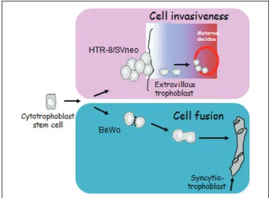
Freshly isolated cytotrophoblast cells can be obtained by enzymatic dissociation of villous placental tissue, followed by Percoll gradient separation. These cells are able to differ-entiate into multinucleated syncytiotrophoblast cells when cul-tured in complete medium. However, they do not proliferate in vitro and thus cannot be cultured for a long time.51,52For these reasons, many immortalized and carcinoma-derived cell lines (BeWo, JAr, JEG-3, and HTR-8/SVneo) have been set up to study selected aspects of the human placenta in vitro.53 For example, the human trophoblast cell lines BeWo and HTR-8/ SVneo cells are representative of specific differentiation path-ways of trophoblast during placentation (Figure 3). In particu-lar, the choriocarcinoma-derived BeWo cell line is representative of the villous pathway since it reveals most of the characteristics of the villous syncytiotrophoblast including cell fusion54and secretion of hormones such as the b-hCG.55 The HTR-8/SVneo cells are representative of the invasive extravillous trophoblast, specifically of those cells which, after detaching from the chorionic villi, migrate to and infiltrate into the maternal decidua. These cells are originated from human first-trimester human placenta and in vitro immortalized by transfection with a complementary DNA construct that encodes the simian virus 40 large T antigen.56Based on the cells’ spe-cific characteristics, any toxic effect observed on BeWo cells may forecast impairment of placenta growth, while any effect toward HTR-8/SVneo cells may contribute to an unsatisfactory blastocyst implantation and placentation in vivo.
However, despite the high reproducibility of the experi-ments provided by these cell lines, the results obtained can be far from the in vivo situation. Furthermore, cell lines enable the study of a single cell type at a time, while the human placenta consists of many cell types. Some ex vivo models offer the opportunity to maintain all the main placental cell types in the same culture.57,58
A suitable in vitro model more closely resembling the in vivo pattern of trophoblast differentiation is offered by primary cultures of placenta explants from fresh human placenta.57This model includes dissection of terminal villi from human placen-tal tissues (Figure 4). A variety of culture conditions can be used to reproduce in utero environments at different times of gestation. Usually, explants from placental tissues at first-trimester pregnancy are cultured on a bed of Matrigel which the tissues adhere to and then expand, mimicking placenta establishment and its development in the maternal uterus. Explants from term placenta are usually cultured on the bottom of the well or as free-floating villi, hanging on a supporting device. This type of culture mainly reflects the syncytialization of the villous trophoblast.57
The model of placental explants presents several advan-tages: First, unlike in the one with isolated cells, this model preserves the topology of intact villi including all the main fetal cell types in the chorionic villi, for example, the syncytio- and the cytotrophoblast, the villous stromal, and endothelial cells, thus preserving each mechanism of paracrine regulation. Sec-ond, this model mimics the trophoblast functions more realis-tically, including production and release of secretory components, proliferation, growth, and differentiation. Third, placental explants can be set up with tissues from early and term placenta, making it possible to compare the effects of substances both at early and term pregnancy. The main disad-vantage of this model is its inability to distinguish among the functional roles of the various cell types present in this organ. Overall, the model of placental explants allows to study all
aspects of placental biochemistry and molecular biology char-acterizing the physiological processes during the organ devel-opment, pharmacology, toxicology, and disease processes.
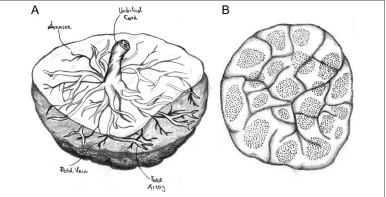
Other in vitro models of human placenta have been set up to study placental transfer.58Some of these models exemplify the transfer across a single cell layer while others retain all the components of the placental barrier, the syncytiotrophoblast, the cytotrophoblast, the basal membrane, and the endothelial layer.32Among the last type of models, the placental perfusion technique can be found. Thanks to the particular anatomy of the human placenta which consists of many circulatory units (coty-ledons; Figure 5), a single cotyledon can be isolated and perfused.59
Potential Biomarkers in Human Placenta
The human placenta is an exquisite tool to study the effect of environmental chemicals on human reproduction, due to the key role of this organ in maintaining pregnancy and providing fetal health. Toxicity of chemicals needs to be tested in order to identify concentrations suitable for evaluating potentially dele-terious effects in the functional processes of the placenta. Indeed, it is important to separate the effects of toxic doses from the ones that nontoxic doses might have on a biological system. To do so, in vitro models can be tested for cell viability and tissue damage in a wide range of chemical concentrations. All the immortalized and carcinoma-derived cell lines are eas-ily cultured and propagated in vitro, and they are generally used for large-scale screening of many chemicals and/or the effects of their concentrations.
Once the curve of toxicity is obtained, functional studies can be conducted using nontoxic doses. Furthermore, in order to make these studies closer to the in vivo situation, concentra-tions of chemicals similar to those detected in the human body should be privileged. As reported in the literature, BPA levels in human pregnancy ranged from 0.3 and 40 nmol/L in adult and fetal serum, follicular and amniotic fluid, and pla-centa.18,20,21The p-NP levels ranged from 0.1 nmol/L in cord blood and 1 nmol/L in maternal plasma and blood.60-62
Among the trophoblast cell types, BeWo and HTR-8/SVneo cells are suitable to detect the specific effects of chemicals with estrogenic activity in human placenta since both of them express ERs.63,64 Moreover, given their different differentia-tion pathways, these cells can be monitored for different biomarkers. Specifically, BeWo cells can be tested for the secretion of b-hCG and tissue expression of cleaved caspase-3, specific markers of endocrine activity of the syncytiotropho-blast and its apoptotic shedding, respectively.62,65The HTR-8/ SVneo cells can be monitored by assaying the passage of these cells through a layer of collagen (cell migration) or matrigel (cell invasion) as well as by the release of metalloprotease (MMP)-2 and MMP-9, the most studied MMPs for trophoblast invasion.66The use of 2 different cell lines representative of the main differentiation pathways in human placenta provides information about the effect of chemicals in the whole process of placentation.
Figure 3. Specific differentiation pathways of the BeWo and the HTR-8/SVneo trophoblast cells.
In the last decade, several studies of BPA and p-NP in in vitro models of human placenta have been conducted. Studies in BeWo and HTR-8/SVneo cells showed that these chemicals caused a decrease in cell viability in the concentration range of 1 mmol/L and 1 mmol/L. Surprisingly, low concentrations (pmol/L-nmol/L), not affecting cell viability, impaired func-tional markers of placentation. More specifically, both chemi-cals, p-NP and BPA, appeared to affect syncytialization of trophoblast as they both altered hCG secretion and cell apop-tosis in BeWo cells.32,47 Interestingly, the effect of estrogen-like chemicals, at low nontoxic concentrations, resulted in a change in hCG release with an hormetic or biphasic behavior.67 The chemicals studied were indeed stimulating or inhibiting hCG release depending on their concentration.67 More recent evidence in HTR-8/SVneo cells showed that cell migration and invasion were also reduced by BPA and p-NP.50 For each chemical, the activity was higher at lower concentrations with a maximum activity between 0.1 and 10 pmol/L. Coculture studies of HTR-8/SVneo with human umbilical cord endothe-lial cells (HUVECs) revealed that trophoblast/endotheendothe-lial inter-action was significantly reduced by p-NP at 10 pmol/L.
All together, the above-mentioned studies on trophoblast cells indicate that low concentrations of the environmental
chemicals BPA and p-NP are able to affect the main differen-tiation pathways of trophoblast, the villous, and extravillous ones.
It is noteworthy that effective concentrations of p-NP in hCG secretion were lower in BeWo cells (0.1-1 pmol/L) than in placental explants (1 nmol/L).47,48This discrepancy can be explained by a different pharmacokinetic behavior in the 2 different models probably due to a more complex organization in a tissue culture with respect to a cell monolayer.
Placental explants give the chance to monitor, with a closer look into the physiological situation, trophoblast differentiation both into syncytiotrophoblast and extravillous trophoblast. This model is also informative about the endocrine secretory activity of placenta (ie, secretion of hCG, placental lactogen, cytokines, GFs, and other paracrine/autocrine molecules produced by the placenta).47,48 In particular, cytokines such as granulocyte macrophage-CSF (GM-CSF), interferon g (IFN-g), 1b, IL-4, and IL-10 were all increased by pmol/L to nmol/L p-NP with a maximum effect at 10 pmol/L, which was statistically signif-icant for GM-CSF and IL-10.47,48Bisphenol A (1 nmol/L) was increasing the secretion of the cytokine MIF while BPA levels ranging from 0.2 to 2 nmol/L, increased tumor necrosis factor-a gene expression, factor-and protein secretion.49,68
Figure 4. Dissection and culture of placental explants. Representative villous explants culture from 9 weeks’ gestation. Fresh placenta is dissected and each villous fragment (15-20 mg wet weight) is placed in a 24-well culture plates (Ø 15.6 mm) previously coated with Matrigel. Villous pieces are then covered with a proper medium and cultured under different experimental conditions. Bar¼ 3 mm.
Perfusion experiments on human placenta are a wonderful tool to investigate the transfer of substances. This model showed the transfer of BPA across the placenta.32Furthermore, placental perfusion can be used to study the placental metabolism of many chemicals and drugs, the role of transporters as well as the effects of acute toxicity in placenta (for detailed review, see Myllynen and Va¨ha¨kangas,58and Myllynen et al studies69).
The effects of representative substances such as BPA and p-NP, in the scenario of environmental contaminants, raise great concern for the environmental risk to the health of the fetus. Even though they do not cause termination of pregnancy, low concentrations of these substances could produce anoma-lies in the functional processes of the placenta, leading to pos-sible disorders and/or abnormalities later on in pregnancy. For example, higher levels of cytokines such as MIF in the mater-nal serum and increased levels of hCG in the second trimester of pregnancy, have been associated with preeclampsia, a seri-ous syndrome of human pregnancy.70-73
In vitro Models of Human Endometrium
The human endometrium is a fertility-determining tissue. It consists of various cell types. Epithelial cells and glandular-epithelial cells are located in the lining of the uterine lumen. Stromal, endothelial, and immune cells are mainly located in endometrial stroma. The cell types that reside in the endome-trium result to be more sensitive to steroid hormones compared to the cell types of other tissues.31,74This raises concern about the possible deleterious effects of hormone-mimicking com-pounds on the cycling and pregnant human endometrium.16,75
Primary cells from human endometrium can be easily cul-tured and are able to proliferate in vitro.76These primary cul-tures of endometrial cells respond to hormonal stimuli but, unfortunately, tend to dedifferentiate after several passages and therefore cannot be cultured for long periods.77,78Immortalized and carcinoma-derived cell lines, such as Ishikawa cells (derived from epithelial endometrial cells) and St-T1b (derived from stromal endometrial cells), are also avail-able.6,78,79These cell lines are easily cultured and maintained for long periods, but they often lose responsiveness to hormo-nal stimuli and this can lead to misleading results. In vitro models of human endometrial epithelium are used to study blastocyst attachment and generally to focus on the very first steps of pregnancy establishment. Cell models of endometrial stroma are a powerful tool to study trophoblast invasion in the maternal uterus. Furthermore, such models are often used to study endometrial pathologies such as endometriosis.80 Endothelial stromal cells can also be used to investigate the physiological and pathological mechanisms that take place in the human endometrium. Unfortunately, primary endothelial cells from human endometrium tend to lose their hormonal responsiveness after few passages.74 Nevertheless, endothe-lial cells of uterine origin are far more sensitive to hormonal stimuli than endothelial cells from other tissues, that is, HUVECs, and thus tissue-specificity should be always con-sidered when selecting an in vitro model.74Models of tissue explants from human endometrium have been also used.81-83 These can be obtained from the decidual fragments remaining in the human placenta after elective termination of pregnancy or after delivery at term.
Figure 5. Anatomy of human placenta. A, Fetal side: the amnion, umbilical cord, and fetal vessels are visible. B, Maternal side: the cotyledons and their circulatory units are visible. Images by Chiara Mannelli.
More recently, three-dimensional (3D) models of human endometrium have been established and are gaining increasing interest since these models are closer to the in vivo situation. Indeed, endometrial cells show different features when grown in 3D matrices compared to cell monolayers.76,77,84
Potential Biomarkers in Human
Endometrium
Among the available in vitro models of human endometrium, cultures of endometrial cell lines are the most commonly used in reproductive toxicology. As reported above about the studies of placenta, toxicity tests are needed to provide guidance for the range of concentrations suitable for functional testing in human endometrial physiology. In order to achieve this, viabi-lity and cell proliferation assays, together with assays of tissue damage (ie, such as lactate dehydrogenase measurement), are recommended. Cell lines, such as Ishikawa cells, are a power-ful tool for screening the effects of environmental chemicals on the human endometrium.6,85,86Using these cells, Schaefer and co-workers6provided very interesting data on the effects that DES, BPA, and p-NP can have on endometrial receptivity, which is a key step for establishment of pregnancy. What is more, these authors investigated the effects of these substances in a wide range of concentrations, thus unraveling the dose– response effects of these chemicals on the endometrium.6 Transfected Ishikawa cells also provided interesting insights into the estrogen-like activity of many environmental pollu-tants, such as DES, BPA, genistein, and o-p0 -dichlorodiphenyl-trichloroethane by monitoring the effect of these chemicals on the activation of estrogen response elements.86
In vitro models of endometrial epithelial cells provide important insights into the effects of exogenous compounds on the binding surface that allows blastocyst attachment. Indeed, the expression of important surface molecules, such as integrins and osteopontin, after exposure to environmental chemicals, could represent a very good marker in order to assess the effect of chemicals on uterine preparation to blasto-cyst attachment. Many studies proved that the expression of such molecules is hormone sensitive and can be altered by exogenous hormone-mimicking compounds in vivo.87-89
On the other hand, endometrial stromal cells are useful to study the intimate mechanisms that prepare the uterus for a gestation and that could be disrupted by hormone-mimicking compounds. Primary cultures of endometrial stromal cells proved to be an effective tool to study the potential deleterious effects of BPA.49,90,91For this purpose, the expression of hor-mone receptors, that is, ERs, PRs, hCG/luteinizing horhor-mone receptor, together with the secretion of paracrine/autocrine fac-tors (ie, cytokines, PGs, and GFs) can be considered a powerful approach to assess the effect of environmental chemicals on the stromal compartment of the endometrium. Many authors inves-tigated the effects of dietary relevant, low doses of phytoestro-gens, such as genistein and daidzein, on endometrial fibroblasts by assessing the ability of these compounds to inhibit aroma-tase activity or cell proliferation.92,93
Cultures of endometrial endothelial cells are useful to study the specific angiogenetic processes that take place in the uter-ine environment. The potentially negative effects of BPA or genistein on the endometrium have been tested on primary cultures of endothelial cells by checking cell viability, cell proliferation, and angiogenic activity via the tube-formation assay.94,95
Furthermore, endometrial coculture systems have been used to investigate the effects of phytoestrogens on the important interactions between epithelial and stromal cells, such as ER activation and cell proliferation.96Endometrial explants are a natural coculture system of endometrial cells, which could be used for reproductive toxicology studies, even if they are, at the moment, applied mostly for physiological studies.97,98
Coculture Models of Feto–Maternal Interface
Coculture models including cells and/or tissues representative of both the fetus and the maternal counterpart have been devel-oped.76,81-83Many of these focus on the mechanisms of blas-tocyst implantation. Usually, human blasblas-tocyst is mimicked by spheroids of placental cells76,77 that are allowed to attach to monolayer cultures of endometrial cells or 3D matrices.
Other coculture models allow the study of a wider spectrum of interactions at the feto–maternal interface.81-83 Such approaches mainly focus on the invasion of human placenta inside the maternal decidua. To mimic this situation, ex vivo explants of human placenta and decidua have been uti-lized.81,82,99These coculture models are a powerful tool as they include all the main cell types involved at the feto–maternal interface. They nevertheless require synchronization of pri-mary cultures from tissues which are sometimes difficult to obtain. In order to overcome these limitations, Mannelli and co-workers49recently applied an in vitro system that could be helpful to study the molecular interactions at the feto–maternal interface even in laboratories that do not have availability of fresh tissues. Indeed, in their article, the authors presented an in vitro model in which explants of chorionic villi were exposed to an endometrial stromal cell-conditioned medium collected in another laboratory.49Such encouraging approach could widen the possibilities of different research groups across the world, and many similar efforts have been taken in the last years. Indeed, Huppertz and co-workers100developed the method of cryogenic preservation of placental explants in order to over-come the paucity of this tissue in other laboratories.
Potential Biomarkers in Coculture Models of
Feto–Maternal Interface
Coculture models of feto–maternal interface could represent a useful end point of toxicological studies, in which the effects of chemicals are tested under more complex and physiological conditions. These models have been largely used to investigate physiological processes of pregnancy76,81-83 while only little has been reported for toxicological studies. Using the model of endometrial stromal cell-conditioned medium and placental
explants, Mannelli and co-workers49 demonstrated that the maternal compartment is able to protect the placenta from the effects of BPA. The study indeed showed that direct exposure of placental explant cultures with very low BPA concentrations (0.5-1 nmol/L) triggered the secretion of MIF and b-hCG and that these effects were abolished/diminished in the placental cultures exposed to cell-conditioned medium from endome-trial stromal cells pretreated with BPA. Although the exact mechanism/s of maternal protection are not known, the data highlight the importance of in vitro models reproducing the complex interactions between the mother and the fetus to verify the effect of environmental chemicals in pregnancy. What is more, the choice to use very low concentrations of BPA revealed how concentrations in the range of 1 nmol/L are able to trigger a significant physiological unbalance in repro-ductive tissues.49
Concluding Remarks
Although not fully respecting the in vivo situation, in vitro models are informative about the effect of chemicals at the maternal–fetal interface and can give fruitful insights about the possible correlations between environmental pollutants and reproductive disorders. However, given the dynamic and com-plex mechanisms responsible for blastocyst implantation and placenta development in the maternal uterus, it is difficult to classify chemically induced alterations as adverse or nonad-verse effects. One can only state that chemicals that are widely distributed in the environment and present in daily used prod-ucts such as BPA and p-NP have the potential to interfere with the physiological processes of preparation of pregnancy and placentation. What raises great concern is that chemical con-centrations lower than those causing cell death can alter fun-damental biological processes such as uterine receptivity and placenta development. This might indicate that maternal con-tamination with these types of chemicals, at concentrations that do not cause termination of pregnancy, may cause dys-function in pregnancy and fetal development. Moreover, as prenatal life is a critical period of life in which the body is formed and develops, any abnormality in its development will have negative consequences for adult life. This review aimed to give an exhaustive overview of all the in vitro models available and of their current use. Our hope is that more com-plex models will be applied to studies of environmental tox-icology, and that the use of low concentrations of chemicals in toxicological tests will become of common use. Indeed, due to the limitations of in vitro models, it is important to prepare an experimental design that is as close as possible to the in vivo situation. The different dose–response effects and the tissue-specific effects of chemicals should be always considered as well.6,91
In conclusion, the data reported here raise great concern for the environmental risk to pregnancy, indicating the need to develop more accurate tests for the protection of both the pre-natal life and the development of the fetus.
Acknowledgments
This study has been supported by NSC Grant Preludium 2013/09/N/ NZ5/03062 and from Funds at the Department of Life Sciences of the University of Siena, Siena (Italy).
Declaration of Conflicting Interests
The author(s) declared no potential conflicts of interest with respect to the research, authorship, and/or publication of this article.
Funding
The author(s) received no financial support for the research, author-ship, and/or publication of this article.
References
1. Caballero-Campo P, Domı´nguez F, Coloma J, et al. Hormonal and embryonic regulation of chemokines IL-8, MCP-1 and RANTES in the human endometrium during the window of implantation. Mol Hum Reprod. 2002;8(4):375-384.
2. Castro-Rendo´n WA, Castro-A´ lvarez JF, Guzma´n-Martinez C, Bueno-Sanchez JC. Blastocyst-endometrium interaction: inter-twining a cytokine network. Braz J Med Biol Res. 2006;39(11): 1373-1385.
3. Saini V, Arora S, Yadav A, Bhattacharjee J. Cytokines in recur-rent pregnancy loss. Clin Chim Acta. 2011;412(9-10):702-708. 4. Paulesu L, Bhattacharjee J, Bechi N, Romagnoli R, Jantra S, Ietta
F. Pro-inflammatory cytokines in animal and human gestation. Curr Pharm Des. 2010;16(32):3601-3615.
5. Huppertz B, Berghold VM, Kawaguchi R, Gauster M. A variety of opportunities for immune interactions during trophoblast development and invasion. Am J Reprod Immunol. 2012;67(5): 349-357.
6. Schaefer WR, Fischer L, Deppert WR, et al. In vitro-Ishikawa cell test for assessing tissue-specific chemical effects on human endo-metrium. Reprod Toxicol. 2010;30(1):89-93.
7. Singh M, Chaudhry P, Asselin E. Bridging endometrial receptiv-ity and implantation: network of hormones, cytokines, and growth factors. J Endocrinol. 2011;210(1):5-14.
8. Tsampalas M, Gridelet V, Berndt S, Foidart JM, Geenen V, Per-rier d’Hauterive S. Human chorionic gonadotropin: a hormone with immunological and angiogenic properties. J Reprod Immu-nol. 2010;85(1):93-98.
9. Chaouat G, Dubanchet S, Led´ee N. Cytokines: Important for implantation? J Assist Reprod Genet. 2007;24(11):491-505. 10. Bowen JM, Chamley L, Mitchell MD, Keelan JA. Cytokines of
the placenta and extra-placental membranes: biosynthesis, secre-tion and roles in establishment of pregnancy in women. Placenta. 2002;23(4):239-256.
11. Makrigiannakis A, Minas V. Mechanisms of implantation. Reprod Biomed Online. 2007;14(1):102-109.
12. Raghupathy R, Kalinka J. Cytokine imbalance in pregnancy com-plications and its modulation. Front Biosci J Virtual Libr. 2008; 13:985-994.
13. Arosh JA, Banu SK, Chapdelaine P, Madore E, Sirois J, Fortier MA. Prostaglandin biosynthesis, transport, and signaling in cor-pus luteum: a basis for autoregulation of luteal function. Endo-crinology. 2004;145(5):2551-2560.
14. Wang H, Li J, Gao Y, et al. Xeno-oestrogens and phyto-oestrogens are alternative ligands for the androgen receptor. Asian J Androl. 2010;12(4):535-547.
15. Roy JR, Chakraborty S, Chakraborty TR. Estrogen-like endocrine disrupting chemicals affecting puberty in humans–a review. Med Sci Monit. 2009;15(6):RA137-RA145.
16. Swedenborg E, Ru¨egg J, Ma¨kela¨ S, Pongratz I. Endocrine disrup-tive chemicals: mechanisms of action and involvement in meta-bolic disorders. J Mol Endocrinol. 2009;43(1):1-10.
17. Berger RG, Foster WG, deCatanzaro D. Bisphenol-A exposure during the period of blastocyst implantation alters uterine mor-phology and perturbs measures of estrogen and progesterone receptor expression in mice. Reprod Toxicol. 2010;30(3): 393-400.
18. Ikezuki Y, Tsutsumi O, Takai Y, Kamei Y, Taketani Y. Determi-nation of bisphenol A concentrations in human biological fluids reveals significant early prenatal exposure. Hum Reprod Oxf Engl. 2002;17(11):2839-2841.
19. Vandenberg LN, Chahoud I, Heindel JJ, Padmanabhan V, Paum-gartten FJ, Schoenfelder G. Urinary, circulating, and tissue bio-monitoring studies indicate widespread exposure to bisphenol A. Cien Saude Colet. 2012;17(2):407-434.
20. Cao XL, Zhang J, Goodyer CG, Hayward S, Cooke GM, Curran IHA. Bisphenol A in human placental and fetal liver tissues col-lected from Greater Montreal area (Quebec) during 1998-2008. Chemosphere. 2012;89(5):505-511.
21. He Y, Miao M, Herrinton LJ, et al. Bisphenol A levels in blood and urine in a Chinese population and the personal factors affect-ing the levels. Environ Res. 2009;109(5):629-633.
22. Baker ME, Chandsawangbhuwana C. 3D models of MBP, a biologically active metabolite of bisphenol A, in human estro-gen receptor a and estroestro-gen receptor b. PloS One. 2012;7(10): e46078.
23. Bouskine A, Nebout M, Bru¨cker-Davis F, Benahmed M, Fenichel P. Low doses of bisphenol A promote human seminoma cell pro-liferation by activating PKA and PKG via a membrane G-protein-coupled estrogen receptor. Environ Health Perspect. 2009; 117(7):1053-1058.
24. Takeuchi T, Tsutsumi O. Serum bisphenol a concentrations showed gender differences, possibly linked to androgen levels. Biochem Biophys Res Commun. 2002;291(1):76-78.
25. Fischer L, Deppert WR, Pfeifer D, et al. Potential hazards to embryo implantation: A human endometrial in vitro model to identify unwanted antigestagenic actions of chemicals. Toxicol Appl Pharmacol. 2012;260(3):232-240.
26. Scippo ML, Argiris C, Van De Weerdt C, et al. Recombinant human estrogen, androgen and progesterone receptors for detec-tion of potential endocrine disruptors. Anal Bioanal Chem. 2004; 378(3):664-669.
27. Prasanth GK, Divya LM, Sadasivan C. Bisphenol-A can bind to human glucocorticoid receptor as an agonist: an in silico study. J Appl Toxicol. 2010;30(8):769-774.
28. Sargis RM, Johnson DN, Choudhury RA, Brady MJ. Environ-mental endocrine disruptors promote adipogenesis in the 3T3-L1 cell line through glucocorticoid receptor activation. Obesity (Silver Spring). 2010;18(7):1283-1288.
29. Wang J, Sun B, Hou M, Pan X, Li X. The environmental obesogen bisphenol A promotes adipogenesis by increasing the amount of 11b-hydroxysteroid dehydrogenase type 1 in the adipose tissue of children. Int J Obes. 2013;37(7):999-1005.
30. Poimenova A, Markaki E, Rahiotis C, Kitraki E. Corticosterone-regulated actions in the rat brain are affected by perinatal expo-sure to low dose of bisphenol A. Neuroscience. 2010;167(3): 741-749.
31. LeBlanc SJ. Interactions of metabolism, inflammation, and repro-ductive tract health in the postpartum period in dairy cattle. Reprod Domest Anim Zuchthyg. 2012;47(suppl 5):18-30. 32. Mørck TJ, Sorda G, Bechi N, et al. Placental transport and in vitro
effects of Bisphenol A. Reprod Toxicol. 2010;30(1):131-137. 33. Soto AM, Justicia H, Wray JW, Sonnenschein C. p-Nonyl-phenol:
an estrogenic xenobiotic released from ‘‘modified’’ polystyrene. Environ Health Perspect. 1991;92:167-173.
34. Guenther K, Heinke V, Thiele B, Kleist E, Prast H, Raecker T. Endocrine disrupting nonylphenols are ubiquitous in food. Environ Sci Technol. 2002;36(8):1676-1680.
35. Monteiro-Riviere NA, Van Miller JP, Simon G, Joiner RL, Brooks JD, Riviere JE. Comparative in vitro percutaneous absorption of nonylphenol and nonylphenol ethoxylates (NPE-4 and NPE-9) through human, porcine and rat skin. Toxicol Ind Health. 2000;16(2):49-57.
36. White R, Jobling S, Hoare SA, Sumpter JP, Parker MG. Envir-onmentally persistent alkylphenolic compounds are estrogenic. Endocrinology. 1994;135(1):175-182.
37. Laws SC, Carey SA, Ferrell JM, Bodman GJ, Cooper RL. Estro-genic activity of octylphenol, nonylphenol, bisphenol A and methoxychlor in rats. Toxicol Sci. 2000;54(1):154-167.
38. Hong EJ, Choi KC, Jung YW, Leung PCK, Jeung EB. Transfer of maternally injected endocrine disruptors through breast milk dur-ing lactation induces neonatal Calbindin-D9 k in the rat model. Reprod Toxicol. 2004;18(5):661-668.
39. Hong EJ, Choi KC, Jeung EB. Induction of calbindin-D9 k mes-senger RNA and protein by maternal exposure to alkylphenols during late pregnancy in maternal and neonatal uteri of rats. Biol Reprod. 2004;71(2):669-675.
40. Choi KC, Leung PCK, Jeung E.-B. Biology and physiology of Calbindin-D9 k in female reproductive tissues: involvement of steroids and endocrine disruptors. Reprod Biol Endocrinol. 2005;3:66.
41. Strucin´ski P, Go´ralczyk K, Ludwicki JK, Czaja K, Hernik A, Korcz W. [Levels of selected organochlorine insecticides, poly-chlorinated biphenyls, phthalates and perfluorinated aliphatic substances in blood–Polish WWF study]. Rocz Pan´stw Zakładu Hig. 2006;57(2):99-112.
42. Fowden AL, Forhead AJ, Coan PM, Burton GJ. The placenta and intrauterine programming. J Neuroendocrinol. 2008;20(4): 439-450.
43. Bromer JG, Zhou Y, Taylor MB, Doherty L, Taylor HS. Bisphenol-A exposure in utero leads to epigenetic alterations in the developmental programming of uterine estrogen response. FASEB J. 2010;24(7):2273-2280.
44. Barker DJP. Maternal nutrition, fetal nutrition, and disease in later life. Nutrition. 1997;13(9):807-813.
45. Barker DJP, Thornburg KL. Placental programming of chronic diseases, cancer and lifespan: a review. Placenta. 2013;34(10): 841-845.
46. Va¨ha¨kangas K, Myllynen P. Drug transporters in the human blood-placental barrier. Br J Pharmacol. 2013;158(3):665-678. 47. Bechi N, Ietta F, Romagnoli R, et al. Estrogen-like response to
p-nonylphenol in human first trimester placenta and BeWo chor-iocarcinoma cells. Toxicol Sci. 2006;93(1):75-81.
48. Bechi N, Ietta F, Romagnoli R, et al. Environmental levels of para-nonylphenol are able to affect cytokine secretion in human placenta. Environ Health Perspect. 2010;118(3):427-431. 49. Mannelli C, Ietta F, Carotenuto C, et al. Bisphenol A alters b-hCG
and MIF release by human placenta: an in vitro study to under-stand the role of endometrial cells. Mediators Inflamm. 2014; 2014:635364.
50. Spagnoletti A, Paulesu L, Mannelli C, et al. Low concentrations of Bisphenol A and para-Nonylphenol affect extravillous pathway of human trophoblast cells. Mol Cell Endocrinol. 2015;412: 56-64.
51. Depoix C, Barret LA, Hubinont C, Debieve F. Viability of pri-mary term cytotrophoblast cell culture in normoxia and hypoxia. Mol Hum Reprod. 2013;19(1):29-34.
52. Hunkapiller NM, Fisher SJ. Chapter 12. Placental remodeling of the uterine vasculature. Methods Enzymol. 2008;445:281-302. 53. Sastry BV. Techniques to study human placental transport. Adv
Drug Deliv Rev. 1999;38(1):17-39.
54. Orendi K, Kivity V, Sammar M, et al. Placental and trophoblastic in vitro models to study preventive and therapeutic agents for preeclampsia. Placenta. 2011;32 suppl:S49-S54.
55. Ringler GE, Strauss JF. In vitro systems for the study of human placental endocrine function. Endocr Rev. 1990;11(1):105-123. 56. Graham CH, Hawley TS, Hawley RG, et al. Establishment and
characterization of first trimester human trophoblast cells with extended lifespan. Exp Cell Res. 1993;206(2):204-211.
57. Miller RK, Genbacev O, Turner MA, Aplin JD, Caniggia I, Hup-pertz B. Human placental explants in culture: approaches and assessments. Placenta. 2005;26(6):439-448.
58. Myllynen P, Va¨ha¨kangas K. Placental transfer and metabolism: an overview of the experimental models utilizing human placental tissue. Toxicol Vitro Int. 2013;27(1):507-512.
59. May K, Grube M, Malhotra I, et al. Antibody-dependent trans-placental transfer of malaria blood-stage antigen using a human ex vivo placental perfusion model. PloS One. 2009; 4(11):e7986.
60. Inoue K, Yoshida S, Nakayama S, Ito R, Okanouchi N, Naka-zawa H. Development of stable isotope dilution quantification liquid chromatography-mass spectrometry method for estima-tion of exposure levels of bisphenol A, 4-tert-octylphenol, 4-nonylphenol, tetrabromobisphenol A, and pentachlorophenol in indoor air. Arch Environ Contam Toxicol. 2006;51(4): 503-508.
61. Kawaguchi M, Inoue K, Sakui N, et al. Stir bar sorptive extraction and thermal desorption-gas chromatography-mass spectrometry for the measurement of 4-nonylphenol and 4-tert-octylphenol in human biological samples. J Chromatogr B Analyt Technol Biomed Life Sci. 2004;799(1):119-125.
62. Tan BLL, Ali Mohd M. Analysis of selected pesticides and alkyl-phenols in human cord blood by gas chromatograph-mass spec-trometer. Talanta. 2003;61(3):385-391.
63. Bukovsky A, Cekanova M, Caudle MR, Wimalasena J, Foster JS, Henley DC, Elder RF. Expression and localization of estrogen receptor-alpha protein in normal and abnormal term placentae and stimulation of trophoblast differentiation by estradiol. Reprod Biol Endocrinol. 2003;1:13.
64. Cervellati F, Valacchi G, Lunghi L, et al. 17-b-Estradiol counter-acts the effects of high frequency electromagnetic fields on tro-phoblastic connexins and integrins. Oxid Med Cell Longev. 2013; 2013:280850.
65. Straszewski-Chavez SL, Abrahams VM, Mor G. The role of apop-tosis in the regulation of trophoblast survival and differentiation during pregnancy. Endocr Rev. 2005;26(7):877-897.
66. Staun-Ram E, Goldman S, Gabarin D, Shalev E. Expression and importance of matrix metalloproteinase 2 and 9 (MMP-2 and -9) in human trophoblast invasion. Reprod Biol Endocrinol. 2004;2: 59.
67. Bechi N, Sorda G, Spagnoletti A, et al. Toxicity assessment on trophoblast cells for some environment polluting chemicals and 17b-estradiol. Toxicol In Vitro. 2013;27(3):995-1000.
68. Benachour N, Aris A. Toxic effects of low doses of Bisphenol-A on human placental cells. Toxicol Appl Pharmacol. 2009;241(3): 322-328.
69. Myllynen P, Pasanen M, Pelkonen O. Human placenta: a human organ for developmental toxicology research and biomonitoring. Placenta. 2005;26(5):361-371.
70. Cardaropoli S, Paulesu L, Romagnoli R, et al. Macrophage migra-tion inhibitory factor in fetoplacental tissues from preeclamptic pregnancies with or without fetal growth restriction. Clin Dev Immunol. 2012;2012:639342.
71. Conrad KP, Benyo DF. Placental cytokines and the pathogenesis of preeclampsia. Am J Reprod Immunol. 1989;37(3):240-249. 72. Todros T, Bontempo S, Piccoli E, et al. Increased levels of
macro-phage migration inhibitory factor (MIF) in preeclampsia. Eur J Obstet Gynecol Reprod Biol. 2005;123(2):162-166.
73. Towner D, Gandhi S, El Kady D. Obstetric outcomes in women with elevated maternal serum human chorionic gonadotropin. Am J Obstet Gynecol. 2006:194(6):1676-1681; discussion 1681-1682.
74. Iruela-Arispe ML, Rodriguez-Manzaneque JC, Abu-Jawdeh G. Endometrial endothelial cells express estrogen and progesterone receptors and exhibit a tissue specific response to angiogenic growth factors. Microcirculation. 1999;6(2):127-1240.
75. Ma L. Endocrine disruptors in female reproductive tract develop-ment and carcinogenesis. Trends Endocrinol Metab. 2009;20(7): 357-363.
76. Wang H, Pilla F, Anderson S, Martı´nez-Escribano S, et al. A novel model of human implantation: 3D endometrium-like cul-ture system to study attachment of human trophoblast (Jar) cell spheroids. Mol Hum Reprod. 2012;18(1):33-43.
77. Evron A, Goldman S, Shalev E. Effect of primary human endo-metrial stromal cells on epithelial cell receptivity and protein expression is dependent on menstrual cycle stage. Hum Reprod Oxf Engl. 2011;26(1):176-190.
78. Samalecos A, Reimann K, Wittmann S, et al. Characterization of a novel telomerase-immortalized human endometrial stromal cell line, St-T1b. Reprod Biol Endocrinol. 2009;7:76.
79. Hombach-Klonisch S, Kehlen A, Fowler PA, et al. Regulation of functional steroid receptors and ligand-induced responses in telomerase-immortalized human endometrial epithelial cells. J Mol Endocrinol. 2005;34(2):517-534.
80. Aghajanova L, Tatsumi K, Horcajadas JA, et al. Unique transcrip-tome, pathways, and networks in the human endometrial fibro-blast response to progesterone in endometriosis. Biol Reprod. 2011;84(4):801-815.
81. Dunk C, Petkovic L, Baczyk D, Rossant J, Winterhager E, Lye S. A novel in vitro model of trophoblast-mediated decidual blood vessel remodeling. Lab Invest. 2003;83(12):1821-1828.
82. Helige C, Ahammer H, Hammer A, Huppertz B, Frank HG, Dohr G. Trophoblastic invasion in vitro and in vivo: similarities and differences. Hum Reprod. 2008;23(10):2282-2291.
83. Moser G, Gauster M, Orendi K, Glasner A, Theuerkauf R, Hup-pertz B. Endoglandular trophoblast, an alternative route of tro-phoblast invasion? Analysis with novel confrontation co-culture models. Hum Reprod. 2010;25(5):1127-1136.
84. Schutte SC, Taylor RN. A tissue-engineered human endometrial stroma that responds to cues for secretory differentiation, decid-ualization, and menstruation. Fertil Steril. 2012;97(4):997-1003. 85. Hashimoto S, Akatsuka Y, Kurihara R, et al. Evaluation of the Ishikawa cell line bioassay for the detection of estrogenic sub-stances from sediment extracts. Environ Toxicol Chem. 2005; 24(7):1587-1593.
86. Xu H, Kraus WL, Shuler ML. Development of a stable dual cell-line GFP expression system to study estrogenic endocrine disrup-tors. Biotechnol Bioeng. 2008;101(6):1276-1287.
87. Casals G, Ordi J, Creus M, et al. Osteopontin and alphavbeta3 integrin as markers of endometrial receptivity: the effect of dif-ferent hormone therapies. Reprod Biomed Online. 2010;21(3): 349-359.
88. Peyghambari F, Salehnia M, Forouzandeh Moghadam M, Reza-zadeh Valujerdi M, HajiReza-zadeh E. The correlation between the endometrial integrins and osteopontin expression with pinopodes development in ovariectomized mice in response to exogenous steroids hormones. Iran Biomed J. 2010;14(3):109-119. 89. Varayoud J, Ramos JG, Bosquiazzo VL, Lower M,
Mun˜oz-de-Toro M, Luque EH. Neonatal exposure to bisphenol A alters rat
uterine implantation-associated gene expression and reduces the number of implantation sites. Endocrinology. 2011;152(3): 1101-1111.
90. Aghajanova L, Giudice LC. Effect of bisphenol A on human endometrial stromal fibroblasts in vitro. Reprod Biomed Online. 2011;22(3):249-256.
91. Mannelli C, Szo´stek AZ, Lukasik K, et al. Bisphenol A modulates receptivity and secretory function of human decidual cells: an in vitro study. Reproduction. 2015;150(2):115-125.
92. Edmunds KM, Holloway AC, Crankshaw DJ, Agarwal SK, Foster WG. The effects of dietary phytoestrogens on aromatase activity in human endometrial stromal cells. Reprod Nutr Dev. 2005; 45(6):709-720.
93. Kayisli UA, Luk J, Guzeloglu-Kayisli O, Seval Y, Demir R, Arici A. Regulation of angiogenic activity of human endometrial endothelial cells in culture by ovarian steroids. J Clin Endocrinol Metab. 2004;89(6):5794-5802.
94. Helmestam M, Davey E, Stavreus-Evers A, Olovsson M. Bisphe-nol A affects human endometrial endothelial cell angiogenic activity in vitro. Reprod Toxicol. 2014;46:69-76.
95. Sha G, Lin S. Genistein inhibits proliferation of human endo-metrial endothelial cell in vitro. Chin Med Sci J. 2008;23(1): 49-53.
96. Sampey BP, Lewis TD, Barbier CS, Makowski L, Kaufman DG. Genistein effects on stromal cells determines epithelial prolifera-tion in endometrial co-cultures. Exp Mol Pathol. 2011;90(3): 257-263.
97. Dudley DJ, Hatasaka HH, Branch DW, Hammond E, Mitchell MD. A human endometrial explant system: validation and potential applications. Am J Obstet Gynecol. 1992;167(6): 1774-1780.
98. Fogle RH, Li A, Paulson RJ. Modulation of HOXA10 and other markers of endometrial receptivity by age and human chorionic gonadotropin in an endometrial explant model. Fertil Steril. 2010; 93(4):1255-1259.
99. Genbacev O, White TE, Gavin CE, Miller RK. Human tropho-blast cultures: models for implantation and peri-implantation toxicology. Reprod Toxicol. 1993;7(suppl 1):75-94.
100. Huppertz B, Kivity V, Sammar M, et al. Cryogenic and low temperature preservation of human placental villous explants -a new w-ay to explore drugs in pregn-ancy disorders. Pl-acent-a. 2011;32 suppl:S65-S76.