Multimodal Imaging as a tool to study the neurovascular
coupling in the spinal cord
Michela Fratini
Scuola Dottorale in Scienze Matematiche e Fisiche: Dottorato di Ricerca in Fisica XXVIII Ciclo
Program Coordinator
Tutor
Prof. Roberto Raimondi
Prof.ssa M.A. Ricci
Cotutor:
Prof. B. Maraviglia
Dr. F. Giove
1
INDEX
INTRODUCTION ... 6
CHAPTER 1 ... 9
SC BIOMEDICAL IMAGING: LIMITS AND FUTURE DEVELOPMENTS IN PRE-CLINICAL AND PRE-CLINICAL FIELD ... 9
1.1 INTRODUCTION TO NON-INVASIVE IMAGING METHODS IN NEUROSCIENCE HISTORICAL OVERVIEW ... 10
1.1.1 Non- invasive SC imaging methods ... 11
1.2 POSITRON EMISSION TOMOGRAPHY: A GENERAL OVERVIEW ... 11
1.3 SPECIFIC MAGNETIC RESONANCE IMAGING METHODS AND APPLICATIONS... 14
1.3.1 Diffusion Tensor Imaging: state of the art, limitations, future developments ... 15
1.3.1.a Limitations DTI in the human SC ... 17
1.3.1.b Future Developments and final considerations ... 17
1.3.2 Functional Magnetic Resonance: general information, applications, state of the art, future developments. ... 18
1.3.2.a General information about the fMRI contrasts ... 18
1.3.2.b State of the art of SC fMRI: general overview, limits and future perspectives ... 20
1.3.2.c Capabilities of human SC imaging applications: ... 22
1.3.2.d Potential Clinical Applications ... 27
1.3.2.e Challenges in clinical application ... 30
1.4 FUTURE DIRECTIONS FOR NON-INVASIVE SC TECHNIQUE ... 31
Summary and conclusions ... 33
CHAPTER 2 ... 34
CENTRAL NERVOUS STYSTEM: SC PHYSIOLOGY ... 34
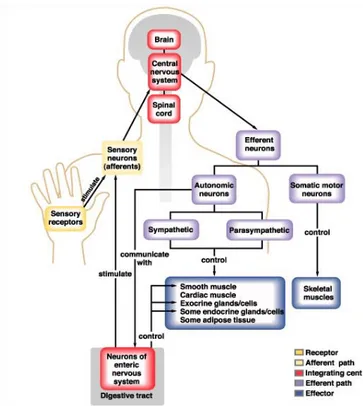
2.1 NEURONAL SYSTEM ... 35
2.1.1 Central Nervous System ... 38
2.1.2 Cell types ... 40
2.1.2.a Neuroglia cell ... 40
2.1.2.b Neurons ... 42
2.2 GROSS ANATOMY OF THE SC ... 48
2.3 INTERNAL STRUCTURE OF THE SC ... 52
2.3.1 Grey matter ... 52
2.3.1.a Structure of the Grey Substance ... 53
2.3.1.b Fine organization of the SC grey matter ... 54
2.3.2 White matter ... 57
2
2.4.1 Ascending Pathways ... 60
2.4.2 Descending Pathways ... 62
2.5 MOTOR CONTROL ... 64
2.5.1 Motor Neurons ... 64
2.5.2 Discending Motor pathways ... 65
2.6 VASCULATURE OF THE SC ... 66
2.6.1 Arterial anatomy ... 67
2.6.2 Venous anatomy ... 69
2.6.3 Capillaries of the SC ... 69
Summary and conclusions ... 70
CHAPTER 3 ... 72
SC FMRI: BASIS AND ISSUES ... 72
3.1 MRI BASIC PRINCIPLES ... 73
3.1.1 Source of the MRI signal ... 73
3.1.2 “Relaxation” times ... 74
3.1.3 Imaging ... 75
3.2 BOLD-BASED FMRI ... 77
3.2.1 Blood susceptibility ... 77
3.2.2 The physical source of the BOLD effect... 78
3.2.3 BOLD and neural events ... 80
3.2.4 BOLD signal timecourse ... 80
3.2.5 Theoretical model of BOLD effect ... 82
3.2.6 Intravascular and Extravascular BOLD Signal Component ... 84
3.2.6.a Extravascular BOLD Signal Component ... 84
3.2.6.b Intravascular effects ... 85
3.3 MAIN CHARACTERISTIC OF THE BOLD CONTRAST IN THE SC ... 87
3.3.1 Sources of physiological noise in the BOLD based fMRI spinal imaging ... 88
3.3.2 SC fMRI time course ... 89
3.3.3 Signal specifity: ... 90
Anatomical localization of the response ... 90
Distribution neuronal activity with motor and sensory stimulation ... 92
3.4 ADDRESSING PHYSIOLOGICAL NOISE IN SC FMRI... 93
3.4.1 Acquisition strategies ... 93
3.4.2 Post-processing strategies ... 95
3.4.3 Noise fitting ... 96
3.4.4 Noise suppression ... 97
3
CHAPTER 4 ... 101
CHARACTERIZATION OF FUNCTIONAL MRI SIGNAL IN THE HUMAN SC: STUDY OF THE LINEARITY IN BOLD CONTRAST ... 101
4.1 INTENSITY DEPENDENCE OF THE BOLD RESPONSE IN SPINAL FMRI ... 102
4.2 MATERIALS AND METHODS ... 105
4.2.1 SC fMRI: technical issues ... 105
4.2.2 Imaging methods ... 106
4.2.2.a Partecipants ... 106
4.2.2.b Stimulation devices ... 107
4.2.2.c Stimulation paradigm ... 108
4.2.2.d Imaging ... 109
4.2.2.e fMRI data analysis ... 110
4.3 RESULTS: BIOPHYSICAL CHARACTERIZATION OF THE BOLD RESPONSE ... 112
4.4 DISCUSSION ... 119
Summary and Conclusions ... 121
CHAPTER 5 ... 122
CHARACTERIZATION OF MOUSE SC VASCULAR AND NEURONAL NETWORK USING HIGH RESOLUTION X-RAY PHASE CONTRAST TOMOGRAPHY TO DEVELOP A SC BOLD MODEL ... 122
5.1 ADVANTAGES OF XRPCT FOR THE STUDY OF VASCULAR AND NEURONAL NETWORK IN THE SC ... 123
5.2 MATERIALS AND METHODS ... 124
5.2.1 Sample preparation ... 125
5.2.1.a Group 1 & 2 sample preparation (for SXPCT): Perfusion with saline solution and with MICROFILL®... 125
5.2.2 High resolution Synchrotron Phase Contrast Tomography ... 127
5.2.2.a XPCT basis ... 127
5.2.2.b Experimental approach used ... 129
5.2.2.c Data analysis. ... 130
5.2.3 Simultaneous submicrometric 3D imaging of the micro-vascular network and the neuronal system in mouse SC... 131
5.2.4 3D imaging of the Vascular network in mouse SC ... 134
5.2.5 3D imaging of the Neuronal network in mouse SC ... 136
4
CHAPTER 6 ... 144
SEGMENTATION OF MOUSE SC VN AS A BASIS TO DEVELOP A REALISTIC SC BOLD MODEL ... 144
6.1 CURRENT HYPOTHESES ABOUT THE ORIGIN OF THE SC FUNCTIONAL CONTRAST ... 145
6.1.1 Origin of functional signal ... 145
6.1.2 General overview of the “work-in-progress” SC BOLD signal Model ... 146
6.2 SC VASCULATURE ... 148
6.3 SKELETONIZE ... 148
6.3.1 General Description ... 149
Implementation with the Marshall algorithm: Longest shortest path estimation ... 150
6.4 VASCULAR SEGMENTATION ... 152
Summary and Conclusions ... 156
CONCLUSIONS ... 157
REFERENCES ... 160
APPENDIX A: MOTOR NEURONS POOLS AND MOTOR UNIT ... 181
APPENDIX B: DESCENDING MOTOR PATHWAYS ... 182
APPENDIX C: PHYSIOLOGICAL NOISE ... 185
APPENDIX D: SEVERAL APPROACHES TO FMRI DATA ANALYSIS CAN ENHANCE THE DETECTION OF SIGNALS WITHIN OR ACROSS SUBJECTS. ... 187
6
INTRODUCTION
Functional Magnetic Resonance Imaging (fMRI) has become one of the most powerful tools in neuroscience research, with promising applications in clinical practice.In particular, fMRI based on blood oxygenation level dependent (BOLD) contrast [1-5] has gained a primary role in the study of human brain, both for the characterization of regular brain activity and in clinical practice. Up to now, however, relatively few studies of the human spinal cord (SC) by functional imaging have been presented, starting from the work published in 1996 by Yoshizawa et al. [6], with a subsequent relevant contribution by Stroman et al. [7-16].
Recently, studies on the task-dependent modulation of the SC fMRI activations signal in response to innocuous and painful sensory stimuli or to motor tasks have been performed, and an hemodynamic response function (hrf) specific for the SC has been proposed [17]. It has been shown that there is a linear relationship between the applied force and the BOLD signal amplitude during isometric exercise [18], and that there is also a movement rate-dependent increase in spinal fMRI signals [19]. All these studies have shown that spinal BOLD fMRI has the potential for becoming a reliable and sensitive tool for studying the modulation of functional activity in the SC.
SC fMRI may be of immediate application in neuroradiology, in particular for the assessment and follow-up of spinal injuries, pain, and neurodegenerative diseases (e.g. multiple sclerosis), as well as in the development and evaluation of new therapies. Indeed, a non-invasive tool capable of monitoring the function, and thus complementing the available anatomical information, is a crucial need in these fields.
Preliminary studies of people with spinal cord injuries (SCI) and multiple sclerosis (MS) have demonstrated an altered activity in the SC, depending on the injury severity and on the advancement of the disease [7, 20-24].
Nonetheless, the application of fMRI to the SC remains confined to a few laboratories, mainly because of the vagaries of the activation patterns and of their characteristics [25-27]. In particular, the potential of SC fMRI is significantly impaired by the limited overall quality of the functional series, emerging in the form of geometrical distortions, signal losses, and poor contrast to noise ratio. These issues can be ascribed, at least in part, to a poor control of physiological noise. In a research environment, on an experiment-to-experiment basis, suitable solutions can be found by carefully
7
choosing the appropriate experimental procedures at the acquisition stage, and by properly treating the data at the post processing stage. However, SC fMRI is still far from defining the standardized protocols required for an everyday clinical use.
To further complicate matters, the exact features –and even the biophysical origins– of the functional response in the SC are still unclear. While for the brain the neurovascular coupling is clear and well characterized, for the SC this is not yet the case. The investigation of the characteristics of the SC functional signal, and in particular of its linear relationship with the underlying neural activity, is therefore crucial for the physiological characterization of the BOLD response, as well as for the analysis of fMRI time series (which is heavily based on the assumption of linearity). Recently, some promising advancements have been made in this field. On the one hand, it was observed by electrophysiological studies that there is a linear relationship between the neuronal activities of the SC and the muscular tensing. Moreover, Madi et al. [18] found the existence of a linear functional relationship between the neuronal activity and the BOLD fMRI signal. However, in spite of the increasing numbers of reports on spinal fMRI studies in the literature, we are still far from a clear and complete knowledge on the characteristics of the functional signal.
Within this context, in the present thesis we study and report the characterization of the functional response of the human SC to a multilevel motor task, designed to assess the linearity of the hemodynamic response as a function of the intensity of a graded task. In addition, we study the structure and distribution of the vascular network (VN) and of the neuronal network (NN), which together contribute to the BOLD fMRI signal, by means of high-resolution X-ray Phase Contrast Tomography (XrPCµT). One of the goals of this study is the definition of a suitable replacement for the vascular model currently used in fMRI, which was developed only for a specific real geometry of the VN (i.e., for the rat brain [4]) and may not be applicable to the SC. Of course, the development of a more accurate vascular model would be of paramount importance for the application of SC fMRI to the clinical practice (for the treatment of SC injuries, pain and neurodegenerative diseases). In addition, a better understanding of the SC could be important for the investigation of neuronal metabolism and of neurodegenerative diseases in an environment that may be regarded as a simplified model of the brain.
8 The present thesis is organized in six Chapters.
In the first Chapter we briefly describe the current state-of-the-art of SC imaging, by reviewing current methodologies and applications. We also identify the greatest current needs, especially from a clinical point of view, that will drive forward future developments of SC imaging.
The second chapter deals with the main aspects of the SC and of its constituents, necessary for a better understanding of this thesis. The SC anatomy has implications for the development and applicability of spinal fMRI. As a matter of fact, the main difficulties facing researchers attempting to image the SC function can be easily traced back to the peculiarities of the SC anatomy. At the same time, the vascular anatomy of the SC is optimally organized for the generation of a strong and spatially specific BOLD effect. This can help in overcoming the numerous other difficulties that characterize SC fMRI.
In the third chapter, we discuss the essential physical concepts at the basis of Magnetic Resonance Imaging, and in particular of BOLD-based fMRI. In addition, we present a selection of recent results in SC fMRI.
The fourth chapter reports the experimental fMRI results obtained on the relationship between the intensity of the stimulation and the amplitude of the SC BOLD response in humans during a controlled motor task (graded isometric force) with the dominant hand. The results confirm the physiological origin of the response, and will be of great help in model-based SC fMRI inference.
The fifth chapter presents the results of our investigation of the vascular and neuronal network of the mouse SC by XrPCµT. The obtained results will be useful to develop an optimised BOLD fMRI signal model for the SC, on the basis of its real vascular geometry.
The sixth chapter discusses the progress made towards the definition of an optimised BOLD signal model for SC fMRI, and outlines the additional steps still required to fulfil this goal.
9
CHAPTER 1
SC B IOMEDICAL IMA GING: L IMITS AND FUTURE
DEVELOP MENTS IN PRE-CL IN ICAL AND
CLIN ICAL FIELD
Non-invasive imaging methods have become an essential tool for assessing the effects of damage or disease processes, with the potential to support the planning of surgical interventions and of other treatments, and to determine if treatments are effective. There are a number of approaches available, including magnetic resonance imaging (MRI), X-ray based imaging (Computer Tomography- CT), nuclear medicine (Positron Emission Tomography–PET), magnetoencephalography (MEG), and each have strengths and weaknesses in terms of what they can show, and what they cannot. These non-invasive approaches provide the only means of accessing the structure and function of the human spinal cord (SC). As a result, there is currently a great need for the development of these methods and of these techniques into pre-clinical and clinical use.
The potential outcomes of advancing these methods are great, enhancing our basic understanding of healthy human SC function, and influencing our ability to accurately diagnose and treat injury and disease, and predict outcomes. In order to support the need for developing of SC imaging methods and advancing the current technology, we report on the key aspects of SC imaging from basic research to clinical practice, while devoting most of our attention to SC functional MRI (fMRI).
In particular the objectives of this chapter are:
1) To describe the current state-of-the-art of SC imaging by reviewing current methodologies and applications;
2) To identify the current greatest challenges, both innate to SC imaging, and relative to hardware and software development;
3) To identify the greatest current needs, especially from a clinical point of view, that will drive forward future developments of SC imaging.
10
1.1 Introduction to non-invasive imaging methods in neuroscience:
Historical overview
The impact of medical imaging on the field of neuroscience has been considerable. The advent of x-ray CT in the 1970's allowed clinicians to see features inside the heads of patients without the need for surgery. By making the small step of placing the source of radiation within the patient, x-ray CT became autoradiography, so that now not only structure but also blood flow and metabolism could be followed in a relatively non-invasive way. A big step forward was made by choosing to use a positron emitter as the radioisotope. Since a positron almost immediately annihilates with an electron, emitting two photons at 180 degrees to each other, much better localisation of the radioisotope within the scanner is obtained. Using labelled water, PET became the first technique to potentially allow researchers to produce maps of the mind, by measuring blood flow during the execution of simple cognitive tasks. Since local blood flow is intimately related to cortical activity, regions of high regional blood flow indicate the area in the cortex responsible for the task being performed.
At around the same time, another technique, which promised even better anatomical pictures of the brain, was being developed. MRI, based on the phenomenon of nuclear magnetic resonance [28], produces images of the human body with excellent soft tissue contrast, allowing neurologists to distinguish between grey and white matter, and brain defects such as tumours. Since MRI involves no ionising radiation, the risks to the subject are minimised. The development of contrast agents suitable for dynamic MRI studies, and improvements in the speed of imaging, opened up the possibility of using the technique for functional brain studies. In 1990 the first experiment using MRI to study brain function was performed, imaging the visual cortex whilst the subject was presented with a visual stimulus [2]. A contrast agent was used in this first study, but it was not much later when the first experiment was carried out using the blood as an endogenous contrast agent. The haemoglobin in the blood has different magnetic properties depending on whether it is oxygenated or not; these differences affect the signal recorded in the MR image. By imaging a subject at rest and whilst carrying out a specific task, it became possible to image brain function in a completely non-invasive way. The functional studies of the Central Nervous System (CNS), that have been produced over the past few years have started to make a big impact on the way neuroscience is approached. There are, however, still areas of the technique of fMRI that require refinement. The mechanisms behind the observed activation response are not well understood, and there are issues in regard to the way that the data from such experiments are analysed. However, the potential of fMRI, alongside that
11
of PET, means that the study of the human CNS has entered a new era, offering new insights into neurology, psychiatry and psychology.
1.1.1Non- invasive SC imaging methods
Non-invasive investigations of human SC function, and of the effects of SC injury (SCI) or disease, are significantly hampered by the inaccessibility of the SC. In order to supplement current methods for assessing residual function, pain, and quality of life factors after SCI or disease, sensitive methods are needed to reveal changes in neurological function and structure. Non-invasive imaging methods such as MRI, PET, and CT provide the only means for accessing the structure and function of the human SC. As a result, there is currently a great need for development of these methods. While progress is being made, only a relatively small number of research labs in the world are actively working on SC imaging methods, and these techniques have yet to be advanced into clinical use. The potential benefits of advancing these methods are tremendous, enhancing our basic understanding of healthy human SC function, and impacting our ability to accurately diagnose and treat injury and disease.
The greatest current challenges for SC imaging were identified as arising from the imaging environment itself, which is made difficult by the bone surrounding the spinal canal, by the physiological motion of the cord and of adjacent tissues, and by the small cross sectional dimensions of the SC, exacerbated by the metallic implants often present in injured patients. Challenges were also identified as a result of a lack of “critical mass” of researchers taking on the development of SC imaging, affecting both the rate of progress in the field, and the demand for equipment and software to manufacturers to produce the necessary tools.
In the following, we will define the current state-of-the-art of SC imaging. Only PET and MRI methods-fMRI and Diffusion Tensor Imaging (DTI) - are described in this chapter, with most of the attention on fMRI. In addition, we will describe the current applications (on SCI, on multiple sclerosis and on pain) of these techniques employed in clinical research, discuss the underlying theory and challenges, and present the evidence for the current and potential power of these methods. In particular, we will discuss all aspects of SC imaging from basic research to clinical practice.
1.2 Positron Emission Tomography: a general overview
PET produces images of the body by detecting the radiation emitted from radioactive substances. These substances are injected into the body and are usually tagged with a radioactive atom, such as Carbon-11, Fluorine-18, Oxygen-15, or Nitrogen-13, that has a short decay time. These radioactive
12
atoms are formed by bombarding normal chemicals with neutrons to create short-lived radioactive isotopes. PET detects the gamma rays given off at the site where a positron emitted from the radioactive substance collides with an electron in the tissue.
PET provides images of blood flow or other biochemical functions, depending on the type of molecule that is radioactively tagged. For example, PET can show images of glucose metabolism in the brain, or rapid changes in activity in various areas of the body. However, PET has high cost of installation and maintenance.
The choice of different injected tracers provides the potential for obtaining unique information regarding the injured SC. PET imaging has at least two distinct uses in the evaluation of traumatic SCI: assessment of the metabolic activity within remaining tissue and monitoring the degree of axonal connectivity. With regard to the first use, 18F-labeled fluorodeoxyglucose (FDG) has been used to monitor nervous system function in a range of preclinical and clinical studies. For example, traumatic brain injury reduces glucose utilization detected by FDG-PET, and a reduced uptake is associated with impaired outcome [29, 30]. This technique has also been applied to compressive and radiation myelopathies of the cervical SC [31-34] (Figure 1.1) as well as to one preclinical study of traumatic SC injury [35]. For cervical compressive myelopathies, some lesions show locally reduced glucose utilization while other cases show increased glucose uptake [32, 34].
13
Figure 1.1: Patient with myelopathy. (A) Sagittal T2-weighted MRI scan shows stenosis with compression of cervical
cord and intramedullary hyperintensity at level C3/C4 (arrow). (B and C) Corresponding sagittal (B) and transaxial
(C) 18F-FDG PET slices demonstrate focally increased uptake at level of stenosis. Patient had good clinical recovery
6 month after decompression of stenosis [31].
There is a positive correlation between a pre-operatively elevated uptake and later improvement after de-compressive surgery in two studies [31, 34]. In one study, the PET data were uncorrelated with magnetic resonance (MR) images [34], providing a distinct means to predict positive outcome from surgery. A challenge for SC PET remains the spatial resolution. Even with high-resolution scanners and CT registration, the spatial resolution is one or more orders of magnitude worse than structural MRI. Regional distinctions between zones of the SC at one segmental level are not possible by PET imaging in axial projections.
There is growing clinical trial activity focused on therapeutic strategies intended to promote axonal growth, regeneration, sprouting and repair.
PET tracers with neurotransmitter specificity have the potential for tracking the degree of damage and repair for specific fiber tracts. Thus, PET ligands may function as biomarkers in the development of new repair strategies. The potential utility of this approach has been demonstrated in one preclinical study [36]. The raphe spinal system is the only serotonergic system in the SC; therefore, all presynaptic serotonin markers depend on the continuity of descending fibers. PET tracers, that bind to the presynaptic serotonin re-uptake sites (such as 11C-labeled 2-(2-(dimethylaminomethyl) phenylthio)-5-fluoromethylphenylamine (AFM)), have been developed [37-39]. In rats, the SC has a
14
clearly defined specific AFM uptake, which is lost caudal to a complete SC transection. With a moderate SC contusion, there is a partial loss of caudal AFM signal. Most critically, treatment with an axon regenerative therapy, namely Nogo receptor (NgR1-Fc) decoy protein, promotes an increase in caudal AFM signal after chronic SC contusion [36].
This increased signal is correlated with behavioural improvement and matches post-mortem histological evidence of serotonin fiber regrowth. In this case, a neurotransmitter-specific PET tracer is a radiological marker that highlights the extent of axonal connectivity across a lesion site and reports the degree of axonal repair with treatment. The use of tracers specific to presynaptic markers of different long tracts in the SC may extend the anatomical specificity of this technique. PET measurements of fiber regrowth do not require the presence of highly ordered fasciculated fiber tracts as DTI-MR measurements. This is critical because the majority of preclinical studies in which interventions promote some fiber regeneration show that new fiber growth is highly branched and irregular. Thus, PET may provide an imaging modality to detect regenerative growth in proof-of-concept clinical trials, Even though, the use of ionising radiation in PET (as opposed to MRI) remain an issue.
1.3 Specific Magnetic Resonance Imaging methods and applications
MRI may be the most widely used method for detecting pathology in the CNS because of the high tissue contrast and relatively high spatial resolution that it provides. MRI also provides several different methods for visualizing tissues and pathology in addition to detailed anatomical imaging, including diffusion-weighted imaging and functional imaging. However, every method has limitations and weaknesses, as detailed below. In particular, imaging of the SC presents inherent challenges that are common to all MR imaging applications. Specifically, these are 1) the spatially non-uniform (inhomogeneous) magnetic field environment when in an MRI system, 2) the small physical dimensions of the cord cross-section, 3) physiological motion. In addition, the application of fMRI to the SC requires specific modifications to the conventional brain methodology and conventional brain tools. Here, we discuss the key challenges and future developments for SC MRI methods (DTI and fMRI), with more attention to spinal fMRI.15
1.3.1 Diffusion Tensor Imaging: state of the art, limitations, future developments
DTI is a MR technique capable of measuring the magnitude and direction of the diffusion of water molecules in various tissues, thereby providing information about the microstructural composition of tissues [40].DTI developed from a technique known as diffusion-weighted imaging, which measures the attenuation of MR signals caused by diffusion [41]. DTI was formally introduced by Basser et al, [42] and subsequent improvements in this technique have led to the development of DTI as a tool to delineate white matter tracts in the brain. In particular, the indices calculated from diffusion MRI experiments -mean diffusivity (MD), axial and radial diffusivity, and fractional anisotropy (FA), which are able to document and quantify orientation changes of water molecules in otherwise normally appearing white matter, are sensitive to changes in microstructural integrity, demyelination, axonal loss, and inflammation. DTI of the SC in humans was initially inadequate because of the small area of the cord, of susceptibility artefacts and of cardiac and respiratory motion artefacts [43, 44]. Improvements in scanning protocols have allowed for obtaining usable diffusion images of the SC, so that this imaging technique can now be applied in healthy and diseased SC in humans in vivo [45]. In particular, in healthy subjects good contrast is observed between grey and white regions, with the highly anisotropic white matter showing much higher FA values than the central grey matter as reported in Figure 1.2. In addition, given the sensitivity and specificity of DTI to white matter integrity, this technique is sensitive to structural changes due to pathology [16, 24]. A groups of researchers has shown that DTI is able to detect cord damage in regions of the cord that appear normal on T2- weighted images [46, 47]. DTI has also proven to be sensitive to Wallerian degeneration through animal models of pericontusional traumatic axonal injury [48], demyelinating lesion [49] and dorsal root axotomy [50].
16
Figure 1.2: Sagittal (gray scale and color) (A, B) and axial (C, D) fractional anisotropy maps from a healthy volunteer
obtained at 3 T. On the axial acquisitions, note the ability to resolve the grey and white matter (the grey matter has lower FA compared with the white matter). The axial acquisition allows for quantitation of DTI metrics, such as FA [51].
The specificity of diffusion measurements to white matter pathology has been studied in animal models of de/dysmyelination. This study suggests that axial diffusivity is more specific to axonal degeneration whereas radial diffusivity is more specific to demyelination [40, 48-50, 52-57]. Other studies however reported possible interactions between axial and radial diffusivities, thereby limiting the specificity of these measures [58-60]. One argument refers to the pathophysiology of axon degeneration, as this process is known to be associated with demyelination in several pathologies such as in Multiple Sclerosis (MS) [61] or in SCI [50, 62]. Another argument is related to the biophysical properties of DTI, in which several physical parameters can influence diffusion metrics including myelination, axonal density, axonal diameter, or orientation of fiber bundles, as well as changes in the tissue matrix surrounding the axons [60, 63, 64].
SC DTI therefore represents an important advancement in the field of neuroimaging, and its use is being expanded both for prognostication and for guiding therapy. Ultimately, it is always important to remember that the diffusion tensor is an imperfect model used to explain data and one must always look at the data and make sure that interpretations in terms of axonal loss and demyelination are well supported by the behavior of the underlying model [60].
In the following paragraphs, we provide a summary of the limitations and future developments of SC DTI.
17
1.3.1.a Limitations of DTI in the human SC
SC DTI in humans still has a number of limitations. Adequate spatial resolution remains a problem, and it is difficult to visualize the individual funiculi on diffusion-weighted images, particularly in the lower thoracic cord [65]. DTI of these segments is affected more by artefacts arising from cardiac and respiratory motion as well as cerebrospinal fluid (CSF) pulsation [51]. The use of faster imaging techniques such as parallel imaging and single shot echo-planar imaging, as well as the use of cardiac pulse-gating, have helped to reduce these artefacts. However, the scan acquisition time is still a limitation for patients with acute SCI, since these patients cannot withstand even 30 minutes of additional scanning time in the MRI suite. The signal to noise ratio in human SCI is sub-optimal in most studies and can lead to overestimation of anisotropy measures, particularly in low-anisotropic tissues such as the central grey matter [66]. The use of 3T MR scanners does improve the signal to noise ratio (SNR) [67], but is still not used universally. The use of DTI postoperatively is hampered significantly by the use of spinal instrumentation, which creates numerous artefacts. Additionally, standardized software to process tensor images is essential to make this a feasible option for routine clinical use.
1.3.1.b Future Developments and final considerations
DTI has given us a unique insight into the pathophysiology and microstructural alterations associated with SC disorders. DTI provides useful information about the direction of fibres and the diffusion anisotropy properties of neural tissue, complementing the information obtained by conventional MRI. DTI in the brain is being used to help diagnose various clinical conditions, but its exact application in spinal imaging is largely unexplored. While initial studies in rat models have primed this modality for human research, more data is required on the accuracy and reliability of DTI indices in defining cord pathology. DTI of the SC does show promise in certain neurosurgical conditions, such as traumatic SCI and intramedullary tumours. However, scanning protocols and image processing need to be refined and standardized. Once these challenges are overcome, we can expect the use of DTI in mainstream clinical practice, both to prognosticate as well as to monitor patients with SC disease. In particular, in the future DTI indices may help surgeons to determine their surgical approach. Correlation between quantitative diffusion measures, such as FA and apparent diffusive constant (ADC), and acute or chronic injuries of the SC may be useful to predict outcome and to monitor the response to treatment. Nevertheless, the clinical use of DTI calls for a careful understanding of acquisition and processing issues. The full potential of DTI will probably not be realized until it is integrated with other modalities to obtain a richer characterization of white matter, in a manner analogous to the combination of perfusion and diffusion imaging data used to demonstrate an
18
ischemic penumbra. Perhaps the most intriguing application is the integration of DTI-tractography [51] with functional imaging. The excellent correlation of BOLD functional MR data with DTI-tractography findings in motor and visual cortex may illustrate the future of structure- function investigations in the brain and SC.
1.3.2 Functional Magnetic Resonance: general information, applications, state of
the art, future developments.
1.3.2.a General information about the fMRI contrasts
fMRI refers specifically to the imaging of neuronal function in the central nervous system (CNS). In fMRI the anatomical images are acquired quickly and repeatedly over time in order to detect changes corresponding to tasks or to sensory stimuli. In fact, during the acquisition a pattern of tasks or stimuli (called the “paradigm”) is applied and tissue changes corresponding to the time-series of the neural response are captured. Image quality, both in terms of spatial resolution and of signal-to-noise ratio, is sacrificed to reduce the imaging time, thereby providing higher temporal resolution in the time-series data.
The fMR signal intensity changes arise from a sequence of related phenomena. A change in neural activity corresponds to a change in metabolic activity and oxygen consumption. An increase in metabolic activity triggers the release of vasoactive substances that cause local relaxation of pre-capillary smooth muscles, thereby increasing the local supply of oxygenated blood. The increase in oxygen supply exceeds the increase in demand, and so the local blood oxygenation level increases at sites of increased metabolism. In 1936, Drs. Pauling and Coryell had discovered that the magnetic properties of haemoglobin are different between the oxygenated and the de-oxygenated states [68]. This effect was observed in the form of dark veins in 𝑇2∗-weighted MR images (𝑇2∗, 𝑇21 are MR signal
relaxation times) and triggered the development of MRI [2]. The local relaxation rate, 1/𝑇2∗, is a function of the concentration of de-oxygenated haemoglobin [69, 70]. In order to detect the changes in blood oxygenation it is therefore necessary to acquire the time series of images with 𝑇2∗-weighting,
using a gradient echo imaging methods, and the echo time optimally set equal to the 𝑇2∗ of the tissues
1 T
2 is the spin-spin relaxation time, while 𝑇2∗ is the spin-spin relaxation time composed of both the contributions from molecular interactions and inhomogeneities in the magnetic field. These relaxation times govern the return of the transverse magnetization to its equilibrium value (zero), and characterize the exponential decay of the NMR signal.
19
being investigated [70]. With this method, it is possible to observe relatively subtle changes in local blood oxygenation, and in the CNS, this is inferred to reflect changes in neural activity.
This is the “blood – oxygenation level dependent” or “BOLD” contrast that is most commonly used for fMRI [69, 70]. The BOLD contrast relies on the paramagnetic properties of deoxyhemoglobin that acts as endogenous contrast agent. Therefore, the increase of metabolism triggered by neuronal activity induces an enhancement of oxygen consumption and a subsequent increase of local blood flow and volume. The overcompensating increase of hemodynamic parameters produces an overall decrease of tissutal deoxyhemoglobin content and thus an increase of 𝑇2∗; consequently, 𝑇2∗-weighted images show an intensity increase in activated regions. Such an increase enables the localization of underlying neuronal activity. After its introduction in the early 1990s [2], BOLD-based functional imaging was widely utilized for non-invasive studies on human brain function. In recent years, similar approaches have been attempted for the study of the SC function [11, 14, 16, 24, 71, 72]; however, obtaining good functional images of SC still represents a technical and scientific challenge.
A second source of functional contrast has been hypothesized as active in the SC and brain fMRI, the so-called signal enhancement by extravascular water protons (SEEP) [8, 10, 73, 74] resulting from an increase in water content in active neural tissues [8, 75]. This mechanism is hypothesized to be related to neuronal swelling and /or glial cells shown to arise at sites of neuronal activity. Also, an increased blood flow to active tissues has been shown to be accompanied by increased intravascular pressure and by on increased production of extra-cellular fluid at sites of neuronal activity. The net effect is a local increase in water content near active neural tissues, causing a higher magnetic resonance signal intensity. Thus, spinal fMRI is generally able to detect neuronal activity in SC grey matter, which allows for the mapping of SC responses to sensory and motor stimulation.
Spinal fMRI reveals neuronal function indirectly by detecting changes in blood flow and blood oxygen levels occurring near metabolically active grey matter. Depending on the scanning parameters, the signal change arises in part from the BOLD contrast and in part from the SEEP contrast.
The application of fMRI to the SC is a logical extension of its use in the brain but has been slow to develop because of the many challenges posed by the application of this method to this specific anatomic region. SC fMRI requires specific modifications to the conventional brain fMRI methodology. Further work is required before the method is optimal for pre-clinical and clinical purposes. The following paragraphs briefly review the recent developments in SC fMRI and discuss the feasibility and potential applications of using SC fMRI in the clinic and pre-clinic field.
20
1.1.1.a State of the art of SC fMRI: general overview, limits and future perspectives
fMRI has become one of the most powerful tools for neuroscience research, and yet its use is limited to studies of the cortex, with relatively few exceptions [14, 19, 71, 76]. It has been demonstrated that important neuronal activity can be identified in the SC using spinal fMRI. Previous studies have shown that BOLD fMRI is feasible in the SC at both 1.5 T and 3 T, and the activation areas detected have good localization at the segmental level [11, 12, 18, 71]. In particular SC fMRI has been able to detect areas of activity in the cervical and lumbar regions with high sensitivity and reliability, in response to thermal, sensory, motor and painful stimuli [7, 19, 22].
Furthermore, spinal specificity for the stimulation of different dermatomes [9], and also the differentiation of motor and sensory areas were observed, and a large overlap was found. Even though substantial advances in knowledge have arisen from these studies, the poor reproducibility of the activation patterns and their characteristics – in terms of amplitude and location – have been invoked as significant concerns by several authors [25-27, 72, 77, 78]. In the clinical area, spinal fMRI has been applied to the study of injured SCs. The lumbar SC was imaged during noxious thermal stimulation of the L4 dermatome of complete and incomplete SC–injured volunteers [7, 13]. Signal intensity changes for the injured groups were of similar magnitude and had a similar time course pattern as those of the healthy group. However, the areas of activity in grey matter were altered. Spinal fMRI was also used to detect neuronal activity elicited by passive and active lower limb movement tasks in regions caudal to the injury site in volunteers with SCI [22]. Activity was detected in all volunteers regardless of the extent of injury. During both active and passive participation, activity was seen caudal to the injury site, although the number of active voxels detected with passive movement was less than with the active movement task. Therefore, spinal fMRI is able to detect a neuronal response in the SC caudal to the injury site during both active and passive lower limb movement tasks, and in response to a noxious stimulus, even when subjects could not feel the stimulus. Thus, spinal fMRI is useful for revealing areas of impaired and preserved activity in SC– injured patients. In addition, patients’ studies provide further evidence of response sensitivity to pathological changes. Studies of people with SCI and MS have demonstrated altered activity in the SC depending on the state of their disease [7, 20-23]. However, obtaining BOLD images of the SC activation still remains a technical challenge. The main difficulties in spinal fMRI, like for the other MRI techniques, arise from the small size of the SC, from the presence of large movements due to cardiac pulsation and to respiration and from the presence of magnetic field inhomogeneities around inter-vertebral disks, [72, 77, 79] which induce significant susceptibility artefacts. In addition, SC motion and the flow of cerebrospinal fluids (CSF) are thought to further confound the analysis, and
21
therefore the interpretation of functional data [25]. Finally, the application of the fMRI to the SC requires specific modification to the conventional brain fMRI methodology, even if the theory of conventional brain fMRI is applied. However, if these challenges can be overcame, SC fMRI may be of immediate application in the clinical framework. Further the study of the SC could also be important because of the possibility to treat the SC as a simplified model of the brain, suitable for experiments aimed at investigating the neuronal metabolism and the neurodegenerative diseases. Finally, the study of the SC system may be of immediate and fruitful application in the treatment of SC injuries, pain and neurodegenerative disease (e.g. multiple sclerosis).
Recently, experiments based on the task-dependent modulation of the SC fMRI activations signal in response to innocuous and painful sensory stimuli or motor tasks have been done and an hemodynamic response function (hrf) specific for SC has been proposed [17]. It has been shown that there is a linear relationship between the applied force and the BOLD signal amplitude during isometric exercise [18], and that there is also a movement rate-dependent increase in spinal fMRI signals [19]. All these studies have shown that spinal BOLD fMRI is a reliable and sensitive tool for studying the modulation of functional activity in the SC.
Nonetheless, the exact features and even the biophysical origins of the functional response are still unclear. In the brain the neurovascular coupling is clear and well characterized, but for the SC this is not the case. Indeed, electrophysiological studies have shown that there is a linear relationship between the neuronal activities of the SC and muscular tensing [80]. In addition, Madi et al. [18] found the existence of a linear functional relationship between the neuronal activity and the BOLD fMRI signal as in the case of the brain, demonstrated by Logothetis et al. [5], but, nowadays there is not a direct demonstration on that relationship in the SC.
Within this context, in this thesis we study and report the characterization of the functional response of the human SC to a multilevel motor task, in order to assess the linearity of the haemodynamic response as a function of the intensity of a graded task. In addition, we study the structure and distribution of the vascular network (VN) and the neuronal network (NN), with high resolution X-ray techniques to refine the vascular model used in fMRI, which was developed only for a specific real geometry of the VN, (i.e. for the rat brain [4]) and may be not be applicable to the SC. As a consequence, the development of a more accurate vascular model would be of paramount importance for the application of fMRI of the SC to the clinical practice.
22
1.3.2.b Capabilities of human SC imaging applications:
In order to provide an overview of the current state of the art and of the capabilities of human SC imaging applications, we provide in the following a general overview of the SC fMRI techniques employed in the clinical research, focusing on the specific applications of SC imaging to SCI, pain and MS.
SC injury:
Improving the ability to assess tissue viability and to detect residual neuronal function in the SC, in order to identify and distinguish morphological and functional changes, is a key step to advancing our capacity for clinical prognosis and management of SCI patients.
Injury to the human SC, regardless of the mechanism, results in a complex cascade of events at the cellular level that may or may not have an effect on the neurovascular coupling mechanisms that underlie the signal changes detected with spinal fMRI.
The pathological processes that occur at the cellular level include ischemia, vasospasm, ion-mediated cellular damage, excitotoxicity, oxidative cellular damage, neuroinflammation, and cell death. Changes in the correspondence between fMRI signal changes and neural activity may therefore also depend on the time since the injury occurred, thus complicating interpretation. There are a number of clinical outcomes that follow traumatic SCI as a consequence of either adaptive or maladaptive plasticity. Adaptive plasticity refers to the concept of natural recovery whereby an individual who sustained motor deficits, sensory impairment or both following SCI recovers to some degree in the months after injury [81]. Maladaptive plasticity refers to neurological symptoms that arise as a result of spinal circuits reorganizing themselves in a way that provides no useful function such as neuropathic pain, spasticity, disruption of autonomic function and the lack of motor and sensory recovery [82]. One potential advantage of spinal fMRI will be to investigate the longitudinal effects of various stimulation paradigms as they relate to clinical symptoms such as the recovery of motor function, sensation or the development of neuropathic pain. A better understanding of the spinal circuit changes that are associated with maladaptive plasticity may lead to more accurate diagnoses and to the ability to objectively monitor treatment.
Studies of the effects of SCI that have been carried out to date using SC fMRI have all focused on the chronic stages at 1 year or more after the injury occurred. Potentially confounding effects include on-going changes in cellular function and ionic metabolism, each of which may alter the mechanisms of neurovascular coupling and the subsequent hemodynamic response. Nonetheless, the studies carried out to date have demonstrated that fMRI of the SC can detect important changes as a result of traumatic injury in both the cervical and lumbar regions of the SC [7, 22, 83]. The first SC fMRI
23
study carried out on participants with SC injuries investigated activity in the lumbar SC, below the level of injury, in response to a noxious cold stimulus applied to the leg (L4 dermatome) [7] . This study demonstrated significant activity in all participants, including SCI patients, in the lumbar segments of the SC, below the level of injury. Moreover, participants with SCI who could feel or had some altered sensation of the cold stimulus, had spatial patterns of activity that were similar to those observed in healthy participants. A more recent study of thermal responses in the cervical SC and brainstem investigated responses to warm thermal stimuli in regions both above and below the level of injury [84]. Results corresponded with the level and extent of injury, with responses above the level of injury being generally similar to those seen in healthy participants, and responses below the level of injury being altered depending on the injury severity. Right- and left-side differences were also demonstrated, as well as corresponding activity in the brainstem (Figure 1.3). Collectively, these results demonstrate the sensitivity of the fMRI method to changes as a result of trauma and the feasibility of using fMRI as a research tool for studying the effects of injury.
Figure 1.3: Example of results obtained in a healthy female participant (top) and an age-matched participant with an
incomplete SC injury in the cervical level. Four dermatomes were stimulated, corresponding to the right and left C5 and C8 SC segments, and results are shown in selected axial and sagittal slices showing the responses to right- and left-side stimulation of the little finger side of the palm (C8 dermatome). Results obtained in the healthy participant show activity in the ipsilateral dorsal horn of the SC in the C8 segment, with a high degree of laterality corresponding to the side of the body being stimulated, as well as activity in the medulla of the brainstem. Results obtained in the participant with SCI show nearly normal responses on the left side of the SC, and slightly altered responses on the right side, as well as corresponding activity in the medulla. In spite of an apparently extensive span of tissue damage in the cervical SC, the neural activity detected is only slightly altered [24, 84].
Future clinical applications will require demonstration that the results obtained in each participant are sufficiently sensitive and reliable to be suitable for diagnostic purposes or for the monitoring of
24
treatment outcomes. As spinal fMRI emerges as a tool to investigate SCI, we must tread cautiously with the interpretation of results. One limitation, for example, is our lack of understanding of how the neurovascular coupling response may change after injury. Currently, the hemodynamic response function utilized in model-based fMRI data analysis is derived from healthy neural tissue. It will be important to investigate if damaged neural tissue also exhibits the same hemodynamic response function (both in temporal as well as spatial aspects) to a given stimulus, because the accuracy of the predicted response can impact on the sensitivity of the fMRI method for detecting neural activity. In addition, the technical challenges of SC fMRI, as discussed in the initial part of this chapter, may need to be reduced or eliminated before its sensitivity and reliability are acceptable for clinical applications.
Pain
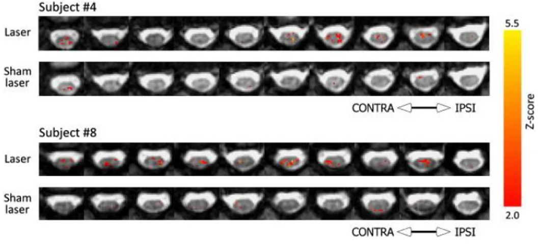
The SC (together with the brainstem) is the first point in the central nervous system that processes the nociceptive signals arriving from the body, and which ultimately may produce a sensation of pain. Functional imaging of the SC aims to record this activity and can help to better understand how these signals are processed and whether an altered SC function underlies chronic or neuropathic pain states in humans. The development of non-invasive imaging techniques to record spinal activity provides critical information needed to interpret nociceptive processing in health and disease, and may help explain the patterns of brain activity observed in response to noxious stimulation in healthy controls and in patients suffering from pain. Furthermore, it has already provided important information about the modulation of spinal nociceptive processing induced by analgesic drugs or by other non-pharmacological treatments. The studies of pain that have been reported in the literature demonstrate that by using spinal fMRI, researchers are already asking challenging questions relating to the interaction of brain, brainstem and SC and of our ability to endogenously control pain. In BOLD fMRI studies of the SC [85, 86], patterns of cervical spinal activity were recorded in response to painful thermal and punctate stimulation and compared across a group of 18 subjects; a predominantly ipsilateral response was demonstrated using a mixed effects model and corrected (voxel-level) statistics [85]. However, not all reports of spinal activity in response to nociceptive stimuli have demonstrated lateralized activity in the cord. In response to painful laser stimulation of the dorsum of the left hand, activity was observed to be bilateral in the cervical SC (see Figure 1.4) [87]. Importantly, this was the first study to directly assess the false-positive detection rates in SC activity, and the number of activated voxels in response to laser stimulation was found to exceed the false-positive level.
25
Figure 1.4: Representative maps from two subjects, showing the spatial location of voxels preferentially responding
to noxious stimulation in the Stimulation run (Laser), or during corresponding time periods in the Rest run (Sham Laser). The uppermost slice is displayed to the right of each row. “Contra” and “Ipsi” refer to the sides of the cord contralateral and ipsilateral to the stimulated hand, respectively [87].
Concerning the absence of lateralized signals in response to nociceptive stimulation, it should be noted that this assessment was made on the basis of sub-dividing the cord into right and left halves, so any bilateral motor-reflex activity in response to stimulation (involving activity in both anterior horns) might have concealed increased ipsilateral dorsal horn activity. By using a combination of BOLD and SEEP contrast, Ghazni and co-workers reported changes in SC and brainstem activity in response to painful and non-painful punctate stimuli [88]. Activity in response to stimulation was observed throughout the SC, brainstem and thalamus. Surprisingly, activity in the ipsilateral cervical dorsal horn and brainstem was highest for the light-weight probe, which was interpreted as reflecting descending modulation from the brainstem. Of direct relevance to the study of descending pain modulation, two recent studies have recorded the brainstem and SC response during endogenous analgesia due to placebo effects [89, 90]. In response to thermal stimulation of the left forearm, in a region treated with an inert cream as a placebo, increased activity in periaqueductal grey matter (PAG) and rostral ventromedial medulla (RVM) was observed [89]. Conversely, BOLD evoked signal increases were reduced in a restricted region of the SC during placebo analgesia; this was interpreted as reflecting decreased nociceptive processing in the SC due to descending pain modulation [90]. The role of attention on patterns of brainstem and SC activity in response to cold stimulation (18 or 15 °C) that produced “mild” to “strong discomfort”, was investigated by Stroman and his colleagues [15]. Similar to previous data examining brainstem responses following painful thermal stimulation [91], increased activity was found in the PAG during distraction conditions, perhaps relating to altered levels of discomfort. However, activity at the level of the SC was opposite to what might be expected on the basis of descending inhibition of nociceptive input, with decreased activity during the no-distraction “rating” period when compared to the no-distraction condition. On the other hand, a recent
26
study by Sprenger et al. [92]has demonstrated that during a mentally demanding distraction task (n-back task), SC activity, in response to a concurrent painful thermal stimulation is reduced when the cognitive demand is high. In addition, the reduction in pain ratings (when compared to the low cognitive demand condition) is significantly correlated with the change in BOLD signal. It is worth noting that developments in MR data acquisition, e.g., the use of slice dependent z-shimming [93] and region-selective radiofrequency (RF) excitation pulses [24, 94, 95], may improve our ability to record BOLD signal changes in the human SC. In particular, z-shimming has been already used to study spinal responses to noxious stimulation [89, 90, 92].
Multiple sclerosis
MS is the disease that has benefitted mostly from advanced quantitative SC imaging techniques, spanning from cord atrophy measurements, fMRI, DTI and also magnetization transfer ratio (MTR). MS is a disease that is being studied quite extensively with fMRI, in both the brain and SC [96]. A key challenge that has been identified for fMRI assessments of MS is that interpretation may be affected by disease-driven differences in task performance. In order to overcome this challenge, studies have been carried out with larger populations, and through the use of several motor, visual and cognitive tasks in groups representing all major clinical phenotypes of the disease. However, this solution will not be effective for individual patient assessments. One key finding from fMRI studies to date is that cortical reorganization occurs in patients affected by MS. As a result, patients adapt and their abilities do not necessarily match the axonal/neuronal loss seen with imaging, until the disease burden becomes too great and they are no longer able to adapt further. This adaptation is seen in fMRI results as recruitment of areas of the brain that are not active with the same task or stimulus in healthy control subjects [96].Studies, performed via fMRI of the SC also showed MS-related changes in the response to proprioceptive and tactile stimulation [20, 21]. Specifically, the signal changes during stimulation were 20% higher, and the areas of activity were altered by on over-recruitment of the ipsilateral posterior cervical cord. Consistently, fMRI results demonstrate the adaptation of regions of the CNS to compensate for the deficits caused by MS lesions in order to maintain motor, sensory, and cognitive abilities.
Also, an increased activation of the cervical cord has been demonstrated in all the major MS clinical phenotypes and has been related to the severity of clinical disability and the extent of tissue damage [23, 24, 97, 98].
27
1.3.2.c Potential Clinical Applications
Currently, the American Spinal Injury Association (ASIA) [99] assessment scale is the standard for classifying SC injuries. This involves a battery of light touch and pin prick examinations to assess where a patient has preserved sensory perception. To determine preserved motor function, the patient’s ability to move specific key muscle groups is assessed. This physical examination technique reveals information regarding SC function but does not reveal information about the condition of the SC caudal to an injury site. Further information regarding the condition of the SC caudal to the level of injury must be obtained by invasive measures or deduced from reflex actions. Electrophysiological techniques such as somatosensory-evoked potential, H-reflex, or stretch reflexes are capable of assessing the residual function after a SCI. The utility of these techniques is limited by the incomplete scope of the information obtained with each measure, such that a combination of measures is required to determine the residual function. For example, somatosensory-evoked potentials examine conduction along large areas of the body, and the results can be affected by peripheral damage, nerve root damage, or SC damage without revealing where along the pathway the damage has occurred. Similarly, an increase in reflexes denotes an upper motor neuron disorder but will not reveal the degree of damage, or whether the damage is complete or incomplete. Although these methods are useful for many research applications, they require specific equipment and are time-consuming. Therefore, these methods are not in routine clinical use. Thus, the ability of spinal fMRI to detect neuronal activity below the injury site in SC–injured patients is of considerable value for those assessing an injury, planning a treatment strategy, or monitoring recovery of function during and after treatment. Although spinal fMRI is not yet ready for routine clinical use in examining SC injuries, it is certainly fit for use in assessing recovery of function strategies. However, spinal fMRI will become more useful as intervention strategies develop, because it can detect differences in neuronal activity pre- and post-treatment. Spinal fMRI could become an important tool for assessing the efficacy of interventions. Currently, the ASIA assessment scale is used to follow progress in research interventions, and this, as mentioned, is unable to detect neuronal changes. Similarly, spinal fMRI could serve in assessing pharmacologic treatment effects on SC and nerve root function. Likewise, spasticity drug trials also could greatly benefit from the use of spinal fMRI, again because presently there are few non-invasive measures available to objectively see changes in neuronal activity. Spinal fMRI is able to show where functional activity occurs in response to a stimulus, regardless of a patient’s ability to feel the stimulus—a feature that the ASIA assessment scale lacks. Therefore, in the case of SCI, the neuronal activity in the SC both above and below the injury site can be monitored after initial injury for prognosis and recovery-of-function strategy decisions (see the paragraph about SC injuries), as well as throughout rehabilitation and in response to pharmacologic treatment. A
28
protocol can be developed such that a standard battery of tests may be conducted to assess ascending, descending, and reflex activity. fMRI has been used to investigate nociceptive processing and central sensitization in the brain to better understand pain [100, 101]. Spinal fMRI can be used to study the structural and functional correlates of pain and advance our understanding of the mechanisms of nociceptive processing, central sensitization, and chronic neuropathic pain. In addition, the neural mechanisms underlying attentional modulation of pain are not known, but data suggest the involvement of multiple levels of the central nervous system, including the dorsal horn of the SC [102]. Dorsal horn involvement has yet to be shown in functional neuroimaging studies. Future research using functional neuroimaging in this area is warranted and is likely to have a significant impact on therapeutic interventions. Combined imaging of the SC, brain stem, and brain will provide information regarding the neuronal activity of the entire central neural axis, advancing scientific knowledge on how clinical chronic pain states are generated and maintained. The ability to image from the SC to the brain, as well as descending modulatory pathways back to the SC, will allow for the investigation of plasticity changes associated with chronic pain conditions and provide the opportunity to evaluate changes in both the disease state and the response to peripherally and centrally acting treatments. With spinal fMRI, objective means of assessing the neural function in patients with chronic pain will thus be available. Spinal fMRI could be useful in identifying the pathogenesis of many chronic pain conditions, whereas presently this is not understood. Spinal fMRI could expose the neuronal abnormalities underlying several pain conditions such as irritable bowel syndrome, chronic lower back pain, or fibromyalgia. It is possible that central sensitization is involved in many chronic pain conditions, and great potential exists for spinal fMRI to reveal the underlying initiation or maintenance of this state.
Although anatomical spinal MRI is used to detect a number of specifically SC– related conditions such as a Chiari malformation, a defect causing Brown-Sequard syndrome, or hydromyelia, a functional image of these conditions would be useful for monitoring the effects of lesion or structural abnormality on physiologic response and subsequent alterations in neuronal function. It might also help differentiate a neurodegenerative disorder such as tabes dorsalis, which involves a breakdown of sensory fibers but has symptoms that include weakness, loss of proprioception, and decreased coordination and reflexes, in addition to the sensory disturbances and pain. The ability of spinal fMRI to capture the activity of dorsal column neurons will aid in the diagnosis, prognosis, and monitoring of disease states such as this. As with many conditions, symptoms may not occur immediately after the initial insult but may appear gradually over time, resulting in a situation in which it may not be immediately clear where to begin investigation. When it is difficult to identify the cause of symptoms, spinal fMRI could indicate areas of abnormal neuronal function or identify the point at which the
29
breakdown is occurring. For example, spinal fMRI could determine whether it is an upper motor neuron disorder, a lower motor neuron disorder, or both, as in amyotrophic lateral sclerosis. Whether neurons are firing normally but the breakdown is at the neuromuscular junction or with the muscle fibers themselves, as in muscular dystrophies, could similarly be determined. MRI is used to locate lesion sites in multiple sclerosis, but use of spinal fMRI would aid in tracking disease progression and prognosis. The functional characterization of disease could be carried out with spinal fMRI. In cases of transverse myelitis, inflammation of a SC segment occurs, which can cause myelin damage, thereby impairing nerve conduction and interfering with neuronal communication. The benefit of functional imaging for these types of conditions is clear. Pre-surgical mapping localizes function in cortical tissue near areas intended for surgery or resection. Functional and anatomically distinct regions of cortex vary between different people. The use of fMRI can identify areas associated with specific functions, so that these areas may be targeted or avoided during resection. This method is a less invasive alternative to electrophysiological cortical mapping commonly used for obtaining this type of information. Functional imaging of the SC could be useful for diagnosis and prognosis of nerve decompression surgery. For example, in patients with cervical or lumbar radiculopathy, it is often unclear which nerve root is irritated or injured. Noxious stimulation of the affected region with simultaneous spinal fMRI would help localize the specific region affected. In another example, for diabetic neuropathy, the relief of pressure on a trunk is achieved by the excision of the constricting band or the widening of the bony canal to alleviate pain symptoms. Based on patient reports, it is not always clear which spinal nerves are implicated, as often a number of nerves are involved in creating the symptomatology. With spinal fMRI, it could be possible to better identify the specific nerves involved in particular symptoms, therefore improving surgery planning and decreasing surgical risk. The ability of spinal fMRI to detect changes in neuronal function in the absence of overtly physical manifestations could be valuable for patients undergoing recovery of function, rehabilitation, or pharmacologic treatment. Aside from physiologic information obtained by the clinician, patients could benefit psychologically. In the initial stages of a treatment strategy, it may be discouraging to patients when signs of improvement are not yet detectable. In the absence of measurable physical improvements, the “proof” of improvement in neuronal activity (increases or decreases in conduction, condition-specific) could provide the encouragement and motivation to continue with rehabilitation strategies. Alternately, spinal fMRI could reveal a strategy to be ineffective and accelerate the search for alternate strategies.


![Figure 2.3: Pictorial view of the seven parts that compose the central Nervous system [111]](https://thumb-eu.123doks.com/thumbv2/123dokorg/2835135.4718/42.892.264.632.542.902/figure-pictorial-view-seven-parts-compose-central-nervous.webp)

![Figure 2.6: view of the neuronal soma. It is represented post-synaptic dendrites and presynaptic neuron [115]](https://thumb-eu.123doks.com/thumbv2/123dokorg/2835135.4718/47.892.247.644.509.891/figure-view-neuronal-represented-synaptic-dendrites-presynaptic-neuron.webp)