Abstract. – OBJECTIVE: The clinical use of mesenchymal stem cells (MSCs) in regener-ative medicine either in tissue repair or tissue reconstruction has given highly interesting re-sults thanks to their particular nature. Sourc-es that have attracted the attention of medical scientists from where stem cells (SCs) in adults could be obtained are different and, dental tis-sues have certainly become an optimal source of MSCs. Dental tissue is a main reservoir of two types of MSCs dental bud (DBSCs) that consti-tute the immature precursor of the tooth and dental pulp (DPSCs) that are derived from den-tal inner pulp and partly from denden-tal follicle tis-sue and can differentiate into several cell pheno-types as osteoblast, chondrocyte, hepatocytes, cardiomyocytes, neuron and β cells.
PATIENTS AND METHODS: Normal impact-ed third molars and tooth buds were collectimpact-ed from adults and adolescents underwent to ex-tractions for orthodontic reasons. The expres-sion of the five stemness genes Nanog, OCT4, Sox2, c-Myc and Klf4 were investigated by qRT-PCR in two different dental stem/progenitor cells: dental pulp stem cells (DPSCs) and stem cells from dental bud (DBSCs), differentiated to-ward osteoblastic phenotype and not.
RESULTS: Both DPSCs and DBSCs are easy to access and we found their expression of the typical mesenchymal stemness makers and os-teogenic capacity due to the effective presence of embryonic gene regulators like Nanog, OCT4, Sox2, c-Myc and Klf4. Both DBSCs and DPSCs could represent a valid tool in regenerative med-icine and translational applications.
CONCLUSIONS: The results depicted here provide, for the first time to our knowledge, a comparative outcome about the stemness prop-erties generated from accessible tissues such as DPSCs and DBSCs. These two types of SCs showed few different distinctive genetic traits supposedly in relation to their origin, location and stage of maturation. Certainly these SCs re-serve solid potential for human clinical appli-cation in autologous procedure for bone, hard tissue and soft tissue regeneration, easy to iso-late, ready availability, high-biocompatibility and safety and no ethical restrictions.
Key Words:
Dental Bud Stem Cells (DBSCs), Dental Pulp Stem Cells (DPSCs), Mesenchymal Stem Cells (MSCs), Stem-ness genes, Osteogenic differentiation.
A. BALLINI
1, S. CANTORE
1,2, S. SCACCO
1,2, L. PERILLO
3, A. SCARANO
4,
S.K. AITYAN
5, M. CONTALDO
3, K. CD NGUYEN
6, L. SANTACROCE
7,2, J. SYED
8,
D. DE VITO
1, G. DIPALMA
6, C. GARGIULO ISACCO
6,9, F. INCHINGOLO
61Department of Basic Medical Sciences, Neurosciences and Sense Organs, University of Bari Aldo Moro, Bari, Italy
2Polypheno srl - Academic Spin Off, University of Bari Aldo Moro, Bari, Italy
3Multidisciplinary Department of Medical-Surgical and Dental Specialties, University of Campania Luigi Vanvitelli, Naples, Italy
4Department of Oral Science, Nano and Biotecnology and CeSi-Met, University of Chieti-Pescara, Chieti, Italy
5Department of Multidisciplinary Research Center, Lincoln University, Oakland, CA, USA 6Interdisciplinary Department of Medicine, University of Bari Aldo Moro, Bari, Italy 7Ionian Department, University of Bari Aldo Moro, Bari, Italy
8Advanced Technology Dental Research Laboratory, Oral Basic and Clinical Sciences, Faculty of Dentistry, King Abdul Aziz University, KSA
9Human Stem Cells Research Center, Ho Chi Minh City, Vietnam
Andrea Ballini and Stefania Cantore equally contributed as co-first Authors
Ciro Gargiulo Isacco and Francesco Inchingolo equally contributed as co-last authors
A comparative study on different stemness
gene expression between dental pulp stem cells
Introduction
Currently different types of stem cells (SCs) have been isolated from dental and surrounding tissue. Their involvement in tissue repair and regeneration, local bone and teeth homeostasis and maintenan-ce has been proved1. It is difficult to characterize
exclusive dental stem cells using just surface pro-tein markers by Flow-cytometry or gene expres-sion by Real-time PCR (qRT-PCR) due to their ubiquitous characteristics, since these markers are equally expressed by all different typology of stem cells2. Gene expression of multipotent and
pluripo-tent markers such as Kruppel-like factor 4 (Klf4), octamer-binding transcription factor 4 (OCT4), ho-meobox transcription factor Nanog (Nanog), v-myc avian myelocytomatosis viral oncogene homolog (c-Myc), SRY (sex determining region Y)-box 2 (Sox2), osteocalcin (OCN), dentin matrix pro-tein-1 (DMP-1) and protein markers such as CD44, CD73, CD90, CD133, CD34, CD45, CD14, Nestin, Stage-specific embryonic antigen-3 (SSEA-3) and Transcription-associated protein 1 (Tra1) are in fact evidence of stemness which is specific feature of stem cells from the bone marrow (BM), umbi-lical cord blood (UCB), placenta, peripheral blo-od (PB) as well from teeth tissue 2-4. Dental tissue
and more specifically dental bud and dental pulp are prominent source of these types of multipotent and pluripotent stem cells, known as dental-derived stem cells (d-DSCs). The focus on these cells has at-tracted highly interest not only within dentists and orthopedics but also in the Regenerative Medicine community. Both dental bud and dental pulp stem cells (DBSCs-DPSCs) have shown great potential in dental tissue repair and regeneration and con-firmed the expression of transcription factors like Nanog, OCT4, Sox2, c-Myc, and Klf4 that play a key regulatory activity in the stem cell self-renewal process in replenishing mature cells that constant-ly die due to normal and constant tissue turnover reprogramming. The DPSCs and DBSCs both di-splayed a similar plasticity and typical features of pluripotency/multipotency typical of other sub-set of mesenchymal stem cells (MSCs)5-7. The human
dental pulp cells from deciduous and permanent teeth can undergo reprogramming to establish plu-ripotent stem cell lines without c-Myc. These surgi-cal residues, usually regarded as medisurgi-cal waste, can be used as an alternative source of pluripotent stem cells for personalized medicine5-7. The role of OCT4,
Nanog and Sox2 in keeping the pluripotency status of stem cells has been well confirmed either in em-bryonic stem cells (ESCs) or in induced pluripotent
stem cells (iPS). Takahashi and Yamanaka9 showed
in 2006 that the introduction of these specific genes encoding transcription factors could convert adult cells into pluripotent SCs8-15. The OCT4, Sox2 and
Nanog are mutually involved in the regulation of em-bryo growth, modulating the cellular differentiation at the very early passages; OCT4, Sox2 and Nanog directly work on ESCs keeping them in a stemness state preventing their differentiation, and sustaining their self-renewal16-20. The active participation of
Klf4 and c-Myc in this stemness activity has been confirmed though the way they interact has not been fully understood yet21-26. The data about c-Myc are
sometimes discordant probably due to the multilevel expression of this gene. In normal cell c-Myc par-ticipate in proliferation and growth mechanism, an interesting feature is that c-Myc is generally low in quiescent cells as could be the case of MSCs in BM niches or in PB circulation; conversely, c-Myc swi-tch on quick and become highly active once induced by growth factors10. Intriguingly, outcomes from
c-Myc knockout mouse lead to embryonic death and can act the same as Klf4 either as an oncogene or tumor suppressor. This redundancy was described by Chang et al26 who reprogrammed human
den-tal pulp cells from deciduous and permanent teeth into pluripotent stem cells without inserting c-Myc. Similarly, Klf4 participate in cell proliferation and growth expressed in different tissues regulating the final commitment of stem cell differentiation as well apoptosis21. These results suggest that both Klf4 and
c-Myc may exert differently depending on general micro-molecular condition, nevertheless they both are involved in the important modulatory function ESCs stemness, self-renewal and pluripotency as it was shown in Yamanaka’s iPS project9,21-26. The aim
of this study was to investigate the natural stemness feature of both DPSCs and DBSCs in the view to considered they as non-conventional, valid alterna-tive source of MSC like cells that might be used for autologous cell therapy and drug screening applica-tions, in particular in oral and maxillofacial practice.
Patients and Methods
Culture of d-DSCs
Dental bud stem cells (DBSCs) and dental pulp stem cells (DPSCs) were cultured as pre-viously reported5,7. In brief, unerupted third
mo-lars and dental buds were obtained from healthy paediatric male donors (8-12 years) underwent to surgical extractions for orthodontics reasons. Written informed consent to tooth extraction by
piezo-surgery equipment (Silfradent Surgybone, S. Sofia, Italy) was obtained from all patient’s guardians and parents.
Culture of MSCs
Dental bud stem cells (DBSCs) and dental pulp stem cells (DPSCs) were cultured for 7 days in a basal medium containing 10% fetal bovine serum (FBS) (Gibco Ltd., Uxbridge, UK), 100 μM L-a-scorbic acid-2-phosphate, 100 U/ml penicillin, 2 mM glutamine and 100 µg/ml streptomycin (Gibco Limited, Uxbridge, United Kingdom), incubated in a Thermo Scientific Heracell CO2 (5%) at 37°C (Thermo Fisher Scientific, Waltham, MA, USA).
For osteogenic experiments, DBSCs and DPSCs cells were cultured for 7 days with oste-ogenic medium containing α-MEM (Sigma-Al-drich, St. Louis, MO, USA), 20% fetal bovine serum (FBS (Invitrogen, Carlsbad, CA, USA) with 10 mM β-glycerophosphate (Sigma-Aldri-ch, St. Louis, MO, USA), 100 nM dexamethasone (Sigma-Aldrich, St. Louis, MO, USA), 0.2 mM L-ascorbic acid-2-phosphate (Sigma-Aldrich, St. Louis, MO, USA), 0.1 mg/mL streptomycin, 0.25 mg/mL amphotericin B and 100 U/mL penicillin (Gibco Limited, Uxbridge, United Kingdom). Total RNA Extraction and qRT-PCR
Total RNA was extracted from DBSCs and DPSCs using the Purelink™ RNA mini kit (Ap-plied Biosystems, Monza, Italy) and RNA were reverse-transcribed using M-MuLV reverse tran-scriptase (Applied Biosystems, Monza, Italy). The cDNA samples were amplified by Real-time PCR
(qRT-PCR) using primers sequences (Table I) speci-fic for the kruppel-like factor 4 (Klf4), octamer-bin-ding transcription factor 4 (OCT4), homeobox transcription factor Nanog (Nanog), v-myc avian myelocytomatosis viral oncogene homolog (c-Myc), SRY (sex determining region Y)-box 2 (Sox2) and hypoxanthine phosphoribosyltransferase (HPRT). The qPCR reactions were performed using a Piko-real 96 system (Thermo Fisher Scientific, Wal-tham, MA, USA). The qRT-PCR conditions were: an initial denaturation step at 95°C for 10 min; 40 cycles of 10 s at 95°C and 1 minute at 60°C. Melting curve analyses were performed at the end of each PCR assay to verify the specificity of the PCR pro-ducts. mRNA expression levels were calculated by the 2−ΔΔCt method with the levels of gene expression
normalized to the housekeeping gene hypoxanthine phosphoribosyltransferase (HRPT).
Statistical Analysis
All experiments were performed in triplicate. Data is shown as means ± standard deviations (SD). Data was evaluated by unpaired two-tailed Student’s t-test (GraphPad Prism software packa-ge, La Jolla, CA, USA). The differences between groups were considered statistically significant when p-values were less than 0.05.
Results
In this study, we compared the expression of five stemness genes (OCT4, Sox2, c-Myc, Nanog and Klf4) DBSCs vs. DPSCs in basal medium and during
Table I. Primer sequences used for quantitative Real-Time PCR.
Gene Sequence (5’-3’) NCBI Accession Number
Klf4 Forward: CCATCTTTCTCCACGTTCG NM_004235.4
Reverse: AGTCGCTTCATGTGGGAG
OCT4 Forward: GTATTCAGCCAAACGACCATC NM_002701.5
Reverse: CTGGTTCGCTTTCTCTTTCG
Sox2 Forward: GACTTCACATGTCCCAGCACTA NM_003106.3
Reverse: CTCTTTTGCACCCCTCCCATT
Nanog Forward: ATTCAGGACAGCCCTGATTCTTC NM_024865.3
Reverse: TTTTTGCGACACTCTTCTCTGC
c-Myc Forward: GCTGCTTAGACGCTGGATTT NM_002467.4
Reverse: TAACGTTGAGGGGCATCG Reverse: TGAAACTCAACCTTCCCTTGGT
HPRT Forward: TGACACTGGCAAAACAATGCA NM_000194.2
Reverse: GGTCCTTTTCACCAGCAAGCT
Klf4, Kruppel-like factor 4; OCT4, octamer-binding transcription factor 4; Sox2, SRY-related HMG-box 2; Nanog, homeobox transcription factor Nanog; c-Myc, v-myc avian myelocytomatosis viral oncogene homolog; HPRT, hypoxanthine phosphoribosyltransferase.
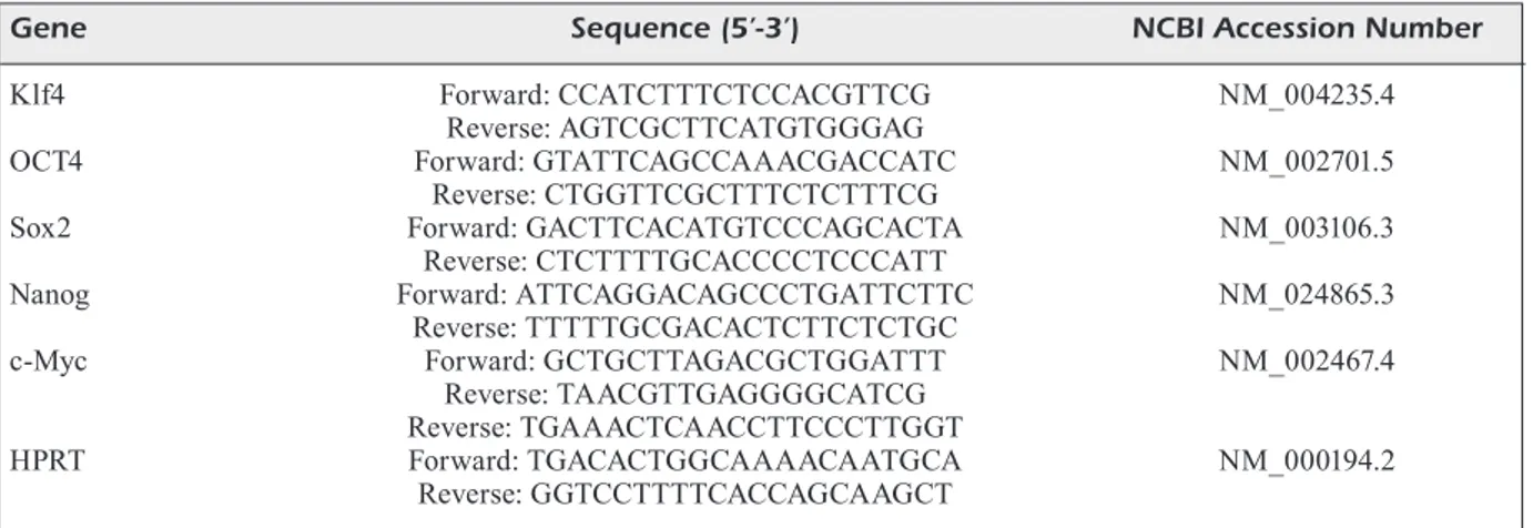
osteogenic differentiation process. Gene expression levels showed that DBSCs cultured in basal medium express significantly higher mRNA levels of Sox2 (2.2-fold) and c-Myc (2.2-fold) than DPSCs.
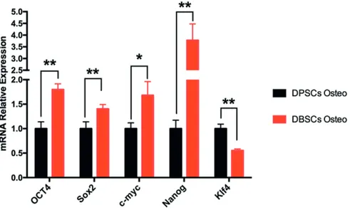
On the contrary, DBSCs express significantly lower levels of Klf4 mRNA (0.21-fold) when com-pared to DPSCs at the same conditions (Figure 1). Furthermore, Real-time PCR (qPCR) analyses were performed to determine if osteogenic
me-dium influenced the expression of the major stem-ness-associated genes OCT4, Sox2, c-Myc, Nanog and Klf4 in DBSCs vs. DPSCs. Gene expression levels showed that DBSCs cultured in osteoge-nic medium express significantly higher mRNA levels of OCT4 (1.8-fold), Sox2 (1.4-fold), c-Myc (1.68-fold) and Nanog (3.77-fold), whereas they express significantly lower levels of KLf4 mRNA (0.55-fold) when compared to DPSCs at the same
Figure 1. Expression of the stemness molecular markers in human DBSCs compared to DPSCs in basal medium. mRNA expression of the stemness genes OCT4, Sox2, c-Myc, Nanog and Klf4 in DBSCs compared to DPSCs cultured in basal me-dium for 1 week. Results are represented as fold increase compared to the level expressed in DPSCs cultured in basal meme-dium. *(p < 0.05); **(p < 0.01); ***(p < 0.001); (NS.= not significant).
Figure 2. Expression of the stemness molecular markers in human DBSCs compared to DPSCs cultured in osteogenic me-dium. mRNA expression of the stemness genes OCT4, Sox2, c-Myc, Nanog and Klf4 in DBSCs compared to DPSCs cultured in osteogenic medium for 1 week. Results are represented as fold increase compared to the level expressed in DPSCs cultured in basal medium *(p < 0.05); **(p < 0.01).
aled that Sox2 is arranged into a heterodimeric com-plex with OCT4 with a spacer of 3bp that is in char-ge of regulating the expression of fibroblast growth factor-4 (Fgf4) gene. This spacer is exactly located in between the Sox and OCT4, CATTGTCATGCA-AAT, important for Fgf4 expression35-37.
Detection of OCT4, Sox2 and Nanog expres-sion in human MSCs from BM, UCB, adipose tis-sue, PB and many other tissues is consistent with earlier reported data by many other authors6,38-42.
Although currently these genes are better un-derstood we still need to know more clear their functional role in different sub-sets of adult stem cells as these genes ubiquitously may refer either to adult stem cells or embryonic like stem cells that are kept in quiescent state in special niches, in circulation or organ tissues.
In fact, our data indicated that both DBSCs and DPSCs cultured in vitro expressed a hetero-gene-ous assortment of markers associated with em-bryonic stemness, showing a well consolidated self-renewal capability and multilineage diffe-rentiation potential to chondrocytes, adipocytes, odontoblasts, and neural-like cells under appro-priate induction conditions32-34.
Despite other sources of MSCs like BM, UCB, PB, skin, brain, liver in the adult body the dental tissues are formed at a later stage and are a sour-conditions (Figure 2). Comparative image of total
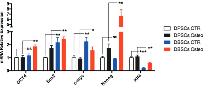
stemness expression pattern for both d-DSCs is reported in Figure 3.
Discussion
Mesenchymal stem cells were first discovered as fast growing, high plasticity and adherent colony for-ming like-fibroblastic cells present in bone marrow stroma; however, they also present in fat tissue, um-bilical cord blood, placenta and peripheral blood. Me-senchymal stem cells sub-set was also obtained from different typology of oral cavity tissues. These MSCs have been studied and used with different typology of scaffolds showing a very interactive activity with the internal scaffold microenvironment27-34.
The co-expression of gene like OCT4, Nanog, Sox2, c-Myc and Klf4 certainly proved the stem-ness attribute of these cells. Of note, in order to appreciate entirely the interactivity of these genes it is crucial to understand that they dynamically intersect each other. For instance, mouse and hu-man cells studies showed that OCT4 as transcrip-tion factors (TF), also includes the homeobox pro-tein of Nanog and homebox transcription factors of Sox2 that maintain pluripotency in ESCs.
Another research performed on blastocytes
reve-Figure 3. Gene expression of OCT4, Sox2, c-Myc, Nanog and Klf4 in DBSCs compared to DPSCs in both basal medium and osteogenic medium. mRNA expression obtained from two lines of cells at day 7 cultured in either basal medium or osteogenic medium (without the adjunct of other osteo-inducers such as vitamin D, Dexamethasone and BMP2) were compared. The DBSCs eventually at day 7 expressed in osteogenic medium environment higher level of OCT4, Nanog and Sox2 with a remarkable de-crease of Klf4 and no valuable changes in c-Myc. Conversely, the DPSCs did not show as much fluctuance in gene expression in comparing; however, in the osteogenic medium it has been noted a decrease in c-Myc whilst OCT4 and Kfl4 showed no valuable changes. The c-Myc down expression in both samples might be related to the fact that this gene need a presence of different in-ducers such as vitamin D, in addition the expression of c-Myc, OCT4 and Klf4 indicate cells osteo-differentiation in later period which is between 21 day for mature osteoblasts and SCs and as early as 14 days for emerging undifferentiated SCs.
ce of large amount of stem cells, caused by late completion of odontogenesis process and tooth eruption.
Additionally wisdom tooth that are mature and at the stage of germ, can be a source of DPSCs and DBSCs. Wisdom teeth are not essential for human masticatory function and frequently ex-tracted for orthodontic reasons or dysodontiasis. The tooth extraction, by piezo-surgery technique, is less invasive when compared to bone marrow or other tissue biopsy.
The challenge of new bone formation and graft integration is strictly dependent on recruitment and adhesion of stem cells on the scaffolds, to attain a successful cell differentiation and inte-raction with the microenvironment.
In this study, the expression of five stemness genes namely OCT4, Sox2, c-Myc, Nanog and Klf4 were compared during expansion for DBSCs and DPSCs (Figures 1, 2, 3). The results conclu-ded that DBSCs cultured in basal medium signi-ficantly expressed higher mRNA levels of both Sox2 and c-Myc almost twice than DPSCs. This was revealed in the course of gene expression analysis. In contrast, when associated to DPSCs in similar conditions, DBSCs showed significant lower levels of Klf4 mRNA. Similarly, real-time PCR (qPCR) analyses was performed to determi-ne if osteogenic medium had any influence on the expression of the major stemness-associated ge-nes OCT4, Sox2, c-Myc, Nanog and Klf4 in DB-SCs with respect to DPDB-SCs.
Gene expression levels showed significantly higher mRNA levels of OCT4 (1.8-fold), Sox2 (1.4-fold), c-Myc (1.68-fold) and Nanog (3.77-fold), however they express significantly lower levels of Klf4 mRNA (0.55-fold) when compared to DPSCs under same culture condition. So, we can speculate that d-DSCs are fitting for stemness potential related clinical applications since their effortless isolation, easiness of culture conditions and reprogramming aptitude. It may be suitable to apply these cells to achieve the regeneration of tissues/organs, in particular for head and neck tis-sues regeneration such as the gingiva, the tooth and salivary gland, which are formed through the interaction of epithelial and mesenchymal tissues during organogenesis43-50. This study evaluated
and confirmed at least in vitro the stemness state, the multipotency and pluripotency of both line of cells obtained from oral tissues, the DPSCs and DBSCs. We are, therefore, highly confident that these SCs could be an alternative and a real va-lid solution to be used in Regenerative Medicine
for soft tissue and bone regeneration due to their distinctive biological features and safety. Howe-ver, any gene variation that has been showed in this study may be the results of few variabilities that, in part, indicate the substantial differences that contradistinguish the two lines of cells, one from the pulp the others from Bud. The differen-ces might be related to the medium used in vitro culture and the intrinsic natural final commitment towards different types of tissues and cell phe-notypes of both DPSCs and DBSCs; the lack of different stimulators in osteogenic medium that has been directly connected with OCT4, c-Myc and Klf4 gene over expression such as vitamin has probably to be considered as an adjunctive cause; eventually we should also consider the pe-riod of culture that in our case was 1 week, which is probably too short to see a fully expression of those genes as showed by different data in which either 14 or 21 days is the right time to either iden-tify iPS formation and osteoblast maturity confir-med by the additional characterization of alkaline phosphatase (AP)51. Last but not least, we should
highlight the structural limitations of the in vitro microenvironment and the lack of molecular and bio-chemical influences that eventually contribu-te to cell behavior and maturity.
Conclusions
We showed for the first time in vitro the genetic expression existing between DPSCs and DBSCs under different media induction and their stem-ness pertinence that put them at the same level of other sub-set of adult SCs and MSCs.
Conflict of Interest
The Authors declare that they have no conflict of interest.
Author Contributions
A.B. made substantial contributions to the conception and design of the study, experiments and coordination, and su-pervised the manuscript. C.I.G. contributed to the data anal-ysis and interpretation, and manuscript revision. S.C. was responsible for cell culture and manuscript first draft and contributed to data analysis. J.S. contributed to molecular biology analyses. S.K.A. contributed to statistical analysis. M.C. and S.S. contributed to the isolation and expansion of mesenchymal stem cells and helped to draft the manu-script. L.S., G.D and K.CD.N. contributed in bibliograph-ic research. L.P., A.S., and F.I. partbibliograph-icipated in the design of the study, collected the biological material, performed da-ta analysis and helped to revise the manuscript. All authors read and approved the final manuscript.
References
1) Mitrano ti, Grob MS, Carrión F, nova-LaMperti e,
Luz pa, Fierro FS, Quintero a, Chaparro a, Sanz
a. Culture and characterization of mesenchymal stem cells from human gingival tissue. J Perio-dontol 2010; 81: 917-925.
2) baLLini a, Cantore S, SCaCCo S, CoLetti D, tatuLLo
M. Mesenchymal stem cells as promoters, enhan-cers, and playmakers of the translational regene-rative medicine 2018. Stem Cells Int 2018; 2018: 6927401.
3) aLLiSon Mr, iSLaM S. Attributes of adult stem cells. J
Pathol 2009; 217: 144-160.
4) MiMeauLt M, hauke r, batra Sk. Stem cells: a
re-volution in therapeutics-recent advances in stem cell biology and their therapeutic applications in regenerative medicine and cancer therapies. Clin Pharmacol Ther 2007; 82: 252-264.
5) Di beneDetto a, poSa F, De Maria S, ravaGnan G,
baLLini a, porro C, trotta t, Grano M, Lo Muzio L,
Mori G. Polydatin, natural precursor of resveratrol,
promotes osteogenic differentiation of mesenchy-mal stem cells. Int J Med Sci 2018; 13: 944-952. 6) LabuSCa L, herea DD, MaShayekhi k. Stem cells as
delivery vehicles for regenerative medicine-chal-lenges and perspectives. World J Stem Cells 2018 26; 10: 43-56.
7) baLLini a, MaStranGeLo F, GaStaLDi G, tettaManti L, buk -viC n, Cantore S, CoCCo t, Saini r, DeSiate a, GherLone e,
SCaCCo S. Osteogenic differentiation and gene
expres-sion of dental pulp stem cells under low-level laser irradiation: a good promise for tissue engineering. J Biol Regul Homeost Agents 2015; 29: 813-822. 8) DaLtoé Fp, MenDonça pp, ManteSSo a, Deboni MC.
Can SHED or DPSCs be used to repair/regene-rate non-dental tissues? A systematic review of in vivo studies. Braz Oral Res 2014; 28: pii: S1806-83242014000100401.
9) takahaShi k, yaManaka S. Induction of pluripotent stem
cells from mouse embryonic and adult fibroblast cul-tures by defined factors. Cell 2006; 126: 663-676. 10) takeDa-kawaGuChi t, SuGiyaMa k, ChikuSa S, iiDa k,
aoki h, taMaoki n, hatakeyaMa D, kuniSaDa t, Shiba -ta t, FuSaki n, tezuka k. Derivation of iPSCs after
culture of human dental pulp cells under defined conditions. PLoS One 2014; 9: e115392.
11) roDDa DJ, Chew JL, LiM Lh, Loh yh, wanG b, nG
hh, robSon p. Transcriptional regulation of
na-nog by OCT4 and SOX2. J Biol Chem 2005; 280: 24731-24737.
12) boyer La, Lee t, CoLe MF, JohnStone Se, Levine SS,
zuCker Jp, Guenther MG, kuMar rM, Murray hL, Jen -ner rG, GiFForD Dk, MeLton Da, JaeniSCh r, younG ra.
Core transcriptional regulatory circuitry in human embryonic stem cells. Cell 2005; 122: 947-956. 13) beLtraMi ap, CeSSeLLi D, berGaMin n, MarCon p, riGo
S, puppato e, D’aurizio F, verarDo r, piazza S, pi -GnateLLi a, poz a, baCCarani u, DaMiani D, Fanin r,
Mariuzzi L, Finato n, MaSoLini p, bureLLi S, beLLuzzi
o, SChneiDer C, beLtraMi Ca. Multipotent cells can
be generated in vitro from several adult human or-gans (heart, liver, and bone marrow). Blood 2007; 110: 3438-3446.
14) rapoSio e, bertozzi n. How to isolate a
ready-to-u-se adipoready-to-u-se-derived stem cells pellet for clinical application. Eur Rev Med Pharmacol Sci 2017; 21: 4252-4260.
15) riekStina u, CakStina i, parFeJevS v, hooGDuiJn M,
JankovSkiS G, MuizniekS i, MuCenieCe r, anCanS J.
Embryonic stem cell marker expression pattern in human mesenchymal stem cells derived from bone marrow, adipose tissue, heart and dermis. Stem Cell Rev 2009; 5: 378-386.
16) kuanG wb, DenG Q, DenG Ct, Li wS, zhanG yG, Shu
Sw, zhou Mr. MiRNA regulates OCT4 expression
in breast cancer cells. Eur Rev Med Pharmacol Sci 2018; 22: 1351-1357.
17) Chen hy, han XL, wanG rG, SonG XF, zhanG hz.
The expression of OCT4 and its clinical significan-ce in laryngeal squamous carcinoma tissues. Eur Rev Med Pharmacol Sci 2017; 21: 4591-4594. 18) aMbroSetti DC, baSiLiCo C, DaiLey L. Synergistic
acti-vation of the fibroblast growth factor 4 enhancer by Sox2 and Oct-3 depends on protein-protein interactions facilitated by a specific spatial arran-gement of factor binding sites. Mol Cell Biol 1997; 17: 6321-6329.
19) zhou L, zhao LC, JianG n, wanG XL, zhou Xn, Luo XL,
ren J. MicroRNA miR-590-5p inhibits breast cancer
cell stemness and metastasis by targeting SOX2. Eur Rev Med Pharmacol Sci 2017; 21: 87-94. 20) SiLva J, niChoLS J, theuniSSen tw, Guo G, van ooSten
aL, barranDon o, wray J, yaManaka S, ChaMberS i,
SMith a. Nanog is the gateway to the pluripotent
ground state. Cell 2009; 138: 722-737.
21) zhanG p, anDrianakoS r, yanG y, Liu C, Lu w.
Krup-pel-like factor 4 (Klf4) prevents embryonic stem (ES) cell differentiation by regulating Nanog gene expression. J Biol Chem 2010; 285: 9180-9189. 22) rowLanD bD, bernarDS r, peeper DS. The KLF4
tu-mour suppressor is a transcriptional repressor of p53 that acts as a context-dependent oncogene. Nat Cell Biol 2005; 7: 1074-1082.
23) rowLanD bD, peeper DS. KLF4, p21 and
context-de-pendent opposing forces in cancer. Nat Rev Can-cer 2006; 6: 11-23.
24) Chen X, JohnS DC, GeiMan De, Marban e, DanG Dt,
haMLin G, Sun r, yanG vw. Krüppel-like factor 4
(gut-enriched Krüppel-like factor) inhibits cell pro-liferation by blocking G1/S progression of the cell cycle. J Biol Chem 2001; 276: 30423-30428. 25) nakaGawa M, koyanaGi M, tanabe k, takahaShi k,
iChiSaka t, aoi t, okita k, MoChiDuki y, takizawa n,
yaManaka S. Generation of induced pluripotent
stem cells without Myc from mouse and human fibroblasts. Nat Biotechnol 2009; 26: 101-106. 26) ChanG yC, Li wC, twu nF, Li hy, Lo wL, ChanG yL,
Lee yy, Lin CF, Shih yh, Chen Mt. Induction of
den-tal pulp-derived induced pluripotent stem cells in the absence of c-Myc for differentiation into neu-ron-like cells. J Chin Med Assoc 2014; 77: 618-625.
27) zippeL n, SChuLze M, tobiaSCh e. Biomaterials and
mesenchymal stem cells for regenerative medici-ne. Recent Pat Biotechnol 2010; 4: 1-22.
28) GuiLLot pv, Cui w, FiSk nM, poLak DJ. Stem cell
differentiation and expansion for clinical applica-tions of tissue engineering. J Cell Mol Med 2007; 11: 935-944.
29) CapLan ai. Adult mesenchymal stem cells for
tis-sue engineering versus regenerative medicine. J Cell Physiol 2007; 213: 341-347.
30) SantiaGo Ja, poGeMiLLer r, oGLe bM. Heterogeneous
differentiation of human mesenchymal stem cells in response to extended culture in extracellular matrices. Tissue Eng Part A 2009; 15: 3911-3922. 31) torMin a, brune JC, oLSSon e, vaLCiCh J, neuMan
u, oLoFSSon t, JaCobSen Se, SCheDinG S.
Characte-rization of bone marrow-derived mesenchymal stromal cells (MSC) based on gene expression profiling of functionally defined MSC subsets. Cytotherapy 2009; 11: 114-128.
32) baLLini a, boCCaCCio a, Saini r, van phaM p, tatuL -Lo M. Dental-derived stem cells, their secretome
and interactions with bioscaffolds/biomaterials in regenerative medicine: from the in-vitro research to translational applications. Stem Cells Int 2017; 2017: 6975251.
33) baLLini a, SCaCCo S, CoLetti D, pLuChino S, tatuLLo M.
Mesenchymal stem cells as promoters, enhancers, and playmakers of the translational regenerative medicine. Stem Cells Int 2017; 2017: 3292810. 34) Di beneDetto a, poSa F, Carbone C, Cantore S, bru
-netti G, Centonze M, Grano M, Lo Muzio L, CavaL -Canti-aDaM ea, Mori G. NURR1 downregulation
favors osteoblastic differentiation of MSCs. Stem Cells Int 2017; 2017: 7617048.
35) yuan h, Corbi n, baSiLiCo C, DaiLey L.
Developmen-tal-specific activity of the FGF-4 enhancer requi-res the synergistic action of Sox2 and Oct-3. Ge-nes Dev 1995; 9: 2635-2645.
36) aMbroSetti DC, baSiLiCo C, DaiLey L. Synergistic
acti-vation of the fibroblast growth factor 4 enhancer by Sox2 and Oct-3 depends on protein-protein interactions facilitated by a specific spatial arran-gement of factor binding sites. Mol Cell Biol 1997; 17: 6321-6329.
37) wei CX, wonG h, Xu F, Liu z, ran L, JianG rD.
IRF4-induced upregulation of lncRNA SOX2-OT promotes cell proliferation and metastasis in cho-langiocarcinoma by regulating SOX2 and PI3K/ AKT signaling. Eur Rev Med Pharmacol Sci 2018; 22: 8169-8178.
38) Dai ww, Liu S, Liu XJ, penG zp. Stemness-related
changes of CD133- cells in nasopharyngeal carci-noma after x-ray radiation at the median lethal dose. Eur Rev Med Pharmacol Sci 2018; 22: 2334-2342. 39) DoMiniCi M, Le bLanC k, MueLLer i, SLaper-CortenbaCh
i, Marini F, krauSe D, DeanS r, keatinG a, proCkop
DJ, horwitz e. Minimal criteria for defining
multi-potent mesenchymal stromal cells. The interna-tional society for cellular therapy position state-ment. Cytotherapy 2006; 8: 315-317.
40) bao CS, Li XL, Liu L, wanG b, yanG Fb, Chen LG.
Transplantation of Human umbilical cord mesen-chymal stem cells promotes functional recovery after spinal cord injury by blocking the expression of IL-7. Eur Rev Med Pharmacol Sci 2018; 22: 6436-6447.
41) Xue zL, MenG yL, Ge Jh. Upregulation of
miR-132 attenuates osteoblast differentiation of UC-MSCs. Eur Rev Med Pharmacol Sci 2018; 22: 1580-1587.
42) wei CX, wonG h, Xu F, Liu z, ran L, JianG rD.
IRF4-induced upregulation of lncRNA SOX2-OT promotes cell proliferation and metastasis in cho-langiocarcinoma by regulating SOX2 and PI3K/ AKT signaling. Eur Rev Med Pharmacol Sci 2018; 22: 8169-8178.
43) Cantore S, baLLini a, De vito D, MarteLLi FS, Ge -orGakopouLoS i, aLMaSri M, DibeLLo v, aLtini v,
Farronato G, DipaLMa G, Farronato D, inChinGoLo
F. Characterization of human apical papilla-de-rived stem cells. J Biol Regul Homeost Agents 2017; 31: 901-910.
44) Miura y, GronthoS S, aLLen Mr, Cao C, uveGeS te, bi
y, ehirChiou D, korteSiDiS a, Shi S, zhanG L.
Defecti-ve osteogenesis of the stromal stem cells predi-sposes CD18-null mice to osteoporosis. Proc Natl Acad Sci U S A 2005; 102: 14022-14027.
45) baLLini a, SCattareLLa a, CrinCoLi v, CarLaio rG, papa
F, periLLo L, roManazzo t, buX Mv, narDi GM, Dituri
a, Cantore S, pettini F, GraSSi Fr. Surgical
treat-ment of gingival overgrowth with 10 years of fol-low-up. Head Face Med 2010 12; 6: 19.
46) brunetti G, Di beneDetto a, poSa F, CoLaianni G, Fa -ienza MF, baLLini a, CoLuCCi S, paSSeri G, Lo Muzio L,
Grano M, Mori G. High expression of TRAIL by
osteoblastic differentiated dental pulp stem cells affects myeloma cell viability. Oncol Rep 2018; 39: 2031-2039.
47) Cantore S, CrinCoLi v, boCCaCCio a, uva ae, Fioren -tino M, Monno G, boLLero p, DerLa C, Fabiano F,
baLLini a, SantaCroCe L. Recent advances in
endo-crine, metabolic and immune disorders: mesen-chymal stem cells (MSCs) and engineered scaf-folds. Endocr Metab Immune Disord Drug Targets 2018; 18: 466-469.
48) Li C, wei GJ, Xu L, ronG JS, tao SQ, wanG yS. The
involvement of senescence induced by the telo-mere shortness in the decline of osteogenic diffe-rentiation in BMSCs. Eur Rev Med Pharmacol Sci 2017; 21: 1117-1124.
49) Mori G, brunetti G, baLLini a, Di beneDetto a, taran -tino u, CoLuCCi S, Grano M. Biological
characteri-stics of dental stem cells for tissue engineering. Key Engineering Materials 2013; 541: 51-59. 50) Chen b, MenG J, zenG yt, Du yX, zhanG J, Si yM,
yuan X. MicroRNA-7-5p regulates osteogenic
dif-ferentiation of hMSCs via targeting CMKLR1. Eur Rev Med Pharmacol Sci 2018; 22: 7826-7831. 51) SinGh u, QuintaniLL a rh, GreCian S, Gee kr, rao
MS, LakShMipathy u. Novel live alkaline
phospha-tase substrate for identification of pluripotent stem cells. Stem Cell Rev 2012; 8: 1021-1029.