UNIVERSITÀ DEGLI STUDI DI ROMA
"TOR VERGATA"
FACOLTA' DI MEDICINA E CHIRURGIA
DOTTORATO DI RICERCA IN EMATOLOGIA
XXII
Investigation on the mechanisms underlying the chromosomal translocations
in therapy-related acute myeloid leukemias
Syed Khizer Hasan
A.A. 2009/2010
Docente Guida/Tutor: Prof. Francesco Lo-Coco
Coordinatore: Prof. Sergio Amadori
3
Index
Chapter 1: Introduction
Page 4
1.1 Acute promyelocytic leukemia (APL) and
therapy related APL (t-APL)
Page 6
1.2 Therapy related acute myeloid leukemia
Page 8
1.3 t-APL following multiple sclerosis and genetic
variants of DNA double strand break repair genes
Page 9
1.4 Aims and objectives
Page 10
1.5 References to chapter 1
Page 11
Chapter 2: Mechanisms of the formation of t(15;17) in mitoxantrone related
therapy related-APL following multiple sclerosis
Page 14
Chapter 3: Molecular analysis of the t(15;17) genomic breakpoints in epirubicin
associated therapy-related APL following breast carcinoma
Page 30
Chapter 4:
Analysis of t(15;17) chromosomal breakpoint sequences in
therapy-related versus de novo APL
Page 41
Chapter 5: Genomic characterization of t(16;21) translocation in therapy-related
acute myeloid leukemia
Page 49
Chapter 6: To study the genetic markers of susceptibility to t-APL and their
association with multiple sclerosis
Page 59
4
Chapter 1
Introduction
5 Introduction
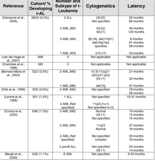
Therapy-related leukemias are well-recognized clinical syndrome occurring as a late complication following cytotoxic therapy. The term "therapy-related" leukemia is descriptive and based on a patient’s history of exposure to cytotoxic agents. Although a causal relationship is implied, the mechanism remains to be proven. These leukemias are thoughtto be the direct consequence of mutational events induced bycytotoxic therapy, or via the selection of a myeloid clone witha mutator phenotype that has a markedly elevated risk for amutational event. These types of leukaemias are becoming an increasing healthcare problem as more patients survive their primary cancers. The nature of the causative agent has an important bearing upon the characteristics, biology, time to onset and prognosis of the resultant leukaemia. Agents targeting topoisomerase II induce acute leukaemias with balanced translocations that generally arise within 3 years, often involving the MLL, RUNX1, PML and RARA loci at 11q23, 21q22 15q22 and 17q21 respectively. Chromosomal breakpoints have been found to be preferential sites of topoisomerase II cleavage, which are believed to be repaired by the nonhomologous end-joining DNA repair pathway to generate chimaeric oncoproteins that underlie the resultant leukaemias. Therapy-related acute myeloid leukaemias occurring after exposure to antimetabolites and/or alkylating agents are biologically distinct with a longer latency period, being characterised by more complex karyotypes and loss of p53. The treatment of therapy-related leukaemias represents a considerable challenge due to prior therapy and comorbidities, however, curative therapy is possible, particularly in those with favourable karyotypic features. Although the transforming function of leukemia-associated fusion proteinshas been widely studied, little is known about the mechanisms that cause the underlying translocations. In this respect, insights can be gained from investigations of secondary leukemias, all of which have counterparts in primary leukemias1.
6
1.1 Acute promyelocytic leukemia (APL) and therapy-related APL
t(15;17)(q22;q21)
In 1977, Rowley et al2 from the University of Chicago reported on the consistent occurrence ofa chromosomal translocation t(15;17)(q22;q21) in APL. This aberration was subsequently found to be uniquely associated to, and therefore pathognomonic of the disease. Upon cloning of the translocation in the late „80s, it was shown that chromosome breakpoints lie within the RAR locus on chromosome 17 and the PML locus on chromosome 15, resulting in the fusion of the two genes3,4. APL is a particular leukemia subset characterized by a unique genetic lesion, i.e the PML-RARα fusion, and an exquisite response to differentiating agents. Until the late 80‟s, APL was considered the most aggressive and rapidly fatal form of acute leukemia. Over the past two decades, important advances have been made into the understanding of APL pathogenesis as well as in its treatment, such that it has been nowadays converted into the most frequently curable leukemia in adults. APL is regarded as a model disease for innovative tailored treatment of human leukemia including differentiation therapy and the use of chromatin remodeling agents and antibody-directed therapy.
The occurrence of APL as a second tumor (sAPL) has been increasingly reported in recent years, most commonly developing in patients receiving chemotherapy and/or radiotherapy for breast cancer or, less frequently, for other primary tumors (including prostate, uterus, ovary)5-8. Chemotherapy associated with development of sAPL usually induces DNA damage through targeting of topoisomerase II (topoII), with mitoxantrone, etoposide and epirubicin being the most commonly implicated agents5-10. More recently, a number of reports have been published describing the occurrence of sAPL in patients with Multiple Sclerosis (MS) most of whom received mitoxantrone given as an immunosuppressive agent for their primary disease 11-16. Because only case reports have
7
been published and no systematic analysis has been performed so far, the true incidence of sAPL in MS patients treated with mitoxantrone is unknown. In addition, it is unclear at present whether factors other than chemotherapy may play a role in sAPL development in patients with MS.
In a study reported by Mistry et al10 breakpoints in sAPL cases arising following mitoxantrone exposure for prior breast carcinoma were found to be clustered in an 8bp region within PML intron 6. In functional assays, this “hotspot” was found to correspond to a preferential site of mitoxantrone-induced topoII-dependent cleavage at PML nucleotide position 1484. These findings suggest a direct causative role of mitoxantrone in stimulating topoII-mediated cleavage within the PML gene thus implying a direct link between this agent and the formation of the t(15;17) chromosome translocation that is the hallmark of APL. However, the precise mechanisms leading to this aberration and therefore to APL development needs to be further clarified. In fact, several reports have described the occurrence of sAPL in patients treated with surgery alone for their primary tumor6. Neither in these cases nor in patients developing sAPL after treatment of MS has the t(15;17) translocation been investigated at the molecular level. Finally, no studies have analysed in details the response to endogenous or exogenous DNA damage in newly diagnosed APL and in sAPL.
DNA double-strand breaks (DSBs) are critical lesions that can result in cell death or in a wide variety of genetic alterations including large or small-scale deletions, loss of heterozygosity, translocations, and chromosome loss. Mitoxantrone creates exogenous DNA double strands breaks (DSBs) and interferes in the cleavage-religation equilibrium of the topoII enzyme17. The maintenance of genomic integrity and the prevention of tumor progression depend on the co-ordination of DNA repair mechanisms and cell cycle checkpoint signaling in an overall DNA damage response. It is believed that double strand
8
breaks (DSBs) are initially detected by specific sensors (e.g. MRN complex) that trigger the activation of transducing kinases (e.g. ATM, ATR, DNAPK). These transducers in co-ordination with mediators (e.g. H2AX) amplify the damage signal, which is then relayed to effector proteins (e.g. p53, SMC1, Kap1, CHK2, CHK1), that directly regulate the progression of the cell cycle and DNA repair. DSBs are normally repaired by non-homologous end-joining (NHEJ) and non-homologous recombination (HR) pathways.
Disruption of the repair proteins may lead the NHEJ pathway to function inappropriately and rejoin DNA ends incorrectly resulting in translocations18. The NHEJ is an error prone pathway in which rejoinings occur at regions of microhomology that are 1–4 nucleotides in length. These are nucleotides that, by chance, are shared between the two ends of different chromosomes. We and others have demonstrated the presence of short homologies at t(15;17) translocation breakpoint junction suggesting that PML and RARA joining may have been mediated by the classical NHEJ pathway10,19-20
1.2 Therapy related acute myeloid leukemia with t(16;21) (q24;q22)
Hematopoietic malignancies are frequently characterized by recurrent chromosomal translocations involving genes that play an important role in the regulation of hematopoietic cell proliferation and differentiation21. The AML1 (RUNX1) gene at cytogenetic band 21q22 is one of the most frequent targets of chromosomal translocations observed in both de novo acute leukemia and therapy-related myelodysplastic syndrome (t-MDS) and acute myeloid leukemia (AML). Translocations involving AML1 have been reported in up to 15% of t-MDS/t-AML cases and the most common chromosome/gene rearrangements described in this clinical context are the t(8;21)(q22;q22), t(3;21)(q26;q22) and t(16;21)(q24;q22) translocations involving the ETO-1 (MTG8), EAP/MDS1/EVI1, and MTG16 (ETO-2) genes, respectively22.
9
The t(16;21)(q24;q22) is a rare but non random chromosome abnormality associated mostly with t-AML23-25. It involves MTG16 (myeloid translocation gene on chromosome 16) which encodes one of a family of novel transcriptional corepressors (MTG proteins) and shows a high degree of homology to the MTG8 gene, the fusion partner in the t(8;21)24 . The evolutionary conserved structural features between AML1-MTG8 and AML1-MTG1624, suggests that the two chimeric proteins are both involved in hematopoiesis, as subsequently demonstrated in functional studies26.
1.3 t-APL following multiple sclerosis and genetic variants of DNA double strand break repair genes
The occurrence of APL as a second tumor has been increasingly reported in patients with Multiple Sclerosis (MS). It is believed that the genetic contribution to leukemia susceptibility is a complex interplay of environmental exposures and many susceptibility alleles, each of which contributes only a small amount to overall risk. Multiple genetic variants, deriving from Single Nucleotide Polymorphisms (SNPs) within coding sequences, have been described for many genes involved in DNA repair processes. Such variants may be associated with functional differences of the encoded proteins, which may in turn be responsible for inefficient DNA repair mechanisms that become evident particularly after exposure to chemotherapeutic agents. Furthermore, specific SNP variants of apoptosis and DNA damage-regulatory genes have recently been described as risk factors for MSand may hence be associated with sAPL occurring in patients with this disease.
We, therefore, intend to investigate the possibility that specific genetic variants in DNA repair genes or genes that predispose to MS are significantly associated with t-APL.
10 1.4 Aims and objectives
We aim to investigate whether specific chromosomal regions are particularly susceptible to DNA damage (either spontaneously or induced by chemotherapeutic agents). In particular, we proposed:
a) To determine the mechanism of chromosomal translocations of t(15;17) and t(16,21) in therapy related acute leukemia through characterization of DNA breakpoint regions using patient samples.
b) To provide evidences of mitoxantrone and epirubicin induced DNA cleavage at translocation breakpoint loci using in vitro DNA cleavage assays.
c) To compare the translocation breakpoint distribution between de novo and t-APL.
d) To analyse genetic variability of genes involved in DNA double strand break repair in multiple sclerosis patients and t-APL developing after multiple sclerosis.
11 References:
1. Larson RA. Is secondary leukemia an independent poor prognostic factor in acute myeloid leukemia? Best Pract Res Clin Haematol 2007; 10: 29-37
2. Rowley JD, Golomb HM, Dougherty C. 15/17 translocation, a consistent chromosomal change in acute promyelocytic leukaemia. Lancet 1977; 1: 549-550. 3. de The H, Chomienne C, Lanotte M, Degos L, Dejean A. The t(15;17) translocation
of acute promyelocytic leukaemia fuses the retinoic acid receptor alpha gene to a novel transcribed locus. Nature 1990; 347: 558-561.
4. Kakizuka A, Miller WH, Jr., Umesono K, Warrell RP, Jr., Frankel SR, et al. Chromosomal translocation t(15;17) in human acute promyelocytic leukemia fuses RAR alpha with a novel putative transcription factor, PML. Cell 1991; 66: 663-674. 5. Pollicardo N, O'Brien S, Estey EH et al. Secondary acute promyelocytic leukemia:
Characteristics and prognosis of 14 patients from a single institution. Leukemia 1996; 10: 27-31
6. Pulsoni A, Pagano L, Lo Coco F et al. Clinico-biological features and outcome of acute promyelocytic leukemia occurring as a second tumor: the GIMEMA experience. Blood 2002;100:1972-76
7. Beaumont M, Sanz M, Carli PM et al. Therapy related acute promyelocytic leukemia. J Clin Oncol 2003; 21: 2123-2137
8. Andersen MK, Larson RA, Mauritzson N et al. Balanced chromosome abnormalities inv(16) and t(15;17) in therapy-related myelodysplastic syndromes and acute
leukemia: Report from an International Workshop. Genes Chromosomes Cancer 2002:33395-400
9. Felix CA, Kolaris CP, Osheroff N. Topoisomease II and the etiology of chromosomal translocations. DNA repair 2006; 5 : 1093-1108
12
10. Mistry AR, Felix CA, Whitmarsh RJ et al. DNA topoisomerase II in therapy-related acute promyelocytic leukemia. N Engl J Med 2005; 352: 1529-1538
11. Vicari AM, Ciceri F, Folli F et al. Acute promyelocytic leukemia following mitoxantrone as single agent for the treatment of multiple sclerosis. Leukemia 1998; 12:441-442
12. Cattaneo C, Almici C, Borlenghi E et al. A case of acute promyelocytic leukemia following mitoxantrone treatment of multiple sclerosis. Leukemia 2003; 17: 985-986 13. B Delisse, J de Seze, A Mackowiak et al. Therapy related acute myeloblastic
leukemia after mitoxantrone treatment in a patient with multiple sclerosis. Mult scler 2004; 10: 92
14. Novoselac AV, Reddy S, Sanmugarajah J. Acute promyelocytic leukemia in a patient with multiple sclerosis following treatment with mitoxantrone. Leukemia 2004; 18: 1561-1562
15. Arruda WO, Montú MB, de Oliveira Mde S et al. Acute myeloid leukemia induced by mitoxantrone: case report. Arq Neuropsiquiatr 2005; 63: 327-329
16. . Ledda A, Caocci G, Spinicci G et al. Two new cases of acute promyelocytic leukemia following mitoxantrone treatment in patients with multiple sclerosis. Leukemia 2006; 20: 2217
17. Fortune JM, Osheroff N. Topoisomerase II as a target for anticancer drugs: when enzyme stops being nice. Prog Nucleic Acid Res Mol Biol 2000;64: 221-53
18. Soutoglou E, Dorn JF, Sengupta K et al. Positional stability of single double strands breaks in mammalian cellls Nat Cell Biol. 2007; 9: 675-82
19. Hasan SK, Mays A, Ottone T et al. Molecular analysis of t(15;17) genomic breakpoints in secondary acute promyelocytic leukemia arising after treatment of multiple sclerosis Submitted to Blood 2008; 112: 3383-90
13
20. Reiter A, Saussele S, Grimwade D et al. Genomic anatomy of the specific reciprocal translocation t(15;17) in acute promyelocytic leukemia. Genes Chromosomes Cancer 2003;36(2):175-88.
21. Renneville A, Roumier C, Biggio V, Nibourel O, Boissel N, Fenaux P, Preudhomme C. Cooperating gene mutations in acute myeloid leukemia: a review of the literature. Leukemia 2008; 22:915-931.
22. Slovak ML, Bedell V, Popplewell L, Arber DA, Schoch C, Slater R. 21q22 balanced chromosome aberrations in therapy-related hematopoietic disorders: report from an international workshop. Genes Chromosomes and Cancer 2002; 33: 379-294.
23. Nucifora G, Rowley JD. AML1 and the 8;21 and 3;21 translocations in acute and chronic myeloid leukemia. Blood 1995 ; 86:1-14.
24. Gamou T, Kitamura E, Hosoda F, Shimizu K, Shinohara K, Hayashi Y, Nagase T, Yokoyama Y, Ohki M. The partner gene of AML1 in t(16;21) myeloid malignancies is a novel member of the MTG8(ETO) family. Blood 1998; 9 : 4028-4037.
25. Roulston D, Espinosa R 3rd, Nucifora G, Larson RA, Le Beau MM, Rowley JD. CBFA2(AML1) translocations with novel partner chromosomes in myeloid leukemias: association with prior therapy. Blood 1998; 92:2879-2885.
26. Rossetti S, Van Unen L, Touw IP, Hoogeveen AT, Sacchi N. Myeloid maturation block by AML1-MTG16 is associated with Csf1r epigenetic down regulation. Oncogene 2006; 24:5325-5332.
14
Chapter 2
Mechanisms of the formation of
t(15;17) in mitoxantrone related
therapy related-APL following
doi:10.1182/blood-2007-10-115600 Prepublished online Jul 23, 2008; 2008 112: 3383-3390
Francesco Lo-Coco
A. Felix, Maria Teresa Voso, Wolfgang R. Sperr, Jordi Esteve, Miguel A. Sanz, David Grimwade and Gnanam Satchi, Anne Lennard, Marta Libura, Jo Ann W. Byl, Neil Osheroff, Sergio Amadori, Carolyn Cattaneo, Erika Borlenghi, Lorella Melillo, Enrico Montefusco, José Cervera, Christopher Stephen, Syed Khizer Hasan, Ashley N. Mays, Tiziana Ottone, Antonio Ledda, Giorgio La Nasa, Chiara
promyelocytic leukemia arising after treatment of multiple sclerosis Molecular analysis of t(15;17) genomic breakpoints in secondary acute
http://bloodjournal.hematologylibrary.org/cgi/content/full/112/8/3383
Updated information and services can be found at:
(2490 articles)
Clinical Trials and Observations
(4221 articles)
Neoplasia
collections:
Blood
Articles on similar topics may be found in the following
http://bloodjournal.hematologylibrary.org/misc/rights.dtl#repub_requests
Information about reproducing this article in parts or in its entirety may be found online at:
http://bloodjournal.hematologylibrary.org/misc/rights.dtl#reprints
Information about ordering reprints may be found online at:
http://bloodjournal.hematologylibrary.org/subscriptions/index.dtl
Information about subscriptions and ASH membership may be found online at:
.
Hematology; all rights reserved Copyright 2007 by The American Society of
200, Washington DC 20036.
semimonthly by the American Society of Hematology, 1900 M St, NW, Suite Blood (print ISSN 0006-4971, online ISSN 1528-0020), is published
For personal use only. at TOR VERGATA on January 10, 2009.
www.bloodjournal.org From
NEOPLASIA
Molecular analysis of t(15;17) genomic breakpoints in secondary acute
promyelocytic leukemia arising after treatment of multiple sclerosis
*Syed Khizer Hasan,1*Ashley N. Mays,2Tiziana Ottone,1Antonio Ledda,3Giorgio La Nasa,3Chiara Cattaneo,4
Erika Borlenghi,4Lorella Melillo,5Enrico Montefusco,6Jose´ Cervera,7Christopher Stephen,8Gnanam Satchi,9
Anne Lennard,10Marta Libura,2Jo Ann W. Byl,11Neil Osheroff,11Sergio Amadori,1Carolyn A. Felix,12Maria Teresa Voso,13
Wolfgang R. Sperr,14Jordi Esteve,15Miguel A. Sanz,7David Grimwade,2and Francesco Lo-Coco1
1Department of Biopathology, University of Tor Vergata, Rome, Italy;2Department of Medical & Molecular Genetics, King’s College London School of Medicine, London, United Kingdom;3Ematologia/Centro Trapianti Midollo Osseo, Ospedale R. Binaghi, Cagliari, Italy;4Ematologia, Spedali Civili, Brescia, Italy; 5Hematology Department, Casa Sollievo della Sofferenza Hospital, S. Giovanni Rotondo, Italy;6Department of Hematology, S. Andrea Hospital, University La Sapienza, Rome, Italy;7Hematology Department, University Hospital La Fe, Valencia, Spain;8Department of Haematology, Pilgrim Hospital, Boston, United Kingdom;9Department of Haematology, Whiston Hospital, Prescot, United Kingdom;10Department of Haematology, Royal Victoria Infirmary, Newcastle, United Kingdom;11Department of Biochemistry, Vanderbilt University School of Medicine, Nashville, TN;12Department of Pediatrics, University of Pennsylvania School of Medicine, Division of Oncology, Children’s Hospital of Philadelphia, Philadelphia, PA;13Istituto di Ematologia, Universita’ Cattolica del Sacro Cuore, Rome, Italy;14Department of Internal Medicine I, Division of Hematology & Hemostaseology, Medical, University of Vienna, Vienna, Austria; and15Hospital Clı´nic, Institut d’Investigacions Biome`diques August Pi i Sunyer, Barcelona, Spain
Therapy-related acute promyelocytic leu-kemia (t-APL) with t(15;17) translocation is a well-recognized complication of can-cer treatment with agents targeting topo-isomerase II. However, cases are emerg-ing after mitoxantrone therapy for multiple sclerosis (MS). Analysis of 12 cases of mitoxantrone-related t-APL in MS pa-tients revealed an altered distribution of chromosome 15 breakpoints versus de novo APL, biased toward disruption within PML intron 6 (11 of 12, 92% vs 622 of 1022, 61%: Pⴝ .035). Despite this intron
span-ning approximately 1 kb, breakpoints in 5 mitoxantrone-treated patients fell within an 8-bp region (1482-9) corresponding to the “hotspot” previously reported in t-APL, complicating mitoxantrone-contain-ing breast cancer therapy. Another shared breakpoint was identified within the ap-proximately 17-kb RARA intron 2 involv-ing 2 t-APL cases arisinvolv-ing after mitox-antrone treatment for MS and breast cancer, respectively. Analysis of PML and RARA genomic breakpoints in functional assays in 4 cases, including the shared
RARA intron 2 breakpoint at 14 446-49, confirmed each to be preferential sites of topoisomerase II␣-mediated DNA cleav-age in the presence of mitoxantrone. This study further supports the presence of preferential sites of DNA damage induced by mitoxantrone in PML and RARA genes that may underlie the propensity to de-velop this subtype of leukemia after expo-sure to this agent. (Blood. 2008;112: 3383-3390)
Introduction
The occurrence of acute promyelocytic leukemia (APL) as a second tumor (sAPL) frequently has been reported as a late complication of chemotherapy and/or radiotherapy (therapy-related APL [t-APL]), although sAPL cases arising in patients whose primary tumors were treated by surgery alone have also been described.1-3 The agents most often associated with
development of t-APL induce DNA damage through targeting of topoisomerase II, with mitoxantrone, epirubicin, adriamycin, and etoposide being most commonly implicated.3,4The latency
period between chemotherapy exposure and the onset of t-APL is relatively short (⬍ 3 years) and typically occurs without a preceding myelodysplastic phase.3,4
In a study by Mistry et al5concerning molecular mechanisms
underlying formation of the t(15;17) in t-APL, breakpoints in cases arising after mitoxantrone exposure for prior breast carcinoma were found to be clustered in an 8-bp region within PML intron 6; this corresponded in functional assays to a preferential site of mitoxantrone-induced topoisomerase II–dependent cleavage at
position 1484. Although these findings highlighted the leukemo-genic role of drug-induced DNA cleavage at specific sites in the genome, the precise mechanism by which secondary leukemias with balanced chromosomal translocations such as the t(15;17) in APL develop remains controversial.6-9This is compounded by the
fact that many patients have been exposed to multiple cytotoxic drugs often accompanied by radiotherapy, making it difficult to categorically ascribe the etiology of therapy-related acute myeloid leukemia (t-AML) in any given case.
Previous studies on t-AML have focused on patient populations that feasibly could have been enriched for persons at particular risk of leukemia, having already developed one form of cancer. Therefore, to investigate whether particular chemotherapeutic agents have a propensity to induce specific molecular subtypes of t-AML, it is of interest to study patients exposed to topoisomerase II targeting drugs used in the treatment of nonmalignant conditions, such as mitoxantrone in the management of multiple sclerosis (MS). MS is a putative autoimmune disease affecting the central
Submitted October 3, 2007; accepted July 5, 2008. Prepublished online as
Blood First Edition paper, July 23, 2008; DOI 10.1182/blood-2007-10-115600.
*S.K.H. and A.N.M. contributed equally to the experimental analyses and should be considered joint first authors.
The publication costs of this article were defrayed in part by page charge payment. Therefore, and solely to indicate this fact, this article is hereby marked ‘‘advertisement’’ in accordance with 18 USC section 1734.
© 2008 by The American Society of Hematology
3383 BLOOD, 15 OCTOBER 2008䡠VOLUME 112, NUMBER 8
For personal use only. at TOR VERGATA on January 10, 2009.
www.bloodjournal.org From
nervous system for which mitoxantrone represents the latest in a long list of general immunosuppressive agents used in the treat-ment of this condition.10,11In recent years, an increasing number of
APL cases have been reported in MS patients treated with mitoxantrone.3,5,12-20However, to date, no attempts have been made
to systematically characterize translocation breakpoints in APL cases that developed in this setting.
In the present study, we analyzed at the genomic level the PML and RARA breakpoints of 14 patients who developed APL on a background of MS, including 12 who received mitoxantrone for their primary disease. Furthermore, we used functional cleavage assays to better elucidate the mechanisms underlying the formation of the t(15;17) in this setting.
Methods
Patients and samples
The main patient characteristics, including demographic data, MS type, and treatments received for MS, are reported in Table 1. Seven patients were diagnosed in 5 Italian institutions, 3 in 2 Spanish institutions, 3 in the United Kingdom, and the remaining patient in Austria. Analyses were undertaken after informed patient consent was obtained in accordance with the Declaration of Helsinki with ethical approval of University Tor Vergata of Rome and St Thomas’ Hospital of London. Bone marrow samples were obtained at the time of diagnosis of APL. Mononuclear cells were collected after centrifugation on a Ficoll-Hypaque gradient and stored at⫺70°C as dry pellets. In all cases, APL diagnosis was confirmed at the genetic level by reverse-transcriptase polymerase chain reaction (RT-PCR) amplification of the PML-RARA hybrid gene.
Amplification of DNA spanning possible break points (PML-RARA): long-range PCR and DNA sequencing
To determine the exact chromosomal breakpoint position in PML and RARA genes, genomic DNA extracted from APL blasts collected at diagnosis was amplified by a 2-step, long-range nested PCR method as reported else-where.5,21Two forward and 8 reverse primers were designed for each step to
cover the PML breakpoint region (bcr1 or bcr3, as previously known based on diagnostic RT-PCR results available for all cases) and the 16.9-kb-long
RARA intron 2. PCR products were purified using the QIAquick PCR
purification kit (Qiagen, Valencia, CA). Samples were loaded in 96-well plates and covered with mineral oil. The amplified products were separated with a capillary electrophoresis-based system (CEQ 8000 Genetic Analysis System; Beckman Coulter, Fullerton, CA) using the “LFR1 Test” default run method and sequenced using appropriate primers.5,21Rigorous
proce-dures were used to reduce risk of PCR contamination,22 and genomic
breakpoints were in all cases confirmed by PCR analysis of a fresh aliquot of DNA. Moreover, in 3 cases, breakpoint analyses were performed independently in parallel in the Rome and London laboratories, yielding identical results.
Amplification and sequence analysis of the reciprocal RARA-PML genomic breakpoint junction
Genomic RARA-PML was amplified using patient specific primers (designed on the basis of PML and RARA breakpoints) and fresh aliquots of DNA. In 13 cases, the reciprocal RARA-PML genomic breakpoint junction was sequenced, provid-ing further confirmation of the t(15;17) translocation breakpoints at the genomic level. In one case (unique patient number [UPN] 10), no DNA was available to carry out sequencing of the reciprocal RARA-PML.
Alignment of sequenced nucleotides using BLAST algorithm
The patients’ genomic PML-RARA junction sequences were aligned against normal PML (GenBank accession number S57791 for bcr1 and S51489 for bcr3) and RARA intron 2 (GenBank accession number AJ297538)
nucleo-tides as a reference text input in BLAST/alignment program. The purpose of alignment was to identify any microhomologies between PML and RARA in the vicinity of the breakpoint.23
In vitro DNA cleavage assays
Human topoisomerase II␣ was expressed in Saccharomyces cerevisiae24
and purified as described previously.25,26 Assays were performed as
described previously.5 Briefly, having identified genomic junction
se-quences, regions of the normal homologs encompassing the breakpoint sites were amplified by PCR and subcloned into the pBluescript SKII(⫹) vector. The optimal insert size for the assay was 200 to 500 bp, with the breakpoint site located approximately 50 to 100 bp from the 5⬘ end of the insert. Substrates containing 25 ng of the normal homologs of the translocation breakpoints were 5⬘end-labeled (30 000 cpm) and incubated with 147 nM of human DNA topoisomerase II␣, 1 mM of ATP in the presence or absence of 20M mitoxantrone.5In all cases, additional reactions were carried out
to evaluate the heat stability of the covalent complexes formed. Cleavage complexes were irreversibly trapped by the addition of sodium dodecyl sulfate, and purified products were resolved in an 8% polyacrylamide-7.0 M of urea gel in parallel with dideoxy sequencing reactions primed at the same 5⬘-end, visualized by autoradiography, and quantified using Phospho-Imager and IMAGEQUANT software (GE Healthcare, Little Chalfont, United Kingdom).
Results
Clinical features
As shown in Table 1, a total of 14 patients with APL developing in a background of MS were studied. The series included 12 cases exposed to a median total dose of 105 mg mitoxantrone (range, 30-234 mg), whereas 2 patients received other treatments for their primary disease (interferon- in UPN 10 and corticosteroids in UPN 12). The median latency period between the first exposure to mitoxantrone and APL diagnosis was 28 months (range, 4-60 months). Patients were treated with all-trans retinoic acid and anthracycline-based chemotherapy, mostly using AIDA-like
(all-trans retinoic acid⫹ idarubicin) protocols27 (Table 2); however,
UPN 13 died of cerebral hemorrhage within 3 hours of APL diagnosis before antileukemic therapy was started. The remaining 13 patients achieved hematologic and molecular remission. Of these, 11 remain in first molecular remission at a median follow-up of 10 months, whereas UPN 7 relapsed at 28 months and achieved second molecular remission after salvage therapy with arsenic trioxide, and UPN 1 died of cerebral hemorrhage while in remission after 7 months.
Location of t(15;17) translocation breakpoints within the PML and RARA loci
RT-PCR showed the bcr1 PML-RARA isoform (PML intron 6 breakpoint) in 12 cases, whereas in the remaining 2 cases the PML breakpoint fell within intron 3 (bcr3; Figure 1). This breakpoint distribution appeared skewed in favor of the bcr1 isoform, which previously has been reported to account for approximately 55% of unselected APL cases.28-30Comparison of the breakpoint
distribu-tion in MS patients with mitoxantrone-related APL relative to a cohort of 1022 consecutive cases of newly diagnosed de novo APL from GIMEMA, PETHEMA, and United Kingdom MRC trials confirmed significant overrepresentation of involvement of PML intron 6 in the former group (11 of 12, 92% vs 622 of 1022, 61%:
P⫽ .035 by Fisher exact test). PML genomic breakpoints within
intron 6 were found to fall between nucleotide positions 1482 and 1489 in 6 patients (UPNs 3, 6, 11, 12, 13, and 14; Figure 1A),
3384 HASAN et al BLOOD, 15 OCTOBER 2008䡠VOLUME 112, NUMBER 8
For personal use only. at TOR VERGATA on January 10, 2009.
www.bloodjournal.org From
T able 1. Main clinical and molecular features of 14 sAPL patients Patient no. Age (y) at time of MS diagnosis Sex Primary disease Therapy of MS Latency between MS and APL, mo Latency between mitoxantrone and APL, mo Bcr subtype PML * breakpoint der(15)-der(17) RARA * breakpoint der(15)-der(17) Type of treatment Mitoxantrone schedule Total dose, mg UPN 1 59 F SPMS Mitoxantrone 10 mg/m 2every 1 mo 30 13 10 1 1496-97 1496-97 6104-05 6104-05 UPN 2 43 F SPMS Mitoxantrone 10 mg/m 2every 2 mo 35 192 37 1 1657-60 1657-60 14446-49 14446-49 UPN 3 56 F PPMS Mitoxantrone 10 mg/m 2every 3 mo 70 30 4 1 1485-87 1485-87 15266-68 15266-68 UPN 4 24 F RRMS Mitoxantrone 12 mg/m 2every 2 mo 147 150 6 1 1922-23 1922-23 12418-19 12418-19 UPN 5 21 F RRMS Mitoxantrone 12 mg/m 2every 2 mo 170 66 18 1 1150-51 1169-70 14082-83 14054-55 UPN 6 53 F RRMS Mitoxantrone 10 mg/m 2(every 1 mo, 3 doses), 10 mg/m 2 (every 3 mo, 7 doses) 234 72 51 1 1483-84 1484-85 12909-10 12908-09 UPN 7 25 M RRMS Mitoxantrone 12 mg/m 2(every 1 mo, 3 doses), 6 mg/m 2 (every 3 mo, 5 doses) 110 37 27 1 1165 1161 7974 7978 UPN 8 44 M SPMS Mitoxantrone 1.9 mg/m 2every 1 mo 100 204 29 1 1409-10 1429-30 718-19 675-76 UPN 9 33 M PPMS Mitoxantrone 10 mg/m 2every 2 mo 176 144 30 3 1286-87 1286-87 15386-87 15386-87 UPN 10 26 M RRMS INF beta NA NA 18 NA 3 1117-22† 14920-25 UPN 11 37 M RRMS Mitoxantrone 13 mg monthly (5 doses) 81 97 17 1 1483-86 1486-1506 11586-89 11683-84 UPN 12 49 M RRMS Prednisone NA NA 216 NA 1 1489-91 1485 14785-87 14117 UPN 13 45 F PPMS Mitoxantrone 8 mg/m 2every 1 mo 64 120 60 1 1485-87 1486-88 11569-71 11569-71 UPN 14 25 F SPMS Mitoxantrone 11.36 mg/m 2every 1 mo 120 237 34 1 1488-89 1483-86 12038-39 12035-38 UPN indicates unique patient number (some details regarding UPNs 1, 4 and 5, and 7 were reported in references 20, 17, and 5, respectively); RRMS, relap sing-remitting multiple sclerosis; SPMS, secondary progressive multiple sclerosis; PPMS, primary progressive multiple sclerosis; and NA, not applicable. *Breakpoint locations are numbered according to the following GenBank accession numbers: PML intron 6 (bcr 1), S57791; PML intron 3 (bcr 3), S51489; and RARA intron 2, AJ297538. †No DNA was available to sequence the reciprocal RARA-PML in this case.
APL IN MULTIPLE SCLEROSIS 3385 BLOOD, 15 OCTOBER 2008䡠VOLUME 112, NUMBER 8
For personal use only. at TOR VERGATA on January 10, 2009.
www.bloodjournal.org From
coinciding precisely with the “hotspot” previously identified in t-APL after mitoxantrone treatment for breast cancer.5
Interest-ingly, one of these patients (UPN 12) had not received mitox-antrone therapy for MS. In the 2 patients (UPNs 9 and 10) with the bcr3 PML-RARA isoform, the breakpoints in PML intron 3 were detected between nucleotides 1286 and 1287 and 1117 through 1122, respectively (Figure 1A). The breakpoints within the RARA locus were distributed across intron 2 without particular clustering in any restricted small region (Figure 1B). However, one break-point (in UPN 2) mapped precisely to a breakbreak-point found in a case of t-APL arising after mitoxantrone therapy for breast cancer, studied previously by Mistry et al.5
Sequence analyses of the reciprocal RARA-PML fusion revealed a balanced translocation in 7 of 13 analyzed cases. Six patients showed size variable deletions and/or insertions at the breakpoint junction (Table 1). Microhomologies at the breakpoint junctions were indicative of DNA repair by the nonhomologous end-joining (NHEJ) pathway.5
t(15,17) translocation breakpoints are preferential sites for mitoxantrone-induced DNA cleavage by human
topoisomerase II␣
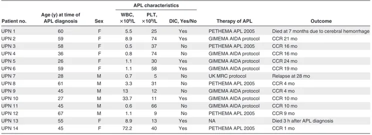
To investigate the mechanisms by which the t(15;17) chromosomal translocation may have been formed in MS patients treated with Table 2. Main clinical features and treatment outcome of sAPL patients
Patient no.
Age (y) at time of
APL diagnosis Sex
APL characteristics
Therapy of APL Outcome
WBC,
ⴛ109/L
PLT,
ⴛ109/L DIC, Yes/No
UPN 1 60 F 5.5 25 Yes PETHEMA APL 2005 Died at 7 months due to cerebral hemorrhage
UPN 2 59 F 8.9 74 Yes GIMEMA AIDA protocol CCR 21 mo
UPN 3 58 F 0.5 37 No PETHEMA APL 2005 CCR 16 mo
UPN 4 36 F 0.8 74 No GIMEMA AIDA protocol CCR 16 mo
UPN 5 26 F 1.1 30 Yes GIMEMA AIDA protocol CCR 24 mo
UPN 6 59 F 1.1 58 Yes GIMEMA AIDA protocol CCR 19 mo
UPN 7 28 M 0.7 5 No UK MRC protocol Relapse at 28 mo
UPN 8 61 M 3.3 31 No PETHEMA APL 2005 CCR 4 mo
UPN 9 45 M 13 12 No GIMEMA AIDA protocol CCR 4 mo
UPN 10 27 M 33.7 11 Yes GIMEMA AIDA protocol CCR 10 mo
UPN 11 45 M 0.6 66 No GIMEMA AIDA protocol CCR 10 mo
UPN 12 67 M 1.1 9 No PETHEMA APL 2005 CCR 9 mo
UPN 13 55 F 8.9 13 Yes NA Died 3 h after APL diagnosis
UPN 14 45 F 72.2 40 Yes PETHEMA APL 2005 CCR 1 mo
DIC indicates disseminated intravascular coagulation; CCR, continuous complete remission; and NA, not applicable.
Figure 1. Characterization of t(15;17) breakpoints within the PML and RARA loci. The location of breakpoints indicated by in the 14 patients (numbers correspond with UPNs in Tables 1 and 2) within the PML gene on chromosome 15 (A; bcr3 region and bcr1/2 region) and intron 2 of RARA on chromosome 17 (B) are shown. Breakpoint
locations are numbered according to the following GenBank accession numbers: PML intron 6 (bcr 1), S57791; PML intron 3 (bcr 3), S51489; and RARA intron 2, AJ297538.23
3386 HASAN et al BLOOD, 15 OCTOBER 2008䡠VOLUME 112, NUMBER 8
For personal use only. at TOR VERGATA on January 10, 2009.
www.bloodjournal.org From
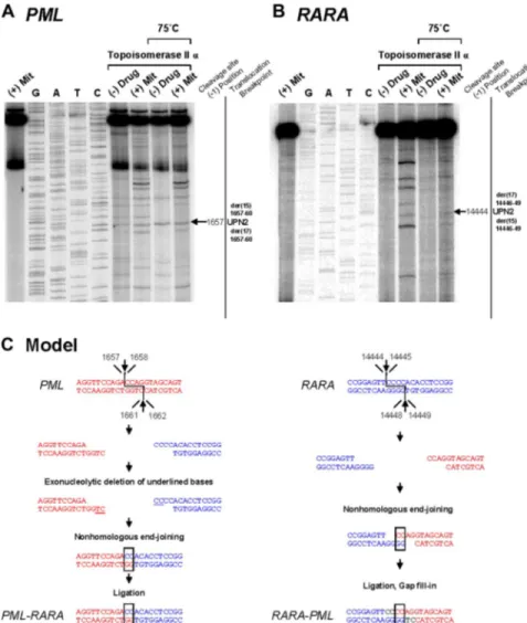
mitoxantrone, we evaluated topoisomerase II␣–mediated cleavage of the normal homologs of PML and RARA encompassing the respective breakpoints detected in 4 cases (UPNs 2, 7, 8, and 14) in the presence or absence of this agent. These included cases (ie, UPN 2 and UPN 14) in which the genomic breakpoint in the RARA or PML locus coincided with those reported previously in cases of t-APL arising in breast cancer patients treated with multiple DNA-damaging agents, including mitoxantrone.5 Few cleavage
sites were observed in the absence of drug; however, bands of various sizes and intensities were observed in the presence of mitoxantrone in a topoisomerase II␣-dependent manner (Figures 2, 3 top panels). Cleavage bands that were significantly enhanced by mitoxantrone corresponding to the location of the observed genomic breakpoints in the PML and RARA loci were detected in each of the cases analyzed (Figures 2A,B, 3A,B; and data not shown). These bands remained detectable after heating, indicating stability of the cleavage complexes. In UPN 2, the case in which the RARA breakpoint was shared with a t-APL case that arose after mitox-antrone-containing breast cancer therapy,5 a functional site of
mitoxantrone-induced cleavage by topoisomerase II was identified at position 14 444 (Figure 2B).
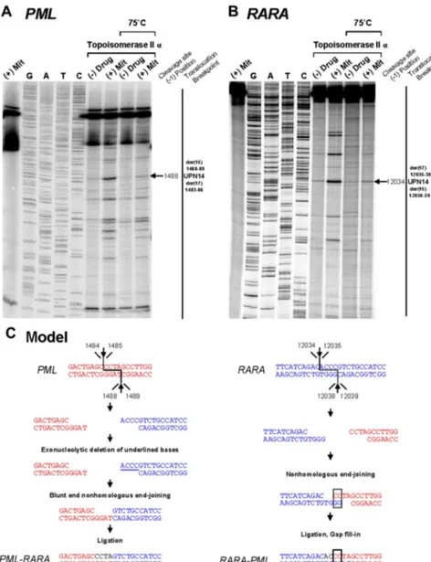
To provide further evidence that the region between positions 1482 and 1489 within PML intron 6 (which was involved in almost half the cases) is also a preferential site of mitoxantrone-induced DNA cleavage mediated by topoisomerase II␣, the reverse comple-ment of the described PML substrate5was used in the cleavage
assay. A strong heat-stable cleavage band was detected in the
presence of mitoxantrone at position 1488, which corresponds to the described functional cleavage at position 1484 on the upper strand5(Figure 3A,C). Given that the chromosome 15 breakpoint in
UPN 12 (in which there was no history of mitoxantrone exposure) also fell within this “hotspot,” it is interesting to note that a weak cleavage band was apparent in the presence of topoisomerase II␣ in the absence of drug (Figure 3A lane 6). This finding suggests that the sequence may be a natural site of topoisomerase II␣–mediated cleavage that could be relevant to the etiology of APL in this case. Based on sequence analysis of PML-RARA and reciprocal
RARA-PML genomic breakpoints, the location of functional
topo-isomerase II␣ cleavage sites in the vicinity of the breakpoints, and known mechanisms by which topoisomerase II induces double-strand breaks in DNA31and their subsequent repair,6it was possible
to generate models as to how the t(15;17) chromosomal transloca-tion could have been formed in the studied cases (Figure 2,3C). Type II topoisomerases introduce staggered nicks in DNA creating 5⬘-overhangs. In the models, repair of the overhangs in PML and
RARA entails exonucleolytic digestion, pairing of complementary
bases, and joining of DNA free ends by the NHEJ pathway, with template-directed polymerization to fill in any gaps.
Discussion
In this study on sAPL that developed after MS, we were able to identify a biased distribution of breakpoints in the PML gene that Figure 2. Investigation of t(15;17) translocation
mechanism in UPN 2 by in vitro topoisomerase II␣
DNA cleavage assay. Chromosomal breakpoint
junc-tions were examined in an in vitro topoisomerase II␣
cleavage assay using substrates containing PML (A) and RARA (B) translocation breakpoints in the APL case of UPN 2. Reactions in lane 1 were performed without DNA topoisomerase II␣ and lanes 2 to 5 show dideoxy sequencing reactions. DNA cleavage reactions were performed in the presence of 147 nM of human DNA topoisomerase II alpha and in the absence (lanes
6 and 8) or presence of 20M mitoxantrone (lanes 7
and 9). Reactions in lanes 8 and 9 were incubated at 75°C to assess the heat stability of the cleavage products seen in lanes 6 and 7. In each case, the location of the relevant heat stable cleavage site is indicated by an arrow on the far right. (C) Native PML and RARA sequences are shown in red and blue, respectively. In the creation of the PML-RARA genomic fusion, processing includes exonucleolytic deletion to form a 2-base homologous overhang that facilitates repair via the error prone NHEJ pathway. In the creation of the reciprocal RARA-PML genomic fusion, 2-base homologies facilitate NHEJ repair, whereas in both instances polymerization of the relevant overhangs fills in any remaining gaps (shown black font).
APL IN MULTIPLE SCLEROSIS 3387 BLOOD, 15 OCTOBER 2008䡠VOLUME 112, NUMBER 8
For personal use only. at TOR VERGATA on January 10, 2009.
www.bloodjournal.org From
clustered in the same “hotspot” region previously identified in APL cases arising after treatment with mitoxantrone for breast cancer.5
In addition, we established in one patient who the breakpoint in
RARA intron 2 at position 14446-49 coincided with a breakpoint
identified by Mistry et al in 1 of 5 t-APL cases arising in breast cancer patients treated with the same agent.5Given that intron 2 is
almost 17 kb in length, such tight clustering of breakpoints between 2 different t-APL cases would be highly improbable to occur by chance. This observation strongly suggests that this is a preferential site of mitoxantrone-induced cleavage of DNA by topoisomerase II␣. The hypothesis is further supported by our functional in vitro data that show that this RARA site, together with the previously identified 8-bp “hotspot” region in PML intron 6, are preferential targets of mitoxantrone-induced DNA damage mediated by topo-isomerase II␣.
Interestingly, of the 6 patients found to have PML breakpoints involving the “hotspot” region, one (UPN 12) did not receive mitoxantrone. Although mitoxantrone may significantly increase the chances of inducing DNA damage at this site, it is conceivable that this region represents a preferential site of cleavage by the native topoisomerase II␣ and could in some instances act in concert with environmental or dietary agents that also target the en-zyme.32-36Accordingly, in the other case of sAPL that arose in the
absence of mitoxantrone exposure (UPN 10), the in vitro DNA cleavage assay also revealed sites of cleavage in the PML and
RARA substrates with topoisomerase II alone, which corresponded
to the observed breakpoints (data not shown).
In some cases, the occurrence of short homologies of 1 or 2 nucleotides at the breakpoint region between the PML and RARA genes precluded precise assignment of the breakpoint within each respective gene. However, further investigation using the in vitro functional assays enabled the location of preferential sites of mitoxantrone-induced topoisomerase II␣–mediated DNA cleavage to be mapped. Taking into account the mechanisms by which type II topoisomerases induce double-strand DNA breaks31 and the
processes mediating their repair, most probably involving the NHEJ pathway,5,30it was possible to model the generation of the
t(15;17) chromosomal translocation underlying the development of APL in these cases. Previous studies have established the presence of functional topoisomerase II cleavage sites at translocation breakpoints in MLL-associated t-AML, indicating that direct DNA damage coupled with aberrant repair by the NHEJ pathway is probably relevant to the formation of translocations that disrupt other genes that are commonly involved in t-AML.37-39
To the best of our knowledge, 20 cases of sAPL occurring in patients with MS have been reported to date,3,5,12-204 of which are Figure 3. Investigation of t(15;17) translocation
mechanism in UPN 14 by in vitro topoisomerase II␣
DNA cleavage assay. DNA cleavage assays are shown for PML (A) and RARA (B) genomic breakpoint regions. For the PML assay, the reverse complement of the substrate containing the “hotspot” region between 1482
and 1489 described by Mistry et al5was used. Lanes 1
to 9 of each cleavage assay are described in the legend to Figure 2. (C) Native PML and RARA sequences are shown in red and blue, respectively. In the creation of PML-RARA, processing includes exonucleolytic dele-tion and repair via the NHEJ pathway. In the creadele-tion of RARA-PML, 2-base homologies facilitate repair via the NHEJ pathway, whereas in both instances polymeriza-tion of the relevant overhangs fills in any remaining gaps (shown in black font).
3388 HASAN et al BLOOD, 15 OCTOBER 2008䡠VOLUME 112, NUMBER 8
For personal use only. at TOR VERGATA on January 10, 2009.
www.bloodjournal.org From
included in the present series (UPNs 1, 4, 5, and 7) [5, 17, 20]. However, this is the first study to systematically analyze such cases at the genomic level. The incidence of sAPL arising in MS patients treated with mitoxantrone is not firmly established, as no system-atic analysis has been undertaken to address this issue and only case reports have been published. On reviewing the records of 2336 MS patients treated with mitoxantrone, Voltz et al40described
5 cases of t-AML and 2 sAPL as a case report. Ghalie et al41
assembled the records of 1378 patients treated with mitoxantrone in 3 MS studies and reported 2 patients who developed t-AML with an observed incidence proportion of 0.15% (95% confidence interval, 0.00%-0.40%). In addition to the reported 20 cases of sAPL, 8 cases of t-AML (non-M3) arising after mitoxantrone treatment for MS have been described, with the majority showing balanced translocations in their leukemic cells.40,42-46 Therefore,
although it appears that an excess of sAPL cases are observed in the MS setting, the reasons underlying this phenomenon remain unclear at present and warrant further basic and epidemiologic investigation. It is unknown, for example, whether factors other than mitoxantrone may play a role in sAPL development in the context of MS. Finally, it would be important to assess prospec-tively the true incidence of APL development in the MS setting (with or without mitoxantrone). Considering the risk of leukemia development and cardiac toxicity, the Therapeutics and Technology Subcommittee of the American Academy of Neurology recently has recommended that mitoxantrone be reserved for patients with progressive MS who have failed other therapies.47
In conclusion, this study lends further support to the presence of preferential sites of DNA damage induced by mitoxantrone within
PML intron 6 and suggests the existence of a further “hotspot” at
the distal end of RARA intron 2. The susceptibility of these regions of the PML and RARA loci to topoisomerase II␣–mediated cleavage by mitoxantrone may underlie the propensity to develop this particular subtype of AML after exposure to this agent. Further
studies are warranted to investigate whether MS patients have a particular predisposition to the development of sAPL.
Acknowledgments
The authors thank Dr M. Boggild for provision of clinical data, Jelena Jovanovic for performance of MRD analyses, and Mireia Camos for kindly providing the DNA from UPN 12.
This work was supported by grants from Associazione Italiana per la Ricerca sul Cancro (F.L.C.) and Italian Ministry of Health (Progetto Integrato Oncologia), Leukemia Research Fund of Great Britain (A.N.M., D.G.), the National Institutes of Health (grant R01CA077683 to C.A.F.; and grant GM33944 to J.A.W.B., N.O.), and the Polish Ministry of Science and Education (grant PBZ KBN 107 P04 2004, to M.L.).
Authorship
Contribution: S.K.H. and A.N.M. performed the experiments, analyzed the data, and contributed to the manuscript; T.O. and M.L. assisted in experimental design and performance of experiments; A. Ledda, G.L.N., C.S., C.C., E.B., L.M., E.M., J.C., G.S., A. Lennard, J.E., M.T.V., and W.R.S. contributed to the samples, clinical data, and interpretation of results; J.A.W.B. and N.O. supplied vital reagents; D.G., M.A.S., C.A.F., and S.A. analyzed the data, critically reviewed the manuscript, and amended the final report; and F.L.C. and D.G. designed the study, supervised the research, and wrote the manuscript.
Conflict-of-interest disclosure: The authors declare no compet-ing financial interests.
Correspondence: Francesco Lo-Coco, Department of Biopathol-ogy, University Tor Vergata, Via Montpellier 1, 00133, Rome, Italy; e-mail: [email protected].
References
1. Pollicardo N, O’Brien S, Estey EH, et al. Second-ary acute promyelocytic leukemia: characteristics and prognosis of 14 patients from a single institu-tion. Leukemia. 1996;10:27-31.
2. Pulsoni A, Pagano L, Lo Coco F, et al. Clinico-biological features and outcome of acute promy-elocytic leukemia occurring as a second tumor: the GIMEMA experience. Blood. 2002;100:1972-1976.
3. Beaumont M, Sanz M, Carli PM, et al. Therapy related acute promyelocytic leukemia. J Clin On-col. 2003;21:2123-2137.
4. Pedersen-Bjergaard J, Christiansen DH, Desta F, Andersen MK. Alternative genetic pathways and cooperating genetic abnormalities in the patho-genesis of therapy-related myelodysplasia and acute myeloid leukemia. Leukemia. 2006;20: 1943-1949.
5. Mistry AR, Felix CA, Whitmarsh RJ, et al. DNA topoisomerase II in therapy-related acute promy-elocytic leukemia. N Engl J Med. 2005;352:1529-1538.
6. Burden DA, Osheroff N. Mechanism of action of eukaryotic topoisomerase II and drugs targeted to the enzyme. Biochim Biophys Acta. 1998;400: 139-154.
7. Zhang Y, Rowley JD. Chromatin structural ele-ments and chromosomal translocations in leuke-mia: DNA repair. 2006;5:1282-1297.
8. Rene B, Fermandjian S, Mauffret O. Does topo-isomerase II specifically recognize and cleave hairpins, cruciforms and crossovers of DNA? Bio-chimie. 2007;89:508-515.
9. Azarova AM, Lyu YL, Lin C, et al. Roles of DNA topoisomerase II isozymes in chemotherapy and secondary malignancies. Proc Natl Acad Sci U S A. 2007;104:11014-11019.
10. Millefiorini E, Gasperini C, Pozzilli C, et al. Ran-domized placebo-controlled trial of mitoxantrone in relapsing-remitting multiple sclerosis: 24-month clinical and MRI outcome. J Neurol. 1997;244: 153-159.
11. Fox EJ. Management of worsening multiple scle-rosis with mitoxantrone: a review. Clin Ther. 2006; 28:461-474.
12. Vicari AM, Ciceri F, Folli F, et al. Promyelocytic leukemia following mitoxantrone as single agent for the treatment of multiple sclerosis. Leukemia. 1998;12:441-442.
13. Cattaneo C, Almici C, Borlenghi E, Motta M, Rossi G. A case of acute promyelocytic leukae-mia following mitoxantrone treatment of multiple sclerosis. Leukemia. 2003;17:985-986. 14. Delisse B, de Seze J, Mackowiak A, et al.
Therapy related acute myeloblastic leukemia af-ter mitoxantrone treatment in a patient with mul-tiple sclerosis. Mult Scler. 2004;10:92. 15. Novoselac AV, Reddy S, Sanmugarajah J. Acute
promyelocytic leukemia in a patient with multiple sclerosis following treatment with mitoxantrone. Leukemia. 2004;18:1561-1562.
16. Arruda WO, Montu´ MB, de Oliveira Mde S, Ramina R. Acute myeloid leukaemia induced by mitoxantrone: case report. Arq Neuropsiquiatr. 2005;63:327-329.
17. Ledda A, Caocci G, Spinicci G, Cocco E,
Ma-musa E, La Nasa G. Two new cases of acut pro-myelocytic leukemia following mitoxantrone treat-ment in patients with multiple sclerosis. Leukemia. 2006;20:2217-2218.
18. Sumrall A, Dreiling B. Therapy-related acute non-lymphoblastic leukemia following mitoxantrone therapy in a patient with multiple sclerosis. J Miss State Med Assoc. 2007;48:206-207.
19. Ramkumar B, Chadha MK, Barcos M, Sait SN, Heyman MR, Baer MR. Acute promyelocytic leu-kemia after mitoxantrone therapy for multiple sclerosis. Cancer Genet Cytogenet. 2008;182: 126-129.
20. Bosca I, Pascual AM, Casanova B Coret F, Sanz MA. Four new cases of therapy-related acute pro-myelocytic leukemia after mitoxantrone. Neurol-ogy. 2008;71:457-458.
21. Reiter A, Saussele S, Grimwade D, et al. Genomic anatomy of the specific reciprocal trans-location t(15;17) in acute promyelocytic leukemia. Genes Chromosomes Cancer. 2003;36:175-188. 22. Flora R, Grimwade D Real-time quantitative
RT-PCR to detect fusion gene transcripts associated with AML. Methods Mol Med. 2004;91: 151-173. 23. Benson DA, Karsch-Mizrachi I, Lipman DJ, Ostell
J, Wheeler DL. GenBank. Nucleic Acids Res. 2008;36:D25-30.
24. Worland ST, Wang JC. Inducible overexpression, purification, and active site mapping of DNA topo-isomerase II from the yeast Saccharomyces cer-evisiae. J Biol Chem. 1989;264:4412-4416. 25. Elsea SH, Hsiung Y, Nitiss JL, Osheroff N. A
APL IN MULTIPLE SCLEROSIS 3389 BLOOD, 15 OCTOBER 2008䡠VOLUME 112, NUMBER 8
For personal use only. at TOR VERGATA on January 10, 2009.
www.bloodjournal.org From
yeast type II topoisomerase selected for resis-tance to quinolones: mutation of histidine 1012 to tyrosine confers resistance to nonintercalative drugs but hypersensitivity to ellipticine. J Biol Chem. 1995;270:1913-1920.
26. Kingma PS, Greider CA, Osheroff N. Spontane-ous DNA lesions poison human topoisomerase IIalpha and stimulate cleavage proximal to leuke-mic 11q23 chromosomal breakpoints. Biochemis-try. 1997;36:5934-5939.
27. Mandelli F, Diverio D, Avvisati G, et al. Molecular
remission in PML/RAR␣-positive acute
promyelo-cytic leukemia by combined all-trans retinoic acid and idarubicin (AIDA) therapy. Blood. 1997;90: 1014-1021.
28. Fenaux P, Chomienne C. Biology and treatment of acute promyelocytic leukemia. Curr Opin On-col. 1996;8:3-12.
29. Kane JR, Head DR, Balazs L, et al. Molecular analysis of the PML/RAR alpha chimeric gene in pediatric acute promyelocytic leukemia. Leuke-mia. 1996;10:1296-1302.
30. Slack JL, Arthur DC, Lawrence D, et al. Second-ary cytogenetic changes in acute promyelocytic leukemia: prognostic importance in patients treated with chemotherapy alone and association with the intron 3 breakpoint of the PML gene. A Cancer and Leukemia Group B study. J Clin On-col. 1997;15:1786-1795.
31. Fortune JM, Osheroff N. Topoisomerase II as a target for anticancer drugs: when enzymes stop being nice. Prog Nucleic Acid Res Mol Biol. 2000; 64: 221-253.
32. Ross JA, Potter JD, Reaman GH, Pendergrass TW, Robison LL. Maternal exposure to potential inhibitors of DNA topoisomerase II and infant leu-kemia (United States): a report from the
Chil-dren’s Cancer Group. Cancer Causes Control. 1996;7:581-590.
33. Strick R, Strissel PL, Borgers S, Smith SL, Rowley JD. Dietary bioflavonoids induce cleav-age in the MLL gene and may contribute to infant leukaemia. Proc Natl Acad Sci U S A. 2000;97: 4790-4795.
34. Felix CA, Kolaris CP, Osheroff N. Topoisomerase II and etiology of chromosomal translocations: DNA repair. 2006;5:1093-1108.
35. Bandele OJ, Osheroff N. Bioflavonoids as poi-sons of human topoisomerase II␣ and II. Bio-chemistry. 2007;46:6097-6108.
36. Bandele OJ, Osheroff N. (–)-Epigallocatechin gal-late, a major constituent of green tea, poisons human type II topoisomerases. Chem Res Toxi-col. 2008;21:936-943.
37. Lovett BD, Strumberg D, Blair IA, et al. Etoposide metabolites enhance DNA topoisomerase II cleavage near leukemia-associated MLL translo-cation breakpoints. Biochemistry. 2001;40:1159-1170.
38. Lovett BD, Lo Nigro L, Rappaport EF, et al. Near-precise interchromosomal recombination and functional DNA topoisomerase II cleavage sites at MLL and AF-4 genomic breakpoints in treatment-related acute lymphoblastic leukemia with t(4;11) translocation. Proc Natl Acad Sci U S A. 2001;98: 9802-9807.
39. Whitmarsh RJ, Saginario C, Zhuo Y, et al. Recip-rocal DNA topoisomerase II cleavage events at
5⬘-TATTA-3⬘ sequences in MLL and AF-9 create
homologous single-stranded overhangs that an-neal to form der(11) and der(9) genomic break-point junctions in treatment-related AML without further processing. Oncogene. 2003;22:8448-8459.
40. Voltz R, Starck M, Zingler V, Strupp M, Kolb HJ. Mitoxantrone therapy in multiple sclerosis and acute leukaemia: a case report out of 644 treated patients. Mult Scler. 2004;10:472-474. 41. Ghalie RG, Mauch E, Edan G, et al. A study of
therapy-related acute leukaemia after mitox-antrone therapy for multiple sclerosis. Mult Scler. 2002;8:441-445.
42. Edan G, Brochet B, Brassat D, Safety profile of mitoxantrone in a cohort of 802 multiple sclerosis patients [abstract]. Neurology. 2002;58(suppl 3): 168.
43. Brassat D, Recher C, Waubant E, et al. Therapy-related acute myeloblastic leukemia after mitox-antrone treatment in a patient with MS. Neurol-ogy. 2002;59:954-955.
44. Heesen C, Bruegmann M, Gbdamosi J, Koch E, Monch A, Buhmann C. Therapy related acute my-elogenous leukemia in a patient with multiple sclerosis treated by mitoxantrone. Mult Scler. 2003;9:213-214.
45. Goodkin D. Therapy-related leukemia in mitox-antrone treated patients. Mult Scler. 2003;9:426. 46. Tanasescu R, Debouverie M, Pitton S, Anxionnat R, Vespignani H. Acute myeloid leukemia induced by mitoxantrone in a multiple sclerosis patient. J Neurol. 2004;251:762-763.
47. Goodin DS, Arnason BG, Coyle PK, Frohman EM, Paty DW; Therapeutics and Technology As-sessment Subcommittee of the American Acad-emy of Neurology. The use of mitoxantrone (Nov-antrone) for the treatment of multiple sclerosis: report of the Therapeutics and Technology As-sessment Subcommittee of the American Acad-emy of Neurology. Neurology. 2003;61:1332-1338.
3390 HASAN et al BLOOD, 15 OCTOBER 2008䡠VOLUME 112, NUMBER 8
For personal use only. at TOR VERGATA on January 10, 2009.
www.bloodjournal.org From
Note: Additional text and figures related to this chapter which are not available to online version of the manuscript.
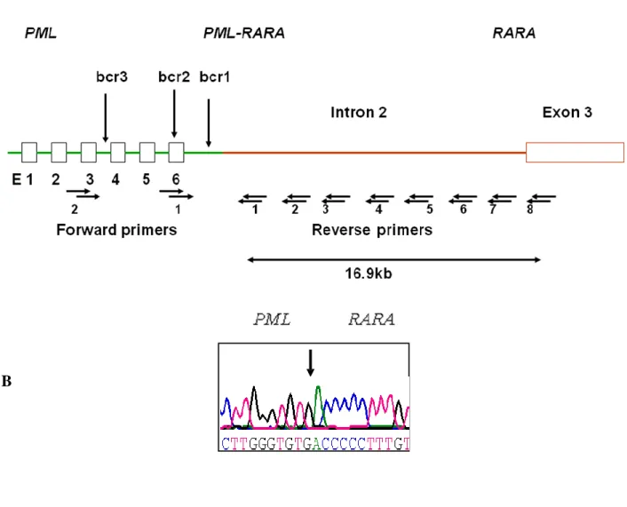
Fig S1: Schematic diagram showing the strategy employed to identify t(15;17) genomic breakpoint junction locations. (A) PML is shown in green, RARA in red, and the locations of the nested primers used to perform long range nested PCR are indicated by the horizontal arrows. Vertical arrows indicate the regions in which breakpoints are most likely to occur. (B) An example of the chromatogram obtained revealing the breakpoint junction sequence.
A
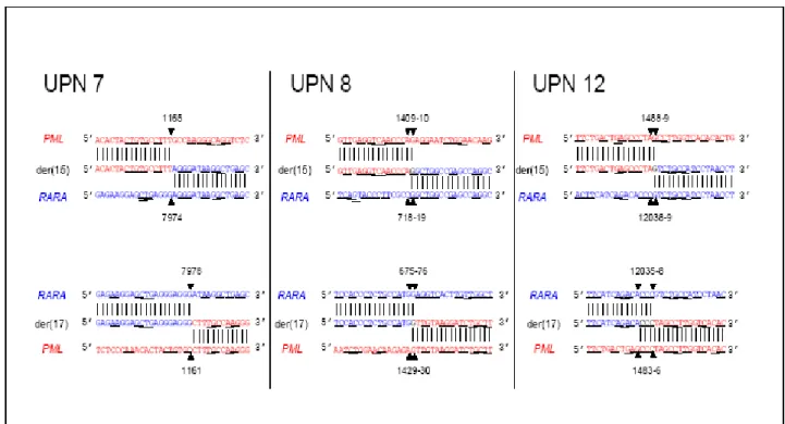
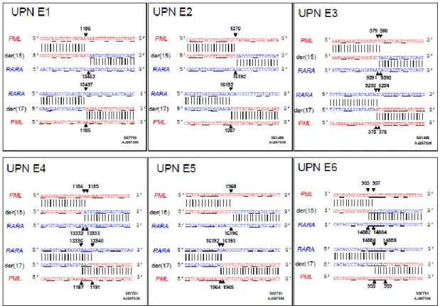
Figure S2. Locations of t-APL breakpoints in multiple sclerosis patients exposed to mitoxantrone. Der(15) and der(17) genomic breakpoint junctions in 3 cases. Native PML sequences are in red and RARA in blue. Vertical lines indicate sequences from the derivative chromosomes, and horizontal lines indicate regions of microhomologies consistent with DNA repair of chromosomal breaks by the non-homologous end joining pathway. Homologies prevented determining precise localization of breakpoint positions (black font).
TableS1: PML and RARA homologues provided the substrate for in vitro topoisomerase II cleavage assays
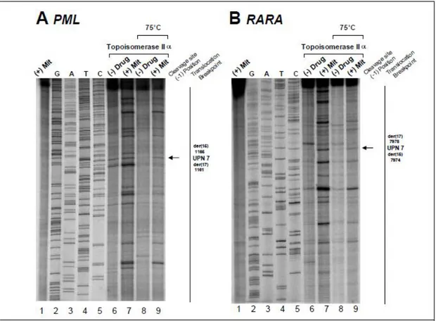
Figure S3: Investigation of t(15;17) translocation mechanism in UPN 7 by in vitro topoisomerase IIα DNA cleavage assay. Cleavage results of PML (A) and RARA (B) translocation breakpoints in the t-APL case of UPN 7. Lanes 1-9 of each cleavage assay are as previously described in the manuscript Figure 3. The location of the relevant heat stable cleavage sites are indicated by an arrow on the far right.
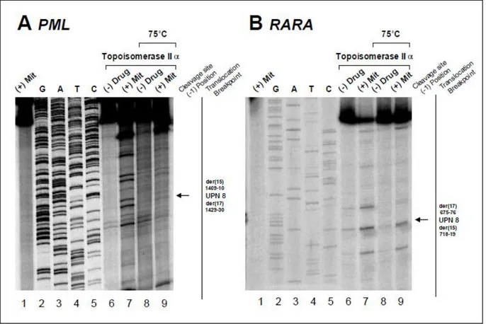
Figure S4. Investigation of translocation mechanism in UPN 8 by in vitro topoisomerase IIα DNA cleavage assay. Cleavage data using PML and RARA substrates corresponding to the locations of UPN 8 breakpoints previously identified. Lanes are as previously described, with relevant heat stable cleavage bands indicated by arrows on the far right.
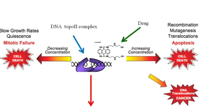
Figure S5: The working model of DNA topoisomerase IIα cleavage assay. Normal homologues encompassing translocation breakpoint regions were end labelled, incubated with human DNA topoisomerase II, ATP, and mitoxantrone. Cleavage complexes were irreversibly trapped upon the addition of SDS (sodium dodecyl sulfate), purified, and resolved in a polyacrylamide gel alongside sequencing to map the sites of cleavage precisely, allowing analysis of the position of the cleavage sites with respect to translocation breakpoint sites. Levels of cleavage complexes are maintained in a critical balance. When levels drop below threshold concentrations, daughter chromosomes remain entangled following replication. As a result, chromosomes cannot segregate properly during mitosis and cells die as a result of catastrophic mitotic failure. When levels of cleavage complexes rise too high, cells also die, but for different reasons. Accumulated topoisomerase II–DNA cleavage intermediates are converted to permanent strand breaks when replication forks, transcription complexes or DNA tracking enzymes such as helicases attempt to traverse the covalently bound protein ‘roadblock’ in the genetic material . The resulting collision disrupts cleavage complexes and ultimately converts transient topoisomerase II-associated DNA breaks to permanent double-stranded breaks that are no longer tethered by proteinaceous bridges. The resulting damage and induction of recombination/repair pathways can trigger mutations, chromosomal translocations and other aberrations.
30