Ai miei nonni , alla mia famiglia
Quando tutte le più belle sinfonie sono state già scritte per un musicista l’ambizione più grande è quella di cambiare una sola nota.
Corso di Dottorato in Neuroscienze di Base e dello Sviluppo
Presidente: Prof. Giovanni Cioni“Environmental Enrichment: effects on the visual
system of an animal model of Retinitis pigmentosa
(RP)”
Candidata Tutor
Dott.ssa. Ilaria Barone Dott.ssa Enrica Strettoi
XXIV CICLO (2009-2011)
SSD BIO/09
SUMMARY
1
RIASSUNTO
1
2
ABSTRACT
3
3
INTRODUCTION AND AIMS OF THE THESIS
5
3.1 The retina 8
3.1.1 Anatomical organization of the retina 8
3.2 The architecture of the mouse retina 11
3.2.1 Photoreceptors 14
3.3 Genetics 16
3.3.1 Genetic mutations affecting photoreceptors 16
3.3.2 Retinitis pigmentosa (RP) 16 3.3.3 Genetics and molecular mechanisms of RP 18
3.3.4 Pdeb mutations and the rd10 mutant mouse 20
3.4 Therapeutic Approaches 23
3.4.1 Therapeutic strategies for RP 23
3.4.2 Vitaminotherapy 24 3.4.3 Pharmacological treatments 25
3.4.4 Artificial devices 26 3.4.5 Gene therapy 30 3.4.6 Optogenetic approach 32
3.4.7 Cell or tissue transplantation 32
3.4.8 Neurotrophic factors 35
3.5 Enviromental Enrichment (EE) 37
3.5.1 Effects of EE on visual system development 38 3.5.2 Maternal enrichment and visual-system development 40
3.5.3 EE influences retinal development 41
3.5.4 EE and pathology 43
4
MATERIALS AND METHODS 44
4.1 Animals 44
4.2 Rearing environments 44
4.2.1 Standard environment 44 4.2.2 Enriched Environment 45
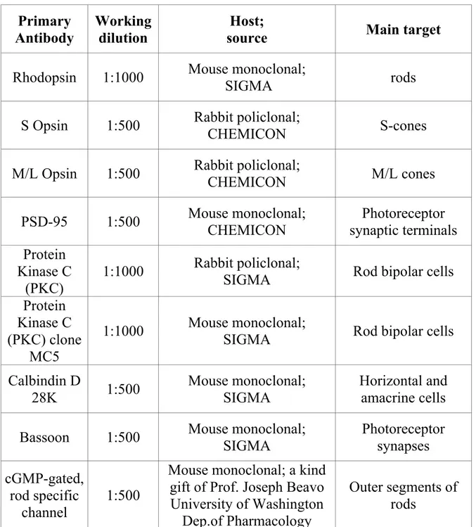
4.4 Immunofluorescence: cone counting 47 4.5 Fluorescence microscopy on retinal vertical sections 48
4.6 Western blot 51
4.7 qPCR 52
4.7.1 Template preparation 54 4.7.2 Determining concentration and purity of nucleic acids 54
4.7.3 Removal of genomic DNA contamination and cDNA synthesis 54
4.7.4 qPCR 54 4.7.5 Detection of a correct amplicon on gel 55
4.7.6 Data analysis 55
4.8 Behavioral test: visual acuity and contrast sensitivity 57 4.9 Electroretinogram recordings (ERG) 60
5
RESULTS 61
5.1 Early EE retards the onset of rod and cone loss during
weaning 61
5.1.1 Photoreceptor survival in EE: pycnotic photoreceptors 61 5.1.2 Photoreceptor survival in EE: quantitative analysis and morphology 62
5.1.3 Photoreceptors survival in EE: cones 65 5.1.4 Inner retina in EE: morphology 67 5.1.5 Photoreceptor survival in EE: western blot. 71
5.1.6 Retinal physiology in EE: electroretinogram recordings 73 5.1.7 Visual system physiology in EE: behavioral tests 74
5.2 EE might change the expression of specific mRNAs in
retinas of adult rd10 mice: preliminary data 77
5.3 Long term effects of EE 80
6
DISCUSSION 84
6.1 Early EE retards the onset of rod and cone loss during
weaning 84 6.2 EE promotes cone survival in RP adult mutant animals 84
6.2.1 Photoreceptor survival in EE: morphology 85 6.2.2 Photoreceptors survival in EE: cones 86 6.2.3 Inner retina in EE: morphology 87 6.2.4 Photoreceptor survival in EE: western blot. 88
6.2.5 Retinal physiology in EE: electroretinogram recordings 88 6.2.6 Visual system physiology in EE: behavioral tests 89
6.2.7 EE and neurotrophins mRNA expression 90
6.3 EE effects endure in the retinas of aged mutant animals 94
6.3.2 Inner retina in old EE: morphology 94 6.3.3 Visual system physiology in old EE: behavioural test 95
7
CONCLUSIONS AND SPECULATIONS 96
8
BIBLIOGRAPHY 99
9
PUBBLICATION LIST 109
10
ABSTRACT LIST 109
1 RIASSUNTO
La Retinite Pigmentosa (RP) comprende una famiglia di degenerazioni ereditarie eterogenee (Kennan et al. 2005) (Paskowitz et al. 2006) caratterizzate dall’iniziale morte apoptotica dei bastoncelli, con sviluppo di insufficiente capacità visiva in condizioni di scarsa luminosità e progressivo restringimento concentrico del campo visivo, cui segue la degenerazione dei coni che comporta il declino dell’acuità visiva, fino alla cecità. Sebbene la causa primaria della RP sia tipicamente un difetto di un gene specifico dei bastoncelli, la degenerazione di queste cellule attiva una cascata di eventi che portano sia alla morte secondaria dei coni che al cosiddetto “remodeling” progressivo dei neuroni della retina interna (Marc et al. 2003). Nella patologia umana come nei suoi modelli animali, questi eventi seguono un pattern spazialmente irregolare sulla superficie retinica, rendendo difficile un’analisi quantitativa della sopravvivenza e della morte cellulare.
Lo scopo principale di questa tesi è quello di far luce sugli effetti precoci e tardivi dell’applicazione del paradigma sperimentale dell’Arricchimento Ambientale (AA) sulla degenerazione dei bastoncelli, sulla sopravvivenza dei coni e sulla morfologia dei neuroni interni della retina del mutante rd10, un modello murino di RP caratterizzato dalla progressiva degenerazione dei fotorecettori retinici secondo un pattern simile a quello che avviene nell’uomo affetto da analoghe mutazioni. L’AA è un paradigma di stimolazione motoria, sensoriale e sociale ampiamente usato come strategia non invasiva di neuroprotezione in diversi modelli di patologie neurologiche, incluse la malattia di Alzheimer e la malattia di Huntington. In questa tesi vengono riportati per la prima volta i risultati dell’esposizione prolungata di topi rd10 ad AA.
Attraverso studi di microscopia del tessuto retinico, registrazioni elettroretinografiche, test comportamentali e analisi molecolari, viene mostrato che l’AA dalla nascita è in grado di preservare in modo
considerevole la morfologia, la fisiologia dei fotorecettori e la percezione visiva negli animali rd10. Studi preliminari suggeriscono che l’effetto protettivo dell’AA possa essere mediato da fattori neurotrofici, tra i quali il CNTF.
Questi risultati possono incoraggiare lo sviluppo di nuove strategie applicative in cui la stimolazione ambientale, da sola o in combinazione con opportuni trattamenti farmacologici, può promuovere la conservazione e l’integrità funzionale dei neuroni retinici.
2 ABSTRACT
Retinitis pigmentosa (RP) is a family of inherited disorders leading to progressive photoreceptor death and is one of the major causes of genetic blindness in the world with no cure yet (Kennan et al. 2005) (Paskowitz et al. 2006). In typical RP, first rods die out, as usual for a mutation in rod-specific genes, and night blindness occurs. Later, secondary degeneration of cones follows until all the useful sight is lost. Both in human RP and in animal models of this disease, photoreceptor death is accompanied and followed by gradual remodeling of the inner retina. Both photoreceptor degeneration and secondary remodeling of the inner retina occur with an irregular pattern over the retinal surface, making quantitative analysis of survival and death difficult.
The general aim of this Thesis is investigate the early and late effects of an Environmental Enriched paradigm upon rod degeneration and viability of cones and inner retinal neurons in a mouse model of RP. Environmental enrichment (EE), a neuroprotective strategy based on enhanced motor, sensory and social stimulation, has already been shown to exert beneficial effects in animal models of various neurological disorders, including Alzheimer and Huntington disease. Here, I report the results of a prolonged exposure of rd10 mice, a mutant strain undergoing progressive photoreceptor degeneration and used to model human RP, to such an enriched environment from birth.
By means of microscopic examination of retinal tissue, electrophysiological recordings, visual behavior assessment and molecular analysis, I show that EE considerably preserves retinal morphology and physiology as well as visual perception over time in rd10 mutant mice. The protective effects of EE are likely mediated by increased production of endogenous protective molecules, including CNTF.
The therapeutic option I applied in this Thesis produced strikingly positive results, also in comparison to other strategies applied to the same animal model. This work opens the exciting possibility that non invasive manipulations of the outer environment can be used, alone or in combination with other treatemtns, to prolong the lifespan of retinal neurons otherwise doomed to death.
3 INTRODUCTION AND AIMS OF THE THESIS
The mature vertebrate retina consists of five neural cell types that have common specializations throughout species.
Photoreceptors are the neurons containing the light-sensitive photo pigment that captures light. These cells pass the information through the entire retina that encodes spatio-temporal and chromatic information transmitted to other parts of the visual processing centers. The basic retina anatomo-physiology involves two kinds of circuitry: the vertical or “through” pathway, which includes photoreceptors, bipolar cells and ganglion cells, and the horizontal or “lateral” pathway, which is made by horizontal cells and amacrine cells. In the mammalian rod pathway, some subtypes of amacrine cells have a function similar to bipolar cells.
At first look, the spatial arrangement of retinal layers seems counter-intuitive, because light rays have to pass through various non-light-sensitive elements of the retina as well as the retinal vasculature before reaching the outer segments of the photoreceptors, where photons are absorbed. The reason of this curious feature of retinal organization lies in the special relationship that exists among the outer segments of the photoreceptors, the pigment epithelium, and the underlying choroid. The disks present in the photoreceptor outer segments are formed near the inner segment of the photoreceptor and move toward the tip of the outer segment, where they are shed. The pigment epithelium plays an essential role in removing the exhausted receptor disks. In addition, the pigment epithelium contains the biochemical machinery that is required to regenerate photo pigment molecules after they have been exposed to light. Finally, the capillaries in the choroid underlying the pigment epithelium are the primary source of sustenance for retinal photoreceptors. These functional considerations explain why rods and cones are found in the outermost rather than in the innermost layer of the retina. They also explain why disruptions in the normal relationships between the choroids, pigment
epithelium and retinal photoreceptors, such as those that occur in some forms of Retinitis pigmentosa, have severe consequences for vision.
Retinitis pigmentosa (RP) refers to a heterogeneous group of hereditary retinal disorders characterized by progressive vision loss due to a gradual degeneration of photoreceptors. An estimated 100,000 people in the United States have RP. In spite of the name, inflammation is not a prominent part of the disease process; instead the photoreceptor cells appear to die by apoptosis (determined by the presence of DNA fragmentation). Multiple mutations have been found, and the heterogeneity of RP at all levels, from genetic mutations to clinical symptoms, has important implications for understanding the pathogenesis of the disease and designing therapies.
Given the complex molecular etiology of RP, it is unlikely that a single cellular mechanism will explain the disease in all cases. Despite the specific mutation or causal sequence, the major vision loss in RP patients is due to the gradual degeneration of cones. Typically, the mutated protein causing RP is not expressed in cones and the loss of these cells is an indirect result of a rod-specific mutation. Hence, understanding and treating this disease presents particularly difficult challenges. Recently, there is increasing excitement about the possibility of using gene therapy to substitute the rod defecting gene, and multiple groups are developing vectors encoding growth factors (Schlichtenbrede et al. 2003; Yang 2009) or antioxidant enzymes (Li et al. 2008; Koilkonda and Guy 2011) to retard the secondary loss of photoreceptors due to a lack of tropic factor or an excessive oxidative stress. There are also other therapeutic strategies under development, such as the optogenetic and the stem cell approaches, or the use of nanoparticles to deliver genes or proteins (Berson et al. 1993; Busskamp and Roska 2011). The endurance of these experimental designs and their effective applicability to RP will certainly be tested in the near future. It is important to recall that combinations of approaches are likely to be more powerful than any individual treatment (Yao et al. 2011).
The main aim of the present Thesis was to investigate the non invasive approach of Enviromental Enrichment (EE) as a strategy to retard the secondary loss of cones in a well known mouse model of RP, the r10 mutant mouse. The rationale is that it is well known that through sensory, cognitive and motor stimulation it is possible to enhance the capability of the central nervous system to respond to different pathological conditions (Nithianantharajah and Hannan 2006; Baroncelli et al. 2010).
The working hypothesis is that EE, a paradigm whereby the animals are raised and kept in large cages and social groups with additional toys specifically devoted to multi-sensory, cognitive and motor stimulation, can retard the secondary events due to the loss of rod photoreceptors in rd10 mutant mice, delaying the degeneration of cones. To test this hypothesis, rd10 mutant mice were kept and grown in either EE conditions or in Standard laboratory (ST) conditions from birth.
More specifically, in this Thesis we focused on two specific aims:
Aim 1: to asses morphological, molecular and functional effects of EE in adult rd10 mutant mice.
To achieve this aim we:
• Compared retinal morphology in ST mice and EE mice using quantitative immunostochemistry to analyze in detail photoreceptor morphology and number, as well as the morphology of the inner retina. • Compared the expression of photoreceptor specific proteins by means
of western blot analysis.
• Compared the viability of photoreceptors by means of electroretinogram (ERG) recordings.
• Compared the visual performance of mice by means of a visual behavioral test used to measure both visual acuity and contrast sensitivity.
• Compared the expression of different trophic factor mRNAs by means of Real Time RT-PCR in ST and EE retinas.
Aim 2: to assess the long run beneficial effects of EE in 1 year old rd10 mutant mice.
To achieve this aim we:
• Compared retinal morphology in ST and EE mice using immunostaining techniques to analyze photoreceptor morphology and number in the inner retina.
• Compared the visual performance of mice by means of a visual behavioral test used to measure both visual acuity and contrast sensitivity.
3.1 The retina
3.1.1 Anatomical organization of the retina
The eye is a sphere filled of fluid surrounded by three layers of tissue. The inner layer of the eye, the retina, contains neurons that are sensitive to light and are capable of transmitting visual signals to central targets. Despite its peripheral location, the retina is part of the central nervous system (CNS).
During the embryonic development, the vertebrate retina and the optic nerve
form as an out pocketing of the diencephalon, called the optic vesicle, which invaginates forming the optic cup. The inner coat of the optic cup gives rise to the retina, while the outer one gives rise to the retinal pigment epithelium. This is a thin melanin-containing layer that reduces backscattering of light that enters in the eye; it also plays a critical role in the maintenance of photoreceptors, renewing photo pigments and phagocyting the photoreceptor disks.
Compared to other portions of the CNS, the retina comprises fewer classes of neurons, and these are arranged in a manner that has been less difficult to unravel than the circuits in other areas of the brain. There are, at
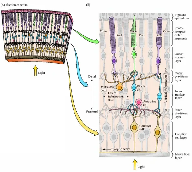
all, five major classes of retinal neurons: photoreceptors, bipolar cells, horizontal cells, amacrine cells and ganglion cells. In the adult retina, these are organized into three main cell body layers that are separated by synaptic terminals (plexiform layers) (Fig. 1). Light passes through the lens and across the whole retinal thickness to stimulate photopigments in the outer segments of rod and cone photoreceptors, which are located adjacent to the Retinal Pigment Epitelium (RPE). The photoreceptors, whose cell bodies costitute the outer nuclear layer (ONL), form synapses in the outer plexiform layer (OPL) with horizontal cells and bipolar neurons, whose cell bodies are located in the inner nuclear layer (INL). Bipolar cells synapse directly or indirectly via amacrine neurons in the inner plexiform (IPL) layer to ganglion cells (GCs) located in the GC layer. GCs are the projection neurons of the retina and transmit visual information via their axons through the optic nerve to visual processing centers in the brain. Amacrine cell bodies are located in the inner part of the INL and comprise roughly half of the cells in the GC layer. The retina also contains a population of radial glial cells called Muller glia, whose cell bodies are located in the INL. Their apical processes form adherents junctions with photoreceptors at the outer limiting membrane and their end feets attach to the basal lamina on the vitreal surface of the retina at the inner limiting membrane. Retinal astrocytes, which develop from a separate cell lineage at the optic disc, form a dense network on the vitreal surface of the retina and are closely associated with the vasculature. Microglial cells, of mesodermal origin, also reside permanently in the retina.
The combination of highly specialized cell types in well-organized networks performing complex modulatory activities results in an amazing and flexible sensory processing system and makes the retina a unique model for the comprehension of the relationship between structure and function inside the CNS.
Fig. 1. Structure of the retina. (A) Section of the retina showing the overall arrangement of retinal layers. (B) Diagram of the basic circuitry of the retina. A three-neuron chain photoreceptor, bipolar cell, and ganglion cell provides the most direct route for transmitting visual information to the brain. Horizontal cells and amacrine cells mediate lateral interactions in the outer and inner plexiform layers, respectively. The terms inner and outer designate relative distances from the center of the eye (inner, near the center of the eye; outer, away from the center, or toward the pigment epithelium). From Purves 3ed 2004.
3.2 The architecture of the mouse retina
The mouse retina resembles that of other vertebrates, so that in a vertical section the following layers and cells can be recognized:
¾ Photoreceptor layer: formed by the outer and inner segments of rods and cones;
¾ Outer Limiting Membrane (OLM);
¾ Outer Nuclear Layer (ONL): constituted by photoreceptor cell bodies; ¾ Outer Plexiform Layer (OPL) where processes of photoreceptors,
bipolar and horizontal cells are synaptically connected;
¾ Inner Nuclear Layer (INL): formed by cell bodies of bipolar, horizontal, amacrine, and Müller glial cells;
¾ Inner Plexiform Layer (IPL) where processes of bipolar, amacrine and ganglion cells synaptically connect;
¾ Ganglion Cell Layer (GCL), containing the cell bodies of ganglion cells and displaced amacrine cells;
¾ Optic nerve Fiber Layer (OFL), constituted by axonal bundles of ganglion cells and supporting astrocytes;
¾ Inner Limiting Membrane (ILM).
The fundamental plan of the mouse retina follows the blue-print common to all mammalians: rods represent the majority of the photoreceptor population; however, the mouse retina is particularly rod-dominated and rods sum up to 97% of all the photoreceptors (Jeon et al. 1998). In the C57B16/J strain of mouse, used for the experiments described thereafter, there are approximately 6.4 million rods and 180,000 cones (Eye, Retina, and visual system of the mouse; chapter 12).
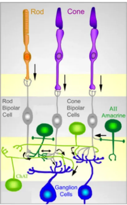
Fig. 2. Schematic representation of the main connections among retinal neurons. The physiology of the retina can be simplified into 4 main processing stages: photoreception, transmission to bipolar cells, transmission to ganglion cells and transmission along the optic nerve. At each synaptic stage there are also laterally connecting horizontal and amacrine cells.
Figure 2 summarizes the main connections among retinal neurons. Photoreceptors - rods and cones - convert light information into electrical and then chemical signals relayed to inter-neurons in the outer retina. At low light levels, only rods have sufficient sensitivity to capture the few photons that are available. Cones permit day and color vision. Photoreceptors are connected to each other by gap junctions (electrical synapses) and make chemical synapses onto bipolar and horizontal cells. Two types of bipolar cells, rod and cone bipolars, are postsynaptic to the homologous photoreceptor populations. Cone bipolar cells are further divided in two functional classes, called “ON” and “OFF” that respectively depolarize and hyperpolarize in response to the illumination of their receptive field centers. Rod bipolar cells belong to the single functional category of ON cells. Bipolar cells are then connected to RGCs to the same type (ON or OFF): this connection can be direct as in the
case of cone bipolar cells, or through a chain of neurons comprising dedicated amacrine cells, as for rod bipolar cells.
Differently from most mammals that have at least two horizontal cell types, mice have only one type of horizontal cells (Peichl and Gonzalez-Soriano 1994). Horizontal cells contribute to enhance contrast between adjacent light and dark regions. This is because they are constituted by two different, functionally distinct portions, by which they can control bipolar cell sensitivity: the dendritic arborization makes synapses with cones, while the axonal arborization is linked to rods. The connection horizontal cells - photoreceptors is bidirectional: not only horizontal cells are post- synaptic to rods, but also pre-synaptic to cones on which they exert a negative feed-back. Amacrine cells (more than 30 types) modulate signals from bipolar cells by supplying inhibition directly onto ganglion cells; moreover, they modulate transmitter release from bipolar cells. Light information leaves the retina and reaches other stations in the brain via axons of RGCs (the only exit neurons of the retina) that collectively form the optic nerve. The commonly employed C57Bl6/J mouse retina contains approximately 50,000 ganglion cells. Most of them project contralaterally, since their axons cross at the optic chiasm; only less than 5% of all ganglion cells project ipsilaterally. Beside these above mentioned five classes of neurons, the retina contains one type of macroglial cells, the Müller cells. These have a radial arrangement spanning the depth of the retina and provide important structural and functional support to retinal neurons.
Within the above described basic organization, many specialized sub-circuits are present in the vertebrate retina: they work both in sequence and in parallel to process different features of the visual image such as luminosity, contrast, chromatic composition and direction of motion (Weng et al. 2005) .
3.2.1 Photoreceptors
The two types of photoreceptors, rods and cones, are distinguished by shape (from which they derive their names), the type of photo pigment they contain, the distribution across the retina, and the pattern of synaptic connections. These properties reflect the fact that the rod and cone systems (both the receptors and their connections within the retina) are specialized for different aspects of vision. The rod system has very low spatial resolution but is extremely sensitive to light; it is therefore specialized for sensitivity at the expense of resolution. Conversely, the cone system has very high spatial resolution but is relatively insensitive to light and it is specialized for acuity at the expense of sensitivity. The properties of the cone system allow humans and many other animals to see colors. The rod-mediated perception is called scotopic vision because at the lowest levels of light, only the rods are activated.
Although cones begin to contribute to visual perception at about the level of starlight, spatial discrimination at this light level is still very poor. As illumination increases, vision becomes more and more dominated by cones and they are the major determinant of perception under relatively bright conditions such as normal indoor lighting or sunlight. The contribution of rods to vision drops out nearly entirely in the so called photopic vision, because their response to light saturates. This means that the membrane potential of individual rods no longer varies as a function of illumination because all of the light-sensitive channels are closed. Mesopic vision occurs in levels of light at which both rods and cones contribute, for example at twilight.
The ability of rods and cones to respond to different ranges of the light absorption spectrum is due to differences in their photo pigments. The spectral sensitivity of mouse rod photoreceptors peaks at 497-500nm (Soucy
et al. 1998; Toda et al. 1999; Fan et al. 2005). In the mouse retina, there are two types of cone photoreceptors: one has peak sensitivity at 360nm (UV light), while the other has peak sensitivity at 508 nm ((M)-wavelength). Most cones express both UV and M photopigments but their maximum sensitivity is at 508 nm (Nikonov et al. 2006). Another difference between the two types of photoreceptors is their sensitivity to light intensity. Rods produce a reliable response to a single photon of light, whereas more than 100 photons are
required to produce a comparable response in a cone (Eye, Retina and visual
system of the mouse; chapter 12). Also, the response of an individual cone does not saturate at high levels of steady illumination, as does the rod response. Although both rods and cones adapt to operate over a range of luminance values, the adaptation mechanisms of cones are more effective. This difference is apparent in the time course of the response of rods and cones to light flashes. The response of a cone, even to a bright light flash that produces the maximum change in photoreceptor current, recovers in about 500 milliseconds, more than four times faster than rod recovery.
The arrangement of the circuits that transmit rod and cone information to retinal ganglion cells also contributes to the different characteristics of scotopic and photopic vision. In most parts of the retina, rod and cone signals converge on the same ganglion cells; i.e., individual ganglion cells respond to both rod and cone inputs, depending on the level of illumination. The early stages of the pathways that link rods and cones to ganglion cells, however, are largely independent. The pathway from rods to ganglion cells involves a distinct class of bipolar cell (rod bipolar) that, unlike cone bipolar cells, does not contact retinal ganglion cells. Rod bipolar cells synapse with the dendritic processes of a specific type of amacrine cell (the AII amacrine) that makes gap junctions and chemical synapses with the terminals of cone bipolars; these processes, in turn, make synaptic contacts on the dendrites of ganglion cells in the IPL. As a consequence, the circuits linking the rods and cones to retinal ganglion cells differ a) in the number of interposed neurons and b) in
their degree of convergence. Each rod bipolar cell is contacted by a large number of rods, and many rod bipolar cells contact a given amacrine cell. In contrast, the cone system is much less convergent and thus less sensitive.
3.3 Genetics
3.3.1 Genetic mutations affecting photoreceptors
A very high number of genetic mutations affect the eye. Those occurring in photoreceptor- or pigment epithelium -specific genes often cause Retinal Degenerations (RDs), a family of inherited dystrophies characterized by photoreceptor dysfunction and death. It is estimated that more than 15 million people worldwide have vision loss due to inherited forms of RD. These include patients suffering from Retinitis pigmentosa (RP), a disease for which there is no cure yet (Chader 2002).
3.3.2 Retinitis pigmentosa (RP)
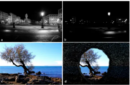
One of the common subtypes of inherited forms of photoreceptors degeneration is Retinitis pigmentosa (RP), which is the second main cause of blindness in 20-64 years olds (Wright et al. 2010). RP represents an heterogeneous group of genetic disorders that lead to loss of vision. Typically, rods, the first cells affected, start to die (as a result of a genetic abnormality) and patients experience night-vision limitations and develop visual field constriction (or tunnel vision) (Fig. 3d). Secondary cone degeneration eventually affects central vision, leading to blindness. RP cause severe visual impairment in as many as 1.5 million patients worldwide (Kennan et al. 2005; Mendes et al. 2005) (Paskowitz et al. 2006).
Fig. 3. Typical examples of night blindness (b) and tunnel vision (d). This is a typical example of eye field degradation in RP patients. Normal night vision (a) and a normal day vision (c) in human.
The progressive demise of photoreceptors also precipitates other pathological symptoms in the retina, including the attenuation of the retinal vasculature and the accumulation of intra-retinal pigment deposits, from which the disease derives its name (Fig. 4).
Fig. 4. Example of fundus views of the retina of a RP patient. A typical brown pigmentation is present in the pathological condition. Pigment deposits, named bone spicules for their typical shape, are responsible for the name of the disease. A clear attenuation of retinal blood vessels is also visible. From:Web Vision.
There is considerable variation in the severity of the conditions of the RP patients: it is unusual for them to become totally blind, while most of them retain some useful vision well into old ages. Classic clinical findings of RP include: bone spicule pigmentation or pigment clumping, retinal arteriolar narrowing, waxy pallor of the optic nerve, epiretinal membrane formation, atrophy of the RPE and choriocapillaris (starting at the mid periphery of the retina with preservation of the RPE in the macula until late in the disease), posterior subcapsular cataract, epiretinal membrane formation, and cystoid macular edema (CME) (Hamel 2006).
3.3.3 Genetics and molecular mechanisms of RP
The genetics of RP is complex: approximately 180 mutations in more than 40 genes have been identified as the cause of different forms of RP. In a segregation analysis of RP, families were divided as autosomal dominant (24%), autosomal recessive (41%) and X-linked (22%), and the remaining 12% of cases were supposed to result from genetic factors, non-Mendelian inheritance (for example, mitochondrial or de novo mutations) or complex inheritance (Wright et al. 2010). The large number of mutations causing RP comprise insertions, deletions, or substitutions, in turn producing missense mutations or truncations (Wang et al. 2005). RP is caused preferentially by different single gene mutations including the following: GTPase regulator (RPGR: 10-20% of cases) rodopsin (RHO: 8-10% of cases) and usherin (USH2A: ≈3% of cases) (Jos´e M. Mill´an and Ascensi ´on Gimenez-Pardo 2011). Some of these genes encode for proteins of the phototransduction pathway, or play a role in the maintenance of cellular structure or in the transcriptional control of other photoreceptor genes (Kennan et al. 2005; Mendes et al. 2005).
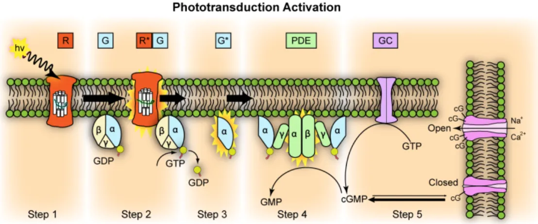
Here I will describe one mutation of the phototransduction cascade: the Pdeb mutation that might cause autosomal recessive RP (arRP) and is also carried by the mouse model used in this study.
Fig. 5. Phototransduction activation. The enzyme phosphodiesterase (Pde) comprises different subunits (alpha, beta, gamma and delta). Mutations in each of these subunits can lead to RP. From: Wikipedia.
3.3.4 Pdeb mutations and the rd10 mutant mouse
For over 30 years, the retina of rodents has provided an invaluable tool to study the dynamics and mechanisms of inherited RD, as mouse photoreceptors undergo dystrophies caused by spontaneous DNA mutations, closely related to those of humans. The so-called rod-less mice (Keeler 1924) expresses a nonsense mutation in the Pdeb gene coding for the β-subunit of cGMP phosphodiesterase of rods. This spontaneous mutation later named rd1 (rd1: Pde6brd1) (Pittler et al. 1993) was subsequently found in humans with arRP (McLaughlin et al. 1993; McLaughlin et al. 1995) and had forms of night blindness (Gal et al. 1994).
There is a surprisingly high degree of evolutionary stability for mutations that cause loss of vision in mice, in fact many of the commonly used inbred strains, including C3H and its derivatives and CBA/J, are homozygous for rd1. In particular, two mutations in the Pdeb gene of rd1 mice have been identified: besides the nonsense mutation that causes truncation of the protein (Bowes et al. 1990), the intronic insertion of an endogenous mouse leukemia virus (Xmv28) causes an incorrect splicing. Xmv28 has also been found in strains derived from wild type mice, as well as from inbred laboratory strains not known to have recent common origins (Bowes et al. 1993). Evidently, there is little or no selective pressure to maintain a functional β-PDE gene, and therefore vision, in these nocturnal animals.
Biochemical studies comparing retinas from normal and rd1 mice have show that the lack of cGMP-PDE activity causes a dramatic increase in cytoplasmic cGMP concentration (Farber and Lolley 1974). The Pdeb gene-triggered cell death alters the expression of genes involved in diverse cellular pathways including Ca2+ homeostasis, catabolism, neuro-inflammation and tissue remodeling (as the blood-retina barrier undergoes a breakdown). In mouse photoreceptors, normal Ca2+ levels range between ~250 nmol/liter (in
complete darkness) and ~60 nmol/liter (in light) (Woodruff et al. 2002). However, in the rd1 photoreceptors, Ca2+ levels are increased up to ~190% over wt levels (Fox et al. 1999). Consequently, messages for Ca2+-binding proteins, and in particular Ca2+ sensors such as calbindin, are up-regulated in the rd1 photoreceptors. So far, the focus has been on Ca2+ as major trigger of the apoptosis cascade, while the role of high cGMP levels remains unanswered yet.
The animal model we used for our studies is another example of autosomic recessive RP, isolated at the Jackson Laboratories in the USA (Chang et al. 2002), and namely the rd10 (retinal degeneration 10) mouse. Genetic analysis shows that this strain carries an autosomal recessive mutation that maps to mouse chromosome (Chr) 5. Sequence analysis shows that the retinal degeneration is caused by a missense point mutation in exon 13 in the Pde6b gene of the β-subunit of the rod cGMP phosphodiesterase (β-PDE) gene (Pde6b). The mutation changes codon 560 from CGC to TGC resulting into an arginine to a cysteine change and in the loss of a CfoI site. The exon 13 missense mutation is the first known occurrence of a re-mutation in the Pde6b gene in mice and represents an additional model for studying the pathogenesis of autosomal recessive RP in humans (arRP). It may also provide a better experimental model for RP because of its later onset and milder phenotype compared to the more common rd1 mutant (Otani et al. 2004). In fact, in rd10 mutant mice, photoreceptor degeneration starts around P18 (about 10 days after the rd1 mutation), when retinal development is far more advanced. Rod death follows a central to periphery gradient; then a slower degeneration of cones occurs. A scattered population of aberrant cones persists up to at least 9 months of age. Atrophic retinal vessels are found at four weeks of age, consistent with retinal degeneration (Chang et al. 2007; Gargini et al. 2007). Electroretinograms (ERGs) of rd10 mice are never normal, but rod and cone ERG a- and b-waves can be measured at P18 while there is no more response at three months of age and later. Interestingly,
rearing rd10 mice in total darkness delays degeneration for at least a week, after which morphological and functional loss progresses irregularly (Chang et al. 2002).
As in other models of retinal degeneration (Chang et al. 1993; Reme et al. 1998), death of photoreceptors in rd10 mice shows the hallmarks of apoptosis: nuclei in the ONL are picnotic and some of them express a form of activated Caspase 3. Apoptotic markers appear in the retina in a centre to periphery gradient.
Secondary changes also affect inner retinal cells. Modifications in their morphology become detectable at around one month of age, and are more widespread at 2 months. Mostly, changes affect rod bipolar and horizontal cells. Their dendritic complement becomes progressively more scant and disorganized ultimately regressing completely. Cone bipolar cells follow, at a slower rate. Dendritic retraction in bipolar cells which carry glutamate mGluR6 receptors is preceded by a decrease in the immunoreactivity for this marker, and by its displacement to the cell bodies and bipolar cell axons in the INL. Morphological changes in bipolar and horizontal cells occurring in the rd10 retina are followed by their death partial death. Other cell types, such as AII and cholinergic amacrine cells, do not seem to be affected in the first 5 months of life (Gargini et al. 2007).
3.4 Therapeutic Approaches
3.4.1 Therapeutic strategies for RP
At present, there is no therapy able to stop the evolution of RP or to restore vision loss in this disease. However, there are several therapeutic strategies aimed at slowing down retinal degeneration or at supplying the loss of cells in this tissue.
In general, prenatal diagnosis can be useful in families in which the responsible gene has been identified, particularly for early onset and severe forms of RP. For other cases, clinical evidence and data from animal studies indicate that some genetic types of RP are partly light-dependent (Wang et al. 1997). Thus, patients with RP are recommended to wear dark glasses outdoor, or yellow-orange spectacles minimizing photophobia. Beside therapies aimed at preserving visual function and preventing cell death, researchers experience the tremendous challenge of trying to restore visual function. However, because mammalian photoreceptors are neurons and do not divide after birth, their loss is irreversible. In addition, the loss of photoreceptors leads to a dramatic remodeling of the inner retina which would probably modify the visual information process. Nevertheless, numerous groups are now working to achieve visual restoration either by photoreceptor replacement or by using artificial prosthesis that substitute their function. A synthesis of the most representative clinical or experimental strategies to treat RP is given below.
3.4.2 Vitaminotherapy
Vitamins A and E seem to protect the photoreceptors because of their trophic and anti-oxidant effects, respectively. It has been shown that long term (5–12 years) vitamin A supplementation slightly slows down the loss in ERG amplitude, while vitamin E seems to have adverse effects. Indeed, based on a study of the natural course of RP, patients who happen to be taking vitamin A, vitamin E, or both were recorded to have slower declines in ERG amplitudes than those not taking such supplements (Berson et al. 1985; Berson et al. 1993). This observation prompted a randomized clinical trial of oral vitamin A and E supplements in 601 patients with dominant, recessive, and X-linked non-syndromic Retinitis pigmentosa and Usher’s syndrome type II. Participants were randomly assigned either daily vitamin A as retinyl palmitate15 000 IU, vitamin E 400 IU as dl-α-tocopherol, the combination, or trace amounts of both vitamins; follow-up was for 4–6 years. In this study patients assigned high-dose of vitamin A showed a significantly slowed decline in cone ERG amplitudes than the other groups; however, in a subgroup in which the effect was higher there was also a significant negative effect of vitamin E. These studies are still debated: vitamin A should not be given to RP patients with mutations in ABCA4 (Berson et al. 1993; Berson et al. 1993), a gene responsible for a subset of recessive Retinitis pigmentosa. Other mutant alleles of ABCA4 cause related diseases, recessive cone-rod dystrophy, and recessive Stargardt macular degeneration. A study on abca4 (-/-) mice indicates that vitamin A supplementation should be avoided in patients with ABCA4 mutations or other retinal or macular dystrophies associated with lipofuscin accumulation in the retinal pigment epithelium because it enhances this accumulation.
Another nutritional treatment assessed for patients with RP is based on docosahexaenoic acid (DHA), an omega-3 fatty acid found in high
concentrations in oily fish such as salmon, tuna, mackerel, herring, and sardines. DHA is apparently important for photoreceptor function, since membranes containing rodopsin and cone opsins in photoreceptor cells have very high concentrations of this fatty acid. In a recent study, patients were given docosahexaenoic acid (DHA) supplementation, in addition to vitamin A. It was shown that the RP degeneration of photoreceptors initially slowed down by the addition of DHA, but the beneficial effect did not last over 2 years (Berson et al. 2010).
Recently, Clemson et al. 2011 demonstrated that the use of retinoids and other small molecules as pharmacological chaperones increases the yield of properly folded RP mutant rhodopsins in heterologous cell culture. Also they tested whether other known small molecules can provide similar effects. Valproic acid (VPA) was identified as a potent inhibitor of histone deacetylase (HDAC) also effective increasing the levels of various neurotrophic factors. VPA and its derivative, divalproex sodium, is used off-label for a variety of indications including chronic pain syndromes, cancer therapy and schizophrenia. In their work Clemson et al. suggested that VPA may be an appropriate agent to treat patients with retinal dystrophies such as RP (Clemson et al. 2011).
3.4.3 Pharmacological treatments
Pharmacological treatments may be a good choice in those cases where some aspects of the pathophysiological mechanisms of RP are known. Pharmacological agents can compensate for a biochemical defect, and enhance or inhibit the activity of various effectors. Calcium-channel blockers have been tried in different animal models of RP (Nakazawa 2011) suggesting that calcium channel antagonists may inhibit photoreceptor apoptosis triggered by abnormally high levels of calcium. Recently an in vivo study on
rd10 mutant mice has demonstrated that photoreceptor loss can be slowed by pharmacologically inhibiting the rate limiting enzyme of the synthesis of ceramide, a known pro-apoptotic messenger (Strettoi et al. 2010).
3.4.4 Artificial devices
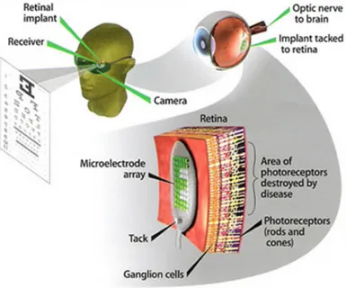
The development of artificial implants inserted in substitution of an entire part of the pathological retina represents an active line of investigation to treat both RP and other retinal diseases, such as macular degeneration. Micro-photodiodes arrays replacing degenerated photoreceptors or more sophisticated devices capturing light and stimulating retina, optic nerve or visual cortex have been developed. Several clinical trials have essentially demonstrated the tolerance of these electronic implants (Zrenner 2002). Some of these approaches have already improved the eyesight of patients with major visual impairments.
Fig. 6. Epiretinic implant: on a normal glasses is mounted a microcamera that supplies the signals, this signals are transmitted to the chip through microcable in the eyes. The chip gives energy to the electrodes in epiretinic space.
From:www.forumsalute.it/community/forum_65_oculistica/thrd_149058_un_microchip_i mpiantato_nella_retina_rida_la_speranza_chi_non_vede_1.html.
Of course, retinal prostheses are only effective if the visual pathway distal to the retinal implant is still intact and functional (Fig. 6). Two types of prostheses have been developed according to the site of retinal implant: epiretinal (on the surface of the retina) and subretinal (between the retina and the RPE) prostheses (Chow et al. 2004) (Hossain et al. 2005), briefly illustrated below subretinal prostheses contain microphotodiodes attached to microelectrodes. These implants, such as the artificial silicon retina (5000 microelectrodes), are placed in the subretinal space between the outer retina and retinal pigment epithelium (Fig. 6). The photodiodes are stimulated by light passing through the retina, and the resulting electric current excites adjacent retinal sensory neurons. The specifications for subretinal implants vary. For example, a typical device measures 50-100 µm wide, has a diameter of 2-3 mm, and carries microphotodiodes on a microelectrode array (Chow et al. 2004). These implants do not require an external electrical source as incident light is sufficient for stimulation (Zrenner 2002). Subretinal devices have been tested in animal experiments (Butterwick et al. 2009). Several species have shown tolerance to the implants, for up to 30 months of time. Histological evidence has shown no relevant changes in the architecture of the retina. Visual perception is improved in six human patients with RP who received an artificial silicon retina, including subjective improvement in appreciation of brightness, contrast color, movement, shape and visual field. Some patients show an improvement in visual acuity.
Two major advantages of subretinal prostheses are the utilization of existing forces between the neural retina and retinal pigment epithelium to maintain their position and the potential of a high spatial resolution, as they are positioned close to retinal nerve cells and can stimulate neurons by means of low electrical currents. The main disadvantages of subretinal implants include impaired nourishment of the inner retina due to the creation of a mechanical barrier between the outer retina and the choroid and the occurrence of trauma to the retina during implanting. These prostheses also
show poor dissipation of heat and therefore could damage the retina. The mechanism of action of these implants may be by direct stimulation of retinal neurons. A specific neurotrophic effect elicited locally onto photoreceptors by means of the surgical procedure cannot be excluded: a study of subretinal artificial silicon implants in rats showed a temporary protective effect on the retina, resulting in decreased degeneration of photoreceptors (Zrenner 2002).
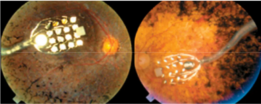
Epiretinal prostheses are composed of an array of electrodes implanted on the surface of the retina between the vitreous and the inner limiting membrane. The implants receive electrical signals from a camera positioned outside the body (Fig. 6 for more details; see also Fig. 7, for in vivo application). In one such device, the camera transmits light signals to a microchip within the camera. This microchip deciphers the signal and relays it, using wireless transmission, to a microchip in the epiretinal implant which in turn stimulates the RGCs (Zrenner 2002). Early studies in dogs and rabbits showed the flexibility of using epiretinal prostheses. A few clinical trials in humans have reported simple visual perception as phosphenes. Perception of light has been only reported by three “completely blind” patients with RP who received the so-called “second sight” model: these implants have survived for up to two years.
Fig. 7. Fundus photography showing an epiretinal electronic implant.
One advantage of epiretinal implants over subretinal devices is that the camera can process signals before they reach the implant: this allows optimization of the signal quality, which may lead to improved visual perception. Epiretinal implants can also use the heat-dissipating properties of the vitreous and are therefore less likely than subretinal implants to damage the retina. The superficial location of the implant reduces risks of trauma during implantation and allows for the implant to be replaced. The main disadvantages of epiretinal implants are the need for complicated microtechnology and surgical techniques that ensure secure fixation of the implant on the retina. The device also requires a higher electrical current than the subretinal implant (Lakhanpal et al. 2003). Implants are being developed to generate their own electrical currents on stimulation.
Some epiretinal prostheses used to attempt restoring sight in RP patients are designed to produce direct or near stimulation of RGCs. The success of such electronic devices is partly based upon the hope that RGCs are still viable after photoreceptors death. Yet, recent data show that some RP subjects lack phosphenes in respond to epiretinal stimulation (Delbeke et al. 2001). In addition, it has been reported that perceptual thresholds for electrical activation of the retina are surprisingly high in RP patients (Rizzo 2011). This is partially confirmed by the work of Stasheff with microelectrode arrays on RGCs in the rd1 mouse (Stasheff 2008). Here, RGCs have a much higher spontaneous frequency than normal, sometimes in rhythmic bursts reminiscent of epileptic discharges. A probable explanation could be that a strong remodeling of RGCs has occurred, and their membrane has become relatively less excitable to exogenous stimulation.
According to the group of Fishman (Stanford Nanofabrication Facility), all the present types of prosthetic chips (either sub or epiretinal) stimulate neurons electrically with limited spatial control and without cell specificity. For example, extracellular electrodes can excite retinal cells but cannot inhibit them (unlike physiological neurotransmitters such as GABA and glycine). An
ideal chip should deliver a chemical stimulation so that different transmitters are recognized by different cells and produce different, and specific, effects. To overcome these limits, researchers are working on the ‘‘artificial synapse chip”, in which the advantages of two technologies, electronic engineering and cell biotechnology, are combined. The prototype chip should drive growth of retinal cell dendrites and axons directly into the chip, essentially mimicking a synapse, by controlled, repeatable release of neurotransmitters, as occurs in natural vision (Peterman et al. 2003; Peterman et al. 2004).
3.4.5 Gene therapy
This therapeutic approach consists in the introduction into the retina of adeno-associated viral vectors carrying correct copies of the gene whose mutation causes the disease. This approach obviously requires the implicated genes to be identified and therefore the availability of efficient genotyping methods. Gene replacement therapy is relatively simple for RP due to loss-of-function mutations (usually recessively inherited). In this case, it is predicted that the expression of wild-type cDNA in the appropriate cell (photoreceptor or RPE) will avoid cell death. The most advanced experimental studies in this direction have been performed in blind dogs mimicking Leber’s congenital amaurosis (LCA). The group of J. Bennett succeeded in surgical administration in the subretinal space of AAV vectors carrying the RPE65 cDNA, allowing restoration of vision in four month-old dogs in USA (Acland et al. 2001). Five years later, the dog vision was still stable, and at present the long-term efficiency of the cure seems ascertained.
Several clinical trials are currently ongoing in RPE65 patients, and more than 30 patients have been treated to date. For example in the Jacobson Study, which employed an adeno-associated virus 2 carrying the RPE65 gene on 15 patients, no ocular adverse events or systemic toxicity were detected
related to surgery or to the viral vector itself. Visual function improved in all patients to different degrees and improvements were localized to treated retinal areas. Cone and rod sensitivities increased significantly in the treated eyes but not in the control eyes. Yet a group of patients with better foveal structure lost retinal thickness and acuity after sub-foveal injections. Even though only a very limited number of patients will greatly benefit from this still experimental treatment protocol, the technique itself has been shown to be safe and will likely be used in other retinal disorders in the near future. At date a canine model for achromatopsia has been treated successfully as well as mouse models for different forms of LCA.
Gene therapy is more complicated for RP due to dominant negative pathogenic mechanisms in which the expression of the mutated gene should be both inhibited and replaced. Experimentally, inhibition has been achieved by use of ribozymes or siRNA. In the last 10 years, studies have been carried out in several animal models with gain-of-function mutations. Although all studies showed a significant rescue of photoreceptors upon gene therapy, there was still progressing photoreceptor death, which could be due to an inappropriate expression level of the therapeutic gene and to an insufficient percentage of transduced photoreceptors. Thus, for patients with autosomal dominant Retinitis pigmentosa (adRP), a combined gene knockdown and gene addition therapy is being developed using RNA interference to block mRNA of the mutant allele (Stieger and Lorenz 2010).
3.4.6 Optogenetic approach
Based on the assumption that photoreceptors work differently than most neurons, since they hyperpolarize when stimulated, Busskamp et al. used an adeno-associated vector (AAV) to deliver halorhodopsin, a light-activated chloride pump of archaebacteria (Nagel et al. 2003), to cone photoreceptors in two mouse models of RP. The aim of this approach was to bypass the need for the normal light sensor (opsin) in cones and the normal phototransduction process. The authors detected light-induced electrical currents in the vector-infected photoreceptor cells, not unlike those measured in normal cones, in which light stimulates cone opsin (Sieving et al. 2006). For those patients suffering from RP with unknown mutations, an AAV-based transfer of bacterial forms of rhodopsin in the central retina might be an option to reactivate residual photoreceptors in the future. In perspective, cones should be treated with a dual gene therapy, a viral vector that delivers a gene to combat the underlying cause of death, along with the halorhodopsin gene. Alternatively, a combination therapy of antioxidants, and/or growth factors, and AAV-halorodopsin might prolong cone survival and function.
3.4.7 Cell or tissue transplantation
The first successful transplant of a mammalian retina happened in 1959, when Royo and Quay transplanted fetal rat retinas into the eyes of adults of the same strain (Royo and Quay 1959). Although the transplanted retinas did not seem to connect with the host retinas, they survived for months. Since then, photoreceptor replacement has been shown to be feasible in RP animal models, but any cell-replacement strategy will require a source of new retinal cells. Retinal cells from fetal or adult retinas have been transplanted in
humans, and layers of photoreceptors or even entire retinas in animal models (rats and rabbits). Generally, the survival of transplanted photoreceptors is rarely observed; moreover, they do not organize in the retina (forming rosettes) and lack, with rare exceptions, functional synapses (Hamel 2006).
Researchers are also becoming interested in using embryonic or adult stem cells, since fetal or embryonic retinal progenitors can be grown in vitro (Anchan et al. 1991) and used for transplantation (Klassen et al. 2004). Neurospheres can be grown from the adult pigmented ciliary epithelium, and these cells can also be transplanted into the retina (Coles et al. 2006). Neural stem cells derived from the hippocampus show a good ability to integrate into the retinal layers and form morphologically normal-appearing retinal neurons (Suzuki et al. 2006).
The best evidence for functional photoreceptor replacement comes from the study of MacLaren et al. (2006), in which freshly dissociated, postmitotic rod photoreceptors were transplanted to the subretinal space; however, the limited number of implanted cells could not be expanded in vitro due to their postmitotic state.
Embryonic stem cells (ESCs) might be a good source for replacement of photoreceptors, but this therapeutic approach is still far from realistic use in a near future. In spite of the possibility that retinal transplantation might constitute a hope for the restoration of vision, getting the transplanted cells to establish right connections within the retina has remained a major problem by far. It appears that an early developmental stage of the retina of acceptor’s represents a key feature of a successful transplant. Despite decades of experimental attempts, transplants have yet to produce better vision in mammals with retinal degeneration because the transplanted cells do not wire up properly.
It has been proven that the RPE grafts may rescue the photoreceptors in Royal College of Surgeons (RCS) rats, in which a mutation of c.Merk causes a retinal dystrophy by lack of outer segment phagocytosis of the RPE. A
similar mutation causes a rare form of RP in humans (Li and Turner 1988). In RP due to RPE defects, RPE transplantation is then theoretically possible, but the immunogenic reaction against allogenic, wild type RPE is a limitation to this approach.
Based on a different rationale, promising results for RP treatment have been obtained by Otani et al. (2004), still using a cell-based approach. These authors have shown that injections of hematopoietic stem cells (lineage-negative called Lin-HSCs) into the eye of mouse models of RP (rd1 and rd10 mice) result into a dramatic rescue of both blood vessels and photoreceptors, mainly cones. These mice show an improvement of the ERG at an age when usually it is completely extinct and the rescue effects are long lasting.
3.4.8 Neurotrophic factors
In humans, it would be sufficient to preserve 10% of all cones to maintain an independent life, while 50% of cone functionality would ensure a normal vision acuity (Hartong et al. 2006). Thus, neuroprotection appears as a promising and feasible approach. In addition, neuroprotection would work independently of the underlying genetic mutation causing RP, and therefore could bypass the tremendous genetic heterogeneity of this disease and the high incidence of sporadic cases.
Various studies tested different neurotrophic factors for their ability to slow retinal cell loss (LaVail et al. 1998). Some of them have been found to be effective when injected into the vitreous or in the subretinal space (Chong et al. 1999), or given as a supplement to transplants (Panni et al. 1994). It has been shown that four different neurotrophic factors, and namely ciliary neurotrophic factor (CNTF), basic fibroblast growth factor (bFGF), brain-derived neurotrophic factor (BDNF) and nerve growth factor (NGF) and one ‘viability’ factor (rod-derived cone viability factor) delay rod degeneration in some animal models of RP (Caffe et al. 2001). Among them, CNTF shows efficacy in 13 different animal models and has progressed to a phase II clinical trial in RP (Chaum 2003). Therefore, CNTF is currently considered the most effective neurotrophin to protect photoreceptors from degeneration (Sieving et al. 2006).
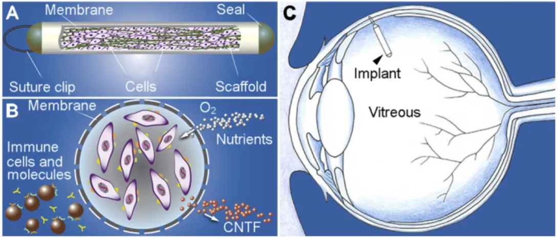
Exogenous administration methods for neurotrophic factor delivery to the retina have mostly relied upon intraocular injections (Whiteley et al. 2001). More recently, a delivery system based on the endovitreal implant of a special capsule (Fig. 8.A) containing engineered cells (Fig. 8.B) has been devised by Tao and co-workers (2002). The encapsulated cells produce CTNF. The results of a recent phase I trial of an encapsulated cell therapy delivering CNTF are presently under evaluation (MacDonald et al. 2007).
Fig. 8. Schematic illustration of CNTF secreting implant using Encapsulated Cell Technology. The implant is composed of a section of semi-permeable membrane capsule which contains CNTF-secreting cells and scaffold. The membrane capsule is sealed at both ends with a suture clip at one end for anchoring on the sclera (A). The membrane allows O2 and nutrients to diffuse in and therapeutic agent (CNTF in this case) to diffuse out. It also keeps components of the immune system out (B). The implant is 6 mm in length and 1 mm in diameter. It is outside the visual axis of the eye when anchored to the sclera (C).From R. Wen et al.2012.
A very important discovery is the existence of intraretinal viability factors. It is postulated that these molecules are normally released by rods (among other cells) and sustain the survival of cones. The latter would undergo secondary degeneration when the density of rods (and therefore the supply of factor) falls below a certain threshold. The French laboratory of Sahel and collaborators has been the first to isolate what has been called the rod-derived cone-viability factor (RdCVf) (Leveillard et al. 2004). These authors observed that in retinal cultures from chicken embryos cones degenerated after a few days. However, the degeneration could be considerably delayed by supplementing the culture with medium derived from wild-type mouse retinal explants. This early experiment led to the biochemical characterization of RdCVF, a secreted protein expressed in rods and necessary for the survival of cones. The injection of antibodies anti RdCVF in the subretinal space of wt mice causes a remarkable reduction in cone number. The administration of RdCVF in rd1 mice preserves about 40% of cones from degeneration. At present, the French group is working on the
mechanism of action of this molecule, also devising pre-clinical trial studies for its application on humans.
3.5 Enviromental Enrichment (EE)
We know that the quality and intensity of environmental stimulation can influence both neural development and plasticity in the adult brain. This concept was first clearly articulated by Hebb in 1949 ( Hebb D.O., The organization of behavior; a neuropsychological theory. Wiley, New York, 1949). Hebb proposed that synapses are strengthened when presynaptic fibers repeatedly participate in activating the postsynaptic neuron. Subsequently, Hubel and Wiesel’s classic experiments (Hubel and Wiesel 1970) demonstrated that the environment influences neural plasticity and neurodevelopment in the cat visual system. Rosensweig and colleagues in the 1960’s (Rosenzweig et al. 1962; Rosenzweig 1966; Rosenzweig et al. 1969) systematically began to examine the influence of environmental enrichment on cognitive functions, brain weight, cortical thickness and the structure of dendrites. In these early studies, Environmental Enrichment was defined as a “combination of complex inanimate and social stimulation” (Rosenzweig et al. 1978). Obviously, this concept is relative: an environmental setting is enriched with respect to another conditions that may lack some stimuli. An essential component of a typical EE setting is the opportunity to perform high levels of voluntary physical activity on running wheels (Spolidoro et al. 2009). Therefore, living in an enriched environment provides the animals with optimal conditions for enhanced exploration, cognitive activity, social interaction and physical exercise.
It is know that EE is a way of rearing the animals in a setting more similar to the wildlife: a sort of naturalistic-like condition. However, the observation of rodents playing in an Enriched Environment, choosing when and how much
to run on the wheel and to explore the new objects show that EE is not only a way to reproduce more natural life conditions. Rather, EE implies a kind of challenge-free interaction with a stimulating surrounding, with few stressful experience then normal. We can also argue that, although the activity of mice in the wild is mostly driven by necessity, and can introduce some stressful experiences, in an EE it is usually prompted by a combination of curiosity and play, because it is carried on with pleasure.
3.5.1 Effects of EE on visual system development
In spite of the vast literature on the effects of EE on the CNS, it was only during the past decade that new light has been shed on the remarkable EE impact on the developmental plasticity of the visual system.
The most remarkable effect that an EE procedure started from birth produces on visual system development is an accelerated maturation of the visual system acuity (VA). This has been initially studied in mice both electrophysiologically, by visual-evoked potentials (VEPs) recordings, and behaviorally, by visual water box task, a visual discrimination task (Cancedda et al. 2004). EE influences also the possibility to induce long-term potentiation (LTP) of layer II-III field potentials after theta-burst stimulation of the white matter (WM/II-III LTP) in the visual cortex, a well established in vitro model of developmental plasticity not present in the adult cortex (Huang et al. 1999). The processes underlying cortical plasticity induced by EE are not well known but different studies agree that there are several molecules involved. One of the most important appears to be BDNF. Indeed, in mice reared from birth in enriched conditions the expression of this neurotrophic factor is significantly higher in their visual cortex at P7 with respect to mice of the same age reared under standard conditions. It is also important to know that such an increase does not persist after P10 (SC) (Sale et al. 2004). Huang
and colleagues in 1999 found, in transgenic animals over-expressing BDNF, that this molecule accelerates the development of the inhibitory GABAergic system. This affects the receptive field development and cortex synaptic plasticity, determining a faster maturation of VA and the accelerated decline of synaptic plasticity. This model is also confirmed in EE mice, in which an increased expression of the GABA biosynthetic enzymes GAD65 and GAD67 has been found in enriched pups at both P7 and P15 (Cancedda et al. 2004; Sale et al. 2004).
Another molecule that seems to be crucial for the effects elicited by EE at visual cortical level is IGF-I and his co-factor mTOR. IGF-I increases postnatally in the visual cortex of enriched rats and post-weaning administration of this molecule in the same structure mimics the EE effects on VA acceleration (Ciucci et al. 2007). Blocking IGF-I action in the visual cortex of developing enriched animals, completely prevents the EE effects on VA acceleration (Ciucci et al. 2007).
One of the targets of IGF-I and BDNF signaling is the activation of the transcription factor CREB. Indeed, it has been demonstrated by Cancedda et al. (2004) that animals enriched from birth display an accelerated time course of CRE/CREB induced gene expression and that treatment of non-EE mice with rolipram, (a specific inhibitor of the high affinity phosphodiesterase type IV that, in turn, activates cAMP system resulting in an increased phosphorilation of the transcription factor CREB), partially mimics EE effects on CREB pathway and on visual acuity development. Moreover, it is very likely that EE effects are not restricted to the visual cortex only, as suggested by the influence on CRE-mediated gene expression observed in the somatosensory cortex as well (Cancedda et al. 2004) .
3.5.2 Maternal enrichment and visual-system development
Experiences acquired between birth and weaning age are essential in promoting and regulating neural development and behavioral traits in the newborn of most mammalian species (Fleming et al. 1999). A key point of this assumption is that newborn animals reared in EE do not interact with the external environment for, at least, the first two weeks of life, spending most of their time in the nest, totally dependent on the mother and, in the case of mammalians that live in large social groups, on the mother helper, which are the most important source of sensory experience for the developing pups (Liu et al. 2000). The thesis that maternal behavior could be the most important source of environmental stimuli for the accelerated development of enriched pups has been directly confirmed by a detailed quantitative analysis of maternal care, which turned out to be higher in pups reared under enriched conditions compared to standard reared pups (Sale et al. 2004).
The enhanced tactile stimulation and the continuous physical contact that enriched pups receive from their mothers seem to greatly influence brain development, providing a source for the earliest changes observed under enriched conditions. All these results are in line with the findings that the offspring of mothers that express higher levels of pup licking and grooming and arched back nursing, show increased expression of NMDA receptor subunit and BDNF-mRNA, increased cholinergic innervation of the hippocampus and enhanced spatial learning and memory (Liu et al. 2000). Moreover, Meaney and co-workers (2001) (Meaney 2001; Meaney 2001) found that naturally occurring variations in maternal care alter the expression of genes that regulate behavioral and endocrine response to stress, as well as hippocampal synaptic development. These effects form the basis for the development of stable, individual differences in stress reactivity and certain forms of cognition. Strikingly, part of these different abilities to respond to