Scuola Dottorale in Biologia
Sezione “Scienze Biomolecolari e Cellulari”
Ciclo di Dottorato XXIV
“Investigation on the origin of biological
enzymatic catalysis”
“Studio sull’origine della catalisi
enzimatica in sistemi biologici”
A.A. 2012/2013
Candidato:
Katarzyna Adamala
Docente Guida:
Prof. Fabio Polticelli
Co-tutors:
Prof. Jack W. Szostak
Acknowledgments
This work would not be possible without generous support, guidance and financing from Professor Jack W. Szostak, who hosted me in his lab and mentored throughout all of my graduate work.
My research was guided by Professor Pier Luigi Luisi, to whom I also owe the privilege of enrolling at Roma Tre.
I am very grateful to Cristiano Chiarabelli for help and guidance in the process of preparing this thesis.
Pasquale Stano was always willing to discuss research ideas, gave me invaluable comments on all the results and also helped me with the bureaucracy behind the doctoral process.
I owe thanks to Rafal Wieczorek, who inspired my work with small catalytic peptides, his excellent research provided background for my own studies.
I want to thank all members of the Szostak Lab, especially Ting Zhu and Christian Hentrich for help with microscope techniques and Yollete Guillen for help with peptide chemistry and analysis. Na Zhang taught me a lot about advanced NMR techniques, and Sheref Mansy introduced me to the work with protocell vesicles.
I am grateful to Sara Walker and Katherine Wright for great discussions on the problems of the prebiotic evolution, and for their help in analyzing my results.
I would like to thank Rachel Massey and Aaron Engelhart for constant support and encouragement.
Riassunto
Nel contesto dell’origine della vita, l’avvento dell’evoluzione Darwiniana fu reso possibile grazie all’insorgere di meccanismi molecolari che consentivano di trasmettere, ereditariamente, le variazioni di fitness dei primi semplici organismi.
Un utile modello teorico-sperimentale che permette di studiare tale scenario evolutivo primordiale consiste in una popolazione di protocellule in competizione tra loro. Ciascuna protocellula è costituita da un polimero genetico in grado di replicarsi all’interno di una vescicola lipidica anch’essa in grado di replicarsi. In questo modello, il polimero genetico incapsulato nella protocellula determina la fitness del sistema, ad esempio perché codifica per un catalizzatore che a sua volta genera un metabolita utile alla vita e alla riproduzione della protocellula.
Le vescicole lipidiche (liposomi) sono i migliori modelli sperimentali di protocellule, grazie alle loro proprietà di auto-assemblamento e incapsulamento di soluti al loro interno (Luisi, Walde et al. 1999). In particolare, è un fatto ben accettato che vescicole lipidiche costituite da acidi grassi a catena lunga rappresentino realisticamente le protocellule più antiche (Deamer, Dworkin et al. 2002).
La seril-istidina (Ser-His) è uno dei più semplici peptidi catalitici noti. E’ stato dimostrato che in particolari condizioni sperimentali, facilita la sintesi di altri peptidi e di RNA (Li, Zhao et al. 2000; Gorlero, Wieczorek et al. 2009; Wieczorek, Dorr et al. 2012).
Lo scopo di questa tesi è quello di sviluppare modelli sperimentali che permettano di studiare i meccanismi alla base dell’evoluzione Darwiniana in tempi primitivi. In particolare, verranno investigate the proprietà di semplici protocellule costituite da acidi grassi, contenenti semplici catalizzatori di natura peptidica.
Innanzitutto verrà mostrato come la Ser-His sia in grado di catalizzare la sintesi di un polimero di tipo “peptide nucleic acid” (PNA) (Gorlero, Wieczorek et al. 2009). Il PNA è un polimero la cui catena principale è costituita da un polipeptide achirale, derivatizzato con gruppi
laterali tipici degli acidi nucleici (basi aromatiche). Il PNA può formare accoppiamenti di tipo Watson-Crick con acidi nucleici naturali, ed è stato proposto come un possibile precursore prebiotico degli acidi nucleici (RNA, DNA), o come polimero complementare ad essi, almeno in alcune fasi del mondo prebiotico (Egholm, Buchardt et al. 1992; Nielsen, Egholm et al. 1994; Nielsen 2007).
Successivamente, si costruirà un modello più complesso mediante la compartimentalizzazione di piccoli catalizzatori come His e Ser-His-Gly all’interno di vescicole di acidi grassi, dimostrando come si possa indurre la competizione tra popolazioni di protocellule.
Infine, verrà illustrato come un altro semplice dipeptide come la cisteil-cisteina (Cys-Cys) possa facilitare la divisione di protocellule indotta fotochimicamente.
Più in generale, il lavoro presentato in questa tesi si concentra sul problema della catalisi in condizioni prebiotiche, e i dati verranno discussi in termini di rilevanza di tali meccanismi nell’origine della vita.
Abstract
The advent of Darwinian evolution required the emergence of molecular mechanisms for the heritable variation of fitness.
One model for such a system involves populations of competing protocells, each consisting of a replicating genetic polymer encapsulated within a replicating membrane vesicle. In this model, the encapsulated genetic polymer imparts enhanced fitness to its protocell by, for example, coding for a catalyst that generates a useful metabolite.
Vesicles are the best known model for testing properties of the self-assembled protocell membranes, and a model of protocell encapsulation. (Luisi, Walde et al. 1999)
It is generally agreed that at the earliest stage of the prebiotic bilayer membrane formation membranes consisted of simple, long chain carboxylic acids. (Deamer, Dworkin et al. 2002)
Ser-His is one of the simplest peptide catalysts, under specific conditions facilitating synthesis of peptides and RNA. (Li, Zhao et al. 2000; Gorlero, Wieczorek et al. 2009; Wieczorek, Dorr et al. 2012)
The aim of this work consists in the development of experimental models to study the primitive mechanisms of Darwinian evolution. In particular, I will investigate the properties of simple fatty acid protocells that contain small catalytic peptides.
First I have shown how Ser-His can catalyze synthesis of peptide nucleic acid polymer. (Gorlero, Wieczorek et al. 2009)
Peptide nucleic acid is peptide bond polymer of notably achiral backbone, capable of standard Watson-Crick based pairing with natural nucleic acids. It has been proposed as a likely prebiotic precursors, or at some stages complementary genetic polymer, to natural RNA and DNA. (Egholm, Buchardt et al. 1992; Nielsen, Egholm et al. 1994; Nielsen 2007)
The work presented in this thesis addresses the problem of prebiotic catalysis on the molecular polymer and the protocell level.
In the studies of catalytic activity of small dipeptides, I showed how prebiotic catalytic dipeptide Ser-His can catalyze synthesis of prebiotically plausible genetic polymer PNA, and how two simple catalysts, Ser-His and Ser-His-Gly can induce competition between populations of protocells. I have also showed that another simple prebiotic dipeptide, Cys-Cys, can facilitate photochemically induced division of protocell vesicles.
Table of contents
Acknowledgments ... 3 Riassunto ... 5 Abstract ... 7 Table of contents ... 9 List of abbreviations ... 11 List of publications and conference contributions ... 13 1. Introduction ... 15 1.1 Origin of life ... 15 1.2 Systems to experimentally explore the origins of life: the model of protocell membrane ... 17 1.3 Exploring the chemical origins of Darwinian Evolution ... 21 2. Results ... 25 2.1. Replication of protocell vesicles ... 25 2.2. Competition between model protocells driven by an encapsulated catalyst ... 33 2.3. Synthesis of genetic polymer PNA catalyzed by the prebiotic catalyst Ser‐His ... 62 3. Discussion ... 67 Conclusions ... 73 References ... 75List of abbreviations
AcPheLeuNH2 – N-acetylphenylalanine leucyl carboxamide
AcPheOEt – ethyl ester of N-acetylphenylalanine AcPheOH – N-acetylphenylalanine
BOC – di-tert-butyl dicarbonate DPH – 1,6-diphenyl-1,3,5-hexatriene DTT – dithiothreitol
FMOC - fluorenylmethyloxycarbonyl
FRET – Förster (fluorescence) resonance energy transfer HPLC – high-performance liquid chromatography HPTS – 8-Hydroxypyrene-1,3,6-trisulfonic acid LeuNH2 – leucyl carboxamide
LUCA – last universal common ancestor MS – mass spectrometry
NMR – nuclear magnetic resonance OA – oleic acid
Phe-Phe – phenylalanyl-phenylalanine PNA – peptide nucleic acid
ROS – reactive oxygen species RPM - revolutions per minute RT – room temperature
Ser-His – Serine-Histidine
Ser-His-Gly – Seryl-Histydyl-Glycine TFA – trifluoroacetic acid
TMAC – tetramethylammonium chloride UV – ultraviolet light
List of publications and conference contributions
M. Gorlero, R. Wieczorek, K. Adamala, A. Giorgi, M. E. Schinina, P. Stano and P. L. Luisi (2009). "Ser-His catalyses the formation of peptides and PNAs." FEBS Lett 583(1): 153-156.
K. Adamala and P. L. Luisi (2011). "Experimental systems to explore life origin: perspectives for understanding primitive mechanisms of cell division." Results Probl Cell Differ 53: 1-9.
T. F. Zhu, K. Adamala, N. Zhang and J. W. Szostak (2012). "Photochemically driven redox chemistry induces protocell membrane pearling and division." Proc Natl Acad Sci U S A 109(25): 9828-9832.
K. Adamala and J.W. Szostak “Competition between model protocells driven by an encapsulated catalyst” manuscript under consideration (submitted November 2012)
Conference contributions during the period of the PhD studies (only meetings where I presented poster or oral contribution are listed)
Bolaamphiphilic bilayer mimics; poster presentation; Mathangi Krishnamurthy, Katarzyna Adamala, Jack W. Szostak; Origin of Life Gordon Conference; January 2008, Ventura, CA, US
Enzymatic reactions in model protocells; poster presentation; Katarzyna Adamala, Jack W. Szostak; ISSOL, August 2008, Florence, Italy
The evolution of protocell membrane, poster presentation and lighting talk; Katarzyna Adamala, Jack W. Szostak; AbGradCon, July 2009, Seattle, WA, US
On the origin of life: natural selection in the protocell population; oral presentation; Katarzyna Adamala, Jack W. Szostak; June 2010, Tällberg, Sweden
Toward the beginning of Darwinian evolution; poster presentation, Katarzyna Adamala, Jack W. Szostak; AbGradCon, June 2011, Bozeman, MT, US
On the origin of life: natural selection in the protocell population; oral presentation; Katarzyna Adamala, Jack W. Szostak; ISSOL, July 2011, Montpellier, France
Vice-chairperson and program organizer, Origin of Life Gordon Research Seminar, January 2012, Galveston, TX, US
The chemical beginning of Darwinian evolution; poster presentation; Katarzyna Adamala, Jack W. Szostak; Origin of Life Gordon Research Conference, January 2012, Galveston, TX, US
Potential mechanisms of prebiotic gene regulation; oral presentation; Aaron E. Engelhart, Katarzyna Adamala, Jack W. Szostak; SERMACS November 2012, Raleigh, NC, US
1. Introduction
1.1 Origin of life
There is no commonly accepted definition of life. Most scientists working on the problem agree that life can be defined by the set of functions and features that must be possessed by the system to be called alive. Yet, the specificity of these functions remains undefined. (Luisi 1998; Cleland and Chyba 2002; Mirazo, Pereto et al. 2004; Ruiz-Mirazo, Pereto et al. 2010; Weber 2010) Since there is no indisputable definition of life, it is also hard to define the event of the origin of life. For the purpose of this work, it will be assumed that the origin of life was the process during which the chemical reactions spontaneously arranged into a homeostatic system undergoing Darwinian evolution. (Oro and Lazcano 1992; Lynn, Burrows et al. 2012)
Life originated on Earth at least 3.6 billion years ago. The oldest known traces of fully-developed life are dated for approximately 3.465 billion years (Schopf 1994), and some evidence show possibility of biochemical cycles existing as early as 3.8 billion years ago (Schidlowski 2001).
The time between the origin of Earth’s crust and primordial ocean 4 billion years ago (Morbidelli, Chambers et al. 2000), and the first known traces of life (date back to 3.8 billion years), is the time when all processes of the origin of life must have occurred. This leaves approximately 200 – 500 million years for the chemical evolution processes. (Orgel 1998)
The first theories postulated that life originated in the water of the prebiotic ocean. The first theory of the primordial ocean, the “prebiotic soup”, is commonly claimed to Aleksandr Ivanovich Oparin (1894-1980) and John B. S. Haldane (1892-1964). Oparin’s book is indeed first well-known scientific publication presenting the theory of origin of life (Oparin 1957). However, the priority in developing backgrounds of modern origin of life scenario belongs to the nineteenth century chemist Ernst H. P. A. Haeckel (Haeckel 1866). In his book, in 1905, he claimed that life originated as a “simple lump of albuminous combination of carbon” (Haeckel 1905). Recently, various possible environments are
prebiotic chemistry reactions, including open water of the prebiotic ocean, lagoons, surfaces of various minerals, thin layers of organic compounds, gaseous phase of the atmosphere or submarine hydrothermal vents. Different prebiotic processes proposed in the literature are placed in different conditions. (Miller and Schlesinger 1983; Miller 1987)
Nevertheless the origin of life on Earth might have not been a singular accident; only one protocell lineage succeeded and survived, proliferating into all known forms of life. There is no reason to assume Figure 1.1.1 General schemes of possible stages of prebiotic evolution. Adapted from (Eigen and Schuster 1982)
Circled in green: focus of section 2.1 of this thesis; circled in blue: section 2.2, and circled in red problems addressed in section 2.3 of this thesis.
single ancestor. (Orgel 1998; Goldman, Bernhard et al. 2013) This organism, LUCA - the Last Universal Common Ancestor of all organisms (Penny and Poole 1999; Delaye, Becerra et al. 2005) – was most likely a population of cells resulting from previous prebiotic, and earliest biological, evolution and selection.
It is not impossible that origin-of-life processes are still occurring, although it is much more difficult on the oxidized environment, and on the planet absolutely possessed by one type of biological organisms it is practically impossible to expect any other form of metabolism to growth enough compete with "our type" of life. Therefore, no effective biogenesis processes are observed today. (Delaye and Lazcano 2005)
Generally, one can distinguish five major fields of research, corresponding to the five major stages of the origin of life. (Eigen and Schuster 1982) (Figure 1.1.1).
The research shown in this thesis was design to address three problems, as marked on figure 1.1.1:
1. the origin of cell division cycle (results described in section 2.1 of this thesis), using lipid vesicles as model protocells;
2. the origin of competition and mechanisms for developing adaptations (results described in section 2.2), with lipid vesicles as model protocell and small dipeptides as prebiotic catalysts;
3. the prebiotic polymerization of small organic molecules (results described in section 2.3), with small dipeptide as a catalyst.
1.2 Systems to experimentally explore the origins of life: the model of protocell membrane
This section was published as part of the chapter of the book Cell Cycle in Development (Adamala and Luisi 2011)
To study the origin of elements of the cell cycle, particularly growth and division of protocell membrane, model protocell vesicles are
2001; Walde 2006) The self-assembled bilayer membranes, semipermeable to small organic molecules and able to encapsulate bigger, polar compounds, are a good model of a prebiotic protocells.
Several authors, including the group of D. Deamer, proposed that at the earliest stage of the prebiotic bilayer membrane formation, membranes consisted of simple, long chain carboxylic acids (Fig. 1.2.1). The open question about the nature of the membrane in Last Universal Common Ancestor leaves many possible routes to the origin of lipid membranes during the earliest stages of protobiological evolution. (Schopf 1994; Goldman, Bernhard et al. 2013)
In modern cells, apart from compartmentation, membranes perform several other functions, including energy transduction and transport of organic and inorganic compounds, and they are the docking site of many enzymes. Presumably, the very first role of the membranes was simple encapsulation – isolation of the reaction cycles (i.e., genetic materials or enzymatic peptides) from the environment. This could be done by the simplest amphiphiles, possibly available under the prebiotic conditions: medium-sized (up to C10) chain carboxylic acids. [Figure 1.2.2] (Luisi, Walde et al. 1999)
The main building blocks of modern cells’ membranes are phospholipids and sterols. Phospholipid glycerol esters and sterols are too complex to be synthesized under abiotic conditions. However, all these compounds can be derived from simplest building block – sterols from isoprene units and lipid derivatives from simple unsaturated carboxylic acids. The simple lipids might have been synthesized under prebiotic Earth conditions (Allen and Ponnamperuma 1967; Yuen, Lawless et al. 1981) including environment of the underwater hydrothermal vents (McCollom, Ritter et al. 1999). Simple amphiphiles were also detected in carbonaceous chondrite meteorites (Yuen and Kvenvolden 1973; Deamer 1985).
Compounds based on these simplest units could have formed the first membranes encapsulating biochemical cycles of the protocell. In a water solution, with the pH close to the polar headgroup pKa, the simplest amphiphiles spontaneously self-organize into bipolar membrane sheets that close into spherical vesicles. (Luisi, Walde et al. 1999; Apel, Deamer et al. 2002; Chen and Walde 2010)
Vesicles are commonly accepted as an approximation of the compartments of the earliest protocells (Walde 2006). Vesicle-like bilayer membranes were even observed in amphiphiles organic material Figure 1.2.1 Protocell vesicles.
A: Vesicles are spontaneously forming from the amphiphilic monomers; B: bilayer membrane of the vesicle, with polar, hydrophilic headgroups directed outside, and aliphatic, hydrophobic chains inside;
C: vesicles can grow upon addition of micelles;
from Murchison carbonaceous chondrite (Deamer 1985; Deamer and Pashley 1989), making its availability on prebiotic Earth more probable.
Vesicle structures can grow (Chen and Szostak 2004; Zhu and Szostak 2009), divide (Hanczyc, Fujikawa et al. 2003; Zhu, Adamala et al. 2012), and selectively take up compounds from the environment (Chen, Roberts et al. 2004). Therefore, investigating properties of the different vesicle systems can give insight into possible routes to the origin of protobiological compartmentalization.
Figure 1.2.2 Amphiphilic compounds building the membranes. A: modern cell’s membrane building block;
1.3 Exploring the chemical origins of Darwinian Evolution Modern cells are thought to have evolved from much earlier protocells – simple replicating chemical systems, composed of a cell membrane and an encapsulated genetic polymer, that were the first cellular systems capable of Darwinian evolution. Evolvability may have emerged in such systems via competition between protocells for a limiting resource. (Szathmáry E 1987; Szostak 2001) Since protocells lacked the complex biochemical machinery of modern cells, such competition was necessarily based on simple chemical or physical processes. (Budin and Szostak 2010) In order to gain further insight into such competitive processes, a variety of model protocell systems have been developed. Fatty acid vesicles have been widely employed as models of early protocellular systems (Hargreaves 1978; Szostak 2001; Apel, Deamer et al. 2002; Noireaux V 2004; Deamer and Dworkin 2005; Mansy, Schrum et al. 2008) because fatty acids can be generated in plausibly prebiotic scenarios, and because membranes based on fatty acids have physical properties that are well suited to primitive forms of life. (Deamer and Dworkin 2005)
The nature of the primordial genetic material remains uncertain; competing schools of thought support either RNA or some alternative nucleic acid as a progenitor of RNA.
Irrespective of the nature of the original genetic material, an important question in considering the origins of cellular competition is how that genetic material could impart a selective advantage to a primitive protocell. An early model (Chen, Roberts et al. 2004) for such a scenario postulated an autocatalytic self-replicating genetic material, such as an RNA replicase, that would accumulate within vesicles at a rate corresponding to its catalytic efficiency. Mutations leading to greater replicase activity would result in a more rapid increase in internal RNA concentration and thus internal osmotic pressure, which would lead to faster vesicle swelling, which in turn would drive competitive vesicle growth. This model was supported by experimental observations that osmotically swollen vesicles could grow by absorbing fatty acid molecules from the membranes of surrounding relaxed vesicles (Chen, Roberts et al. 2004). Although the simple physical link between mutations leading to faster replication of the genetic material, and the consequent osmotic swelling and vesicle growth is attractive, this model
suffers from the lack of a plausible mechanism for the division of osmotically swollen vesicles.
More recently, we have observed that low levels of phospholipids can drive the competitive growth of fatty acid vesicles, in a manner that circumvents this problem by causing growth into filamentous structures that divide readily in response to mild shear stresses (Budin and Szostak 2011). This model implies that a catalyst for phospholipid synthesis, such as an acyltransferase ribozyme, would impart a large selective advantage to its host protocell because faster growth, coupled with division, would result in a shorter cell cycle. A model system illustrating the potential of an encapsulated catalyst to drive vesicle growth in a similar manner would therefore be a significant step towards realizing a complete model of the origin of Darwinian evolution.
Here, we show that the simple dipeptide catalyst seryl-histidine (Ser-His) can drive vesicle growth through the catalytic synthesis of a hydrophobic dipeptide, N-acetyl-L-phenylalanine leucinamide (AcPheLeuNH2), which localizes to the membrane of model protocells
and drives competitive vesicle growth in a manner similar to that previously demonstrated for phospholipids. Ser-His has previously been shown to catalyze the formation of peptide bonds between amino acids and between PNA monomers. (Gorlero, Wieczorek et al. 2009) Although Ser-His is a very inefficient and non-specific catalyst, we have found that Ser-His generates higher yields of peptide product in the presence of fatty acid vesicles.
As a result, vesicles containing the catalyst generate sufficient reaction product to exhibit enhanced fitness, as measured by competitive growth, relative to those lacking the catalyst [Figure 1.3.1]. We can therefore observe how a simple catalyst causes changes in the composition of the membrane of protocell vesicles and enables the origin of selection and competition between protocells.
Figure 1.3.1 Schematic representation of adaptive changes and competition between protocell vesicles.
A: Synthesis of AcPheLeuNH2 by catalyst encapsulated in fatty acid
vesicles. A1: dipeptide catalyst Ser-His catalyzes the reaction between substrates LeuNH2 and AcPheOEt, generating the product of the reaction,
AcPheLeuNH2. A2: product dipeptide AcPheLeuNH2 localizes to the
bilayer membrane.
B: Vesicles with AcPheLeuNH2 in the membrane grow when mixed with
vesicles without dipeptide, which shrink.
C: Vesicles with AcPheLeuNH2 in the membrane grow more following
2. Results
2.1. Replication of protocell vesicles
The results shown in this chapter have been published in “Photochemically driven redox chemistry induces protocell membrane pearling and division” by Ting F. Zhu, Katarzyna Adamala, Na Zhang, and Jack W. Szostak, PNAS vol. 109 no. 25 p. 9828-32. (Zhu, Adamala et al. 2012)
During the course of our imaging studies of vesicle growth and division, we observed an unexpected artifact: long threadlike oleate vesicles containing an encapsulated fluorescent dye quickly (in approximately 5 s) round up into large spherical vesicles under intense illumination. The vesicles contained 2 mM HPTS (8-hydroxypyrene-1,3,6-trisulfonic acid trisodium salt, a water-soluble, membrane-impermeable fluorescent dye) (Zhu and Szostak 2009) and were illuminated by an EXFO 120 W metal halide lamp. We reasoned that since most fluorescent dyes, such as HPTS, generate reactive oxygen species (ROS) under illumination, this phenomenon might be caused by the radical mediated oxidation and fragmentation of the internal buffer solute, bicine, leading to increased internal osmolarity, as previously seen in the case of spherical vesicles that explode under similar conditions. (Zhu and Szostak 2011) The fact that the sphere-to-filament transition is reversible shows directly that growth into a filamentous form does not involve topological changes in vesicle structure. In an effort to obtain better images of filamentous vesicles, we added 10 mM dithiothreitol (DTT) to a vesicle suspension to scavenge ROS, in an attempt to block the rounding-up artifact.
To our surprise, intense illumination in the presence of 10 mM DTT caused the thread-like fatty acid vesicles to go through pearling and subsequent division (Fig. 2.1.1 and Movie S2.1.1). The long thread-like membrane first transformed into a periodic string of smaller ellipsoidal vesicles connected by narrow necks, and the smaller vesicles eventually separated into independent vesicles that moved apart by Brownian motion.
Figure 2.1.1 Oleate vesicle pearling and division. (A) Radical-mediated oxidation of DTT.
(B) An oleate vesicle (containing 2 mM HPTS, in 0.2 M Na-glycinamide, pH 8.5, 10 mM DTT) 30 min after the addition of 5 equivalents of oleate micelles.
(C, D) Under intense illumination (for 2 sec and 12 sec, respectively), the long thread-like vesicle went through pearling and division (Movie S2.1.1). Scale bar, 10 μm
To understand the mechanisms responsible for the pearling and division of thread-like fatty acid vesicles, we examined oleate vesicles labeled with different dyes, in either the internal aqueous space or in the membrane itself. When we prepared vesicles containing 2 mM calcein (bis[N,N-bis(carboxymethyl) aminomethyl] fluorescein) in the internal aqueous space and added 10 mM DTT 30 min after the addition of five equivalents of oleate micelles, vesicle pearling and division were again observed upon illumination.
In contrast, we found that long thread-like oleate vesicles labeled with a low concentration of membrane-localized dye [0.5 mol% Rh-DHPE (Lissamine rhodamine B 1,2-dihexadecanoyl-sn-glycero-3- phosphoethanolamine)] did not go through pearling and division (Fig. S3 A and B). This may be because there are fewer dye molecules associated with the vesicles in the latter case, since in a control experiment where the encapsulated water-soluble fluorescent dye was adjusted to a lower concentration (0.05 mM) comparable to that of the membrane dye used, no vesicle pearling and division were observed. We also observed that with equal concentrations (2 mM) of HPTS inside and outside of the filamentous vesicles (labeled with 1 mol% Rh-DHPE for imaging), vesicle pearling and division still occurred (Fig. 2.1.2). This result indicates that a cross-membrane concentration gradient of the fluorescent dye is not a prerequisite for the pearling and division phenomenon.
It has been shown that vesicle pearling and division can occur as a result of laser tweezers induced surface tension changes which induce a Rayleigh instability in vesicle membranes. (Bar-Ziv and Moses 1994) Recent experiments have also shown that encapsulated cationic nanoparticles can induce vesicle pearling. (Yu and Granick 2009) We hypothesized that the photochemically induced oxidation of thiols into its oxidized form, trans-4,5-dihydroxy-1,2-dithiane, resulted in an increased hydrophobicity of the compound (logP 0.12 for DTT vs. logP 0.52 for trans-4,5-dihydroxy-1,2-dithiane), and consequently increased interaction of the molecule with the membrane.
Figure 2.1.2 Vesicle pearling and division with equal concentrations of HPTS inside and outside the vesicles.
(A) An oleate vesicle with 2 mM HPTS inside and
outside the membranes (labeled by 1 mol% Rh-DHPE for imaging, in 0.2MNa-bicine, pH 8.5, 10mMDTT) 30 min after the addition of five equivalents of oleate
micelles.
(B–D) Under intense illumination (for 2 s, 7 s, and 11 s, respectively), the long thread-like vesicle went through pearling and division. Scale bar, 10 μm.
We used NMR methods to examine the interaction of DTT and its oxidation product with fatty acid vesicle membranes. Because the proton chemical shifts of both DTT and trans-4,5-dihydroxy-1,2-dithiane overlap with the strong background peaks of bicine buffer at 2–4 ppm (and other organic buffers such as tricine and TRIS), which are difficult to selectively suppress, we used oleate vesicles prepared without buffer (15mMoleate in 7.5mMNaOD solution in D2O, pD approximately 8.5)
for the following experiments.
We added 15 mM DTT and 15 mM trans-4,5-dihydroxy-1,2-dithiane to vesicles prepared as above and used two independent NMR methods to examine the interaction of these small molecules with the oleate membranes. We first used saturation transfer difference (STD) spectroscopy, a widely used method for examining the interaction of small ligands with large receptors. (Mayer and Meyer 2001; Haselhorst, Lamerz et al. 2009) Following selective saturation irradiation of the oleate vinyl protons, we observed saturation transfer to the CH protons of trans-4,5-dihydroxy-1,2-dithiane but not to the CH protons of DTT. Thus, only the oxidized form of DTT appears to interact with oleate membranes. As a further test, we used the waterLOGSY method, another sensitive NMR method commonly used for screening ligand-receptor interactions. (Dalvit, Fogliatto et al. 2001)
Again, only oxidized and not reduced DTT was observed to interact with the oleate bilayer membrane.
To further understand the effect of thiols on vesicle pearling and division, we asked whether other thiols can also cause this phenomenon.
Thiols, such as 3-mercaptopropionic acid (10 mM), 1-propanol (50 mM), 1-mercapto-2-1-propanol (50 mM), and 3-mercapto-1,2,4-triazole (50 mM), all caused thread-like oleate vesicles containing 2 mM HPTS to undergo pearling and division. At lower thiol concentrations (2 mM 3-mercaptopropionic acid, 10 mM 3-mercapto-1-propanol, 10 mM 1-mercapto-2-3-mercapto-1-propanol, and 10 mM 3-mercapto-1,2,4-triazole), vesicle pearling was observed but without subsequent division. Recent evidence suggests that the thiol-containing amino acid cysteine may have been prebiotically available, as cysteine oxidation products have been found in the material from a 1958 Miller H2S-rich spark
When we tested cysteine, we observed vesicle pearling but not division. We then considered the possibility that the cys-cys dipeptide, Figure 2.1.3 Oleate vesicle pearling and division mediated by the dipeptide di-Lcysteine.
(A) An oleate vesicle (containing 2 mM HPTS, in 0.2 M Na-bicine, pH 8.5, 20 mM di-L-cysteine) 30 min after the addition of five equivalents of oleate micelles. (B–D) Under intense illumination (for 3 s, 8 s, and 12 s, respectively), the long thread-like vesicle went through pearling and division.
bond. When we prepared filamentous oleate vesicles containing 20 mM di-L-cysteine and 2 mM HPTS in the internal aqueous phase, we observed both vesicle pearling and division upon illumination (Fig. 2.1.3).
As an initial step toward exploring prebiotically plausible scenarios in which photochemically driven pearling and division might operate, we tested the idea that polycyclic aromatic hydrocarbons (PAHs) might replace the synthetic fluorescent dyes used in the above experiments. (Deamer 1992) PAHs are the most abundant polyatomic organic molecules in the universe, have been postulated to play important roles in the origin of life (Deamer 1992; Ehrenfreund, Rasmussen et al. 2006), and have been shown to stabilize short-chain fatty acid membranes (18).
We therefore asked whether oxygenated derivatives of PAHs, such as 1-hydroxypyrene (while HPTS itself is also a hydroxypyrene derivative, it is unlikely to be prebiotically abundant), can be incorporated into vesicle membranes and act as photosensitizers that can absorb UV radiation, generate ROS, and facilitate the division of protocells. To test this idea experimentally, we dissolved a high concentration of 1-hydroxypyrene (20 mol%), oleic acid and a low concentration of Rh-DHPE (1 mol% for imaging) in a chloroform solution, followed by rotary evaporation and resuspension in buffer (0.2 M Na-bicine, pH 8.5). After vesicle growth into filamentous form, and in the presence of 15 mM DTT and UV illumination, vesicle pearling and division were indeed observed (Fig. 2.1.4 and Movie S2.1.2).
Figure 2.1.4 Pearling and division of an oleate vesicle containing 1-hydroxypyrene in the membrane.
(A) An oleate vesicle (with 20 mol %1-hydroxypyrene and 1 mol % Rh-DHPE in the membrane, in 0.2 M Na-bicine, pH 8.5, 15 mM DTT) 30 min after the addition of 5 equivalents of oleate micelles. (B-D) Under intense illumination (57 sec, 153 sec, and 213 sec after the start of illumination, respectively), the long thread-like vesicle went through pearling and division.
2.2. Competition between model protocells driven by an encapsulated catalyst
We have shown that activity of the prebiotic catalytic dipeptide Ser-His can be enhanced by the presence of the bilayer membrane of the protocell, and the product of the reaction catalyzed by Ser-His can cause the emergence of competitive growth of the protocell.
Vesicles Enhance Ser-His synthesis of AcPheLeuNH2
The dipeptide Ser-His has been reported to synthesize the hydrophobic dipeptide AcPheLeuNH2 from the ethyl ester of
N-acetylphenylalanine (AcPheOEt), and leucyl carboxamide (LeuNH2).
(Gorlero, Wieczorek et al. 2009) However, the catalytic efficiency is extremely low, and quantitation is difficult due to precipitation of the hydrophobic product. In addition, Ser-His appears to be a more effective catalyst of the hydrolysis of the AcPheOEt ethyl ester than of peptide bond formation, which further limits product yield. We decided to characterize the Ser-His catalyzed synthesis of AcPheLeuNH2 in the
presence of fatty acid membranes, which we suspected would dissolve the product and prevent precipitation. We also suspected that co-localizing the hydrophobic substrates for the synthesis of the AcPheLeuNH2 dipeptide within or on fatty acid membranes might
improve the yield and possibly minimize substrate hydrolysis.
We therefore tested the above reaction in the presence of different concentrations of oleic acid vesicles. In these experiments, both the Ser-His catalyst and the AcPheOEt and LeuNH2 substrates were present both
inside and outside of the oleate vesicles. We found that the Ser-His catalyst produced progressively more AcPheLeuNH2 in the presence of
increasing concentrations of oleate vesicles, with the conversion of substrate to product increasing from 25% in free solution to 44% in the presence of 50 mM oleate vesicles [Figure 2.2.1A]. The presence of vesicles also diminished substrate hydrolysis from 65% in free solution to 10% in the presence of 50 mM oleate vesicles [Figure 2.2.1B].
Figure 2.2.1A-B Ser-His activity in presence of fatty acid vesicles. A: Ser-His catalyzed synthesis of AcPheLeuNH2 in the presence of
different concentrations of oleate vesicles.
B: Hydrolysis of substrate AcPheOEt in the same reactions.
All experiments: 10mM of each substrate, 5 mM Ser-His catalyst, 0.2 M Na+-bicine pH 8.5, 37 °C.
Figure 2.2.1C-D Ser-His activity in presence of fatty acid vesicles. C: yield of dipeptide AcPheLeuNH2 vs. concentration of oleate vesicles.
D: yield of hydrolyzed substrate AcPheOH vs concentration of oleate vesicles in the same reactions.
All experiments: 10mM of each substrate, 5 mM Ser-His catalyst, 0.2 M Na+-bicine pH 8.5, 37 °C.
One possible explanation for these results is that the hydrophobic substrates partition to the membrane, allowing the reaction to occur at the solvent-lipid bilayer interface, or even within the bilayer, thereby minimizing ester hydrolysis and enhancing product formation.
We also studied the catalysis of AcPheLeuNH2 synthesis by the
tripeptide Ser-His-Gly, which is a less effective catalyst than Ser-His, producing about half as much product dipeptide in the presence of oleate vesicles (with 50 mM vesicles, the yield of dipeptide AcPheLeuNH2 synthesis from 10 mM of each substrate AcPheOEt and LeuNH2 and with 5mM Ser-His-Gly was 27% after 96 hours in 0.2 M Na+-bicine, pH 8.5 at 37°C, compared to 44% for Ser-His).
Slow exchange of Ser-His and AcPheLeuNH2 peptides between
vesicles
Competition between protocells, and the origin of Darwinian evolution, leads to the selection of adaptations that are beneficial to the protocells in which those adaptations originated. Thus, the adaptive property must not be shared with other protocells in a population. It was, therefore, important to establish that the Ser-His catalyst and the dipeptide product AcPheLeuNH2 remain localized within the vesicles
that originally contained the catalyst.
We first asked whether the hydrophobic peptide AcPheLeuNH2
remained localized within an initial set of vesicles, or exchanged between vesicles. We prepared two sets of vesicles, both at a concentration of 50 mM oleate, one with 5 mol% of AcPheLeuNH2 and one without the
dipeptide. After placing samples of each vesicle suspension on opposing sides of a dialysis membrane, we removed aliquots of each solution at a series of time points, and measured the concentration of AcPheLeuNH2
in each solution using HPLC analysis.
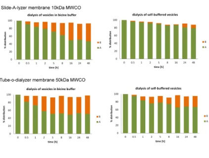
Our results indicate that dipeptide AcPheLeuNH2 exchanges only
mainly in the original vesicles over a period of several hours [Figure 2.2.2A]. In contrast the presence of additional buffer (0.2 M Na+-bicine, pH 8.5) leads to accelerated exchange of peptide between vesicles. All subsequent experiments were therefore carried out over time scales that were short relative to peptide exchange. We performed similar experiments to show that encapsulated Ser-His and Ser-His-Gly were also retained within vesicles and did not exchange between vesicles [Figure 2.2.1B and 2.2.2C].
Figure 2.2.2A Exchange of compounds between vesicles
Figure 2.2.2BC Exchange of compounds between vesicles B: Exchange of Ser-His between two populations of vesicles.; C: exchange of Ser-His-Gly between two populations of vesicles. Vesicles prepared separately, with and without AcPheLeuNH2, and with
and without bicine buffer. After 24 h of tumbling, extrusion, and another 5 h of tumbling, vesicles were transferred to a dialysis chamber. Two different dialysis membranes were tested in two different conditions: self-buffered (50 mol% NaOH) or 0.2 M Na+-bicine pH 8.5. Vesicles with peptide contained 5 mol% AcPheLeuNH2.
Equal volumes were present on both sides of the dialysis membrane. AcPheLeuNH2 distribution between vesicles on either side of the dialysis
Interaction of AcPheLeuNH2 with Membranes
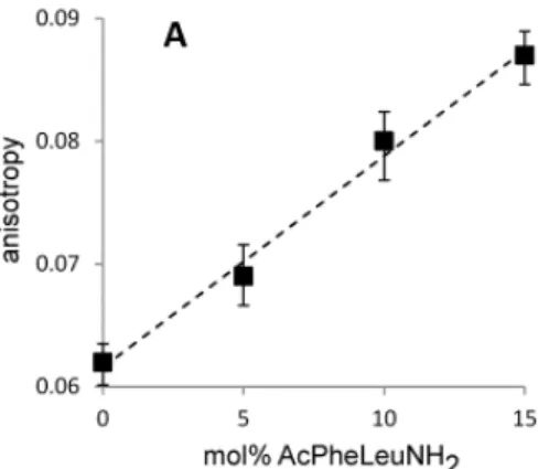
As a means of examining the interaction of AcPheLeuNH2 with the vesicle membrane, we measured the fluorescence anisotropy of the molecular probe 1,6-diphenyl-1,3,5-hexatriene (DPH) as a probe of membrane order.
This assay has previously been used to demonstrate that the addition of phospholipids to fatty acid membranes leads to an increase in membrane viscosity (Budin and Szostak 2011) and has frequently been used to determine viscosity changes in biological membranes resulting from the presence of different peptides. (Juhan-Vague I 1986 ; Miyamoto A 1990) We have observed that the fluidity of oleate membranes also decreases in the presence of the dipeptide AcPheLeuNH2 [Figure 2.2.3],
demonstrating the interaction of this compound with the membrane.
Figure 2.2.3A DPH anisotropy in oleate membranes with and without AcPheLeuNH2
A: self-buffered vesicles (0.5 eq NaOH)
Dashed lines are linear regression fits, R2 > 0.98. Error bars indicate SEM (N=5).
Consistent with previous phospholipid effects (Budin and Szostak 2011), the presence of the hydrophobic dipeptide also decreases the off-rate of fatty acids from fatty acid membranes [Figure 2.2.4]. The off-rate of desorption of lipids from fatty acid vesicles containing dipeptide AcPheLeuNH2 was measured using a population of phospholipid reporter vesicles containing the pH sensitive dye HPTS encapsulated in their internal aqueous space.
After oleic acid vesicles were mixed with reporter vesicles, oleic acid monomers desorbed from the fatty acid vesicles and were then rapidly adsorbed into the reporter phospholipid vesicles, followed by flip-flop and ionization of the oleic acid, causing acidification of the interior of the phospholipid vesicle.
Figure 2.2.3B DPH anisotropy in oleate membranes with and without AcPheLeuNH2
B: vesicles in high salt buffer (0.2M Na+-bicine, pH 8.5).
Dashed lines are linear regression fits, R2 > 0.98. Error bars indicate SEM (N=5).
and Methods). We found that the desorption rate of oleate monomers from fatty acid vesicles decreases with increasing concentration of dipeptide AcPheLeuNH2 in the membrane. This is consistent with
previously reported observations of the effect of phospholipid on fatty acid vesicles. The similar effect of AcPheLeuNH2 and phospholipid on
fatty acid desorption suggested that the dipeptide might, like phospholipid, also drive vesicle growth at the expense of surrounding pure fatty acid vesicles.
We also examined the effects of the peptide on membrane permeability; neither AcPheLeuNH2, nor reaction substrates or catalyst
affected vesicle permeability to the small molecule calcein or to oligonucleotides [Figure 2.2.5].
Figure 2.2.4 The desorption rate of oleate from oleate vesicles as a function of AcPheLeuNH2 content.
Fatty acid desorption rates are derived from the rate of pH change inside reporter phospholipid vesicles. The pH change inside reporter vesicles was measured with the pH-sensitive dye HPTS.
The dotted lines indicate linear regression fits, R2 ≥ 0.98. Error bars indicate SEM (n=4).
Figure 2.2.5 Stability of vesicles with AcPheLeuNH2 in the
membrane.
A: Leakage of small molecules; vesicles in high salt buffer (0.2M bicine Na+ pH=8.5) were prepared with 5mM calcein dye. Unencapsulated dye was removed on size exclusion column. Vesicles were then tumbled for the specified amount of time and each sample was purified again on size exclusion column. Leakage was calculated by quantifying amount of encapsulated vs. free dye.
B: Leakage of oligonucleotide; vesicles were prepared as described above, but with d(Cy3-CAGCAG) oligonucleotide encapsulated instead of calcein dye.
Competitive Growth of Vesicles Containing AcPheLeuNH2
Given that AcPheLeuNH2 decreases membrane fluidity and fatty
acid dissociation in a manner similar to that previously observed for phospholipids, we investigated whether AcPheLeuNH2 also affected
membrane growth dynamics. The dipeptide AcPheLeuNH2 is practically
insoluble in water, so the dilution of the insoluble peptide fraction present in fatty acid membrane is entropically favored. That, in addition to the decreased fatty acid off rate (monomer efflux from the membrane), could lead to the accumulation of fatty acids in the dipeptide-containing membrane, when fatty acid vesicles without the dipeptide are present to provide the oleate monomer.
We monitored vesicle growth using a FRET-based assay for the real-time measurement of membrane surface area (Chen and Szostak 2004), in which the dilution of membrane localized fluorescent donor and acceptor dyes causes decreased FRET (see the Materials and Methods for details). We found that vesicles containing AcPheLeuNH2 grew at the
expense of those lacking this dipeptide, when the two were incubated together [Figure 2.2.6A, Table 2.2.1A].
Similarly, when “fed” with added fatty acid micelles, AcPheLeuNH2-containing vesicles grew preferentially, taking up more
micelles than vesicles without dipeptide [Figure 2.2.6B, Table 2.2.1B]. The time course of competitive vesicle growth after mixing 1 equivalent of vesicles with and without peptide, and the corresponding time course of shrinking of vesicles without the dipeptide, match previously reported phospholipid-driven growth, suggesting a similar fatty acid exchange mechanism [Figure 2.2.7].
Figure 2.2.6A Competition between vesicles in different environments.
A: In the absence of excess salt (self-buffered oleate), vesicles with AcPheLeuNH2 mixed with 1 or 2 equivalents of empty vesicles; surface
area measured after 15min. Filled markers: with FRET dyes on vesicles with AcPheLeuNH2 growth is observed; open markers: with FRET dyes
Figure 2.2.6B Competition between vesicles in different environments.
B: In a high-salt environment (0.2M Na+-bicine, pH=8.5), equal amounts of vesicles with and without AcPheLeuNH2 were mixed, and to that
sample oleate micelles were added surface area measured after 15min. Filled markers: FRET dyes on vesicles with AcPheLeuNH, growth is
Figure 2.2.7A Time course of competitive vesicle growth and shrinking.
A: dye labeled oleic acid vesicles with 5 mol% of dipeptide AcPheLeuNH2 in the membrane were mixed with: 1 equivalent of oleic
acid vesicles without the dipeptide (black trace), 1 equivalent of oleic acid vesicles with 5 mol% of dipeptide AcPheLeuNH2 (red trace), or
Figure 2.2.7B Time course of competitive vesicle growth and shrinking.
B: dye labeled oleic acid vesicles were mixed with: 1 equivalent of oleic acid vesicles with 5 mol% of dipeptide AcPheLeuNH2 (black trace), 1
equivalent of oleic acid vesicles without the dipeptide (red trace), or with water (green trace).
Table 2.2.1 Changes of vesicle size
A: during competitive growth and shrinkage, following 1:1 mixing of indicated vesicle populations, and
B: during competitive oleate uptake after addition of 6 equivalents of oleate micelles.
Indicated populations of vesicles contained either Ser-His (S-H), Ser-His-Gly (S-H-G), or no catalyst. All vesicle populations were incubated separately with amino acid substrates for 48 h to allow for synthesis of hydrophobic dipeptide product prior to mixing.
Surprisingly, we only observed competitive growth between vesicles with and without peptide in self-buffered vesicles. Following the addition of ½ molar equivalent of NaCl to self-buffered vesicles (relative to oleate), less growth was observed, and when 1 equivalent of NaCl was added to self-buffered vesicles, no significant competitive growth was observed [Figure 2.2.8].
The addition of 1 equivalent of tetramethylammonium chloride (TMAC) affected the yield of lipid transfer (observed surface area change) to a lesser extent [Figure 2.2.8], suggesting that surface ionic interactions strongly affect fatty acid exchange processes. In contrast to competitive vesicle-vesicle growth, we were only able to measure competitive micelle-induced growth in high-salt Na+-bicine buffered vesicle samples [Figure 2.2.6B].
Without additional buffer, the alkaline oleate micelles (with 1 equivalent of NaOH per fatty acid, vs. the ½ equivalent per fatty acid in a vesicle) quickly and excessively changed the pH of the mixture, destabilizing the pre-formed vesicles.
In a preliminary effort to correlate structure and activity, we examined the effect of several different small peptides on vesicle growth. While other small hydrophobic dipeptides (e.g., Phe-Phe) led to some competitive growth, none was as efficient as AcPheLeuNH2 [Figure
2.2.9]. N-terminal peptide acetylation was quite important, as Phe-LeuNH2 was less effective than AcPheLeuNH2.
LogPcalc (calculated logarithm of octanol-water partition coefficient) values suggest that the more lipophilic peptides induce a greater competitive growth effect [Figure 2.2.9].
Figure 2.2.8 Effect of salt/buffer on competitive growth of vesicles. In black columns, vesicles containing AcPheLeuNH2 were labeled with
FRET dyes (growth was monitored); in white columns, vesicles without AcPheLeuNH2 were labeled with FRET dyes (shrinking was monitored).
Figure 2.2.9 Competitive growth with other small hydrophobic peptides, and corresponding partition coefficients (logPcalc).
Two populations of self-buffered oleate vesicles prepared as described in Materials and methods, one with 5 mol% of tested peptide and with FRET dye pair, and one population of empty vesicles without any additives. Equal amounts of each vesicle population were mixed and the size of the vesicles with peptide was measured after 1h using the FRET assay.
Partition coefficients logPcalc were calculated using Molinspiration Property Calculation Service (www.molinspiration.com).
Competitive growth can facilitate protocell vesicle division It has previously been shown that the growth of oleate vesicles following micelle addition results in the development of fragile, thread-like structures that can easily fragment, producing daughter vesicles. (Zhu and Szostak 2009) Similar filamentous growth of phospholipid containing vesicles is observed following mixing with excess pure fatty acid vesicles. (Budin and Szostak 2011) We therefore asked whether competitive growth caused by the presence of the hydrophobic AcPheLeuNH2 dipeptide can also result in the formation of filamentous
vesicles and subsequent division.
The development of thread-like filamentous vesicles from initially spherical vesicles is caused by the more rapid increase of surface area relative to volume increase, which is osmotically controlled by solute permeability. To recreate this effect in the absence of additional buffer, we used sucrose, a non-ionic osmolyte providing an osmotic constraint on vesicle volume. We found that initially spherical vesicles with 10 mol% of AcPheLeuNH2 in their membranes develop into
thread-like filamentous vesicles after mixing with 100 equivalents of empty oleate vesicles [Figure 2.2.10A-B].
Gentle agitation of the filamentous vesicles resulted in vesicle division into multiple smaller daughter vesicles [Figure 2.2.10C]. We have previously shown that division of filamentous vesicles into daughter vesicles occurs without significant loss of encapsulated content, and therefore is a plausible mechanism for spontaneous protocell division. (Budin and Szostak 2011; Zhu, Adamala et al. 2012)
Figure 2.2.10 Vesicle growth and division.
A: Large multilamellar vesicles with 0.2 mol% Rh-DHPE dye and 10 mol% of dipeptide AcPheLeuNH2 in the membrane are initially spherical.
B: 10 minutes after mixing with a 100 equivalents of unlabeled, empty oleic acid vesicles without the dipeptide; thread-like filamentous structures develop.
C: after gentle agitation of the sample, threads break producing small daughter vesicles.
Competitive vesicle growth induced by Ser-His catalyzed synthesis of AcPheLeuNH2
The results described above show that Ser-His can catalyze the formation of AcPheLeuNH2 in vesicles and that AcPheLeuNH2 can cause
or enhance vesicle growth. We sought to combine these phenomena to effect Ser-His-driven vesicle growth. We prepared oleate vesicles containing encapsulated Ser-His dipeptide and FRET dyes in their membranes. Unencapsulated catalyst was removed either by dialysis against a vesicle solution of the same amphiphile concentration lacking the catalyst, or, alternatively, by purification on a Sepharose 4B size exclusion column.
Purified vesicles containing 5 mM Ser-His were then mixed with 10 mM of the amino acid substrates AcPheOEt and LeuNH2, and
incubated at 37 °C for 24 to 48 hours to allow for synthesis of the dipeptide AcPheLeuNH2. HPLC analysis of vesicles after 48h of
incubation showed that inside vesicles with Ser-His the dipeptide product AcPheLeuNH2 was synthesized with 28% yield; parallel experiments
with vesicles containing Ser-His-Gly showed that the product was synthesized in 16% yield [Figure 2.2.11].
After incubation, samples with catalyst were mixed with one equivalent of oleate vesicles that had been incubated with amino acid substrates, but without Ser-His catalyst. Vesicle size changes were measured following mixing using the FRET assay for surface area, as described above. We found that vesicles containing the peptide catalyst Ser-His that had been incubated with substrates to allow for the Figure 2.2.11 Synthesis of dipeptide AcPheLeuNH2 in vesicles.
A: HPLC analysis of sample with catalyst Ser-His incubated for 48h at 37°C. Product dipeptide AcPheLeuNH2 elutes at 19.8min.
B: HPLC analysis of sample with catalyst Ser-His-Gly incubated for 48h at 37°C. Product dipeptide AcPheLeuNH2 elutes at 19.8min, marked with
an arrow.
empty oleate vesicles were labeled with FRET dyes, we saw a decrease in their surface area, as expected. In parallel experiments, vesicles lacking Ser-His, or incubated without substrates, did not grow following the addition of oleate vesicles [Table 2.2.1A]. We also examined vesicles containing the less active catalyst Ser-His-Gly, which grew, but to a lesser extent than the Ser-His containing vesicles [Table 2.2.1A].
We then examined the ability of Ser-His (and Ser-His-Gly) to enhance vesicle growth following AcPheLeuNH2 synthesis and then
oleate micelle addition. As expected from previous experiments in which vesicles were prepared with AcPheLeuNH2 directly, vesicles in which
AcPheLeuNH2 was synthesized internally also showed enhanced oleate
uptake from added micelles, and they therefore grew more than empty vesicles [Table 2.2.1B]. Vesicles prepared with Ser-His-Gly showed a similar but smaller growth enhancement.
AcPheLeuNH2-containing Vesicles Exhibit Enhanced
Chemical Potential Generation During Competitive Micelle Uptake As fatty acid vesicles grow, protonated fatty acid molecules flip across the membrane to maintain equilibrium between the inner and outer leaflets. Subsequent ionization of a fraction of the protonated fatty acids that flipped to the inside of the vesicle acidifies the vesicle interior, generating a pH gradient across the membrane.
This pH gradient normally decays rapidly due to H+/Na+
exchange, but we have previously shown that by employing a membrane-impermeable counterion, such as arginine, the pH gradient can be maintained for many hours (t1/2 ≈ 16h). (Chen and Szostak 2004) We
have therefore used arginine as a counterion (arg+) to study the effect of AcPheLeuNH2 on the generation of a trans-membrane electrochemical
potential during micelle-mediated vesicle growth.
In order to monitor the effect of the peptide on vesicle growth-induced pH gradient formation, we prepared two populations of vesicles in arg+-bicine buffer, one with and one without the hydrophobic dipeptide AcPheLeuNH2 in the membrane. In one experiment, the
peptide-containing vesicles also carried the pH-sensitive water-soluble dye HPTS encapsulated in the vesicle interior; in a second, separate
experiments, the two vesicle populations were mixed together and incubated for 30 min. No competitive growth between those two populations occurred, since vesicles were in a high-salt arg+-bicine buffer. We then added 1 equivalent of oleate-arg+ micelles to the mixed vesicle sample, triggering growth of both sets of vesicles.
We measured the pH change inside the vesicles by monitoring the change in the fluorescence emission of the HPTS dye (see Materials and Methods). In the two parallel experiments, pH change was monitored either inside the vesicles containing AcPheLeuNH2 or inside vesicles
without dipeptide (by having HPTS dye encapsulated in either of those populations). Consistent with the enhanced surface area growth of AcPheLeuNH2-containing vesicles [Figure 2.2.6B], the
peptide-containing vesicles developed a larger transmembrane pH gradient than the vesicles lacking peptide, following growth induced by addition of oleate-arg+ micelles [Figure 2.2.12].
Competition between vesicles containing different catalysts In order to examine competition between vesicles harboring peptides of varying catalytic efficiencies, we prepared self-buffered Ser-His-containing vesicles and Ser-His-Gly-containing vesicles and incubated them separately in the presence of LeuNH2 and AcPheOEt to
allow for the synthesis of the hydrophobic dipeptide AcPheLeuNH2.
These vesicles were then mixed, and after 60 min incubation the change in the surface area of vesicles labeled with dye was determined from the Figure 2.2.12 Transmembrane pH gradient generated by growth of vesicles during competitive micelle uptake.
Equal amounts of vesicles with and without 10 mol% AcPheLeuNH2
were mixed in high salt buffer (arg+ -bicine, pH=8.1), and 1 equivalent of arginine-oleate micelles was added to the mixture. The internal pH of the vesicles was measured using the pH-sensitive fluorescent dye HPTS.
surface area, while those containing Ser-His-Gly shrunk. This directly demonstrates competition between protocells containing two catalysts of varying efficiency [Table 2.2.2].
In a separate experiment, we mixed vesicles with both catalysts and added one equivalent of empty oleate vesicles, to serve as a “feedstock” for the vesicles with the dipeptide AcPheLeuNH2. In this
case, both vesicles with Ser-His and with Ser-His-Gly increased in size, but vesicles with Ser-His grew more than those with Ser-His-Gly. This demonstrates that in case where “feedstock” is readily available (from empty vesicles), both populations of protocell grow, but the more efficient catalyst allows for more growth [Table 2.2.2].
Table 2.2.2 Competition between vesicles with two different catalysts.
Vesicle size changes during competitive growth and shrinking in populations of vesicles with and without different catalysts. Vesicle populations were incubated separately with substrates for 48 h, as described in Materials and Methods, prior to mixing.
Each sample contained two or three populations of vesicles, as indicated, in a 1:1 or 1:1:1 ratio. In each case, the FRET dye pair was placed in one
2.3. Synthesis of genetic polymer PNA catalyzed by the prebiotic catalyst Ser-His
We have shown that the dipeptide catalyst Ser-His can catalyze synthesis of a prebiotically plausible genetic polymer PNA.
The results shown in this chapter have been published inGorlero, M., R. Wieczorek, K. Adamala, A. Giorgi, M.E. Schinina, P. Stano, and P.L. Luisi, “Ser-His catalyses the formation of peptides and PNAs” FEBS Lett, 2009. 583(1): p. 153-6. (Gorlero, Wieczorek et al. 2009)
This thesis contains only results from the part of the article that have been prepared by the author of this thesis.
Figure 2.3.1 Synthesis the PNA monomer a) (BOC)2O, THF, 0°C -> RT, 4h; 91%;
b) BrCH2COOEt, K2CO3, CH2Cl2; 79%;
c) Thymine acetic acid, EDC*HCl, Et3N, DMF, 61%;
As part of the project of investigating catalytic activity of Ser-His and other simple prebiotically plausible peptides, we have asked would Ser-His catalyze synthesis of peptide nucleic acids.
Since PNA monomers were not commercially available at the time, we have synthesized PNA thymine monomer, with ethyl ester activating group on the carbonyl end.
The PNA monomers were synthesized according to the previously described procedure. (Fader, Boyd et al. 2001)
Ser-His catalyzed synthesis of the PNA oligomers
The thymine PNA monomer was readily soluble in water. Upon addition of Ser-His to the PNA monomer, at pH 8.3, after 35h we have observed formation of PNA oligomers.
The PNA monomer polymerized to the corresponding PNA dimer Figure 2.3.2 Oligomerization of the PNA monomer
(1) the monomer oligomerizes to give PNA dimer (2), trimer (3) and tetramer (4).
It is interesting to note that the tetramer product is formed in a very high yield compared to the lower products.
The control reaction, carried out under the same conditions but with alpha-chymotrypsin enzyme instead of Ser-His catalyst, gave about 30% substrate conversion, compared to 19% with Ser-His. In the chymotrypsin control reaction we observed the unusually high tetramer product accumulation again.
In all reactions, we have observed the accumulation of the internally cyclized PNA T monomer, the ketopiperazine product.
Table 2.3.1 Results of the Ser-His catalyzed oligomerization of PNA monomers.
Figure 2.3.3 HPLC analysis of PNA oligomers 1: monomer; 2: dimer; 3: trimer; 4: tetramer
3. Discussion
3.1 On the replication of the protocells
Our observations strongly suggest that oxidized, but not reduced, thiols can associate with membranes, triggering division in our protocell model. While the mechanism by which disulfide-containing compounds cause membrane pearling and division is not entirely clear, it is plausible that disulfide-membrane interactions lead to changes in surface tension. Changes in membrane surface tension due to externally applied force from optical tweezers have been shown to induce pearling in tubular vesicles (9, 19), as well as vesicle expulsion (20).
Vesicle pearling has also been demonstrated by insertion of amphiphilic polymers in the outer leaflet of the membrane, inducing changes in spontaneous curvature (21, 22). In addition, lipid phase separation has been shown to drive vesicle budding and division as a consequence of minimizing interdomain edge energy (23). While our results suggest that the oxidation of dithiothreitol to trans-4,5-dihydroxy-1,2- dithiane results in increased interaction of the molecule with the membrane, how such interactions lead to membrane pearling and division remains unclear. Future studies will be required to investigate the location and homogeneity or heterogeneity of disulfide association with the bilayer membrane in order to quantitatively understand the resulting changes in morphology of the vesicle membrane. The observation that thiols and UV-absorbing PAHs facilitate vesicle pearling and division may have important implications for understanding the origin of cellular life.
Thiols are likely to have been abundant in prebiotic hydrothermal systems, such as nearsurface hydrothermal vents (24). Oxygenated PAH fragments could be generated by hydrothermal processing of meteoritic kerogen or by pyrolysis of organic materials. Such hydrophobic molecules are likely to have been selectively solubilized and concentrated in primitive membranes. Indeed, hydroxypyrene, a simple oxygenated PAH, has been shown to integrate into and stabilize membranes composed of short chain fatty acids (18).
In a high UV surface environment, membrane-localized PAHs would be expected to absorb UV radiation and generate ROS, even under anaerobic conditions (16). Our observations show that in the presence of thiols, generation of ROS could lead to the pearling and subsequent division of filamentous vesicles composed of fatty acids and PAHs, thus providing an independent pathway for protocell division that is distinct from the previously described shear force-dependent pathway (6).The control of the timing of cell division by thiol redox state provides a mechanism by which an initial dependence on environmental fluctuations could transition to an internally controlled process, through the evolution of cellular metabolism. For example, we have shown that the dipeptide cys–cys can result in vesicle pearling and division, whereas the amino acid cysteine by itself cannot. Therefore, metabolic control of dipeptide synthesis and degradation could potentially mediate the control of the timing of cell division in primitive cells. Alternatively, as cellular metabolism evolved to the point that cells derived energy from environmental redox disequilibrium, they could potentially use the control of their internal thiol:disulfide redox state as a means of controlling cell division. Finally, it would be interesting to investigate the possibility that intracellularly produced small molecules, peptides, or structured RNAs that interact with protocell membranes might also lead to pearling and division. Such processes would result in competition between protocells on the basis of faster or more appropriately timed cell division, and thus control of cell size, foreshadowing the rise of more complex and more tightly regulated modes of cell division.
3.2. On the catalytic activity of protoenzymes, leading to the origin of protocell competition and synthesis of genetic polymer
The ubiquitous role of proteins as the catalysts of metabolic reactions raises the question of the origins of protein enzymes. The fact that all modern proteins are synthesized through the catalytic activity of the RNA component of the large ribosomal subunit (Moore and Steitz 2011) suggests that primitive enzymes might have been peptides
one or more ribozymes acting sequentially. If short, prebiotically available peptides played useful roles in the growth or division of early protocells, then there would have been a strong selective advantage conferred by ribozymes able to synthesize more of such useful peptides. We have found that a short hydrophobic peptide, whether supplied exogenously or synthesized internally, can confer a growth advantage to fatty acid vesicles. Thus, a ribozyme capable of synthesizing short hydrophobic peptides could accelerate protocell growth, thereby conferring a strong selective advantage. The short peptide Ser-His confers similar effects through its catalytic synthesis of the hydrophobic peptide product, supporting the idea that a ribozyme with similar catalytic activity would confer a selective advantage, but also raising the possibility that a ribozyme that made a catalytic peptide product could amplify its own efficacy through the indirect synthesis of the functional end-product. Similarly, a ribozyme that synthesized a peptide with nascent phospholipid synthase activity would confer a strong selective advantage through phospholipid-driven growth. (Budin and Szostak 2011)
Because of the polarity of nucleic acids, ribozymes might have difficulty catalyzing reactions between membrane-localized substrates; thus the synthesis of intermediate catalytic peptides could be an effective strategy for the synthesis of membrane-modifying products. The hydrophobic environment found in the interior of vesicle bilayers could provide a favorable reaction milieu for many chemical reactions, similar to that afforded (in much more sophisticated fashion) by the interior of folded proteins in contemporary biochemistry. This speculation is supported by our observation that the presence of vesicles increases the yield of AcPheLeuNH2 from amino acid substrates in the presence of
Ser-His as a catalyst. This increased yield most likely results from the greatly decreased extent of AcPheOEt substrate hydrolysis when it is localized to the hydrophobic membrane interior and is thereby protected from attack by water [Figure 2.2.1], much as labile intermediates are protected from water within the active sites of enzymes. Membrane localization of the leucyl-carboxamide substrate may also decrease the pKa of the N-terminal amino group, enhancing its reactivity by increasing the fraction of nucleophilic deprotonated amine. The phenomena of enhancing yields of chemical reactions by co-localizing substrates, altering pKas, and limiting side-reactions are likely to be observed for many other membrane-associated substrates, allowing