University of Rome “Sapienza”
Department of Environmental Biology
Lipid mediated cross-kingdom communication
among hosts and pathogens
A thesis submitted for the degree of
Doctor of Philosophy
in Environmental and Evolutionary Biology
October 2017
Author
Supervisor
Index
Introduction
Review: Fatty acids, sphingolipids and oxylipins: structural and signaling
component
A lipidomic approach to evaluate the host pathogen interaction
Beccaccioli M., Salustri M., Scala V., Reverberi M.
5
Introduction
5
Crucial lipids: fatty acids
Fatty acid biosynthesis
Fatty acid in host-pathogen interaction
5
Oxidised fatty acids: the oxylipins
Oxylipin biosynthesis
Oxylipins in host-pathogen interaction
8
Sphingolipids: signaling mediators and membrane component
Sphingolipid biosynthesis
Sphingolipids in host-pathogen interaction
11
Conclusion
12
References
13
Collection of articles
1. Sphingolipids in Fusarium verticillioides – Zea mays interaction
Beccaccioli M., Salustri M., Ludovici M., Battilani P., Scala V., Reverberi M.
18
Introduction
18
Results
Maize and F. verticillioides sphingolipidome characterization
Sphingolipids level evaluation in infected maize
Fumonisin production analysis
Gene expression analysis and SA quantification
Localization of Fusarium verticillioides in maize during seed colonization and
consequences on seed structure
Discussion
25
Materials and Methods
27
Literature cited
30
Supplementary materials
34
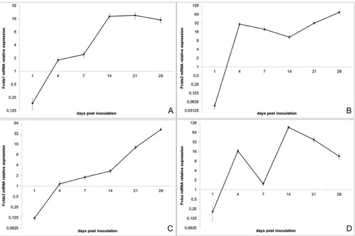
2. Analysis of the expression of genes related to oxylipin biosynthesis in
Fusarium verticillioides and maize kernels during their interaction
Scala V., Beccaccioli M., Dall’Asta C., Giorni P. and Fanelli C.
Edizioni ETS Pisa, 2015 Journal of Plant Pathology (2015), 97 (1), 193-197
40
Summary
40
References
44
3. Fatty acid composition and metabolism in the maize pathogen Fusarium
verticillioides
Beccaccioli M., Scala V., Reverberi M.
46
Introduction
46
Results
Evaluation of free fatty acid composition during the interaction
Fad and elo gene expression during the interaction
Investigation on transcriptional factor (TF) crazy1
Molecular profile of Fv_Crz1Δ
Phenotypic characterization of crz1Δ
Fad, elo, lds and lox gene expression in crz1Δ mutant
Evaluation of fatty acid composition in crz1Δ
Oxylipin profile in crz1Δ
47
Discussion
54
Materials and Methods
56
References
58
4. Fungal secreted oleic acid represents a signal for reinforcing the
host-oriented growth in Fusarium oxysporum
Beccaccioli M., Vitale S., Scala V., Reverberi M.
64
Introduction
64
Results
Oxylipins and fatty acids quantification after POX induction by LC-MS/MS analysis
Susceptibility of oleic acid to Ferrous Iron/Hydrogen Peroxide (Fenton-) mediated
oxidation
Chemotropism assays
65
Discussion
68
Materials and methods
70
References
72
Supplementary matherials
74
Review
Fatty acids, sphingolipids and oxylipins: structural and signaling components
A lipidomic approach to evaluate the host pathogen interaction
Beccaccioli M.1, Salustri M.1, Scala V.2, Reverberi M1. 1
Department of Environmental Biology, University of Rome “Sapienza”, 00185 Rome, Italy. 2CREA-DC, 00156 Rome, Italy.
Which are the main biological functions of lipids? Lipids have a structural role in forming cell membranes and play a role in energy storage. Current knowledge indicates that the lipids have an additional role in cell signaling. When a fungal pathogen contacts the host, the cell surfaces can exchange signals, among which, several are lipids. Lipids occur in fungi not only as major constituents of the membrane system, but also as cell wall components, as storage material in abundant and readily observed lipid bodies, and in some cases, as extracellular products. Fungi contain a various set of lipids, including fatty acids, oxylipins, sphingolipids, phospholipids, glycolipids and sterols. We are going to examine some fungal lipid classes involved in the pathogenic interaction with the host. These signals can confer different information. In some cases can trigger host immune responses; conversely, they may function as virulence factors altering the normal homeostasis of the host or even causing the death.
Introduction
Lipids are classified thereby as being sparingly soluble in water but readily soluble in organic solvents. Structures of the more common lipids can be divided into two categories: structures based on long-chain fatty acid and their derivatives and structure derived from an isoprene unit (Fahy et al., 2009). Fatty acid (FA) either can be in the free form (FFA) or linked to other molecules, most commonly to glycerol. Fatty acid may be fully saturated and may be linear, branched, or contain alicyclic rings. Unsaturated fatty acids occur as frequently as the saturated ones; they may contain several double bond, though 1 or 2 is the most usual. A wide variety of fatty acids with a second oxygen-containing functional group generates what are termed oxylipins. FFAs do not usually accumulate intracellularly because of their toxic effects: they can bind to and inactivate many enzymes and other proteins. FA may form complex with various alcohols and amines, glycerol being the prime example of the former. The fatty acids can be esterified with glycerol and with sugar such as glucose.
One distinctive group of lipids, the sphingolipids, is based on long-chain, polyhydroxy amines (sphingoid bases). Other structural variations include homology and branching of the carbon skeleton. N-Acylation of the 2-amino group of the bases by a fatty acid produces compounds collectively known as ceramides. The ceramides in turn can be further derivatized through the 1-hydroxy group, leading to families of phospholipids and glycolipids comparable with, but less abundant than, those derived from diacylglycerols. There are also several glycosylated sphingolipids.
Second lipid group is that of terpenoids. They are characterized by the repetition of the isoprene unit incorporated in the basic molecular skeleton.
Lipids are the major constituent of fungi plasma membrane and intracellular membranes, but they also have a biological function in energy storage, signal transduction, and stress response. The role of lipids in host-fungus interactions has strongly increased since the availability of highly sensitive analytical technologies, including gas chromatography and high-pressure liquid chromatography coupled to mass spectrometry (Han et al., 2012).
This review aims to give an overview of the status of our knowledge on the involvement of lipids in host-pathogen interaction, indicating that lipids play a key role in infection processes.
Crucial lipids: fatty acids
Fatty acid biosynthesis. Long-chain fatty acids are an important metabolic energy source and the building blocks of membrane
(SE) stored in lipid droplets. Fatty acids synthesis (Figure 1) in Saccharomyces cerevisiae is initiated by the acetyl-CoA carboxylase Aac1 and continued by the cytosolic multi-enzyme fatty acid synthase complex consisting of Fas1p and Fas2p, yielding acyl-CoA with an acyl chain length of 16 or 18 carbon atoms (Tehlivets et al., 2007). Fatty acid elongation up to 26 carbon atoms in contrast to de novo fatty acid synthesis takes place in the endoplasmatic reticulum (ER) presumably due to the
hydrophobic nature of the very long chain fatty acids (VLCFA) of C20-C26 carbon atoms that may be inserted directly into
ceramide by the ceramide synthase. The major VLCFA species in yeast is C26, predominantly present in an amide linkage of
the ceramide backbone of sphingolipids (Raffaele et al., 2009). De novo synthesis uses acetyl-CoA as primers, while the elongation requires malonyl-CoA, which is provided by Aac1. S. cerevisiae encodes different fatty acid elongation enzymes, the elo1 gene and his paralog elo2 encode enzymes involved in lengthening fatty acids from 14 to 16 carbons (Toke et al., 1996), ELO3 is involved in the formation of VLCFA (Oh et al., 1997). Membrane fluidity has an important physical property, which can depend by the ratio of saturated versus unsaturated fatty acids (Los et al., 2004), unsaturated fatty acids are the most abundant
acyl group in the membrane glycerolipids and consist of a wide range of C14-C26 species with one to six double bonds. In S.
cerevisiae approximately 70-80% of their total glycerolipid acyl chains consist of the monounsaturated fatty acids. The Δ9-fatty acid desaturase Ole1p synthesizes monounsaturated fatty acid (i.e. oleic acid C18:1), recently it was shown that Sct1p, a glycerol-3-phosphate acyltransferase catalysing the production of lyso-phosphatidic acid, competes with Ole1p for the saturation (De Smet et al., 2012). The monounsaturated fatty acids can be desaturated further by the action of other desaturases producing PUFA (Polyunsaturated Fatty Acids). PUFAs are a family of lipids that contain more than one double bond in their backbone. PUFAs are biosynthesized via an extension of the saturated-fatty acid pathway, in which stearate C18:0 is converted to oleate C18:1 and then to linoleate C18:2, which is the central precursor for the ω-6 and ω-3 series.
Figure1. Fungal fatty acid metabolism. Fatty acids are derived from de novo synthesis and external uptake. The free fatty acids can be incorporated into complex membrane lipids. Enzymes catalyzing these processes are described in the text.
Fatty acid in host-pathogen interaction. FA are not only membrane molecules, the free fatty acid (FFA), can also function as
transcriptional regulators and signaling molecules and can be involved in post-translational modification of proteins.
The fungi can grow over a range of physiological and nutritional conditions that require different adaptations of membrane lipids. The expression of the crucial gene ole1 is regulated and responds to a number of different stimuli, including a feedback by the FFAs. ole1 expression is regulated by the import of long chain fatty acids from the environment, the supplemented species
s De novo synthesis acetil-CoA 16:0-CoA 18:0-CoA External uptake 16:0, 18:0 Faa1,2,3,4 Acc1 malonyl-CoA Fas1,2 Elo1,2 16:1, 18:1 Sct1 vs Ole1 Elo3 VLCFA Endoplasmatic Reticulum Cytoplasm Complex lipids (phospholipids, acylglycerides, sphingolipids) PUFA Fad Free Fatty Acids
(i.e. the monounsaturated palmitoleic acid C16:1 and oleic acid C18:1) increase in membrane lipids. The linoleic acid C18:2 added was incorporated into microsomal lipids but after several generation of growth replaces the monounsaturated fatty acids 16:1 and 18:1 produced by Ole1p. The enzyme Ole1p is repressed when the cells were grown in presence of monounsaturated fatty acids or PUFAs (Bossie et al., 1989). Ole1 mRNA levels are regulated at transcriptional (Choi et al., 1996) and post-transcriptional level (Gonzalez et al., 1996). The addition of unsaturated fatty acids to the growth medium decreases the desaturase activity (Bossie et al., 1989). The transcription of the ole1 gene can be repressed by cis-unsaturated fatty acids products of Ole1p and polyunsaturated species (McDonough et al., 1992; Fujiwara et al., 1999). It should be noted that only the fatty acids with the cis conformation maintain the repression of the gene. When the fungus contacts the host, an alteration of the fatty acid composition occurs. This alteration can be caused by a re-modulation of the fatty acid uptake. A second way to modify the fatty acid composition is by modulating the activity or expression of enzymes involved in fatty acid synthesis. It was demonstrated that the fatty acids can influence the growth of the fungi. In vitro assays indicate that the unsaturated fatty acid, linoleic 18:2 and linolenic 18:3 acids, reduce the mycelial growth in several fungi species, suggesting an antifungal activity (Agoramoorthy et al., 2007).
The fatty acids can be imported from the environment through a simple diffusion or an active transport, and they can be catabolized after the conversion to a coenzyme A (coA) derivate. The extracellular fatty acid transport across the plasma membranes occurs through transporters and receptors (Daum et al., 1998). Several enzymes can mediate the transport and the activation: Fat1p, Faa1p, Faa2p, Faa3p and Faa4p. These enzymes can be localized not only to the plasma membrane but also to ER, lipid droplets and peroxisomes (Di Russo et al., 1999). Fat1p can be involved in the maintenance of VLCFAs homeostasis; Fat1p appears to be involved to fatty acid transport and very long chain acyl-CoA synthetase activity, therefore it is involved in both the transmembrane movement of fatty acids and in the turnover of very long chain fatty acids (Zou et al., 2002; Black & Di Russo, 2007). The exogenous fatty acids are activated by the acyl-CoA synthetases (Faa1-4p) (Johnsos et al., 1994). Faa1 and faa4 genes encode acyl-CoA synthetases required for activation of imported exogenous fatty acids. Faa1p
exhibits a preference for fatty acids C12-C16 and can import sphingoid chain bases (Narita et al., 2016). Faa2p and Faa3p can
activate only the fatty acids synthesized within the cell (Johnson et al., 1994). Faa2p has been localized to the matrix side of peroxisomal membranes and it is mainly active toward the fatty acids with 9-13 carbon atoms, while Faa3p prefers fatty acids with 16-18 carbons (Knoll et al., 1994).
Membrane lipids and lipid-derived molecules from the plant or the fungal organism play important roles during the infection process. Lipid-derived molecules are crucial for intracellular signaling in the plant as well as in the fungal cell. The free fatty acids derived from the action of some enzyme (see below), and are the precursors for oxylipins (Siebers et al., 2016).
A major portion of the intracellular lipids of fungi reside in the membrane elements, some lipids, however, tend to accumulate as droplets or globules in the cytoplasm. The fatty acids may be part of more complex lipids, as acylglycerides, phospholipids and sphingolipids (that will be detailed subsequently).
The expression of several genes encoding enzymes of lipid metabolism is up-regulated after infection of plant cells, resulting in the synthesis, modification, or re-allocation of lipid-derived molecules. Lipid-modifying enzymes are essential regulators of the spatial and temporal production of lipid metabolites involved in signaling and membrane proliferation for the establishment of intracellular compartments or compositional changes of lipid bilayers.
Acylglycerides are the ester of fatty acids link to the glycerol, this class of lipids includes monoacyl, diacyl- and triacyl-esters; since fatty acids are the major carbon source of energy in living system triacylglycerol are the major constituent of the lipid droplets. The esterases are a class of hydrolase which catalyse the hydrolysis of triglycerides to glycerol and free fatty acids. Several works indicate that the secreted lipases, that release the fatty acids, might serve as virulence factors in plant pathogenic fungi (Commenil et al., 1998; Voigt et al., 2005).
Phospholipids (PL) contain two fatty acids esterified to the sn-1 and sn-2 positions of a glycerol backbone, and a polar head group attached to the sn-3 position. The phospholipid class comprises phosphatidic acid (PA), phosphatidylserine (PS),
phosphatidylcholine (PC), phosphatidylethanolamine (PE), phosphatidylglycerol (PG), and phosphatidylinositol (PI). Each phospholipid class includes many molecular species due to a large number of fatty acids varying in chain length and degree of desaturation. Phospholipids are predominantly synthesized in the ER.
Phospholipases and phospholipid-derived molecules are involved in signaling and plant immunity during plant–pathogen interactions (Arisz et al., 2009; Pleskot et al., 2013). Phospholipases can catalyze the conversion of phospholipids into fatty acids free form, they can function as a modulator of many signal transduction pathways.
It was seen that changes in phospholipid content and phospholipase activities during host–pathogen interactions elicit the activation of defense and resistance response to the necrotrophic pathogen (Zhao et al., 2013). Different sites of phospholipid hydrolysis are noted as shown in figure 2.
Figure 2. Sites of phospholipid hydrolysis by PLD, PLC, PLA1, PLA1.
The phospholipase D (PLD) catalyze the hydrolysis of PC to generate choline and phosphatidic acid, the phospholipase C (PLC) produces two intracellular messengers, diacylglycerol (DAG) and inositol 1,4,5-trisphosphate (IP3), which mediate the activation of protein kinase C and intracellular calcium release, respectively. The phospholipases A1 (PLA1) and A2 (PLA2) can remove acyl groups from both sn-1 and sn-2 positions yielding to the free fatty acids and lysophospholipids formation. Plant PLD and PLA1-2 release the fatty acids during the host-pathogen interaction, the fatty acids are the substrates for the formation of the plant oxylipins implicated in the defense (Blée et al., 2002).
Furthermore, exist a link between oxidative burst and PLA activity, it was demonstrated that extracts of the pathogenic fungus Verticillium dahliae induce PLA activity and ROS production in soybean cells (Chandra et al., 1996).
Fatty acids can be produced by de novo synthesis or are released from complex lipids by the enzymes indicated above, the fatty acid can be converted into bioactive lipid mediators, as the oxylipins (see below). Changes in the oleic acid C18:1 levels result in alterations of the defense responses (Kachroo et al., 2005), also the trienoic acids (Hexadecatrienoic acid C16:3 and α-linolenic acid C18:3) are involved in defense responses against avirulent bacterial pathogens (Yaeno et al., 2004).
We can say that the knowledge of the fatty acid biosynthesis and the study of their metabolism is useful in understanding the lipid signals that mediate the plant-pathogen interaction.
Oxidised fatty acids: the oxylipins
Oxylipin biosynthesis. Oxylipins constitute a large family of oxidized fatty acids and metabolites derived therefrom. These
bioactive lipids are abundant in mammals (Funk et al., 2001) plants, bacteria and fungi.
The plant oxylipins have been studied more in-depth and can provide valuable insights into the description of the biosynthesis of the fungal oxylipins. These compounds can regulate developmental processes and mediate responses to biotic and abiotic stresses (Howe et al., 2009). Plant-oxylipins include fatty acid hydroperoxides, hydroxyl-, epoxy-, keto- and oxo- fatty acids,
O
=
CH
2O
-O
P
|
|
__
__
__ CH
H
2C
| |
O O
| |
C
C
=
=
O
O
| |
FA FA
PLD
PLC
PLA
1PLA
2|
Head group
and epoxy alcohols, divinyl-ethers, volatile alcohols or aldehydes, and jasmonic acid (JA) and its corresponding derivatives (Mosblech et al., 2009). These compounds can be formed through two routes: by non- enzymatic or enzymatic processes. The biological systems are subjected continually to autoxidation and the double bond of fatty acids are particularly susceptible of modifications by reactive oxygen species. In the higher plants, linoleic acid C18:2 and α-linolenic acid C18:3 represent the most
abundant polyunsaturated fatty acids (PUFA). PUFAs easily react with O2 generating hydroperoxides into membrane bound
lipids, in turn affecting membrane fluidity (Girotti 1998). Plants have to develop a broad range of defense responses to cope with the pathogenic infections. The oxidative burst, a rapid and transient production of reactive oxygen species (ROS), is one of the earliest observable aspects of a plant’s defense strategy. The term ROS is used to describe the products of the sequential reduction of molecular oxygen as shown in the scheme:
O2 → •O2- → H2O2 → •OH → H2O2
The superoxide radical (•O2-), hydrogen peroxide (H2O2) and hydroxyl radical (•OH) are the species predominantly detected
in plant-pathogen interaction (Wojtaszek, 1997). The ROS are able to oxidase the PUFAs on the plasma membrane, and biologically active oxylipins are (Grun et al. 2007, Mosblech et al., 2009). To limit these oxidation reactions, thus reducing potentially damaging effects, cells have evolved a wide battery of antioxidant defense systems (Reverberi et al., 2012).
We describe some examples to understand the activity. The superoxide radical (O2•) can behave either as a reducing agent
(often reducing Fe3+ to Fe2+) or as a weak oxidizing agent as in its interaction with ascorbic acid. Superoxide radicals are
detoxified partially in cells to hydrogen peroxide through the enzyme superoxide dismutase that is then detoxified by a group
of heme-containing protein, the catalases, to water and oxygen. Hydrogen peroxide (H2O2) is detoxified through the actions of
both superoxide dismutase and ascorbate peroxidase (Asada et al., 1999). Hydrogen peroxide is a weak oxidizing agent but in
the presence of cellular Fe2+ or other transition metals, forms hydroxyl radicals (•OH), which lacking charge, are able to enter
the interior of cell membranes and are the most reactive and damaging of the reactive oxygen species. The decomposition of
hydrogen peroxide by Fe2+ and some Fe3+ complexes is referred to as Fenton reaction (H
2O2 + Fe2+ + H+ → H2O + Fe3+ + ˙OH).
All these compounds alter the oxidant/antioxidant balance which plays a critical role in lipid peroxidation. Autoxidation is the direct reaction of molecular oxygen with organic compounds; the lipid oxidation involves a free radical mechanism consisting of initiation, propagation and termination steps, frequently overlapping (Halliwell et al., 2005). The relative rates of oxidation of C18-series reveal that more double bonds C=C corresponding to more reactivity, C18:3 is more instable of C18:2, which in turn is more instable of C18:1 (Frankel, 2014).
In plants, the formation of non-enzymatic oxylipins also takes place in response to pathogen attack and levels of esterified hydroxyl-fatty acids increase significantly in esterified lipids, up to 30 times more abundant than free oxylipins suggesting that they are predominantly generated in membranes from which they can be released (Grun et al., 2007).
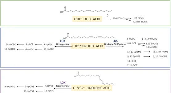
The enzymatic routes (Figure 3) act primarily on linoleic acid C18:2 and α-linolenic acid C18:3. The biosynthetic oxylipin pathway starts with an initial peroxidation reaction of a polyunsaturated fatty acid (PUFA) that is catalyzed by lipoxygenases (LOXs). LOXs are non-heme iron-containing enzymes that catalyze the stereospecific and regiospecific oxidation of PUFA containing a 1Z,4Z-pentadiene system (Andreou and Feussner, 2009). The hydroperoxy fatty acid can be converted to different products by enzymes belong to the family of cytochrome P450 enzymes (Pinot, F and Beisson 2011). Some work suggests that LOXs utilize non-esterified fatty acids and are capable of acting on esterified acyl chains in triacylglycerols or phospholipids (Andreou and Feussner, 2009). Plant oxylipins are produced by distinct plant LOX isozymes that preferentially introduce molecular oxygen into linoleic and linolenic acids either at C-9 (9-LOX) or at C-13 (13-LOX) of the hydrocarbon backbone of the fatty acid.
e- e- e- e-
H+
H+
Oxylipins in host-pathogen interaction. The structural similarity of plant and fungal oxylipins has given rise to a hypothesis
that they are important molecules in cross-kingdom communication based (Tsitsigiannis & Keller, 2007). The Linoleate Diol Synthase (LDS), which catalyzes the enzymatic conversion of linoleic acid into dihydroxy-linoleate was the first oxylipin biosynthetic enzyme that was biochemically characterized from the fungus, in particular from Gaeumannomyces graminis (Su et al., 1996). Based on sequence homology studies, LDS of G. graminis led to the discovery of three oxylipin biosynthetic genes in Aspergillus nidulans named Ppo: psi (precocious sexual inducer)-producing oxygenases (Tsitsigiannis et al., 2005
Microbiology) and in Aspergillus fumigatus (Tsitsigiannis et al., 2005 Infection and immunity). PpoA, PpoB and PpoC are able
to regulate reproduction, dispersal of spores and mycotoxins production (Tsitsigiannis et al., 2004 a; Tsitsigiannis et al., 2004
b; Brodhagen & Keller 2006; Tsitsigiannis & Keller 2006). Three different LDS isoforms were identified in Fusarium
verticillioides too (Scala et al., 2013); the deletion of the one of these LDS generates a mutant that produces more mycotoxins, more conidia and resulted more virulent in maize ears (Scala et al., 2014).
Figure 3. Fungal oxylipins formation by enzymatic route. Principal substrates of oxylipins (oleic, linoleic and α-linolenic acid)
and relative hydroperoxides.
In fungi, the oxylipins modulate sexual and asexual sporulation, coordinate the quorum sensing and regulate the density-dependent sporulation (Brown et al., 2009). The density can determine the colonization of the host and the consequent mycotoxins production. Also in yeast, in human pathogen C. albicans, the oxidised farnesol regulates the quorum sensing and the biofilm formation (Langford et al., 2009). Even within prokaryotes, lipid molecules regulate some crucial event of the life cycle as the virulence and the biofilm formation (Martínez & Campos-Gómez, 2016).
The oxylipins are among the molecules capable of signaling; they can drive signals between different organism, and studies demonstrate that the oxylipins can be sensed by mean of G-protein coupled receptors (Tag et al., 2000). Christensen & Kolomiets in 2011 proposed a hypothetical model of oxylipin-mediated signal communication between plants and fungi. They suggest that the: “fungal lipases could be secreted into plant cells where fatty acid substrates are cleaved and processed by fungal secreted lipoxygenases and/or plant lipoxygenases for oxylipin production. Plant-produced oxylipins are perceived and exploited by fungi to regulate GPCR-, PkaA-, and Ppo-mediated growth, sporogenesis, and mycotoxin production. Host manipulation through oxylipin mimicry (i.e. fungal oxylipins binding to host GPCRs and secretion of JA-Ile-like coronatine into host cells) is also implicated”. The model is still valid and the missing dots are less and less.
C18:1 OLEIC ACID C18:2 LINOLEIC ACID C18:3 α -LINOLENIC ACID 10-HPOME 7, 10 Di-HOME 10-HOME 8-HODE 8-HpODE 12, 13 EpOME 10-HODE 8,13 diHODE 8,11 diHODE 5, 8 diHODE 12, 13 Di HOME 9, 10 EpOME 9, 10 Di HOME 11-HpODE ? LDS
Linoleate Diol Syntase
LOX Lipoxygenase 9-HpODE 13-HpODE 9-HODE 13-HODE 9-oxoODE 13-oxoODE LOX Lipoxygenase 9-HOTrE 13-HOTrE 9-HpOTrE 13-HpOTrE 9-oxoOTrE
Sphingolipids: signaling mediators and membrane component
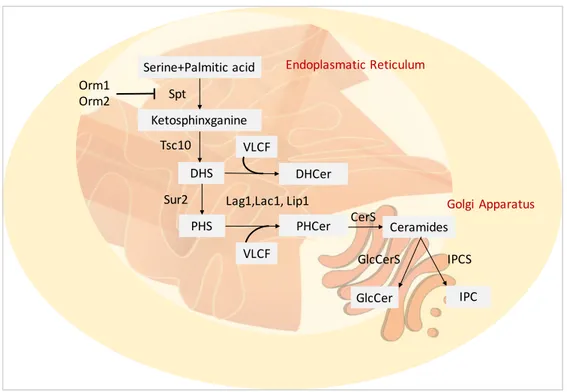
Sphingolipid biosynthesis. A class of lipids gaining new prominence in both mammalian and fungal research is the sphingolipid
family. In mammals, these lipids are involved in several processes, like cell proliferation, apoptosis, and stress response. In recent years, the role of sphingolipids in pathogenic fungi was amply studied. The main themes are the sphingolipids involvement in signaling, growth and virulence. Sphingolipids play an important role in the regulation of the balance between the pathogen and the host. ER is the site of sphingolipid biosynthesis (Figure 4), and they can be transported to other organelles especially to the plasma membrane. The structure of sphingolipids consists of a long-chain base (LCB) or sphingoid base and a saturated fatty acid that is amide-linked to the LCB to form a ceramide (Cer). The Ceramide synthase (CerS) is the enzyme that catalyses the amide bond, exist different isoform on the base of the fatty acids length. Another variability factor is the grade of hydroxylation of FAs and LCBs (Breslow et al., 2010; Merril et al., 2011). Complex sphingolipids are the glucosylceramides (GlcCer) and phosphoinositol ceramides (IPC), they are produced by the respective synthase (GlcCerS and IPCS).
Referring to the model organism S. cerevisiae de novo synthesis of sphingolipids starts with condensation of a serine and a fatty acyl-CoA, normally palmitoyl-CoA or stearyl-CoA, catalysed by the serine palmitoyl-transferase (SPT) complex (Buede et al., 1991; Nagiec et al., 1994) generates 3-ketodihydrosphingosine, and the reductase Tsc10p catalyses the formation of dihydrosphingosine (DHS) (Beeler et al., 1998). DHS can be hydroxylated by Sur2p to another LCB, the phytosphingosine (PHS) (Bae et al., 2004).
The association between the LCB and VLCFA is catalysed by another ceramide synthase complex formed of Lag1p, Lac1p and Lip1p (D’mello et al., 1994; Valée et al., 2005), leading to dihydroceramide (DHCer) and phytoceramide (PHCer). Complex yeast ceramides provide the addiction of head groups; the IPC synthase Aur1 synthesise IPC (Hashida-Okado et al., 1996). Two ER’s proteins Orm1p and Orm2p that bind to SPT and inhibit its activity (Han et al., 2010) regulate the biosynthesis of the sphingolipids.
The catabolism of sphingolipids can be regulated by the phospholipase C encoded by ISC1; this enzyme hydrolysed the ceramides DHCer and PHCer by phospholipids. The ceramides are reduced in FFA and LCB by Ydc1p and its paralog Ypc1 (Mao et al., 2000). The free LCBs can be phosphorylated by Lcb4 and Lcb5 leading to DHS-P and PHS-P (Funato et al., 2003).
Figure 4. Fungal sphingolipid synthesis. Enzymes catalyzing these processes are described in the text. Serine+Palmitic acid Ketosphinxganine DHS PHS PHCer Lag1,Lac1, Lip1 Spt Tsc10 Sur2 VLCF Orm1 Orm2 DHCer VLCF CerS Ceramides GlcCer GlcCerS IPC IPCS Endoplasmatic Reticulum Golgi Apparatus
Sphingolipids in host-pathogen interaction. Sphingolipids play an important role in the regulation of the delicate balance
between the pathogen and the host. Pathogens can produce sphingolipid species that are not present in the host (Heung et al., 2006). The fungal GlcCer presents C-9 methyl group and it is very important for the pathogenesis potentially inducing different plant defence mechanisms (Ramamoorthy et al., 2007). GclCers sprayed on rice leaves function as elicitors and induce the pathogenesis related (PR) protein synthesis (Umemura et al., 2002).
Some plant necrotrophic pathogens can regulate the CerS through the mycotoxins production. Alternaria alternata f. sp. lycopersici, tomato’s pathogen, produce the AAL toxin (Abbas et al., 1995). This toxin is structurally similar to sphingosine
and inhibits the CerS activity accumulating LCB into cell sap (Shier et al., 1995); the accumulation of LCBs triggers PCD in
tomato (Spassieva et al., 2002). The fumonisins (FBs) are fungal toxins produced by the Fusarium genus, in particular by F.
verticillioides and F. proliferatum; FBs are able to inhibit the CerS activity due to the structure similar to sphingosine (Sydenham
et al., 1990; Thiel et al., 1991). The two fungal toxins, AAL and FB, are therefore able to induce PCD inhibiting the CerS, and this mechanism can be seen as a virulence strategy of necrotrophic pathogens suggesting a close link between sphingolipid metabolism and plant PCD.
The sphingolipids are important molecules because are the main component of the lipid rafts (LRs).
In the eukaryotic plasma membranes, these structures are rich in sterols and sphingolipids, in mammal, the main sterol is the cholesterol. In fungal LRs, the main components are the glycol-sphingolipids and ergosterol, these areas are implicated in the secretory pathway. In yeast, it was demonstrated the existence of functional lipid rafts composed of glycosphingolipids and the ergosterol (Bagnat et al., 2000). The LRs play important role in the dynamic processes, including protein sorting, cell polarity, and signal transduction. In the budding yeast S. cerevisiae LRs are detected with filipin staining on the hyphal tips of the cells induced with mating pheromone (Bagnat et al., 2002; Proszynski et al., 2006). The LRs at the tips of pheromone-induced cells plays an important role in cell polarization and proper localization of components needed for cell and membrane fusion during mating. LRs have also been reported at sites of polarized morphogenesis in Cryptococcus neoformans and Aspergillus nidulans (Alvarez et al., 2007). These domains may be formed by the clustering of lipid rafts and may therefore function as organizing centers in the membrane.
It was reported that the sphingolipids, in particular the ceramides, can stabilize the LR formation (Massey et al.,2001), this may have important physiological consequences, the ability of ceramide to participate in raft formation, stability, organization is important in signaling, as proposed in several studies (Grassmé et al., 2001, Cremesti et al., 2001).
Conclusion
The knowledge of lipid-mediated signaling in fungi is still fragmented. Nevertheless, the area of lipid-mediated signal communication is a new and fast growing field with implications in quorum sensing in bacteria, microbe niche interactions, insect–plant communication and mammalian disease defenses. Fatty acid biosynthesis and their release phospholipase-mediated regulate the balance between the free fatty acids and the complex lipids, this mechanism is closely linked to the pathogenesis induction and defense responses. Fatty acids can be converted in the modulators of many signal transduction pathways. They
can be oxidised and form oxylipins able to mediate the cross-talk between a host and a pathogen. We have seen that also the
sphingolipids are crucial in cell signaling, furthermore they can reshape the membrane, forming the lipid rafts that perceive stimuli and trigger response. Thus, lipids play a role that goes further beyond the mere constituents of membranes. Moreover, even their role within the membrane is not “static” by highly dynamic contributing, for instance, to the instant and transient assembly of the intriguing Lipid Rafts. Their chameleonic nature made these molecules fabulous drivers of information exchange. In relation to this, lipids act as shapers of the host-pathogen interaction, allowing to each contenders to develop fine-tuned strategies of defense or virulence.
References
• Abbas, H. K., Tanaka, T., & Shier, W. T. (1995). Biological activities of synthetic analogues of Alternaria alternata
toxin (AAL-toxin) and fumonisin in plant and mammalian cell cultures. Phytochemistry, 40(6), 1681-1689.
• Agoramoorthy, G., Chandrasekaran, M., Venkatesalu, V., & Hsu, M. J. (2007). Antibacterial and antifungal activities
of fatty acid methyl esters of the blind-your-eye mangrove from India. Brazilian Journal of Microbiology, 38(4), 739-742.
• Alvarez, F. J., Douglas, L. M., & Konopka, J. B. (2007). Sterol-rich plasma membrane domains in fungi. Eukaryotic
cell, 6(5), 755-763.
• Andreou A, Brodhun F & Feussner I (2009) Biosyn- thesis of oxylipins in non-mammals. Prog Lipid Res 48, 148–
170.
• Andreou, A., & Feussner, I. (2009). Lipoxygenases–structure and reaction mechanism. Phytochemistry, 70(13),
1504-1510.
• Andreou, A., & Feussner, I. (2009). Lipoxygenases–structure and reaction mechanism. Phytochemistry, 70(13),
1504-1510.
• Arisz, S. A., Testerink, C., & Munnik, T. (2009). Plant PA signaling via diacylglycerol kinase. Biochimica et
Biophysica Acta (BBA)-Molecular and Cell Biology of Lipids, 1791(9), 869-875.
• Asada, K. (1999). The water-water cycle in chloroplasts: scavenging of active oxygens and dissipation of excess
photons. Annual review of plant biology, 50(1), 601-639.
• Bae, J. H., Sohn, J. H., Park, C. S., Rhee, J. S., & Choi, E. S. (2004). Cloning and functional characterization of the
SUR2/SYR2 gene encoding sphinganine hydroxylase in Pichia ciferrii. Yeast, 21(5), 437-443.
• Bagnat, M., & Simons, K. (2002). Cell surface polarization during yeast mating. Proceedings of the National Academy
of Sciences, 99(22), 14183-14188.
• Bagnat, M., Keränen, S., Shevchenko, A., Shevchenko, A., & Simons, K. (2000). Lipid rafts function in biosynthetic
delivery of proteins to the cell surface in yeast. Proceedings of the National Academy of Sciences, 97(7), 3254-3259.
• Beeler, T., Bacikova, D., Gable, K., Hopkins, L., Johnson, C., Slife, H., & Dunn, T. (1998). The Saccharomyces
cerevisiae TSC10/YBR265w gene encoding 3-ketosphinganine reductase is identified in a screen for temperature-sensitive suppressors of the Ca2+-temperature-sensitive csg2Δ mutant. Journal of Biological Chemistry, 273(46), 30688-30694.
• Black, P. N., & DiRusso, C. C. (2007). Yeast acyl-CoA synthetases at the crossroads of fatty acid metabolism and
regulation. Biochimica et Biophysica Acta (BBA)-Molecular and Cell Biology of Lipids, 1771(3), 286-298.
• Blée, E. (2002). Impact of phyto-oxylipins in plant defense. Trends in plant science, 7(7), 315-322.
• Bossie, M. A., & Martin, C. E. (1989). Nutritional regulation of yeast delta-9 fatty acid desaturase activity. Journal of
bacteriology, 171(12), 6409-6413.
• Breslow, D. K., Collins, S. R., Boden- miller, B., Aebersold, R., Simons, K., Shevchenko, A., Ejsing, C. S., and
Weissman, J. S. (2010). Orm family proteins mediate sphin- golipid homeostasis. Nature 463, 1048–1053.
• Brodhagen, M., & Keller, N. P. (2006). Signalling pathways connecting mycotoxin production and sporulation.
Molecular Plant Pathology, 7(4), 285-301.
• Brown, S. H., Scott, J. B., Bhaheetharan, J., Sharpee, W. C., Milde, L., Wilson, R. A., & Keller, N. P. (2009).
Oxygenase coordination is required for morphological transition and the host–fungus interaction of Aspergillus flavus. Molecular plant-microbe interactions, 22(7), 882-894.
• Buede, R., Rinker-Schaffer, C., Pinto, W. J., Lester, R. L., & Dickson, R. C. (1991). Cloning and characterization of
• Chandra, S., Heinstein, P. F., & Low, P. S. (1996). Activation of phospholipase A by plant defense elicitors. Plant Physiology, 110(3), 979-986.
• Choi, J. Y., Stukey, J., Hwang, S. Y., & Martin, C. E. (1996). Regulatory elements that control transcription activation
and unsaturated fatty acid-mediated repression of the Saccharomyces cerevisiae OLE1 gene. Journal of Biological Chemistry, 271(7), 3581-3589.
• Christensen, S. A., & Kolomiets, M. V. (2011). The lipid language of plant–fungal interactions. Fungal Genetics and
Biology, 48(1), 4-14.
• Comménil, P., Belingheri, L., & Dehorter, B. (1998). Antilipase antibodies prevent infection of tomato leaves
byBotrytis cinerea. Physiological and Molecular Plant Pathology, 52(1), 1-14.
• Cremesti, A., Paris, F., Grassmé, H., Holler, N., Tschopp, J., Fuks, Z., ... & Kolesnick, R. (2001). Ceramide enables
fas to cap and kill. Journal of Biological Chemistry, 276(26), 23954-23961.
• D. Fujiwara, O. Kobayashi, H. Yoshimoto, S. Harashima, Y. Tamai, Molecular mechanism of the multiple regulation
of the Saccharomyces cerevisiae ATF1 gene encoding alcohol acetyltransferase, Yeast 15 (1999) 1183–1197.
• D'mello, N. P., Childress, A. M., Franklin, D. S., Kale, S. P., Pinswasdi, C., & Jazwinski, S. M. (1994). Cloning and
characterization of LAG1, a longevity-assurance gene in yeast. Journal of Biological Chemistry, 269(22), 15451-15459.
• Daum, G., Lees, N. D., Bard, M., & Dickson, R. (1998). Biochemistry, cell biology and molecular biology of lipids
of Saccharomyces cerevisiae. Yeast, 14(16), 1471-1510.
• De Smet, C. H., Vittone, E., Scherer, M., Houweling, M., Liebisch, G., Brouwers, J. F., & de Kroon, A. I. (2012). The
yeast acyltransferase Sct1p regulates fatty acid desaturation by competing with the desaturase Ole1p. Molecular biology of the cell, 23(7), 1146-1156.
• DiRusso, C. C., & Black, P. N. (1999). Long-chain fatty acid transport in bacteria andyeast. Paradigms for defining
the mechanism underlying this protein-mediated process. Molecular and cellular biochemistry, 192(1), 41-52.
• Fahy, E., Subramaniam, S., Murphy, R. C., Nishijima, M., Raetz, C. R., Shimizu, T., ... & Dennis, E. A. (2009). Update
of the LIPID MAPS comprehensive classification system for lipids. Journal of lipid research, 50(Supplement), S9-S14. ISO 690
• Frankel, E. N. (2014). Lipid oxidation. Elsevier.
• Funato, K., Lombardi, R., Vallée, B., & Riezman, H. (2003). Lcb4p is a key regulator of ceramide synthesis from
exogenous long chain sphingoid base in Saccharomyces cerevisiae. Journal of Biological Chemistry, 278(9), 7325-7334.
• Funk, C. D. (2001). Prostaglandins and leukotrienes: advances in eicosanoid biology. science, 294(5548), 1871-1875.
• Girotti, A. W. (1998). Lipid hydroperoxide generation, turnover, and effector action in biological systems. Journal of
lipid research, 39(8), 1529-1542.
• Gonzalez, C. I., & Martin, C. E. (1996). Fatty Acid-responsive Control of mRNA Stability UNSATURATED FATTY
ACID-INDUCED DEGRADATION OF THE SACCHAROMYCES OLE1 TRANSCRIPT. Journal of Biological Chemistry, 271(42), 25801-25809.
• Grassmé, H., Jekle, A., Riehle, A., Schwarz, H., Berger, J., Sandhoff, K., ... & Gulbins, E. (2001). CD95 signaling via
ceramide-rich membrane rafts. Journal of Biological Chemistry, 276(23), 20589-20596.
• Grun, C., Berger, S., Matthes, D., & Mueller, M. J. (2007). Early accumulation of non-enzymatically synthesised
oxylipins in Arabidopsis thaliana after infection with Pseudomonas syringae. Functional plant biology, 34(1), 65-71
• Han, S., Lone, M. A., Schneiter, R., & Chang, A. (2010). Orm1 and Orm2 are conserved endoplasmic reticulum membrane proteins regulating lipid homeostasis and protein quality control. Proceedings of the National Academy of Sciences, 107(13), 5851-5856.
• Han, X., Yang, K., & Gross, R. W. (2012). Multi-dimensional mass spectrometry-based shotgun lipidomics and novel
strategies for lipidomic analyses. Mass spectrometry reviews, 31(1), 134-178.
• Hashida-Okado, T., Ogawa, A., Endo, M., Yasumoto, R., Takesako, K., & Kato, I. (1996). AUR1, a novel gene
conferring aureobasidin resistance onSaccharomyces cerevisiae: a study of defective morphologies in Aur1p-depleted cells. Molecular and General Genetics MGG, 251(2), 236-244.
• Heung, L. J., Luberto, C., & Del Poeta, M. (2006). Role of sphingolipids in microbial pathogenesis. Infection and
immunity, 74(1), 28-39.
• Hörnsten, L., Su, C., Osbourn, A. E., Garosi, P., Hellman, U., Wernstedt, C., & Oliw, E. H. (1999). Cloning of linoleate
diol synthase reveals homology with prostaglandin H synthases. Journal of Biological Chemistry, 274(40), 28219-28224.
• Howe, G. A., & Jander, G. (2008). Plant immunity to insect herbivores. Annu. Rev. Plant Biol., 59, 41-66.
• Johnson DR, Knoll LJ, Rowley N, Gordon JI. 1994. Genetic analysis of the role of Saccharomyces cerevisiae
acyl-CoA syn- thetase genes in regulating protein N- myristoylation. J. Biol. Chem. 269:18037– 46
• Johnson, D. R., Knoll, L. J., Levin, D. E., & Gordon, J. I. (1994). Saccharomyces cerevisiae contains four fatty acid
activation (FAA) genes: an assessment of their role in regulating protein N-myristoylation and cellular lipid metabolism. The Journal of Cell Biology, 127(3), 751-762.
• Kachroo, P., Venugopal, S. C., Navarre, D. A., Lapchyk, L., & Kachroo, A. (2005). Role of salicylic acid and fatty
acid desaturation pathways in ssi2-mediated signaling. Plant physiology, 139(4), 1717-1735.
• Knoll LJ, Johnson DR, Gordon JI. 1994. Biochemical studies of three Saccha- romyces cerevisiae acyl-CoA
synthetases, Faa1p, Faa2p, and Faa3p. J. Biol. Chem. 269:16348–56
• Langford, M. L., Atkin, A. L., & Nickerson, K. W. (2009). Cellular interactions of farnesol, a quorum-sensing
molecule produced by Candida albicans. Future microbiology, 4(10), 1353-1362.
• Los, D. A., & Murata, N. (2004). Membrane fluidity and its roles in the perception of environmental signals.
Biochimica et Biophysica Acta (BBA)-Biomembranes, 1666(1), 142-157.
• Mao, C., Xu, R., Bielawska, A., Szulc, Z. M., & Obeid, L. M. (2000). Cloning and characterization of a Saccharomyces
cerevisiae alkaline ceramidase with specificity for dihydroceramide. Journal of Biological Chemistry, 275(40), 31369-31378.
• Martínez, E., & Campos-Gómez, J. (2016). Oxylipins produced by Pseudomonas aeruginosa promote biofilm
formation and virulence. Nature communications, 7.
• Massey, J. B. (2001). Interaction of ceramides with phosphatidylcholine, sphingomyelin and
sphingomyelin/cholesterol bilayers. Biochimica et Biophysica Acta (BBA)-Biomembranes, 1510(1), 167-184.
• McDonough, V. M., Stukey, J. E., & Martin, C. E. (1992). Specificity of unsaturated fatty acid-regulated expression
of the Saccharomyces cerevisiae OLE1 gene. Journal of Biological Chemistry, 267(9), 5931-5936.
• Merrill, A. H. Jr. (2011). Sphin- golipid and glycosphingolipid meta- bolic pathways in the era of sphingolipidomics.
Chem. Rev. 111, 6387–6422.
• Mosblech, A., Feussner, I., & Heilmann, I. (2009). Oxylipins: structurally diverse metabolites from fatty acid
• Nagiec, M. M., Baltisberger, J. A., Wells, G. B., Lester, R. L., & Dickson, R. C. (1994). The LCB2 gene of Saccharomyces and the related LCB1 gene encode subunits of serine palmitoyltransferase, the initial enzyme in sphingolipid synthesis. Proceedings of the National Academy of Sciences, 91(17), 7899-7902.
• Narita, T., Naganuma, T., Sase, Y., & Kihara, A. (2016). Long-chain bases of sphingolipids are transported into cells
via the acyl-CoA synthetases. Scientific reports, 6.
• Oh, C. S., Toke, D. A., Mandala, S., & Martin, C. E. (1997). ELO2 and ELO3, Homologues of the Saccharomyces
cerevisiae ELO1 Gene, Function in Fatty Acid Elongation and Are Required for Sphingolipid Formation. Journal of Biological Chemistry, 272(28), 17376-17384.
• Pearson, C. L., Xu, K., Sharpless, K. E., & Harris, S. D. (2004). MesA, a novel fungal protein required for the
stabilization of polarity axes in Aspergillus nidulans. Molecular biology of the cell, 15(8), 3658-3672.
• Pinot, F., & Beisson, F. (2011). Cytochrome P450 metabolizing fatty acids in plants: characterization and
physiological roles. The FEBS journal, 278(2), 195-205.
• Pleskot, R., Li, J., Žárský, V., Potocký, M., & Staiger, C. J. (2013). Regulation of cytoskeletal dynamics by
phospholipase D and phosphatidic acid. Trends in plant science, 18(9), 496-504.
• Proszynski, T. J., Klemm, R., Bagnat, M., Gaus, K., & Simons, K. (2006). Plasma membrane polarization during
mating in yeast cells. The Journal of cell biology, 173(6), 861-866.
• Raffaele, S., Leger, A., & Roby, D. (2009). Very long chain fatty acid and lipid signaling in the response of plants to
pathogens. Plant signaling & behavior, 4(2), 94-99.
• Ramamoorthy, V., Cahoon, E. B., Li, J., Thokala, M., Minto, R. E., & Shah, D. M. (2007). Glucosylceramide synthase
is essential for alfalfa defensin-mediated growth inhibition but not for pathogenicity of Fusarium graminearum. Molecular microbiology, 66(3), 771-786.
• Reverberi, M., Fabbri, A. A., & Fanelli, C. (2012). Oxidative stress and oxylipins in plant-fungus interaction. In
Biocommunication of Fungi (pp. 273-290). Springer Netherlands.
• Scala, V., Camera, E., Ludovici, M., Dall'Asta, C., Cirlini, M., Giorni, P. A. O. L. A., ... & Reverberi, M. (2013).
Fusarium verticillioides and maize interaction in vitro: relationship between oxylipin cross-talk and fumonisin synthesis. World Mycotoxin Journal, 6(3), 343-351.
• Shier, W. T., Abbas, H. K., & Badria, F. A. (1995). Complete structures of the sphingosine analog mycotoxins
fumonisin B 1 and AAL toxin T A: Absolute configuration of the side chains. Tetrahedron letters, 36(10), 1571-1574.
• Siebers, M., Brands, M., Wewer, V., Duan, Y., Hölzl, G., & Dörmann, P. (2016). Lipids in plant–microbe interactions.
Biochimica et Biophysica Acta (BBA)-Molecular and Cell Biology of Lipids, 1861(9), 1379-1395.
• Spassieva, S. D., Markham, J. E., & Hille, J. (2002). The plant disease resistance gene Asc-1 prevents disruption of
sphingolipid metabolism during AAL-toxin-induced programmed cell death. The Plant Journal, 32(4), 561-572.
• Su, C., & Oliw, E. H. (1996). Purification and characterization of linoleate 8-dioxygenase from the fungus
Gaeumannomyces graminis as a novel hemoprotein. Journal of Biological Chemistry, 271(24), 14112-14118.
• Sydenham, E. W., Gelderblom, W. C., Thiel, P. G., & Marasas, W. F. (1990). Evidence for the natural occurrence of
fumonisin B1, a mycotoxin produced by Fusarium moniliforme in corn. Journal of Agricultural and Food Chemistry, 38(1), 285-290.
• Tag, A., Hicks, J., Garifullina, G., Ake, C., Phillips, T. D., Beremand, M., & Keller, N. (2000). G-protein signalling
mediates differential production of toxic secondary metabolites. Molecular microbiology, 38(3), 658-665.
• Tehlivets O, Scheuringer K, Kohlwein SD (2007). Fatty acid synthesis and elongation in yeast. Biochim Biophys Acta
• Thiel, P. G., Marasas, W. F., Sydenham, E. W., Shephard, G. S., Gelderblom, W. C., & Nieuwenhuis, J. J. (1991). Survey of fumonisin production by Fusarium species. Applied and Environmental Microbiology, 57(4), 1089-1093.
• Toke, D. A., & Martin, C. E. (1996). Isolation and characterization of a gene affecting fatty acid elongation in
Saccharomyces cerevisiae. Journal of Biological Chemistry, 271(31), 18413-18422.
• Tsitsigiannis, D. I., & Keller, N. P. (2006). Oxylipins act as determinants of natural product biosynthesis and seed
colonization in Aspergillus nidulans. Molecular microbiology, 59(3), 882-892.
• Tsitsigiannis, D. I., & Keller, N. P. (2007). Oxylipins as developmental and host–fungal communication signals.
Trends in microbiology, 15(3), 109-118.
• Tsitsigiannis, D. I., Bok, J. W., Andes, D., Nielsen, K. F., Frisvad, J. C., & Keller, N. P. (2005). Aspergillus
cyclooxygenase-like enzymes are associated with prostaglandin production and virulence. Infection and immunity, 73(8), 4548-4559.
• Tsitsigiannis, D. I., Kowieski, T. M., Zarnowski, R., & Keller, N. P. (2005). Three putative oxylipin biosynthetic genes
integrate sexual and asexual development in Aspergillus nidulans. Microbiology, 151(6), 1809-1821.
• Tsitsigiannis, D. I., Kowieski, T. M., Zarnowski, R., & Keller, N. P. (2004). A) Endogenous lipogenic regulators of
spore balance in Aspergillus nidulans. Eukaryotic Cell, 3(6), 1398-1411.
• Tsitsigiannis, D. I., Zarnowski, R., & Keller, N. P. (2004). B) The lipid body protein, PpoA, coordinates sexual and
asexual sporulation in Aspergillus nidulans. Journal of Biological Chemistry, 279(12), 11344-11353.
• Umemura, K., Ogawa, N., Koga, J., Iwata, M., & Usami, H. (2002). Elicitor activity of cerebroside, a sphingolipid
elicitor, in cell suspension cultures of rice. Plant and cell physiology, 43(7), 778-784.
• Vallée, B., & Riezman, H. (2005). Lip1p: a novel subunit of acyl-CoA ceramide synthase. The EMBO journal, 24(4),
730-741.
• Voigt, C. A., Schäfer, W., & Salomon, S. (2005). A secreted lipase of Fusarium graminearum is a virulence factor
required for infection of cereals. The Plant Journal, 42(3), 364-375.
• Wojtaszek, P. (1997). Oxidative burst: an early plant response to pathogen infection. Biochemical Journal, 322(3),
681-692.
• Yaeno, T., Matsuda, O., & Iba, K. (2004). Role of chloroplast trienoic fatty acids in plant disease defense responses.
The Plant Journal, 40(6), 931-941.
• Zhao, J., Devaiah, S. P., Wang, C., Li, M., Welti, R., & Wang, X. (2013). Arabidopsis phospholipase Dβ1 modulates
defense responses to bacterial and fungal pathogens. New Phytologist, 199(1), 228-240.
• Zou, Z., DiRusso, C. C., Ctrnacta, V., & Black, P. N. (2002). Fatty Acid Transport in Saccharomyces cerevisiae
DIRECTED MUTAGENESIS OF FAT1 DISTINGUISHES THE BIOCHEMICAL ACTIVITIES ASSOCIATED WITH Fat1p. Journal of Biological Chemistry, 277(34), 31062-31071.
d
1. Sphingolipids in Fusarium verticillioides – Zea mays interaction
Beccaccioli M.1, Salustri M.1, Ludovici M.3, Battilani P.4, Scala V.2, Reverberi M1. 1Department of Environmental Biology, University of Rome “Sapienza”, 00185 Rome, Italy.
2
CREA-DC, 00156 Rome, Italy. 3Laboratory of Cutaneous Physiopathology and Integrated Center of Metabolomics, San Gallicano Dermatologic Institute (IRCCS), Rome, Italy.4 Istituto di Entomologia e Patologia Vegetale, Università Cattolica del SacroCuore, Piacenza, Italy.
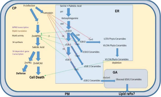
The sphingolipidome, i.e. the whole ensemble of sphingolipids, goes through perturbation during the interaction between the caryopses of Zea mays and the mycotoxigenic fungus Fusarium verticillioides. Fumonisins, mycotoxins produced by Fusarium
verticillioides, should inhibit the activity of ceramide synthase in plant host cell, leading to sphingoid bases accumulation and
complex sphingolipids depletion. Sphingoid bases accumulation should trigger, in turn, programmed cell death (PCD) activating MPK6-Salycilic acid pathway, while complex sphingolipids depletion could cause cell death because of membrane and vesicle impairment. Maize sphingolipidome could be perturbed also in the early phases of infection, when the cell membrane could go through a rearrangement leading to the formation of defense related lipid rafts enriched in complex sphingolipids.
The level of variation of several sphingolipids and salicylic acid was evaluated, by MRM approach in LC-MS/MS, in maize ears artificially infected with F. verticillioides at different times post inoculation. We compared this metabolic profile with the expression of genes related to PCD onset and execution. We hypothesize that F. verticillioides could jeopardize the sphingolipids metabolism of the wounded caryopses of Z. mays for switching its pathogenic growth from endophytic to necrotrophic.
Introduction
Sphingolipids are a wide group of compounds, having both structural and signaling roles in every eukaryote. The common backbones shared by all sphingolipids are long-chain bases, even called sphingoid bases: they are hydroxylated hydrocarbon chains with an amino group on carbon two. LCBs, eventually hydroxylated or unsaturated, are building blocks of sphingolipids together with fatty acyl-CoA. They are substrates of Ceramide Synthases (CerS), which catalyze amide bond formation between sphingoid base amine group and fatty acid carboxylic group. These N-acyl-derivatives are called generally ceramides (Merrill
et al. 2005, Merrill et al., 2011). There are different isoforms of CerS preferring very long chain fatty acids (VLCFA), i.e. fatty
acids from 20 up 36 carbons chain length, or long chain fatty acids. Another factor affecting sphingolipids variability is, in addition to fatty acids length, their hydroxylation degree: they can be α-hydroxylated or Δ9 unsaturated (Markham et al. 2013;
Merrill et al., 2011).
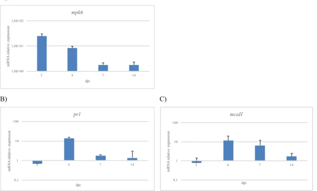
Ceramides are backbones of more complex sphingolipids such as glucosylceramides (GluCers) or glycosil-inositol-phospho-ceramides (GIPCs). GIPCs and GluCers are aconstitutive element of plant plasma membrane and endo-membranes and have a pivotal role in defining membrane microdomains known as “lipid rafts” (Malinsky et al., 2013; Simon-Plas et al., 2011). There is an intimate connection between the unbalance of sphingolipid metabolism and the onset of PCD in plants disease caused by necrotrophic pathogens (Wolpert et al. 2002). Sphinganine analog mycotoxins – SAM (SAM) such as AAL toxin and fumonisins inhibit the host ceramide synthases causing LCB accumulation and cell death in Arabidopsis cell (Stone et al., 2000). The mutant strain of F. verticillioides being deficient for FUM1 polyketide synthase gene are not virulent (Sanchez-Rangel et al., 2012). Fumonisin-induced PCD in Arabidopsis is triggered by increased LCB levels (Shi et al., 2007) and reveals association with ROS generation, callose and phenolic compounds deposition, phytoalexins production and generally with pathogenesis-related genes expression (Nishimura et al., 2003; Stone et al., 2000). It requires ethylene, jasmonate and salicylic acid (SA) dependent signaling pathways (Asai et al., 2000; Nishimura et al., 2003; Sanchez-Rangel et al., 2015). SA synthesis is modulated by MPK6, a crucial molecular hub for different signaling pathways (Bartels et al., 2009). MPK6 is involved in PAMP-triggered
d
immunity (PTI) (Pitzsche et al., 2009; Zhang et al., 2007), in effector-triggered immunity (ETI) (Menke et al., 2004) and, very interestingly, it is probably downstream to long chain bases in SA-dependent PCD provoked by ceramide synthases inhibitors such as fumonisins (Sanchez-Rangel et al., 2015). A recent work in Arabidopsis demonstrates that fumonisin FB1 or sphinganine administration increased the activity of MPK6; while in the MPK6-deficient mutant, neither FB1 nor exogenous sphinganine can induce PCD. It is not clear up today if sphingoid bases modify MPK6 directly or not (Saucedo-Garcìa et al., 2011). Another fundamental feature of fumonisins is the activation of the SA-dependent PCD inducing hydrolytic enzymes. Among these, vacuolar processing enzyme (VPE) and caspase-like proteins are active (Kuroyanagi et al., 2005; Li et al., 2008). As regards last ones, a special mention is for metacaspases. Several metacaspases were characterized in Arabidopsis and there are increasing researchers about monocots. Particularly, TaMCA4 type II wheat metacaspase was found to give resistance against the biotrophic pathogen Puccinia striiformis f.sp. tritici (Wang et al., 2012).
To summarize, it seems clear that toxin perturbation of sphingolipid metabolism leads to a SA signaling dependent PCD, but it is necessary to understand what molecules are definitively responsible for it. Contrasting results are reported about recognizing the main role as PCD inducer for sphinganine or phytosphingosine. Some studies suggest the first one (Saucedo-Garcìa et al.,
2011), some other the second (Shi et al., 2007). Surely, the increase of endogenous LCBs is related generally to AAL toxin and
Fumonisins (especially FB1) derived PCD, consistently with the action mechanism of such sphinganine-like toxins (Akamatu
et al., 1997 Brandwagt et al., 2002, Gechev et al., 2004, Islam et al., 2012).
PCD is not triggered only by LCBs, but by ceramides too. Arabidopsis deficient mutant for ceramide kinase acd5 showed ceramide accumulation leading to PCD, while phosphate ceramides could suppress it (Liang et al., 2003).
Another factor to be considered in ceramide-triggered PCD is fatty acids length. It seems that VLCFA ceramides are involved in vesicle trafficking and lipid rafts formation. VLCFA sphingolipids define both membrane microdomains, in which typical pathogenic response protein take place, and specific cargo vesicle membranes, which definitely concur to microdomains formation (Berkey et al., 2012, Markham et al., 2011). Consequently, their depletion can provoke plasma membrane organization impairment and finally cell death (Markham et al., 2011).
Over-expression of a specific ceramide synthase (i.e. LOH2) in Arabidopsis leads to d18/16:0 accumulation and triggering of SA accumulation and PCD (Siebers et al. 2016). Pathogen-specific sphingolipids similarly to plant ones may elicit a immune response in the host. This is the case of cerebrosides, classified as glycosphingolipids, from Magnaporthe oryzae. Cerebrosides treatment in rice leaves causes phytoalexins accumulation, HR, increased resistance to subsequent infection with compatible pathogens. As intact molecules, their unglycosylated cognate ceramides and long chain bases have been found to induce an immune response (Umemura et al., 2000; Umemura et al., 2004). Anyway, the underlying mechanism has to be clarified. It is likely that PCD may be driven by long-chain bases “perception” as previously seen for toxin-induced sphingolipid metabolism perturbation; nonetheless, another fascinating hypothesis should be proved, i.e. the possibility that exogenous sphingolipid could act as PAMP (Kurusu et al., 2011). Concerning this, the presence in the host of a receptor for fungal sphingolipids remained elusive.
Taking in account this whole background, we tried to define a model describing the host-pathogen interaction between Zea mays (maize) and Fusarium verticillioides. Our ex vivo experimental system, reproducing naturally occurring field infection, consisted in artificially infected maize ears. Before evaluating sphingolipidome perturbation in infected maize along disease progression, we proceeded in maize and F. verticillioides sphingolipidome characterization, in order to find species specific compounds or molecules showing abundance variation along the different infection steps. At the same time, we tried to shed light on maize PCD-related genes expression during the interaction with F. verticillioides.
d
RESULTS
Maize and F. verticillioides sphingolipidome characterization
Extracted sphingolipids were separated and characterized by LC-MS/MS, as described in Materials and Methods section. For each compound class fragmentation spectra have been chosen. After fragmentation, in positive ionization, sphingoid bases lose water moieties. Parental ion (or molecular ion) [M+H+] should be the less abundant ion, while di-dehydrate one [M+H+ -2H2O] the most present. Saturated C18 LCBs, i.e. sphinganine and phytosphingosine, can exhibit a typical 60 m/z fragment indicative of a trimethyl-ammonium moiety (Supplementary figure 1).
As regards ceramides and phytoceramides, their molecular ion undergoes water losses as well as sphingoid bases. Furthermore, their fragmentation pattern clearly includes cognate sphingoid base fragments with their associated water losses, and eventually the amide bounded fatty acid ion (Supplementary Figure 1). Glucosyl-ceramides harbor an additional glucose moiety constituting a neutral loss, hence not detectable as positive ion. The sphingolipidome profile presented significant differences between Z. mays and F. verticillioides, referring to two implemented MRM methods. The first MRM method includes some glucosyl d18:2 ceramides, various types of ceramides and fungal d19:2 glucosyl-ceramides, the second one phytoceramides and dehydro-phytoceramides.
In Supplementary Figure 2 (A and B), non-infected maize kernels and F. verticillioides mycelia overlaid total ion chromatograms, are shown. Just at glance, without the need of further plot magnification, it is possible to note different profiles. This is because some compounds are detected only in F. verticillioides (m/z 506, 680, 710, 736, 754, 822), some other only in maize (m/z 536, 562, 564, 566, 596, 696, 698, 724, 770, 780, 808).
Specifically, in Supplementary Tables 1-6 all characterized compounds are listed and grouped according to their detection into the sole maize, in F. verticillioides or shared by maize and F. verticillioides.
Sphingolipids level evaluation in infected maize
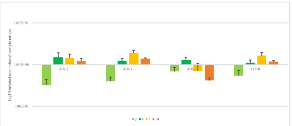
Sphingolipidome perturbation is observed among maize-specific and maize-fungus shared sphingolipids. Firstly, we evaluated LCBs level along infection, calculating infected /control samples ratios, and we kept proceeding the same way for all characterized compounds. The LCBs are down-represented at 2 dpi (days post inoculation) in particular the levels of d18:2, d18:1, d18:0 and t18:0. Following infection, LCB levels increased: at 4 dpi all LCBs are over-represented. At the same time point, d18:0 and d18:2 reached their maximum, while top level of d18:1 and t18:0 is reached at 7 dpi. Afterwards they tend to decrease but remaining over-represented, except for d18:0 (Figure 1).
Figure 1. Long chain bases level variation at different days post inoculation. Results represented the mean of 6 values originated
by two biological replicates technically repeated trice (n=3; ± SE).
As regards the rest of sphingolipidome, i.e. sphingolipids stricto sensu, we observed different trends. d18:2 and d18:0 ceramides apparently had a trend similar to LCB, with a slight down-representation at 2 dpi and a maximum amount achieved between
1,00E-01 1,00E+00 1,00E+01 d18:2 d18:1 d18:0 t18:0 log10 inf ec te d/ non inf ec te d sa m pl e ra ti oos 2 4 7 14
d
and 14 dpi (Figure 2A). The sole d18:1-ceramide considered is the d18:1/16:0. Notably, this ceramide is over-represented into the infected samples at 4-7 dpi (Figure 2B).
A)
B)
Figure 2A and B. Ceramides level variation at different days post inoculation. Plots A refers to ceramides characterized
only in maize; B) ceramides characterized both in maize and Fv10027; Y axis: log10 of infected/non infected sample ratios. X axis: time points, i.e. days post inoculation. Results represented the mean of 6 values originated by two biological replicates technically repeated trice (n=3; ± SE).
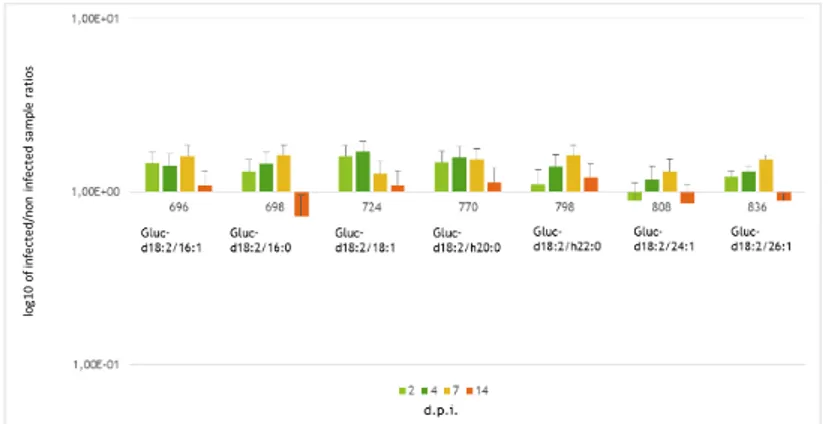
Similarly d18:1/16:0, glucosyl-ceramides are constantly over-represented from 2 to 7 dpi and then decrease with a compound-specific level of significance at 14 dpi (Figure 3).
Figure 3. Glucosyl Ceramides level variation at different days post inoculation. Each compound of this group was characterized
only in maize. Y axis: log10 of infected/non infected sample ratios. X axis: time points, i.e. days post inoculation. Results represented the mean of 6 values originated by two biological replicates technically repeated trice (n=3; ± SE).
1,00E-01 1,00E+00 1,00E+01 534 536 538 562 564 566 596 618 log10 inf ec te d/ non inf ec te d sa m pl e ra ti oos d.p.i. 2 4 7 14 d18:1/16:0 d18:2/18:1 d18:0/18:2 d18:0/18:1 d18:0/20:1 d18:2/22:1 d18:2/16:1 d18:2/16:0 1,00E-01 1,00E+00 1,00E+01 540 552 log10 inf ec te d/ non inf ec te d sa m pl e ra ti oos d.p.i. 2 4 7 14 d18:0/16:0 d17:1/18:0 log 10 of in fe ct ed /n on in fe ct ed sa m pl e ra tio s
d
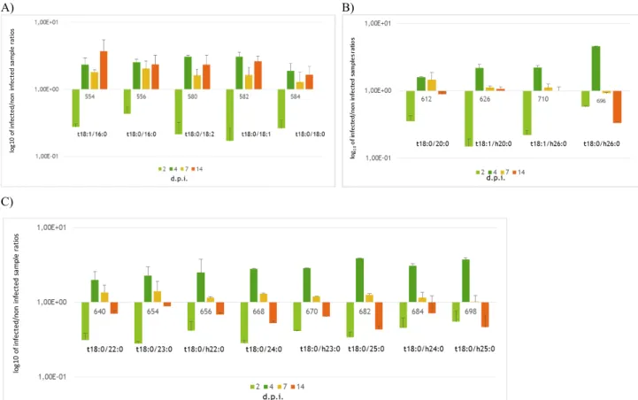
Phytoceramides and dehydrophytoceramides cluster into two different trends; first one including m/z from 554 to 584 (Figure
4A), the second one from 612 to 696 (Figure 4B). The first group includes sphingolipids harboring long chain fatty acids
(LCFA), as palmitic, stearic, linoleic, linolenic, whereas the second group compounds include into their skeleton very long chain fatty acids (VLCFA) such as nervonic acid. Both classes were under-represented at 2 dpi, but while the first group showed a following over- representation persisting for the rest of infection period, the second group exhibited a maximum at 4 dpi followed by a marked decrease leading to an under-representation at 14 dpi (Figure 4 A-B). Compounds with m/z 656, 684 and 698 were previously identified as up-regulated in naturally infected maize hybrids (Figure 4C; Dall’Asta et al., 2014). A) B)
C)
Figure 4 A-C. Phytoceramides and DehydroPhyoCeramides level variation at different days post inoculation. A) Sphingolipids
harboring long chain fatty acids, 554 (t18:1/16:0) is the only dehydrophytoceramide of this group. B) Sphingolipids harbouring very long chain fatty acids (VLCFA). Compounds shown in A and B have been characterized only in maize. C) Compounds characterized both in maize and in F. verticillioides. Y axis: log10 of infected/non infected sample ratios. X axis: time points, i.e. days post inoculation. Results represented the mean of 6 values originated by two biological replicates technically repeated trice (n=3; ± SE)
Fumonisin production analysis
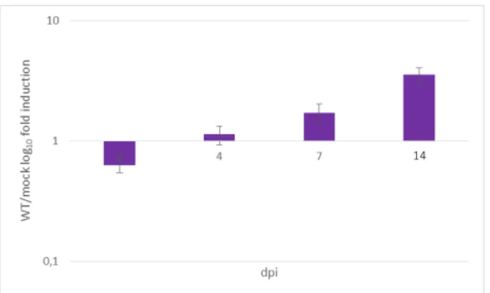
The total amount of fumonisins B (B1+B2+B3) production in maize ears during F. verticillioides infection was quantified by LC-MS/MS. Figure 5 showed an increasing trend in FBs total production along the whole time course of infection in Fv10027-infected maize ears. No significant amount of FBs was detected into mock.
log 10 of in fe ct ed /n on in fe ct ed sa m pl e ra tio s 696 log 10 of in fe ct ed /n on in fe ct ed sa m pl e ra tio s