CLINICAL TRIALS AND OBSERVATIONS
BCR-ABL–specific T-cell therapy in Ph
1
ALL patients on
tyrosine-kinase inhibitors
Patrizia Comoli,1,2,* Sabrina Basso,1,2,* Giovanni Riva,3Patrizia Barozzi,3Ilaria Guido,1,2Antonella Gurrado,1,2
Giuseppe Quartuccio,1,2Laura Rubert,1Ivana Lagreca,3Daniela Vallerini,3Fabio Forghieri,3Monica Morselli,3
Paola Bresciani,3Angela Cuoghi,3Ambra Paolini,3Elisabetta Colaci,3Roberto Marasca,3Antonio Cuneo,4Lorenzo Iughetti,5
Tommaso Trenti,6Franco Narni,3Robin Fo `a,7Marco Zecca,1Mario Luppi,3,* and Leonardo Potenza3,*
1Pediatric Hematology/Oncology and2Cell Factory, Fondazione Istituto di Ricovero e Cura a Carattere Scientifico Policlinico S. Matteo, Pavia, Italy;3Section
of Hematology, Department of Medical and Surgical Sciences, University of Modena and Reggio Emilia, Azienda Ospedaliera Universitaria (AOU) Policlinico, Modena, Italy;4Section of Hematology, University of Ferrara, Ferrara, Italy;5Section of Pediatric Hemato-Oncology, Department of Medical and
Surgical Sciences, University of Modena and Reggio Emilia, AOU Policlinico, Modena, Italy;6Department of Laboratory Medicine and Pathology, Unit `a
Sociosanitaria Locale, Modena, Italy; and7Hematology, Department of Cellular Biotechnologies and Hematology, Policlinico Umberto 1, Sapienza
University, Rome, Italy
Key Points
• BCR-ABL–specific CTLs may be obtained by stimulation with peptides derived from BCR-ABL junctional region and alternative splicing. • T-cell therapy with
BCR-ABL–specific CTLs from healthy donors or patients mediates molecular or hematologic CR in patients with Ph1ALL.
Although the emergence of bone marrow (BM)–resident p190BCR-ABL–specific
T lymphocytes has been correlated with hematologic and cytogenetic remissions in patients with Philadelphia chromosome–positive acute lymphoblastic leukemia (Ph1 ALL) undergoing maintenance tyrosine-kinase inhibitor treatment, little is known about the possibility of culturing these cells ex vivo and using them in T-cell therapy strategies. We investigated the feasibility of expanding/primingp190BCR-ABL–specific T cells in vitro by stimulation with dendritic cells pulsed withp190BCR-ABL peptides derived from the
BCR-ABL junctional region and alternative splicing, and of adoptively administering them to patients with relapsed disease. We report on the feasibility of producing clinical-grade BCR-ABL–specific cytotoxic T lymphocytes (CTLs), endowed with antileukemia activity, from Ph1ALL patients and healthy donors. We treated 3 patients with Ph1ALL with autologous or allogeneic p190BCR-ABL–specific CTLs. No postinfusion toxicity was
observed, except for a grade II skin graft-versus-host disease in the patient treated for hematologic relapse. All patients achieved a molecular or hematologic complete remission (CR) after T-cell therapy, upon emergence ofp190BCR-ABL–specific T cells in the BM. Our results show thatp190BCR-ABL–specific CTLs are capable of controlling treatment-refractory Ph1ALL in vivo, and
support the development of adoptive immunotherapeutic approaches with BCR-ABL CTLs in Ph1ALL. (Blood. 2017;129(5):582-586)
Introduction
Philadelphia chromosome–positive acute lymphoblastic leukemia (Ph1
ALL) was formerly burdened by uniformly poor prognosis.1
Wide-spread application of allogeneic hematopoietic stem cell (HSC) trans-plantation (alloHSCT) and advent of targeted BCR-ABL–specific
tyrosine-kinase inhibitors (TKIs) have significantly improved complete
response rates and disease-free survival.1,2Despite these therapeutic
advances, some unresolved issues remain, including the high prevalence
in older patients,3often ineligible for alloHSCT, and the extremely poor
prognosis of relapsed Ph1ALL, particularly following alloHSCT.1,4
Prolonged hematologic and cytogenetic remissions have been observed with imatinib mesylate (IM) alone, even in the presence
of persisting levels of minimal residual disease (MRD).5-7Our group
was able to demonstrate that attainment of such clinical responses
directly correlated with the emergence of BCR-ABL–specific T cells
in the bone marrow (BM) and, to a lesser extent, in the peripheral
blood of nonallografted Ph1ALL patients undergoing postremission
maintenance treatment with either IM or other second-generation
TKIs.8,9These observations extended previous evidence of functional
leukemia-specific cellular immune responses developing in patients receiving IM, and possibly acting in synergy with IM to reach disease
control,10,11and represent the basis for a combined TKI and T-cell
therapy approach to Ph1 ALL in elderly patients, or in patients
relapsing after alloHSCT.
We report on the feasibility of inducing durable MRD clearance and leukemia control, without additional toxicity, by transfer of donor-derived or autologous cytotoxic T lymphocytes (CTLs) specific for the BCR-ABL fusion product in patients receiving TKI treatment of leukemia relapse after alloHSCT, or for molecular relapse in patients ineligible for alloHSCT. In addition, we describe the immunological parameters correlated with clinical response.
Submitted 29 July 2016; accepted 29 November 2016. Prepublished online as Blood First Edition paper, 7 December 2016; DOI 10.1182/blood-2016-07-731091.
*P.C., S.B., M.L., and L.P. contributed equally to this study. The online version of the article contains a data supplement.
There is an Inside Blood Commentary on this article in this issue.
The publication costs of this article were defrayed in part by page charge payment. Therefore, and solely to indicate this fact, this article is hereby marked “advertisement” in accordance with 18 USC section 1734. © 2017 by The American Society of Hematology
582 BLOOD, 2 FEBRUARY 2017xVOLUME 129, NUMBER 5
Study design
Patient 1 was a 61-year-old man in second molecular recurrence after matched unrelated donor (MUD) alloHSCT and unmanipulated donor lymphocyte infusions (DLIs). Patient 2 was a 30-year-old man diagnosed with Ph1ALL with hyperleukocytosis and central nervous system (CNS) involvement, in third hematologic relapse (BM blast 66%, F317L mutation) after MUD-HSCT, DLI,
and rescue therapy with Nilotinib. Patient 3 was a 62-year-old woman diagnosed with Ph1ALL with CNS involvement, showing persistent molecular disease (last MRD before T-cell therapy 0.1% BCR-ABL/ABL) after induction, maintenance chemotherapy, and prolonged TKI treatment. She was not eligible for alloHSCT due to comorbidities. Details on patients’ clinical histories are reported in supplemental Methods (available on the Blood Web site).
Methods for p190BCR-ABL–specific CTL preparation and testing are
detailed in supplemental Methods. The BCR-ABL peptide pool used in the 200
150
100
50
0
mix1int mix1est mix2 mix3
SFU/10 5 cells
C
50 40 30 20 10 0 20:1 10:1 5:1 2:1 0.1:1 % specific lysisE
8 6 4 Cell numbers (×10 8) 2 0 d 0 d 28 d 40A
80 60 40 20 0BCR-ABL-PHAtargets 5hBCR-ABL-PHAtargets 12h
P815 allo-PHA auto-PHA % specific lysis
D
B
CD3+ CD8+ CD4+CD3-CD56+CD4/45RA+CD4/45RO+CD8/45RA+CD8/45RO+
100 80 60 40 20 0 % positive cells
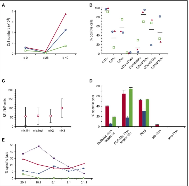
Figure 1. Characteristics of thep190BCR-ABL–specific T cells used in the 3 patients. (A) T-cell expansion ofp190BCR-ABL–specific T-cell lines achieved over a 40-day period
based on cell counting using trypan blue exclusion (green square, patient 1; blue circle, patient 2; red triangle, patient 3). (B) Phenotype ofp190BCR-ABL–specific T-cell lines, reported
as the percentage of positive cells (green square, patient 1; blue circle, patient 2; red triangle, patient 3). (C) Response, measured as IFNg production in a enzyme-linked immunospot (ELISPOT) assay, to the different peptide pools used in the activation/expansion process. Mix1int indicates 9- and 10-mer peptides spanning the internal p190 breakpoint region; Mix1est, 9- and 10-mer peptides spanning the external p190 breakpoint region; Mix3, 9-mer peptides derived from the alternative BCR-ABL splice variants. (D) Cytotoxic activity of
T-cell lines, measured as the percentage of specific lysis at a effector-to-target (E:T) ratio of 5:1, against autologous phytohemagglutinin (PHA) blasts pulsed withp190BCR-ABL
peptides (BCR-ABL-PHA targets, cytotoxicity measured at 5 hours and 12 hours, calculated after subtraction of background, consisting of cytotoxicity against autologous PHA blasts pulsed with irrelevant peptides), P815 cell line, nonpulsed PHA blasts from patients 1 and 2 (allo-PHA), nonpulsed autologous PHA blasts (auto-PHA) (red column, patient 1; blue
column, patient 2; green column, patient 3). (E) Cytotoxicity profile ofp190BCR-ABL–specific CTLs obtained from patient 2. The figure reports the percentage of specific lysis against
patient ALL blasts (solid line and triangle), autologous PHA blasts pulsed withp190BCR-ABL peptides (dashed line and solid circle), P815 cell line (dotted line and solid squares),
nonpulsed PHA blasts of the patient (dashed-dotted line and empty circles). The mean percentage of lysis of duplicate wells for 5 different E:T ratios is shown. SFU, spot-forming unit.
allogeneic BCR-ABL-specific CTL infusions 1,0 0,1 0,01 0,001 0,0001 NEG. -2 -1 +1 +2 +3 +4 +5 BCR-ABL-specific BM T lymphocites (%) MRD (%) +6 +9 +12 +54 0,2 0,4 0,6 0,8 1,0 65% 80% 90% 95% 100% DLI Pt. 1 Donor Chimerism 0,25 IMATINIB 600 mg/die EM 99% EM 99% EM 99% EM 99% EM 99% EM 95% EM 90% EM 90% EM 90% EM 90% EM 75% EM 80% EM 80% EM 80% EM 55% EM 91% EM 99% EM 97% EM 98% EM 88% CM 70% CM 75% CM 73% CM 98% EM 60% CM 90% CM 90% CM 90% CM 90% CM 90% CM 90% CM 90% CM 70% CM 80%
I˚ II˚ III˚ IV˚ V˚ VI˚ IX˚ XII˚
0,5 0,5 0,5 0,5 0,2 0,45 0,4 (x106/Kg)
EM 60%
A
Month from initiation of CTL therapy
allogeneic BCR-ABL-specific CTL infusions
100 10 1 0,1 -5 -4 -3 -2 -1 +1 +2 +3 +4 +5 +6 0,2 0,4 0,6 0,8 1,0 2,0
Month from initiation of CTL therapy
BCR-ABL-specific BM T lymphocites (%) MRD (%) 95% 85% 60% 40% 45% 85% 95% 100% 100% 1,0 0,15 0,5 1,0 1,0 2,3 2,5 DLI Pt. 2 Donor Chimerism BM leukemic blasts: 25% BM leukemic blasts: 66% BM leukemic blasts: 28% BM leukemic blasts: 2,5%
IMATINIB 600 mg/die NILOTINIB 800 mg/die PONATINIB 45 mg/die
EM 85% EM 99% EM 99% EM 99% EM 99% EM 97% EM 85% EM 80% EM 98% EM 85% EM 90% EM 99% EM 99% EM 99% I˚ II˚ III˚ IV˚ V˚ VI˚ (x106/Kg)
B
IFNγ CD8 CD4 TNFα CD8 CD4 IL-2 CD8 CD4 MRDautologous BCR-ABL-specific CTL infusions
0,1 0,01 0,001 0,0001 NEG. -2 -1 +1 +2 +3 +4 +5 +6 +7
Month from initiation of CTL therapy
BCR-ABL-specific BM T lymphocites (%) MRD (%) Pt. 3 IMATINIB 800 mg/die EM 99% EM 50% EM 99% CM 88% CM 90% CM 90% EM 99% EM 99% EM 97% EM 95% EM 92% EM 90% EM 87% EM 65% EM 55%
I˚ II˚ III˚ IV˚ V˚ VI˚
3,0 3,0 1,0 0,8 0,6 0,4 0,2 3,0 3,0 2,0 1,0 1,0 (x106/Kg)
C
Figure 2. Clinical and immunological responses to
p190BCR-ABL–specific CTL infusion in 3 patients
with molecular or hematological relapse of Ph1
ALL. Longitudinal data tracking MRD kinetics (left y-axis) and frequency of IFNg-, interleukin 2 (IL-2)-, and
tumor necrosis factor a (TNFa)-producing, p190
BCR-ABL–specific, CD81and CD41T cells in the BM of
patients, measured by flow cytometry and reported as the percentage of positive cells (right y-axis) are summarized in a single time-course graph for each patient. For each cytokine-producing T-cell subset, memory profiles are depicted over the related time
points, defined as following: CD62L2CD45RA2(effector
memory [EM]), CD62L1CD45RA2(central memory
[CM]). On each patient’s graph, data on the percentage of donor chimerism, TKI treatment, and cell therapy
(unmanipulated DLI; p190BCR-ABL–specific CTLs)
timing and dose are also reported.
stimulation procedure has been previously reported.8 BCR-ABL–specific
treatment was administered on a compassionate basis according to bioethical committee approval.
MRD values were measured sequentially on BM mononuclear cell (MC) samples at baseline, and after each CTL infusion, by means of a previously described reverse transcriptase–polymerase chain reaction quantification of BCR-ABL transcripts.12Immunological responses were evaluated sequentially
byflow cytometry (supplemental Methods).8,13
Results and discussion
BCR-ABL–specific CTLs were expanded from peripheral blood MCs collected from the patient (case 3) or from HSC donors (cases 1-2) (Figure 1A). CTL lines were polyclonal (supplemental Figure 1) and
included both CD31CD81and CD31CD41T cells (Figure 1B). Each
CTL line produced interferon g (IFNg) in response to at least 1 BCR-ABL peptide pool (Figure 1C), and recognized autologous targets pulsed with BCR-ABL peptides and/or patient leukemia blasts (Figure 1D-E).
Cytotoxic activity was likely mediated by both CD81and CD41T cells
because we observed lysis in both 5-hour and 12-hour assays, the latter being HLA class II–restricted (supplemental Figure 2). Regarding
specific activity against BCR-ABL peptides, we observed a broad
response to the different peptide pools; of note, all CTLs recognized mix3 peptides, which included epitopes derived from products of BCR-ABL alternative splicing, confirming data on the suitability of
these proteins as leukemia-specific antigens capable of eliciting an
effective tumor-specific CTL response in vitro and also in vivo.14
As
previously reported,15,16by stimulating with dendritic cells pulsed with
BCR-ABL peptides that included long peptides along with 9-mer
epitopes, and using homeostatic cytokines in the culture process,17we
were able to prime leukemia-specific responses also in healthy donors.
Cells were administered in a dose-escalating manner on a monthly schedule. The 3 patients received a mean of 10 monthly infusions (range, 6-13). No immediate infusion-related adverse events were observed, and no grade 2-4 toxicities, including development of cytokine release syndrome, attributable to the T-cell infusions were recorded during follow-up. Patient 2, treated for hematologic relapse after alloHSCT, showed grade II skin graft-versus-host disease after administration of dose-level 2, success-fully treated with topic steroids.
Molecular or hematologic complete remission (MCR or HCR) was
obtained in all patients, associated with emergence of p190
ABL–specific T-cells (Figure 2). Patient 1, who had detectable BCR-ABL transcripts in the BM prior to donor CTL treatment, achieved MCR
after 4 weeks from thefirst CTL dose. He was maintained in MCR with
monthly T-cell infusions, associated with IM treatment, for 12 months. After T-cell therapy discontinuation, he persists in MCR at 57-month follow-up (Figure 2A). Patient 3, who had persistent molecular disease and was ineligible for an alloHSCT, was treated with autologous
p190BCR-ABL–specific CTLs. She achieved MCR at month 16 from
cell therapy initiation (Figure 2C). In the patient with hematologic relapse
(no. 2), we obtained HCR after 6 monthlyp190BCR-ABL–specific CTL
infusions at lower doses (0.1-0.53 106T cell/kg), in combination with
ponatinib treatment. Of note, in this last patient, the sole administration
ofp190BCR-ABL–specific CTLs, before the introduction of ponatinib,
reduced BM blasts from 66% to 25%. Leukemia-specific T-cell
responses were undetectable in the BM prior to CTL administration in all 3 patients. Progressive emergence of BM-resident, polyfunctional
(cases 1, 3), or IFNg-secreting (case 2)p190BCR-ABL–specific CD41
and CD81T cells was associated with clearance of residual disease
(Figure 2). Parallel epitope spreading to WT-1-antigen was observed in
the patients’ BM (supplemental Figure 3).
Despite concerns on deleterious effects of TKIs on immune effectors, experimental and clinical evidence obtained in patients on long-term imatinib treatment suggests that the immune system is functional, and may be harnessed toward antitumor
surveil-lance.18 After underscoring the role ofp190BCR-ABL–specific
T cells, emerging during TKI treatment, in controlling Ph1ALL,8
we show the feasibility of expanding/priming these T cells from patients and HSCT donors, and demonstrate their excellent safety
profile and in vivo antileukemic activity, in combination with TKI
therapy.
In the last 5 years, impressive results have been obtained in the control of relapsed/refractory ALL by administration of T
lympho-cytes genetically modified to express chimeric antigen receptors
(CARs) targeting B-cell–associated antigens.19-22
The increase in
CAR–T-cell clinical efficacy, however, has been paralleled by the
potential to induce severe adverse events, such as cytokine release
syndrome, and on-target off-tumor toxicities.19,21,23In this regard,
BCR-ABL–specific CTLs may represent a valuable
immunotherapeu-tic option for patients not amenable to or experiencing severe adverse
events after CAR–T-cell infusion, or for patients with persistent levels
of MRD during TKI maintenance treatment after HSCT.
Clinical trials using immune checkpoint inhibitors (ICIs) have shown enhancement of naturally occurring T-cell immunity against
cancers, with promising therapeutic results.24 In perspective,
combination treatments with ICIs or the bispecific T-cell antibody
blinatumomab25could allow the prevention of T-cell anergy and
exhaustion after p190BCR-ABL–specific CTL infusions, thus
improving persistence of antileukemia CTLs and restoring a
durable immune surveillance against Ph1ALL. At the same time,
leukemia-specific T cells mediating tumor lysis may expose neoantigens, further potentiating the activity of ICIs.
Acknowledgments
This work was supported by grants from the Ministero della Salute (Ricerca Finalizzata, GR-2010-2313609 [L.P.]; RF-2009-1548666 [P.C.]), the Associazione Italiana per la Ricerca sul Cancro (AIRC),
Milan, Italy (IG 14797-2013) (M.L.), AIRC 53 1000 (MCO1007)
(R.F.), and the Associazione Italiana Lotta alle Leucemie, Linfoma
e Mieloma–Sezione ‘Luciano Pavarotti’–Modena-ONLUS (L.P. and
F.F.); Fondazione Istituto di Ricovero e Cura a Carattere Scientifico Policlinico San Matteo (Ricerca Corrente 08069113 [P.C.]; 08045801/ 10 and 08045801/11 [M.Z.]).
Authorship
Contribution: P.C., S.B., G.R., P. Barozzi, M.L., and L.P. conceived and designed the study, analyzed results, and wrote the manuscript; S.B., I.G., A.G., G.Q., L.R., I.L., D.V., and A.P., produced and controlled CTL lines, processed samples, and executed experiments; F.F., M.M., P. Bresciani, A. Cuoghi, A.P., and E.C., provided clinical care, collected patient data, and commented on the manuscript; and R.M., A. Cuneo, L.I., T.T., F.N., R.F., and M.Z. supervised the study and critically revised the manuscript.
Conflict-of-interest disclosure: The authors declare no competing financial interests.
ORCID profiles: P.C., 0000-0001-5964-0553; S.B., 0000-0003-2377-815X; P. Barozzi, 8936-1114; M.Z., 0000-0002-8818-1744; M.L., 0000-0002-0373-1154; L.P., 0000-0002-2738-6105.
Correspondence: Patrizia Comoli, Oncoematologia Pediatrica, Fondazione IRCCS Policlinico San Matteo, Viale Golgi 19, 27100 Pavia, Italy; e-mail: [email protected].
References
1. Fielding AK. How I treat Philadelphia chromosome-positive acute lymphoblastic leukemia. Blood. 2010;116(18):3409-3417. 2. Thomas DA, Faderl S, Cortes J, et al. Treatment
of Philadelphia chromosome-positive acute lymphocytic leukemia with hyper-CVAD and imatinib mesylate. Blood. 2004;103(12): 4396-4407.
3. Chiaretti S, Vitale A, Cazzaniga G, et al. Clinico-biological features of 5202 patients with acute lymphoblastic leukemia enrolled in the Italian AIEOP and GIMEMA protocols and stratified in age cohorts. Haematologica. 2013;98(11): 1702-1710.
4. Oriol A, Vives S, Hern ´andez-Rivas JM, et al;
Programa Espa~nol de Tratamiento en
Hematologia Group. Outcome after relapse of acute lymphoblastic leukemia in adult patients included in four consecutive risk-adapted trials by the PETHEMA Study Group. Haematologica. 2010;95(4):589-596.
5. Potenza L, Luppi M, Riva G, Marasca R, Martinelli S, Torelli G. Efficacy of imatinib mesylate as maintenance therapy in adults with acute lymphoblastic leukemia in first complete remission. Haematologica. 2005;90(9): 1275-1277.
6. Vignetti M, Fazi P, Cimino G, et al. Imatinib plus steroids induces complete remissions and prolonged survival in elderly Philadelphia chromosome-positive patients with acute lymphoblastic leukemia without additional chemotherapy: results of the Gruppo Italiano Malattie Ematologiche dell’Adulto (GIMEMA) LAL0201-B protocol. Blood. 2007;109(9): 3676-3678.
7. Chiaretti S, Vitale A, Vignetti M, et al. A sequential approach with imatinib, chemotherapy and
transplant for adult Ph1 acute lymphoblastic
leukemia: final results of the GIMEMA LAL 0904 study. Haematologica. 2016;101(12):1544-1552. 8. Riva G, Luppi M, Barozzi P, et al. Emergence of BCR-ABL-specific cytotoxic T cells in the bone
marrow of patients with Ph1 acute lymphoblastic
leukemia during long-term imatinib mesylate treatment. Blood. 2010;115(8):1512-1518. 9. Riva G, Luppi M, Quadrelli C, et al.
BCR-ABL-specific cytotoxic T cells in the bone marrow of
patients with Ph(1) acute lymphoblastic leukemia
during second-generation tyrosine-kinase inhibitor therapy. Blood Cancer J. 2011;1(7):e30. 10. Chen CI, Maecker HT, Lee PP. Development and
dynamics of robust T-cell responses to CML under imatinib treatment. Blood. 2008;111(11): 5342-5349.
11. Bocchia M, Gentili S, Abruzzese E, et al. Effect of a p210 multipeptide vaccine associated with imatinib or interferon in patients with chronic myeloid leukaemia and persistent residual disease: a multicentre observational trial. Lancet. 2005;365(9460):657-662.
12. Scheuring UJ, Pfeifer H, Wassmann B, et al. Early minimal residual disease (MRD) analysis during treatment of Philadelphia chromosome/Bcr-Abl-positive acute lymphoblastic leukemia with the Abl-tyrosine kinase inhibitor imatinib (STI571). Blood. 2003;101(1):85-90.
13. Comoli P, Pedrazzoli P, Maccario R, et al. Cell therapy of stage IV nasopharyngeal carcinoma with autologous Epstein-Barr virus-targeted cytotoxic T lymphocytes. J Clin Oncol. 2005; 23(35):8942-8949.
14. Volpe G, Cignetti A, Panuzzo C, et al. Alternative BCR/ABL splice variants in Philadelphia chromosome-positive leukemias result in novel tumor-specific fusion proteins that may represent potential targets for immunotherapy approaches. Cancer Res. 2007;67(11):5300-5307. 15. Bocchia M, Korontsvit T, Xu Q, et al. Specific
human cellular immunity to bcr-abl oncogene-derived peptides. Blood. 1996;87(9):3587-3592. 16. Doubrovina E, Carpenter T, Pankov D,
Selvakumar A, Hasan A, O’Reilly RJ. Mapping of novel peptides of WT-1 and presenting HLA alleles that induce epitope-specific HLA-restricted
T cells with cytotoxic activity against WT-1(1)
leukemias. Blood. 2012;120(8):1633-1646.
17. Montagna D, Maccario R, Locatelli F, et al. Ex vivo priming for long-term maintenance of antileukemia human cytotoxic T cells suggests a general procedure for adoptive immunotherapy. Blood. 2001;98(12):3359-3366.
18. Zitvogel L, Rusakiewicz S, Routy B, Ayyoub M, Kroemer G. Immunological off-target effects of imatinib. Nat Rev Clin Oncol. 2016;13(7):431-446. 19. Brentjens RJ, Rivi `ere I, Park JH, et al. Safety and persistence of adoptively transferred autologous CD19-targeted T cells in patients with relapsed or chemotherapy refractory B-cell leukemias. Blood. 2011;118(18):4817-4828.
20. Cruz CR, Micklethwaite KP, Savoldo B, et al. Infusion of donor-derived CD19-redirected virus-specific T cells for B-cell malignancies relapsed after allogeneic stem cell transplant: a phase 1 study. Blood. 2013;122(17):2965-2973. 21. Maude SL, Teachey DT, Porter DL, Grupp SA.
CD19-targeted chimeric antigen receptor T-cell therapy for acute lymphoblastic leukemia. Blood. 2015;125(26):4017-4023.
22. Turtle CJ, Hanafi LA, Berger C, et al. CD19
CAR-T cells of defined CD41:CD81 composition in
adult B cell ALL patients. J Clin Invest. 2016; 126(6):2123-2138.
23. Morgan RA, Yang JC, Kitano M, Dudley ME, Laurencot CM, Rosenberg SA. Case report of a serious adverse event following the administration of T cells transduced with a chimeric antigen receptor recognizing ERBB2. Mol Ther. 2010; 18(4):843-851.
24. Armand P. Immune checkpoint blockade in hematologic malignancies. Blood. 2015;125(22): 3393-3400.
25. Topp MS, Kufer P, G ¨okbuget N, et al. Targeted therapy with the T-cell-engaging antibody blinatumomab of chemotherapy-refractory minimal residual disease in B-lineage acute lymphoblastic leukemia patients results in high response rate and prolonged leukemia-free survival. J Clin Oncol. 2011;29(18):2493-2498.