Arbuscular mycorrhizal symbiosis
affects the grain proteome of Zea
mays: a field study
Elisa Bona1,*, Alessio Scarafoni2,*, Francesco Marsano1, Lara Boatti1, Andrea Copetta1, Nadia Massa1, Elisa Gamalero1, Giovanni D’Agostino3, Patrizia Cesaro1, Maria Cavaletto1 & Graziella Berta1
Maize is one of the most important crops worldwide and is strongly dependent on arbuscular mycorrhiza (AM) fungi, organisms that form a mutualistic association with land plants. In maize, AM symbiosis enhances spike dry weight, spike length, spike circumference, and the dry weight and dimensions of the grain. Notwithstanding its ubiquitous nature, the detailed relationship between AM fungal colonization and plant development is not completely understood. To facilitate a better understanding of the effects of AM fungi on plants, the work reported here assessed the effects of a consortium of AM fungi on the kernel proteome of maize, cultivated in open-field conditions. To our knowledge, this is the first report of the modulation of a plant seed proteome following AM fungal inoculation in the field. Here, it was found that AM fungi modify the maize seed proteome by up-regulating enzymes involved in energetic metabolism, embryo development, nucleotide metabolism, seed storage and stress responses.
Mycorrhiza represent a widespread mutualistic association between most land plants, including agriculturally relevant species1, and arbuscular mycorrhizal (AM) fungi, a monophyletic group of soil microorganims belonging
to the Glomeromycota phylum2. When the symbiosis is established, the fungus grows within the cells of the roots
forming arbuscules, which is the main site of nutrient exchange between the fungus and the plant. Moreover, the fungus develops an extensive extraradical mycelium that enhances the absorption ability of the plant root system3. The success of AM symbiosis is mostly due to the benefits that both partners gain from this relationship.
The fungal partner takes up both water and mineral nutrients, mainly phosphorus and nitrogen, from the soil, through its mycelium, and transfers these compounds via the symbiotic interface to the plant root cells4,5. In turn,
the plant supplies the fungus with about 10–20% of the plant’s photosynthates. This symbiosis directly influences plant responses and plant physiology, both in the target organ (roots) and in shoots, and as recently demonstrated in fruits6–9. As a consequence of this plant-fungal relationship, the AM symbiosis enhanced yield and improved
fruit quality (taste and vitamin concentration) in strawberry fruits6–9; modulated sugar and carotenoid
concentra-tions in tomato fruits10; increased the accumulation of carotenoids, chlorophylls and tocopherol in green and red
leaf lettuces11; improved the yield and quality of saffron (Crocus sativus L.)12; increased growth, flavour and yield
in Allium sativum L. cultivated in field conditions13; impacted the phenolic content and antioxidant properties
of artichoke leaves14; and modulated essential oil production in Artemisia annua L.15 and in Ocimum basilicum
L.16,17. A large body of evidence has shown that the protein profile of Pteris vittata and Medicago truncatula
root18–20, and P. vittata, Populus alba and Zea mays leaf21–23, are affected by AM symbiosis. The above mentioned
studies found that the plant traits that were positively affected by AM fungi included photosynthesis, carbon fixation and energy production in leaves, and glycolysis in roots. Notwithstanding these results, there is very little data regarding the impact of AM fungi on the plant seed proteome.
Maize (Zea mays L.) is one of the most important crops worldwide. Its economic and nutritional value is mainly due to the high starch content that represent about 75% of mature seed weight24. Maize is strongly
depend-ent on mycorrhizae25. For example, in maize, the AM symbiosis enhances spike dry weight, spike length, spike 1Dipartimento di Scienze ed Innovazione Tecnologica, Università del Piemonte Orientale Amedeo Avogadro, Viale
eresa ic e 11 1 121 essan ria Ita . 2Dipartimento di Scienze per gli Alimenti, la Nutrizione e l’Ambiente,
ni ersit i i ano ia e oria 2 20133 i ano Ita . 3 aso sr ia enti ini 3 1 121 essan ria Ita . *These
aut ors contri ute e ua to t is wor . orrespon ence an re uests for materia s s ou e a resse to . . emai : e isa. ona uniupo.it
recei e : 22 e ruar 2016 Accepte : 26 pri 2016 Pu is e : 24 a 2016
OPEN
circumference, and the dry weight and dimensions of the grain26. To better understand the effect of AM fungi on
maize, the present study was undertaken with the aim of assessing the effects of a consortium of AM fungi on the maize kernel proteome, cultivated in open-field conditions.
Results and Discussion
According to FAO, cereals are defined as a group of species generally, but not exclusively, belonging to the grami-neous family (i.e. Poaceae) that produce dry seeds rich in starch. Of the cereals, the most commonly cultivated plant is maize; this is because of its multiple uses, as a food and feed, and as a source of raw materials for indus-trial applications, such as the production of bioplastics and biofuels. While it has been known for some time that maize is a mycorrhiza-dependent plant25, the effects of the interaction between AM fungi and seeds has not been
exhaustively investigated. To the best of our knowledge, this is the first report describing the effects of AM fungal inoculation, in open field conditions, on seed protein composition using a proteomic approach. As reported previously26, maize plant roots are naturally colonized by autochthonous AM fungi. Perhaps not surprisingly,
using field soil, the frequency and the intensity of the mycorrhizal colonization, as well as arbuscule abundance, were significantly higher in plants treated with an AM fungal inoculum than in control plants. For example, the mycorrhizal colonization degree (M%) in mycorrhiza inoculated plants (MIC) was 27.7 ± 4.6 while in control plants (CTRL) was 6.9 ± 0.9. Moreover, it has been demonstrated26 that AM fungal inoculation increased maize
plant growth and grain yield. In particular, spikes produced by MIC plants were greater in both number and size than those produced by the CTRL plants. In addition, the number, the dry weight, the size and the morphology of kernels were also increased by mycorrhizal inocula.
In the present work, seeds from CTRL and MIC plants collected 20 days after flowering (DAF) and 60 DAF were used for biochemical and proteomic analyses. Maize seeds accumulate large amount of proteins beginning with the first phases of seed development (Table 1), however, the differences between the amounts of protein in four protein different classes in CTRL and MIC plants were not statistically significant (p > 0.05). This data is con-sistent with previous results27 in a study examining the early accumulation of proteins in developing kernels. We
subsequently investigated the possible modulation of the relative amounts of the different seed protein classes as a consequence of the AM inoculation. The results are shown in Table 1. According to Osborne28, seed proteins may
be classified into groups according to their solubility in a series of solvents as albumins (water), globulins (dilute aqueous salt solutions), prolamins (alcohol solutions) and glutelins (dilute alkali or acid). At 20 DAF, the albumin fraction represented the majority of the kernel proteins in both CTRL and MIC samples. At 60 DAF, when the seeds were fully mature, the albumin content was significantly reduced with more albumin fraction in CTRL than in MIC plants. The albumin fraction consits largely of metabolically active proteins and, thus, a greater amount of this kind of proteins was expected in the first sampling, when intensive seed filling occurs. Mature seeds contain a limited set of enzymes, with the majority of them necessary to sustain the ability of the seed to resume metabolic activities during germination. Globulins, which are deposited in the embryo and in the outer aleurone layer29,
were accumulated in a larger amount in mature seeds than in the 20 DAF seeds; they were positively affected by AM treatment. The globulin fraction is a heterogeneous group that includes the 7S proteins (also called vicilins), which include the major maize storage protein globulin30, the 11S storage proteins (legumins), various kind of
defence proteins and the lipid transfer protein (LTP), one of the main maize seed allergens31. Zeins are prolamins
and are the main storage proteins in the starchy endosperm tissue32, accounting for about 45–50% of the total
maize seed proteins33. Zeins are classified according to structural features as α -, β -, γ - and ω -zeins, the first of
which is the most abundant and is encoded by at least four gene families34. It has been shown that the
accumula-tion of zeins begins very early (15 DAF) and continues through most of seed development34. In these experiments,
the presence of zeins reached about 32% in 20 DAF CTRL plant. Moreover, AM treatment boosted their relative amount up to 42%. In mature seeds, zeins accounted for about 45–47% of the total seed proteins, independent of the presence of the AM fungal symbiosis. Glutelins, together with zeins, are major storage proteins of the seed endosperm. They represent the second largest protein fraction in mature seeds and show sequence similarities to other cereal storage proteins, such as gliadins and glutenins35. Overall, these results indicate that AM treatment
does not greatly influence the accumulation of the analysed protein fractions either at the beginning of the seed filling process or in mature seeds.
CTRL MIC CTRL MIC
Albumin 34.7 ± 5.3 aA 31.1 ± 6.2 bA 15.1 ± 1.7 aB 10.3 ± 2.4 bB Globulin 8.1 ± 4.4 aA 5.3 ± 2.6 aA 9.9 ± 2.2 aA 16.8 ± 2.2 bB Prolamins 32.1 ± 6.1 aA 41.9 ± 5.7 aA 44.6 ± 3.1 aB 47.4 ± 2.6 aA Glutelins 20.0 ± 5.4 aA 21.6 ± 5.2 aA 27.9 ± 3.4 aA 25.2 ± 3.9 aA
Table 1. Relative amounts (%) of maize seed proteins. Seed proteins were classified in groups according to their solubility in a series of solvents including albumin (soluble in water), globulins (soluble in dilute aqueous salt solution), prolamins (soluble in alcohol solution) and glutelins (soluble in dilute alkali). Data are expressed as means ± standard error (Three biological samples were analysed twice in duplicate). ANOVA followed by Fisher’s probable least-squares difference test used a cut-off significance at p = 0.05. Different letters indicate significantly different values based on one-way ANOVA (P < 0.05). Small letters indicate comparison between treatments (CTRL vs MIC) at the same time (20 or 60 DAF); capital letters indicate comparison between different times (20 DAF vs 60 DAF) in the same treatment (CTRL or MIC).
The 2D maps of seed proteins, stained with Colloidal Coomassie, showed a mean of 750 reproducible spots (Figs 1(a,b) and 2(a,b)). Significant variations were detected for 141 spots, of which 131 were MS/MS identified (93%). Table 2 lists the information regarding modulated proteins: spot number, number of identified peptides, sequence coverage, optical density variation using colour code, ANOVA P-value, protein name and Blast results when present, theorical molecular weight and pI, accession number and reference organism, and the biolog-ical process in which the identified protein is believed to be involved. Supplementary Tables S1, S2 and S3 list optical density raw data (as well as the statistical differences and P values), MS/MS results and BLAST results, respectively.
Figure 1. ((a) (CTRL), (b) (MIC)). 2D maps of seed proteins extracted from seeds at 20 days after flowering (DAF), stained with Colloidal Coomassie. The assigned spots in the map were those modulated by AM symbiosis (green, up-regulated spots; red, down-regulated spots).
Spot Peptides Coverage 20 DAF 60 DAF on CTRL on MIC P-Value Protein name/Blast result Theor organism Biological process 114 8 47% • • • < 0.0001 Unknown/Adenine phosphoribosyl transferase 19336/5.14 gi|194701624/Zea mays Adenine salvage 381 11 18% • 0.0357 Acetolactate synthase 1 68887/6.69 gi|75102649/Zea mays Amino-acid biosynthesis 219 7 22% • • • • < 0.0001 ATP synthase beta chain 45679/4.92 gi|149798689/Eriosorus cheilanthoides ATP synthesis coupled proton
transport 388 3 8% • 0.0344 IAA-glu synthetase 49679/5.75 gi|162460991/Zea mays Auxin conjugation 24 5 7% • • < 0.0001 Putative aconitate hydratase 98021/5.67 gi|75225211/Oryza sativa Carbohydrate metabolism 164 9 22% • • < 0.0001 Phosphoglucomutase 2 63002/5.47 gi|162459678/Zea mays Carbohydrate metabolism 274 12 34% • • 0.0135 Sorbitol dehydrogenase 39063/6.27 gi|77378040/Zea mays Carbohydrate metabolism 380 13 37% • • 0.0018 Fructokinase 2 35459/5.34 gi|162460525/Zea mays Carbohydrate metabolism 54 11 22% • • < 0.0001 Phosphoglucomutase 1 63058/5.46 gi|162463106/Zea mays Carbohydrate metabolism/glucose
metabolism 67 17 36% • • < 0.0001 Phosphoglucomutase 2 63002/5.47 gi|162459678/Zea mays Carbohydrate metabolism/glucose
metabolism 245 3 8% • • 0.0272 Phosphoglucomutase 2 63002/5.47 gi|162459678/Zea mays Carbohydrate metabolism/glucose
metabolism 112 12 53% • • 0.0002 Unknown/Aldolase 1 38566/7.52 gi|194690156/Zea mays Carbohydrate metabolism/
Glycolysis 136 16 41% • • 0.0002 Phosphoglycerate mutase 60592/5.29 gi|551288/Zea mays Carbohydrate metabolism/
Glycolysis 145 20 62% • • < 0.0001 3-phosphoglycerate kinase 42413/5.65 gi|194707626/Zea mays Carbohydrate metabolism/
Glycolysis 163 14 63% • • 0.0002 Glyceraldehyde-3-phosphate dehydrogenase 24930/8.44 gi|293887/Zea mays Carbohydrate metabolism/
Glycolysis 196 18 61% • • 0.0004 Glyceroldehyde-3-phosphate dehydrogenase 36428/6.61 gi|162458671/Zea mays Carbohydrate metabolism/
Glycolysis 205 17 64% • 0.0045 Glyceroldehyde-3-phosphate dehydrogenase 36519/6.41 gi|162461501/Zea mays Carbohydrate metabolism/
Glycolysis 249 4 7% • • < 0.0001 Phosphoglycerate mutase 60592/5.29 gi|551288/Zea mays Carbohydrate metabolism/
Glycolysis 251 10 49% • • 0.0003 Glyceroldehyde-3-phosphate dehydrogenase 36428/6.61 gi|162458671/Zea mays Carbohydrate metabolism/
Glycolysis 278 10 35% • • 0.0070 Enolase1 48033/5.20 gi|162458207/Zea mays Carbohydrate metabolism/
Glycolysis 305 15 67% • • 0.0002 Glyceraldehyde-3-phosphate dehydrogenase 24930/8.44 gi|293887/Zea mays Carbohydrate metabolism/
Glycolysis 309 4 9% • 0.0112 Unknown/Phosphofructokinase 60980/5.96 gi|194700662/Zea mays Carbohydrate metabolism/
Glycolysis 311 17 51% • • 0.0013 Unknown/Phosphoglycerate kinase 42413/5.65 gi|194707626/Zea mays Carbohydrate metabolism/
Glycolysis 321 17 48% • • 0.0079 Enolase1 48033/5.20 gi|162458207/Zea mays Carbohydrate metabolism/
Glycolysis 326 2 8% • • < 0.0001 Enolase1 48033/5.20 gi|162458207/Zea mays Carbohydrate metabolism/
Glycolysis 368 10 31% • • 0.0100 Enolase2 48132/5.70 gi|162460735/Zea mays Carbohydrate metabolism/
Glycolysis 36 7 42% • • • < 0.0001 Prohibitin3 30580/7.00 gi|162462359/Zea mays Cell growth
Spot PeptidesN. CoverageSeq. Fungus effect at 20 DAF Fungus effect at 60 DAF Ripening effect on CTRL Ripening effect
on MIC P-Value Protein name/Blast result Mr (kDa)/pI Theor AC number/reference organism Biological process 45 4 22% • • • < 0.0001 Prohibitin 2 30702/6.55 gi|162462211/Zea mays Cell growth 358 26 55% • • 0.0018 Protein disulfide isomerase 56838/5.01 gi|162461063/Zea mays Cell redox homeostasis 29 4 40% • • • < 0.0001 Actin depolymerizing factor 15890/5.46 gi|162459533/Zea mays Cytoskeleton 170 5 40% • • • < 0.0001 Actin depolymerizing factor 15890/5.46 gi|162459533/Zea mays Cytoskeleton 252 11 51% • 0.0003 Actin 41699/5.24 gi|53759189/Saccharum officinarum Cytoskeleton 340 7 21% • • 0.0033 Hypothetical protein LOC100191561/Actin 41699/5.24 gi|212274479/Zea mays Cytoskeleton 398 7 40% • 0.0409 Hypothetical protein LOC100193683/Proteasome
subunit alpha type 2 25848/5.53 gi|212720956/Zea mays
Defense response to bacterium 382 2 8% • • 0.0145 Unknown/Ankyrin repeat domain-containing protein 2 36227/4.50 gi|194707992/Zea mays
Defense response to bacterium, incompatible interaction 61 15 91% • • • < 0.0001 Late embryogenesis abundant protein Lea14-A 16078/8.05 gi|195658529/Zea mays Defense response to dessiccation 104 3 14% • • 0.0007 Unknown/Dessication-related protein 34010/4.82 gi|194708240/Zea mays Defense response to dessiccation 1 3 16% • • < 0.0001 Late embryogenesis abundant protein, group 3 18588/7.85 gi|195605580/Zea mays Embryo development ending in seed dormancy 363 9 44% • 0.009 Unknown/APx1 - Cytosolic Ascorbate Peroxidase 27368/5.65 gi|195654277/Zea mays Embryo development ending
in seed dormancy 385 10 22% • 0.0314 Unknown/Vacuolar ATP synthase catalytic subunit A 68376/5.30 gi|195658441/Zea mays Embryo development ending
in seed dormancy 387 10 48% • • • • 0.0012 Unknown/APx1 - Cytosolic Ascorbate Peroxidase 27368/5.65 gi|195654277/Zea mays Embryo development ending
in seed dormancy 403 7 36% • 0.0189 Unknown/APx2 - Cytosolic Ascorbate Peroxidase 27211/5.28 gi|194707280/Zea mays Embryo development ending
in seed dormancy 328 4 8% • 0.0273 Unknown/2-isopropylmalate synthase B 67138/7.02 gi|195604800/Zea mays Glucosinolate biosynthesis process 339 17 30% • 0.0463 Unknown/2-isopropylmalate synthase B 67138/7.02 gi|195604800/Zea mays Glucosinolate biosynthesis process 185 2 3% • • 0.0003 Putative aconitate hydratase 98021/5.67 gi|75225211/Oryza sativa Glyoxylate and dicarboxylate
metabolism 186 3 6% • • 0.0009 Putative aconitate hydratase 98021/5.67 gi|75225211/Oryza sativa Glyoxylate and dicarboxylate
metabolism 147 5 16% • • < 0.0001 Catalase isozyme 1 56841/7.40 gi|115679/Zea mays Hydrogen peroxide 399 3 7% • 0.0451 Non-photosynthetic NADP-malic enzyme 70622/6.46 gi|37147841/Zea mays Malate metabolic process 9 2 17% • • • < 0.0001 Unknown/Splicing factor 19898/11.53 gi|194695412/Zea mays Nucleic acid binding 266 2 10% • • 0.0093 Glycine-rich RNA binding protein 15908/5.22 gi|20257707/Zea mays Nucleic acid binding 282 2 9% • • 0.0006 Unknown/Plasminogen activator inhibitor 1
RNA-binding protein 40439/5.72 gi|194701098/Zea mays Nucleic acid binding 111 3 7% • • • • 0.0001 Nucleoside diphosphate kinase 1 16835/6.30 gi|50096951/Oryza sativa Nucleotide metabolism 319 4 19% • • 0.0144 Nucleoside diphosphate kinase 1 16835/6.30 gi|50096951/Oryza sativa Nucleotide metabolism 69 16 54% • • < 0.0001 Unknown/Glucose and ribitol dehydrogenase
homolog 32924/5.78 gi|194699516/Zea mays
Oxidation-reduction process 334 6 29% • 0.0023 Carbonyl reductase 1 32662/6.16 gi|195650645/Zea mays Oxidation-reduction process 335 3 20% • • • < 0.0001 Unknown/Peroxiredoxin 17312/4.85 gi|194698866/Zea mays Oxidation-reduction process 43 17 27% • • < 0.0001 C4-specific pyruvate orthophosphate dikinase 102343/5.50 gi|31322754/Miscanthus x giganteus Photosynthesis 52 34 51% • • < 0.0001 Chain A, Pyruvate Phosphate Dikinase 95132/5.27 gi|62738111/Zea mays Photosynthesis
Spot Peptides Coverage 20 DAF 60 DAF on CTRL on MIC P-Value Protein name/Blast result Theor organism Biological process 38 28 43% • • < 0.0001 Pyruvate orthophosphate dikinase 102444/5.71 gi|168586/Zea mays Photosynthesis 53 12 22% • • < 0.0001 Pyruvate orthophosphate dikinase 102444/5.71 gi|168586/Zea mays Photosynthesis 74 13 18% • • • < 0.0001 Pyruvate orthophosphate dikinase 102471/5.52 gi|6274486/Saccharum officinarum Photosynthesis 96 13 19% • • 0.0002 Pyruvate orthophosphate dikinase 102471/5.52 gi|6274486/Saccharum officinarum Photosynthesis 107 17 21% • • < 0.0001 Pyruvate orthophosphate dikinase 102471/5.52 gi|6274486/Saccharum officinarum Photosynthesis 207 2 10% • 0.0047 QM protein 24903/10.27 gi|162458844/Zea mays Photosynthesis 276 5 10% • • 0.0014 Pyruvate, orthophosphate dikinase 102444/5.71 gi|168586/Zea mays Photosynthesis 108 14 24% • • 0.0006 Os02g0519900/Elongation factor 2 93961/5.85 gi|115446385/Oryza sativa Protein biosynthesis 139 2 10% • • • < 0.0001 Translation initiation factor 5A 17486/5.61 gi|162458009/Zea mays Protein biosynthesis 174 13 35% • • 0.0003 Unknown/Eukariotic translation initiation factor
3 subunit 7 64846/5.51 gi|194704818/Zea mays Protein biosynthesis 178 12 23% • • < 0.0001 Os02g0519900/Elongation factor 2 93961/5.85 gi|115446385/Oryza sativa Protein biosynthesis 360 12 38% • • 0.0041 Translational initiation factor eIF-4A 46952/5.37 gi|162458395/Zea mays Protein biosynthesis 286 5 27% • 0.0055 Unknown/Proteasome subunit alpha type 5 25961/4.76 gi|195635461/Zea mays Protein catabolic process 66 5 39% • • < 0.0001 Unknown/Putative chaperonin 21 precursor 25739/8.49 gi|194688414/Zea mays Protein folding 265 10 27% • • 0.0005 Os02g0102900 /RuBisCO large subunit-binding
protein 63759/5.77 gi|115443643/Oryza sativa Protein folding 284 16 33% • 0.0145 Unknown/T-complex protein 1 subunit alpha 59158/5.78 gi|195636596/Zea mays Protein folding 287 11 22% • • 0.0211 Os06g0114000/Chaperonin 60 Beta 64046/5.60 gi|115466004/Oryza sativa Protein folding 318 9 48% • • 0.0003 Peptidyl-prolyl cis-trans isomerase 18337/8.91 gi|118104/Zea mays Protein folding 86 13 22% • • 0.0029 OSJNBa0039C07.4/ATP dependent Clp protease
ATP-binding subunit 98436/5.79 gi|38347158/Oryza sativa
Protein metabolic process 148 7 13% • • < 0.0001 Os12g0230100/ATP dependent Clp protease 101954/6.62 gi|115487910/Oryza sativa Protein metabolic process 338 3 34% • • 0.0100 Unknown/NADH ubiquinone oxidoreductase
B22-like subunit 13346/8.01 gi|195605254/Zea mays Respiratory chain 214 4 15% • • < 0.0001 Glyoxalase I 32336/5.59 gi|162461576/Zea mays Response to salt stress 292 13 25% • • 0.0416 Unknown/Ketol-acid reductoisomerase 62963/6.31 gi|194693902/Zea mays Response to salt stress 337 11 27% • 0.0084 Alanine aminotransferase 2 53000/6.23 gi|195625602/Zea mays Response to salt stress 357 7 18% • 0.0293 Adenosine kinase 36009/5.23 gi|4582787/Zea mays Response to salt stress 12 6 41% • • • < 0.0001 Unknown/Transcription factor homolog 17757/6.62 gi|194695608/Zea mays Response to salt stress 134 13 19% • • < 0.0001 Putative aconitate hydratase 1 106913/6.63 gi|92429669/Sorghum bicolor Response to salt stress 189 3 27% • • 0.0034 Unknown/Mitochondrial F0 ATP synthase D chain 19915/5.19 gi|194701816/Zea mays Response to salt stress 192 10 20% • 0.0014 Methionine synthase protein 83736/5.93 gi|18483235/Sorghum bicolor Response to salt stress 273 7 44% • • 0.0003 Unknown/Superoxide dismutase 3 25571/7.11 gi|194689068/Zea mays Response to salt stress 395 8 29% • • 0.019 Hypothetical protein LOC100191638/Salt
tolerance protein 35252/4.92 gi|212274681/Zea mays
Response to salt stress 15 3 6% • • < 0.0001 Vicilin-like embryo storage protein 66122/6.23 gi|22284/Zea mays Seed storage
Spot PeptidesN. CoverageSeq. Fungus effect at 20 DAF Fungus effect at 60 DAF Ripening effect on CTRL Ripening effect
on MIC P-Value Protein name/Blast result Mr (kDa)/pI Theor AC number/reference organism Biological process 21 4 19% • • 0.0004 Zein-alpha 19D1 precursor 26616/9.21 gi|162458484/Zea mays Seed storage 26 3 6% • • • < 0.0001 Vicilin-like embryo storage protein 66122/6.23 gi|22284/Zea mays Seed storage 44 9 20% • • < 0.0001 Vicilin-like embryo storage protein 66122/6.23 gi|22284/Zea mays Seed storage 51 3 6% • • < 0.0001 Vicilin-like embryo storage protein 66122/6.23 gi|22284/Zea mays Seed storage 188 4 14% • • 0.0031 Vicilin-like embryo storage protein 66122/6.23 gi|22284/Zea mays Seed storage 242 7 19% • • 0.0010 Vicilin-like embryo storage protein 66122/6.23 gi|22284/Zea mays Seed storage 261 1 10% • 0.0229 Zein protein precursor 19448/8.05 gi|168664/Zea mays Seed storage 354 4 6% • • 0.0187 Vicilin-like embryo storage protein 66122/6.23 gi|22284/Zea mays Seed storage 64 1 23% • • 0.0003 z1B alpha zein protein 16047/8.00 gi|157780962/Zea mays Seed storage (nutrient reservoir
activity) 179 10 34% • • 0.0135 Legumin 1 52798/6.20 gi|162460908/Zea mays Seed storage (nutrient reservoir
activity)
50 11 51% • • < 0.0001 Unknown/Chitinase 29193/8.44 gi|194702870/Zea mays Somatic embryogenesis/fruit development 210 11 22% • 0.0232 Granule-bound starch synthase precursor 66567/6.59 gi|33321047/Zea mays Starch metabolic process 342 10 22% • • 0.0036 Granule-bound starch synthase precursor 66567/6.59 gi|33321047/Zea mays Starch metabolic process 83 10 38% • • 0.0002 Unknown/Stress responsive protein 38371/6.30 gi|194707628/Zea mays Stress response 4 6 30% • • • < 0.0001 22.0 kDa class IV heat shock protein precursor 22872/6.01 gi|195644560/Zea mays Stress response 13 3 26% • • < 0.0001 Heat shock protein 17.2 17152/5.54 gi|162459222/Zea mays Stress response 49 4 16% • • 0.0018 Activator of 90 kDa heat shock protein ATPase 38577/5.33 gi|195651993/Zea mays Stress response 55 10 21% • • • < 0.0001 Heat shock 70 kDa protein 72704/5.62 gi|195649437/Zea mays Stress response 59 14 54% • • • • < 0.0001 Unknown/Stress responsive protein 38371/6.30 gi|194707628/Zea mays Stress response 87 2 3% • 0.0059 Putative heat shock protein 82531/5.43 gi|37718900/Oryza sativa Stress response 149 15 26% • • 0.0027 Heat-shock protein 101 101069/5.85 gi|162458166/Zea mays Stress response 154 10 21% • 0.0036 Unknown/NADH ubiquinone oxidoreductase
75 kDa subunit 80628/6.10 gi|194688928/Zea mays Stress response 172 8 31% • • < 0.0001 Lactoylglutathione lyase 35140/6.62 gi|195639070/Zea mays Stress response 197 8 42% • • < 0.0001 Unknown/Stress responsive protein 37857/6.70 gi|194703432/Zea mays Stress response 213 4 42% • 0.0002 Hypothetical protein Z477F24.14/
Lactoylglutathione lyase 15597/4.94 gi|48374986/Zea mays Stress response 272 6 41% • 0.0012 Hypothetical protein LOC100191552/Heat shock
protein 17.9 17869/6.86 gi|212276212/Zea mays Stress response 312 11 41% • • < 0.0001 Unknown/Stress responsive protein 37857/6.70 gi|194703432/Zea mays Stress response 356 18 29% • 0.0149 Unknown/Stromal 70 kDa heat shock-related protein 74625/5.08 gi|195657157/Zea mays Stress response 376 15 21% • • 0.0134 Unknown/Heat shock 70 kDa protein 72704/5.62 gi|195649437/Zea mays Stress response 352 7 19% • • < 0.0001 Unknown/Putative aminotransferase 49566/6.55 gi|195634861/Zea mays Systemic acquired resistance 336 4 32% • • 0.0030 Unknown/40S ribosomal protein S7 22198/9.76 gi|195605060/Zea mays Translation 268 6 32% • • < 0.0001 Malate dehydrogenase 5 35567/5.77 gi|162464321/Zea mays Tricarboxylic acid cycle 394 5 12% • • 0.0150 Succinate dehydrogenase flavoprotein subunit 67941/6.08 gi|195647178/Zea mays Tricarboxylic acid cycle
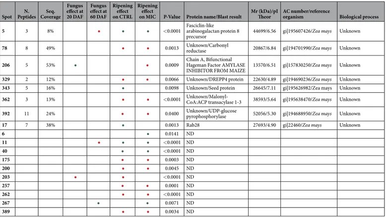
Spot Peptides Coverage 20 DAF 60 DAF on CTRL on MIC P-Value Protein name/Blast result Theor organism Biological process 5 3 8% • • • < 0.0001 Fasciclin-like arabinogalactan protein 8
precursor 44699/6.56 gi|195607426/Zea mays Unknown 78 8 49% • • 0.0013 Unknown/Carbonyl reductase 20867/6.84 gi|194701990/Zea mays Unknown 206 5 53% • • 0.0009 Chain A, Bifunctional Hageman Factor AMYLASE
INHIBITOR FROM MAIZE 13570/6.51 gi|157830250/Zea mays Unknown 329 2 12% • • 0.0066 Unknown/DREPP4 protein 22630/4.89 gi|194690236/Zea mays Unknown 343 5 16% • 0.0098 Unknown/Seed protein 26645/7.11 gi|195626982/Zea mays Unknown 362 3 13% • • < 0.0001 Unknown/Malonyl-CoA:ACP transacylase 1-3 38593/5.64 gi|195638470/Zea mays Unknown 392 11 24% • • 0.0400 Unknown/UDP-glucose pyrophosphorylase 52056/5.30 gi|194688950/Zea mays Unknown
17 7 38% • 0.0013 Rab28 27693/4.90 gi|22460/Zea mays Unknown
6 • 0.0141 ND 11 • • • < 0.0001 ND 40 • • < 0.0001 ND 175 • • 0.0003 ND 200 • • 0.0045 ND 203 • • < 0.0001 ND 257 • • 0.0001 ND 262 • • < 0.0001 ND 267 • • 0.0071 ND 389 • • 0.0034 ND
Table 2. Information regarding modulated proteins spot number, number of identified peptides, sequence coverage, optical density variation using colour code, ANOVA P-value, protein name and Blast results when present, theoretical molecular weight and pI (experimental pI data were not reported because the isoelectrofocusing was performed on non-linear IPG strips and image analysis software was not able to precisely calculate the pI), accession number and reference organism, biological process in which the identified protein was involved.
Despite the fact that the root is the organ that is colonised by AM fungi, the physiology of the entire plant is affected by the symbiosis, with interaction with the fungus having been reported to modulate photosynthesis, leaf hydration, reproduction and fruit quality in both maize and other plant species6,26,36,37. This modulation of the
plant physiology changes according to the different stages of a plant’s development and which plant organs are being analyzed. As shown in Table 2, the maize seed proteome of mycorrhizal plants differed either at the begin-ning of seed development (20 DAF) or at the end of maturation (60 DAF).
Effects during seed development. A detailed examination of the results revealed that at 20 DAF the AM symbiosis induced the up-regulation of enzymes involved in energetic metabolism, the latter stages of embryo development, nucleotide metabolism, seed storage and stress responses. AM fungi enhances primary metabolism by up-regulating ATP synthase (spot 219); this protein is a key enzyme whose expression is linked to respira-tory and photosynthetic phosphorylation, both of which are major processes in the energetic metabolism of above-ground plant tissues. The up-regulation of the cytosolic ascorbate peroxidase (spot 387), a major enzyme involved in detoxification of hydrogen peroxide, was also induced by AM fungi; its expression may be linked with embryo development. Thus, Méchin et al.24 reported that this enzyme is modulated in maize seeds 14 days after
pollination24, whereas Finnie et al.38 showed that a cytosolic form of this enzyme was only detectable in an early
developmental stage of barley seeds.
The overexpression of nucleoside diphosphate kinase 1 (spot 111) may lead to reduced constitutive reactive oxygen species (ROS) levels and enhaced tolerance to multiple environmental stress39. The expression of
nucle-oside diphosphate kinase has been reported to increase in response to drought and salinity, thus it is expected to accumulate in the late phases of embryogenesis. This enzyme also plays significant roles in hormone responses, heat stress and, in general, growth and development39. An increase of ROS could induce the observed increase of
heat shock 70 kDa protein (spot 55) expression.
The accumulation of Legumin 1 (spot 179), a storage protein found in maize seeds, is a confirmation of the data of globulin quantification and is linked with the seed storage process. The AM symbiosis induced down reg-ulation of three starch granule-associated proteins, namely phosphoglucomutase 2 (spot 245), phosphoglycerate mutase (spot 136), and a pyruvate ortophosphate dikinase (spot 74) as well as seven proteins involved in cellular metabolic processes, an elongation factor 2 (spot 108), a translational initiation factor eIF-4A (spot 360), an ATP-dependent Clp protease ATP-binding subunit (spot 86), a ketol-acid reductoisomerase (spot 292), a stress responsive protein (spot 59) and succinate dehydrogenase flavoprotein subunit (spot 394). In order to use their
stored carbon reserves, plants must be able to degrade their starch granules to oligosaccharides and monosac-charides. In particular, as previously reported, phosphoglucomutase 2 converts glucose 1-phosphate to glucose 6-phosphate facilitating the use of this compound in glycolysis40. The orthophosphate dikinase partly controls the
composition of the storage protein fractions and the starch-protein balance24. The classical role of orthophosphate
dikinase in both C3 and C4 plants involves catalyzing the reversible reaction of pyruvate, ATP and phosphate to phosphoenol-pyruvate, AMP and diphosphate. In rice, the expression of ortophosphate dikinase was found to be highest at 5–15 days after pollination; after that time this enzyme was likely rapidly degraded or inactivated through phosphorylation41. This pool of inactivated orthophosphate dikinase was also present in mature seeds,
suggesting a role in developmental processes during seed germination39,42.
Effects at seed maturation. At 60 DAF, the presence of AM fungi induced the modulation of 33 maize seed proteins, 4 up-regulated and 29 down-regulated. The degradation of the reserves (starch and storage pro-teins) and of some functional proteins could provide enough energy and amino acids for seed germination and for embryo development43. This could explain the up-regulation of proteasome proteins (spot 398) and the strong
down regulation of different enzymes in AM-treated plants, such as adenine phosphoribosyl transferase (spot 114), ATP synthase beta chain (spot 219), sorbitol dehydrogenase (spot 274), prohibitin 3 and 2 (spots 36 and 45, respectively), two actin depolymerizing factor (spots 29 and 70), ankyrin repeat domain-containing protein 2 (spot 382), and late embryogenesis abundant protein Leal 4-A (spot 61).
Maize seeds acquire the ability to germinate during the stage of maturation drying44. The decreased water
content plays an important role for the seeds to acquire the ability to germinate and for protection against fun-gal infection. Germination is a potentially stressful process and the reactivation of metabolism may provide an important source of ROS44. This can explain the increase in the abundance of proteins linked to the ROS
response and AM symbiosis an overexpression of the same proteins such as salt tolerance protein (395) and down-regulation of APx-1 cytosolic ascorbate peroxidase (387), splicing factor (9), two spots belonging to nucle-oside diphosphate kinase 1 (spots 111 and 319) that can lead to decreased constitutive reactive oxygen species (ROS) levels and enhanced tolerance to multiple environmental stress39, a peroxiredoxin (335), superoxide
dis-mutase3 (273), a 22 kDa heat shock protein (4), the activator of a 90 kDa heat shock protein ATPase (49), and a stress responsive protein (59). The down regulation, in AM-treated seeds, of different isoforms of storage proteins such as Zein-alpha 19D1 precursor (21), vicilin-like embryo storage protein (spots 26, 51 and 242), z1B alpha zein protein (64) and lactoylglutathione lyase (213) could be linked with the seed protein turnover induced by embryo maturation.
Ripening effect on maize seed proteome. Maize is an excellent model for research on cereal seed development because of the relatively large size of both its embryo and endosperm. Despite the importance of seed maturation information for agricultural purpose, there is scant data available in literature regarding the effects of root AM fungal inoculation on seed maturation. Kegg maps (Fig. 3 and Table S4) summarizes the main biochemical pathways involved in the maize seed proteome modification during ripening, i.e. carbon fixation; starch and sucrose metabolism; the pentose phosphate pathway; the citrate cycle; glycolysis/gluconeogenesis; valine, leucine and isoleucine biosynthesis; alanine, aspartate and glutamate metabolism; glyoxylate and dicar-boxylate metabolism; pyruvate metabolism; purine metabolism; and cysteine and methionine metabolism. Both in control and in mycorrhizal plants, seed maturation induced the same proteome evolution with the exception of: malate dehydrogenase, succinate dehydrogenase, adenosine kinase, adenylate kinase, acetolactate synthase, ketol-acid reductoisomerase, homocysteine S-methyltransferase and methionine synthase (down-regulated in CTRL and not modified in MIC); alanine transaminase, alanine-glyoxylate transaminase, polygalacturonase, 6-phosphofructokinase, glycine transaminase, 1-aminocyclopropane-1-carboxylate synthase (up-regulated in CTRL and not modified in MIC); nucleoside diphosphate kinase (up-regulated in CTRL and down-regulated in MIC); and zein-protein precursor (up-regulated in MIC and not modified in CTRL). These changes in protein abundance could be linked with the higher content of starch in the seeds of plants treated with mycorrhizal fungi.
Our results are in agreement with those of Huang et al.45, who reported, on the basis of the metabolic and
functional features of maize embryos, the identification of proteins classified into 7 major categories belonging to 3 functional groups: protein metabolism (26%), stress response (21%) and carbohydrate and energy metabolism (17%). At maturity, the maize seed accumulates large amounts of starch and storage proteins45. However, proteins
involved in stress response (24%) were often up-regulated during seed maturation45.
A large body of literature describes the effects of AM fungi on the physiology of whole plants6,7,16,17, with a
particular focus on fruit composition. The work presented here is a first step in filling the gap in the knowledge of the effect of AM fungi on seed composition. In the work described here, it was demonstrated that AM fungi strongly modify the seed proteome, particularly up-regulating enzymes involved in energy metabolism, embryo development, nucleotide metabolism, seed storage and stress responses.
Finally, this work underlines the importance of using soil microorganisms as inocula in field production to sustainably improve crop quality.
Materials and Methods
Experimental Field, Plant Growth And Seed Sampling. The experiment was conducted as described in Berta et al.26. In accordance with standard agricultural practices, field soil was fertilized with potassium sulfate
(400 Kg/ha) and 18/46 N/P (350 Kg/ha). Corn seeds (Zea mays var. Ostiglia) were sown on 14th March 2013 in double rows. Three double lines (200 plants each) were treated with AM inoculum. An uninoculated double row was selected ramdomly as a control. The AM inoculum (Mybasol s.r.l., Alessandria, Italy), consisted of root frag-ments, spores, and hyphae of Rhizophagus intraradices, Glomus aggregatum, Glomus viscosum, Claroideoglomus
etunicatum, and Claroideoglomus claroideum produced on sorghum, containing about 85,000 infective prop-agules l−1, was applied every 6 cm using a drip irrigation tube. During the growth period, diseases and insects
were adequately controlled. Caryopsis harvest started from the 26th of July, 20 DAF and ended on the 4th of
September, 60 DAF. During each sampling date, tillers of three kernels of control (CTRL) and mycorrhizal (MIC) plants were open, 25 g of grains from half of each ear, were collected, immediately frozen in liquid nitrogen, and stored at −80 °C.
Five, randomly selected, roots per treatment were used to evaluate frequency (F%), mycorrhizal degree (M%) and arbuscule abundance (A%)46.
Figure 2. ((a) (CTRL), (b) (MIC)). 2D maps of seed proteins extracted from seeds at 60 days after flowering (DAF), stained with Colloidal Coomassie. The assigned spots in the map were those modulated by AM symbiosis (green, up-regulated spots; red, down-regulated spots).
Selective extraction of different protein classes. Ten grams of seeds were ground in a mortar using liquid nitrogen and extracted twice with milliQ water containing a protease inhibitor cocktail (Sigma-Aldrich), in the ratio 1:10 (p/v), at 4 °C for 2 hours. The slurries were centrifuged at 10,000 × g for 15 min. The two superna-tants (albumin fraction) were pooled and stored at − 20 °C, whereas the pellet was extracted twice with Tris-HCl 50 mM, pH 8.0, containing 0.3 M NaCl. The slurries were centrifuged at 10,000 × g for 15 min. The supernatants (globulin fraction) were pooled and stored at − 20 °C. The insoluble pellet was extracted twice with 70% eth-anol containing 0.2% 2-mercaptoetheth-anol. After stirring for 3 hours at 4 °C, the suspension was centrifuged at 10,000 × g for 30 min at 4 °C. The supernatants (prolamin fraction), were pooled and dried with a Rotavapor device. The insoluble pellet was then resuspended in 0.1 M NaOH to extract the glutelin fraction at 4 °C for 2 hours.
Three biological replicates were analysed in triplicate. Protein concentrations were determined according to Bradford47.
Seed water content was determined by placing one gram of ground seeds at 110 °C and then in a jar containing silica gel. Samples were analyzed twice in duplicate.
Proteomic analysis. Proteins were extracted according to Bona et al.48. The pellet was resuspended in 1 ml
of solubilization buffer containing 7 M urea, 2M thiourea, 4% CHAPS, 100 mM DTT, 1% IPG buffer (3–11 NL) and quantified by the method of Bradford47. Aliquots of 700 µ g of protein extracts were mixed with a rehydration
buffer (8 M urea, 4% w/v CHAPS, 18 mM DTT, 0.5% 3–11 IPG Buffer), focused at 60 kVh at 20 °C on precast 13 cm NL pH 3–11 strips in an IPG-Phor unit (GE Healthcare Bio-Sciences) and separated on 12% gels at 10 °C under constant amperage (30 mA per gel) with a Protean Plus Dodeca gel (BioRad). At least ten replicates were run, two analytical replicates per five biological replicates.
Gels were stained according to Candiano et al.49, and then digitized in a GS 710 densitometer (Bio-Rad). The
gel images were analyzed using SameSpot (Progenesis v. 2006) (build 3419. 12870). Differential expression anal-ysis was performed: i) comparing the quantity of matched spots in the CTRL at 20 DAF versus MIC plants at 20 DAF (to evaluate the effect of AM fungus addition at the beginning of maturation); ii) comparing the quantity of matched spots in the CTRL at 60 DAF versus MIC plants at 60 DAF (to evaluate the effect of AM fungus addition at the end of the maturation period); iii) comparing the quantity of matched spots in the CTRL at 20 DAF versus CTRL plants at 60 DAF (to evaluate protein changes during maturation); iv) comparing the quantity of matched spots in the MIC plants at 20 DAF versus MIC plants at 60 DAF (to evaluate protein changes during maturation in AM plants). The software created a quantitative table with all normalized optical spot densities that allowed Figure 3. Kegg maps summarizing the main biochemical pathways involved in proteome modification during ripening: carbon fixation, starch and sucrose metabolism, pentose phosphate pathway, citrate cycle, glycolysis/gluconeogenesis, valine, leucine and isoleucine biosynthesis, alanine, aspartate and glutamate metabolism, glyoxylate and dicarboxylate metabolism, pyruvate metabolism, purine metabolism, cysteine and methionine metabolism.
all replicates.
Protein identification by nano-LC-Q-TOF MS/MS. For MS analysis, spots of interest were cut from the gel and destained overnight with a solution of 25 mM ammonium bicarbonate and 50% acetonitrile. The proteins were digested with trypsin (Roche, Segrate, Milano, Italy) in-gel digested as described by Hellmann et al.50. All nano-HPLC-MS/MS experiments were performed on a Q-TOF mass spectrometer Q-Star XL (AB
Sciex, Concord, Ontario, Canada) controlled by the Analyst QS 1.1 software (AB Sciex) connected to an Ultimate 3000 nano-HPLC system. The peptide pellets were resuspended in 10 µ l of solvent A (95% v/v water, 5% v/v acetonitrile, 0.1%v/v formic acid). Five microliters of each sample were loaded onto the precolumn, 300 µ m i.d. × 5 mm, C18 PepMap, 5 µ m beads, 100 Å, (LC-Packings) and washed for 5 min using a flow rate of 40 µ l min−1
solvent A. The peptides were subsequently eluted at 300 nl min−1 from the precolumn over an analytical column,
15 cm × 75 µ m, C18 PepMap100, 3 µ m beads, 100 Å (LCPackings) using a 35 min gradient from 5 to 60% solvent B (5% v/v water, 95% v/v acetonitrile, 0.1% v/v formic acid) delivered at 300 µ l min−1. The analytical column was
connected with a 15 µ m inner diameter Silica Tip (Pico Tip) nanospray emitter (New Objective, Woburn, MA). The spray voltage (set between 1800 and 2000 V) was applied to the emitter through a stainless steel union and tuned to get the best signal intensity using a standard BSA tryptic digest before every sample’s batch submission. The QStar-XL was operated in information-dependent acquisition (IDA) mode. Mass spectra were acquired from 400 to 1800 m/z. The two most intense ions with charge states between 1 and 4 in each survey scan were selected for the MS/MS experiment. MS/MS data were acquired from 60 to 1800 m/z. Each acquisition cycle was com-prised of a 1 s MS and a 3 s MS/MS. The MS to MS/MS switch threshold was set to 15 counts per second (c.p.s.). All precursor ions subjected to MS/MS in the previous cycle were automatically excluded for 60 s using a 3 amu.
Homology-driven proteomics. Mascot Distiller (Matrix Science, London, UK) was used to create peak lists from MS and MS/MS raw data. Mascot Server (Matrix Science) was used for database searching versus NCBInr. The last check for proteins homology assignments was made versus NCBInr 20151214 (78002046 sequences; 28422168805 residues). Carbamidomethylation of cysteine residues, oxidation of methionine, deam-idation of asparagine and glutamine were set as possible variable modifications and trypsin was selected as the protease. One missed cleavage site was allowed, and the peptide MS and MS/MS tolerance was set respectively to 100 ppm and 0.2 Da. Positive identifications were assigned with a minimum of two unique peptides with at least one peptide having a significant ion score (underlined in red in Table S2 in the supporting information). Considering the scarce number of corn sequences in the databases, if we obtained an automatic hit without a sig-nificant score, sequence tags were manually interpreted from the ESI-MS/MS spectra to confirm the hypothetical assignment. We also accepted hits identified by at least one peptide with a significant ion score according to the MASCOT MS/MS ion search algorithm as being confident assignments. When a protein has only one spectrum with a significant Mascot score, but in the results there are more spectra with lower scores, they were manually inspected and if they had a pattern compatible with the theoretical peptide, they were considered for homology searching. The sequence obtained from the manually reconstructed peptide was submitted to MS homology and if the first positive hit was the same protein or a homologue sequence of the one automatically recognized, the peptide was inserted in the table as assigned to the protein. This approach allow the use of partial “de novo” sequences that can be more fitting to the sequences in the database51.
Blast2GO data analysis. To perform the Blast2GO analysis (http://www.blast2go.com/b2ghome) we downloaded the protein FASTA sequences from http://www.ncbi.nlm.nih.gov using the GI code ID.
Data analysis was performed with Blast2GO standard parameters.
The EC annotations, obtained by mapping from equivalent GO annotations, were visualized reconstruct-ing the structure of the Gene Ontology relationships and ECs on KEGG maps (http://www.genome.jp/kegg). In KEGG maps were displayed the enzymatic functions of sequences in the context of the metabolic pathways in which they participate.
Statistical analysis. Data were analyzed by a one-way ANOVA followed by Fisher’s test with cut-off signif-icance at p = 0.05 using Stat View 4.5 (Abacus Concepts) software.
References
1. Smith, S. E. & Read, D. Mycorrhizal symbiosis 3rd edn (eds Smith, S. E., Read, D.) The symbionts forming arbuscular mycorrhizas, 13–41 (Academic press, New York, 2008).
2. Schlussler, A., Schwarzott, D. & Walker, C. A new fungal phylum, the Glomeromycota: phylogeny and evolution. Mycol. Res. 105, 1413–1421 (2001).
3. Sawers, R. J. H., Gutjahr, C. & Paszkowski, U. Cereal mycorrhiza: an ancient symbiosis in modern agriculture. Trends Plant Sci. 13, 93–7 (2008).
4. Javot, H., Pumplin, N. & Harrison, M. J. Phosphate in the arbuscular mycorrhizal symbiosis: transport properties and regulatory roles. Plant. Cell Environ. 30, 310–22 (2007).
5. Bonfante, P. & Genre, A. Mechanisms underlying beneficial plant-fungus interactions in mycorrhizal symbiosis. Nat Commun 1, 1–11 (2010).
6. Lingua, G. et al. Arbuscular mycorrhizal fungi and plant growth-promoting pseudomonads increases anthocyanin concentration in strawberry fruits (Fragaria x ananassa var. Selva) in conditions of reduced fertilization. Int. J. Mol. Sci. 14, 16207–16225 (2013). 7. Bona, E. et al. AM fungi and PGP pseudomonads increase flowering, fruit production, and vitamin content in strawberry grown at
low nitrogen and phosphorus levels. Mycorrhiza 25, 181–193 (2015).
8. Castellanos-Morales, V., Villegas-Moreno, J., Vierheilig, H. & Cárdenas-Navarro, R. Nitrogen availability drives the effect of Glomus intraradices on the growth of strawberry (Fragaria x ananassa Duch.) plants. J. Sci. Food Agric. 92, 2260–2264 (2012).
9. Castellanos-Morales, V. et al. Root colonisation by the arbuscular mycorrhizal fungus Glomus intraradices alters the quality of strawberry fruits (Fragaria × ananassa Duch.) at different nitrogen levels. J. Sci. Food Agric. 90, 1774–1782 (2010).
10. Copetta, A., Bardi, L., Bertolone, E. & Berta, G. Fruit production and quality of tomato plants (Solanum lycopersicum L.) are affected by green compost and arbuscular mycorrhizal fungi. Plant Biosyst. 145, 106–115 (2011).
11. Baslam, M., Esteban, R., García-Plazaola, J. I. & Goicoechea, N. Effectiveness of arbuscular mycorrhizal fungi (AMF) for inducing the accumulation of major carotenoids, chlorophylls and tocopherol in green and red leaf lettuces. Appl. Microbiol. Biotechnol. 97, 3119–3128 (2013).
12. Aimo, S. et al. Use of arbuscular mycorrhizal fungi and beneficial soil bacteria to improve yield and quality of saffron (Crocus sativus L.). ISHS Acta Hortic. 850, 159–162 (2010).
13. Borde, M., Dudhane, M. & Jite, P. K. Role of bioinoculant (AM Fungi) increasing in growth , flavor content and yield in Allium sativum L. under field condition. Not. Bot. Horti Agrobot. Cluj-Napoca 37, 124–128 (2009).
14. Ceccarelli, N. et al. Mycorrhizal colonization impacts on phenolic content and antioxidant properties of artichoke leaves and flower heads two years after field transplant. Plant Soil 335, 311–323 (2010).
15. Chaudhary, V. & Kapoor, R. & Bhatnagar, a. K. Effectiveness of two arbuscular mycorrhizal fungi on concentrations of essential oil and artemisinin in three accessions of Artemisia annua L. Appl. Soil Ecol. 40, 174–181 (2008).
16. Copetta, A., Lingua, G. & Berta, G. Effects of three AM fungi on growth, distribution of glandular hairs, and essential oil production in Ocimum basilicum L. var. Genovese. Mycorrhiza 16, 485–94 (2006).
17. Copetta, A., Lingua, G., Bardi, L., Masoero, G. & Berta, G. Influence of arbuscular mycorrhizal fungi on growth and essential oil composition in Ocimum basilicum var. Genovese. Caryologia 60, 106–110 (2007).
18. Bona, E. et al. Proteomic analysis as a tool for investigating arsenic stress in Pteris vittata roots colonized or not by arbuscular mycorrhizal symbiosis. J. Proteomics 74, 1338–50 (2011).
19. Aloui, A. et al. On the mechanisms of cadmium stress alleviation in Medicago truncatula by arbuscular mycorrhizal symbiosis: a root proteomic study. Proteomics 9, 420–33 (2009).
20. Valot, B., Negroni, L., Zivy, M., Gianinazzi, S. & Dumas-Gaudot, E. A mass spectrometric approach to identify arbuscular mycorrhiza-related proteins in root plasma membrane fractions. Proteomics 6 Suppl 1, S145–55 (2006).
21. Bona, E. et al. Proteomic analysis of Pteris vittata fronds: two arbuscular mycorrhizal fungi differentially modulate protein expression under arsenic contamination. Proteomics 10, 3811–3834 (2010).
22. Lingua, G. et al. Effects of heavy metals and arbuscular mycorrhiza on the leaf proteome of a selected poplar clone: a time course analysis. PLoS One 7, e38662 (2012).
23. Requejo, R. & Tena, M. Maize response to acute arsenic toxicity as revealed by proteome analysis of plant shoots. Proteomics 6 Suppl 1, S156–62 (2006).
24. Méchin, V., Thévenot, C., Le Guilloux, M., Prioul, J.-L. & Damerval, C. Developmental analysis of maize endosperm proteome suggests a pivotal role for pyruvate orthophosphate dikinase. Plant Physiol. 143, 1203–19 (2007).
25. Willmann, M. et al. Mycorrhizal phosphate uptake pathway in maize: vital for growth and cob development on nutrient poor agricultural and greenhouse soils. Front. Plant Sci. 4, 1–15 (2013).
26. Berta, G. et al. Maize development and grain quality are differentially affected by mycorrhizal fungi and a growth-promoting pseudomonad in the field. Mycorrhiza 24, 161–70 (2014).
27. Cully, D. et al. Endosperm protein synthesis and L-[35S] methionine incorporation in maize kernels cultured in vitro. Plant Physiol.
74, 389–394 (1984).
28. Osborne, T. B. Monographs on Biochemistry 2nd edn (eds Longmans, Green and Co) The vegetable proteins XIII-154 (London, 1924).
29. Shewry, P. & Halford, N. Cereal seed storage proteins: structure, properties and role in grain utilization. J. Exp. Bot. 53, 947–958 (2002).
30. Kriz, A. L. Seed proteins (eds Shewry P. R. & Casey R.) 7S Globulins of cereals 477–498 (Kluwer Academic Publishers, Dordrecht, 1999).
31. Egger, M., Hauser, M., Mari, A., Ferreira, F. & Gadermaier, G. The role of lipid transfer proteins in allergic diseases. Curr. Allergy Asthma Rep 10, 326–335 (2010).
32. Woo, Y., Hu, D., Larkins, B. & Jung, R. Genomics analysis of genes expressed in maize endosperm identifies novel seed proteins and clarifies patterns of zein gene expression. Plant Cell 13, 2297–2317 (2001).
33. Soave, C., Tardani, L., Di Fonzo, N. & Salamini, F. Zein level in maize endosperm depends on a protein under control of the opaque-2 and opaque-6 loci. Cell 27, 403–410 (1981).
34. Feng, L. et al. Expressional profiling study revealed unique expressional patterns and dramatic expressional divergence of maize α -zein super gene family. Plant Mol. Biol. 69, 649–659 (2009).
35. Prat, S., Cortadas, J., Puigdomènech, P. & Palau, J. Nucleic acid (cDNA) and amino acid sequences of the maize endosperm protein glutelin-2. Nucleic Acids Res. 13, 1493–1504 (1985).
36. Taylor, J. & Harrier, L. A. Expression studies of plant genes differentially expressed in leaf and root tissues of tomato colonised by the arbuscular mycorrhizal fungus Glomus mosseae. Plant Mol. Biol. 51, 619–29 (2003).
37. Liu, J. et al. Arbuscular mycorrhizal symbiosis is accompanied by local and systemic alterations in gene expression and an increase in disease resistance in the shoots. Plant J 50, 529–544 (2007).
38. Finnie, C., Melchior, S., Roepstorff, P. & Svensson, B. Proteome analysis of grain filling and seed maturation in barley. Plant Physiol
129, 1308–1319 (2002).
39. Salekdeh, G. H. & Komatsu, S. Crop proteomics: aim at sustainable agriculture of tomorrow. Proteomics 7, 2976–96 (2007). 40. Koziol, A. G., Marquez, B. K., Huebsch, M. P., Smith, J. C. & Altosaar, I. The starch granule associated proteomes of commercially
purified starch reference materials from rice and maize. J. Proteomics 75, 993–1003 (2012).
41. Chastain, C. & Challet, R. Regulation of pyruvate, orthophosphate dikinase by ADP-/Pi-dependent reversible phosphorilation in C3 and C4 plants. Plant Physiol. Biochem. 41, 523–532 (2003).
42. Chastain, C., Heck, J., Colquhaun, T., Voge, D. & Gu, X. Posttranslational regulation by pyruvate, orthophosphate dikinase in developing rice (Oryza sativa) seeds. Planta 224, 924–934 (2006).
43. Yang, P., Li, X., Wang, X., Chen, H., Chen, F. & Shen, S. Proteomic analysis of rice (Oryza sativa) seeds during germination. Proteomics 7, 3358–3368 (2007).
44. Wang, W.-Q. et al. Proteomic comparison between maturation drying and prematurely imposed drying of Zea mays seeds reveals a potential role of maturation drying in preparing proteins for seed germination, seedling vigor, and pathogen resistance. J. Proteome Res. 13, 606–26 (2014).
45. Huang, H., Møller, I. M. & Song, S.-Q. Proteomics of desiccation tolerance during development and germination of maize embryos. J. Proteomics 75, 1247–62 (2012).
46. Trouvelot, A., Kough, J. & Gianinazzi-Pearson, V. Mycorrhizae: physiology and genetics (eds Gianninazzi-Pearson, V. & Gianinazzi, S.) Mesure du taux de mycorhization VA d’un système radiculaire. Recherche de méthodes d’estimation ayant une signification fonctionnelle, 217–221 (INRA, Paris, 1986).
47. Bradford, M. M. A rapid and sensitive method for the quantitation of microgram quantities of protein utilizing the principle of protein dye binding. Anal. Biochem. 72, 248–254 (1976).
Proteomics 60, 96–101 (2007).
49. Candiano, G. et al. Blue silver: a very sensitive colloidal Coomassie G-250 staining for proteome analysis. Electrophoresis 25, 1327–1333 (2004).
50. Hellman, U., Wernstedt, C., Gonez, J. & Heldin, C. Improvement of an ‘In-Gel’ digestion procedure for the micropreparation of internal protein fragments for amino acid sequencing. Anal. Biochem. 224, 451–455 (1995).
51. Shevchenko, A. et al. Charting the proteomes of organisms with unsequenced genomes by MALDI-quadrupole time-of-flight mass spectrometry and BLAST homology searching. Anal. Chem. 73, 1917–1926 (2001).
Acknowledgements
This work was supported by Dipartimento di Scienze e Innovazione Tecnologica, Università del Piemonte Orientale and by Mossi and Ghisolfi s.r.l. The authors wish to thank the farm Mossi and Ghisolfi S.r.l. and Dr. Alessandro Ausano for his precious help throughout the experiments; Dr. Giorgia Novello for the help during paper revisions; Prof. Bernard Glick for the English Language revision.
Author Contributions
B.E. organized the sampling, performed protein extractions, two-DE analysis, image analysis, protein digestion, MS data elaboration and wrote the paper; S.A. performed the extraction and the quantification of the different protein categories and wrote the paper; M.F. performed MS analysis and cooperated to MS data elaboration; B.L. performed BLAST 2GO analysis; C.A., G.D. and C.P. performed biological experiments in field; N.M. cooperated in statistical data analysis and in the manuscript revision; G.E. cooperated in the paper writing; C.M. cooperated in data elaboration and manuscript revision; G.B. coordinated biological experiments, data analysis and paper writing. All authors revised the manuscript.
Additional Information
Supplementary information accompanies this paper at http://www.nature.com/srep Competing financial interests: The authors declare no competing financial interests.
How to cite this article: Bona, E. et al. Arbuscular mycorrhizal symbiosis affects the grain proteome of Zea mays: a field study. Sci. Rep. 6, 26439; doi: 10.1038/srep26439 (2016).
This work is licensed under a Creative Commons Attribution 4.0 International License. The images or other third party material in this article are included in the article’s Creative Commons license, unless indicated otherwise in the credit line; if the material is not included under the Creative Commons license, users will need to obtain permission from the license holder to reproduce the material. To view a copy of this license, visit http://creativecommons.org/licenses/by/4.0/