Scuola Normale Superiore di Pisa
Ph.D. Thesis
Molecular Biology
Localization and dynamics of homeotic
oncogenic protein HOXC13 in pre-initiation
complex of human DNA replication origins
Laura Comelli
ADVISOR
Prof. Arturo Falaschi
Contents
Publications.
...5Abstract
...61.Introduction
...91.1.DNAreplication...9
1.1.1. Eukaryotic DNA replication...9
1.1.2. The Replicon model...9
1.1.3. Replication complex...12
1.2.Eukaryotic origins of DNA replication...17
1.2.1. Replication origins...17
1.2.2. Origins spatio-temporal organization...20
1.2.3. Origin activity and transcription...21
1.2.4. Chromatin structure influence on origin activity...24
1.2.5. Histone modification and Trichostatin A...25
1.2.6. Chromatin remodelling factors...28
1.2.7. Role of DNA topology...29
1.2.8. Cruciforms structures...30
1.3. Hox proteins...31
1.3.1. HOX genes...31
1.3.2. Hox cluster in Drosophila and Human...33
1.3.3. HOX and its cofactors...34
1.4.Hox genes and cancer...39
1.4.1. Hox as oncogenes...39
1.4.2. HOX, NUP98 and leukaemia...41
1.4.3. Oncogenic potential of homeobox genes...43
1.4.4. Role of HOX-Pbx interaction in cancer...45
2.Results
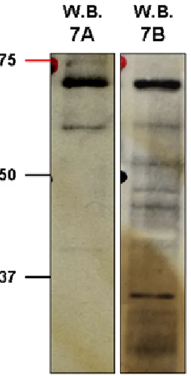
...512.1. Characterization of polyclonal anti-HOXC13 antibody...51
2.1.1. Western blot detection of endogenous and double tagged HOXC13 in cells extracts...52
2.1.2. Partial proteolytic peptide maps...53
2.1.3. Immunoprecipitation of tagged HOXC13...55
2.2. Expression of HOXC13 in different human cell lines...56
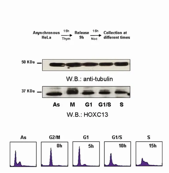
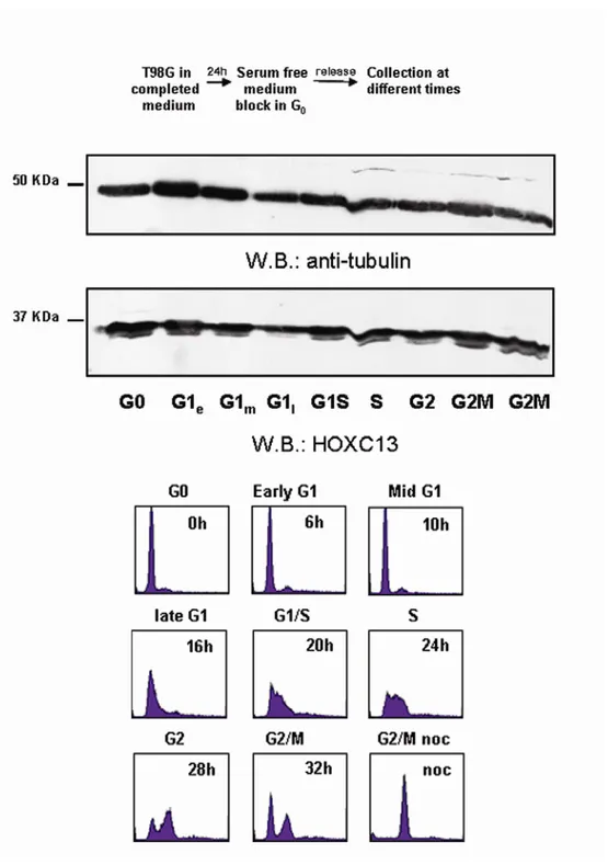
2.3. Protein expression of HOXC13 throughout the cell cycle...57
2.4. Interactions of HOXC13 with other replication protein complexes...61
2.5. Binding of HOXC13 to lamin B2 origin...66
2.5.1. Cell cycle dependent binding of HOXC13 to lamin B2...70
2.5.2. Analysis of the lamin B2 origin structure by footprinting assay...72
2.6. Interaction of HOXC13 with other origins...74
2.6.1. Cell cycle dependent binding of HOXC13 to TOP1 and MCM4...77
2.7. UV-photofootprinting of origin structure...79
2.7.1. Effects of disruption of origin chromatin...81
2.8. Dispensability of HOXC13 on the lamin B2 origin...83
2.8.1. HOXC13 silencing do not alter the cell cycle progression...86
3.Discussion
...923.1. Conclusion and future perspective....99
4.Materials and Methods
...1014.1. Cell culture, synchronization and TSA treatment...101
4.2. Antibodies...102
4.3. Preparation of Nuclear Extracts...103
4.4 Western blot analyses...103
4.5. Gradient gel...104
4.6. Partial proteolytic peptide maps...104
4.7. Chromatin Immunoprecipitation...105
4.9. GST pull-down assay...107
4.10. Competitive PCR analysis...108
4.11. PCR with multiple pairs of primers...108
4.12. ChIP and dimethylsulfate (DMS) treatment...109
4.13. RNA depletion and stable clones production...111
4.14. Brdu incorporation experiment...111
Acnowledgments
...114Publications
Marchetti L; Comelli L; D'Innocenzo B; Puzzi L; Luin S; Arosio D; Calvello M; Mendoza-Maldonado R; Peverali F; Trovato F; Riva S; Biamonti G; Abdurashidova G; Beltram F; Falaschi A. "Homeotic proteins participate in the function of human DNA replication origins". Nucleic Acid Research 2010 Ago 6
Comelli L, Marchetti L, Arosio D, Riva S, Abdurashidova G, Beltram F, Falaschi
A. "The homeotic protein HOXC13 is a member of human DNA replication complexes". Cell Cycle. 2009 Feb 1;8(3):454-9. Epub 2009 Feb 19
Abstract
In metazoan cells the DNA replication origins are not well defined. Differently from what observed for bacteria cells and for budding yeast, in metazoan the origins does not show a conserved sequence and they appear to be specified by many factors. In order to better understand the mechanisms involved in the origin specification, many studies have been done to identify the proteins involved in the recognition and activation of the origins. From these kind of analysis is emerging that, beside the well-known proteins of the pre replicative complex, also other factors might be involved. Between these, the HOX proteins seem to be able to play a role in the origin activity. One of the first studies of this involvement was done by our group and leads to the identification of three homeotic proteins able to specifically bind in vitro the human lamin B2 origin. Thus, in the study conducted during this PhD program, was investigated the involvement of one of these homeotic proteins, namely HOXC13, with human DNA replication origins and with replicative complexes.
We found an interaction of HOXC13 with two crucial factors of the pre Replication Complex (pre-RC), ORC1 and Cdc6 and that HOXC13 binds a good fraction of the origins, in particular the early replicating ones, like the lamin B2 origin and other known human origins. The HOXC13 protein is bound to origin chromatin, at least for the lamin B2 origin, at a precise site within the pre-RC at specific moments of the cell cycle. Interaction with the origin occurs within the area protected by the pre-RC in G1, very close to the start sites of leading strand synthesis and to the binding sites of ORC1, ORC2, Cdc6, topoisomerase (topo) I and topo II. The protein is absent from the origin in M and appears on it at the beginning of G1, reach a peak at G1/S and as synthesis starts, the interaction of HOXC13 with the origin fades, in parallel with the transition from this large pre-RC to a smaller and differently organized post-RC.
Recently also other HOX proteins have been identify as proteins involved in regulation processes of DNA replication, suggesting that the interaction of HOXC13 with the origins might occur in a multi-homeotic proteins complex. Depletion of one of these proteins however is compatible with the continuation of the cell cycle and, according with what observed for the other homeotic proteins, we found that also the depletion of HOXC13 does not alter cell cycle progression or S phase entry. This is probably due to the redundancy of homeotic proteins and indicates a relatively generic
Among the identified elements influencing the choice and the activity of a sequence as DNA replication origin, much relevance is assumed by the chromatin structure and topology of DNA. Therefore, we analysed the effects of chromatin structure disruption using Tricostatin A, a histone deacetylase inhibitor. The alteration of chromatin caused by this treatment not only sharply reduces origin function, but also disturbs the binding of replication complex members like HOXC13 and the well known Cdc6 to the DNA replication origins, while does not affect the binding of other unrelated proteins like USF1. On the basis of this finding, we infer that an appropriate chromatin organization and DNA topology strongly influence the binding between factors of the pre Replication Complex and DNA replication origins. This influence could be a key element in origin specification.
The described interactions are not restricted to a single origin nor to a single homeotic protein, leading us to conclude that HOX proteins, probably in the context of a multi-protein homeotic effectors, contribute to recruit and stabilize the replicative complexes onto early replicating origins, in presence of specific chromatin and topological configurations.
The relevance of HOXC13 in DNA replication is also underlined by its involvement in oncogenesis, clearly demonstrated in acute myeloid leukaemia when HOXC13 is fused with NUP98 protein.
1.Introduction
1.1. DNA replication
1.1.1. Eukaryotic DNA replication
Cells begin DNA replication from specifically selected chromosomal sites termed replication initiation sites or replication origins. The chromosomal DNA is subdivided into a number of tandemly organized replicons, ranging in number from ~400 in yeast to ~30,000 in humans. Each replicon contains a replication origin, a sequence that is the final target of specific proteins that lead to the recognition of the specific origin, unwinding of DNA, formation of replication forks and synthesis of new strands. The two opposite moving replication forks progress through the replication until they merge with those issued from the adjacent ones. In metazoans DNA replication is a tightly regulated process, ensuring that the genome is duplicated only once each cell cycle, before chromosome segregation and cytokinesis.
The initial event in DNA replication is constituted by the origin selection mediated by the binding of the Origin Recognition Complex (ORC) into origin DNA sequences. The ORC complex provides a "landing-pad" for other proteins including Cdc6, Cdt1, DNA helicase complex Mcm2-7 and others. All together these proteins form the so called pre-Replication Complex (pre-RC) that is formed in G1 phase of the cell cycle and marks the potential sites for the initiation of DNA replication. Each replication origin spaced apart from 50 to 250 kb, depending on the development stage, growth conditions and cell transformation status.
1.1.2. The Replicon model
The main principles characterizing the events that initiate the DNA replication are conserved in evolution and were described in replicon model, formulated in 1963 by
Jacob and Brenner1. This model postulates the presence of specific cis-acting sequence
elements, termed replicator, that genetically determine replication-initiation sites on DNA molecules. The replicators interact with trans-acting regulatory factors, termed initiators, able to recognize the replicators on the genome in response to appropriate
cellular signals (Figure1). The first event in the initiation of DNA synthesis is the chromatin decondensation and local opening of the duplex strands to provide the access for the initiators.
The replicon model was proposed to explain the regulation of the Escherichia coli DNA replication and provided a foundation to understand how the initiation of DNA replication occurs in all organisms. With this model two new elements were introduced: the existence of specific site where the double helix of DNA is opened and the involvement of proteins other than polymerases in DNA replication.
In bacteria the initiation of DNA replication at oriC requires origin binding by the initiator protein dnaA. This binding alters the structure of the origin and provides an associative platform for targeting additional proteins as dnaB, an helicase involved in the progression of the replication fork. DnaB also allows the loading of an other replication-fork enzyme called primase, which is a replication-priming RNA
polymerase2. Together, the helicase and primase form the core of the so called
"primosome"3. During the entire course of replication, the dnaB helicase catalyzes the
unwinding of genomic DNA in an ATP dependent reaction4. This model was validated
in numerous prokaryotic and viral systems.
The unwinding of the double stranded genomic DNA is energetically unfavorable, thus the cells have evolved helicases enzymes, that couple the energy of NTP binding
and hydrolysis to the unwinding5. Families of helicases share several sequences and
structural motifs, implying that there is a common unwinding mechanism used by these enzymes.
The first extension of this model from prokaryotes to eukaryotic chromosomes was the identification of Autonomously Replicating Sequences (ARS) in budding yeast. Analysis of the ARS revealed that these are about 100bp sequences and consist of a 17bp consensus A-domain region with an 11bp ARS Consensus Sequences (ACS) that is AT rich and flanked by poorly conserved B domains. The A and the B1 domains are binding sites for the proteins involved in DNA replication, while others B elements act as enhancers for the origin efficiency. The identification of consensus ARS elements has permitted the isolation of the Origin Recognition Complex ORC, the protein complex that binds to the origin sequences and allows the DNA replication in all eukaryotes.
Actually, in many eukaryotes the consensus sequence conservation at replicons may not be completely faithful because of the very little sequence specificity among
origins, differently from what observed in budding yeast. AT richness is generally retained feature and is assumed to be important for facilitate the opening of DNA strands at the origin. However, in the other eukaryotes as in fission yeast, the origins are generally longer, without any identifiable conserved consensus sequences analogous to ACS and in many cases, in contrast to the budding yeast, inefficient origins, that fire
randomly, are spread in the genome6. Thus, S. cerevisiae appears to be an exception in
the eukaryotes. The eukaryotic origins seem to be defined by different combinations of elements depending on the context that license them to be recognized by "initiators" including several features like DNA sequence, but there is not a single consensus combination that define an origin.
Figure 1. The replicon model. A trans-acting protein encoded by the initiator gene was proposed
to recognize a cis-acting sequence (the replicator) that controls the initiation of DNA replication in the replicon.
1.1.3. Replication complex
The mechanism of initiation of eukaryotic replication is based on in the "origin licensing" model. In this model, origins are licensed once the pre Replication Complex (pre-RC)is entirely loaded onto them, and the ORC complex is the first to be involved in this assembly (Figure 2).
The ORC complex consists of six subunits (ORC1-6) discovered by specific isolation of ACS binding proteins in budding yeast. These proteins are conserved in evolution but their DNA specific binding dependence, present in budding yeast, is lost in the other eukaryotes. In S. pombe, ORC-origin binding to origins is mediated uniquely by ORC4, which is able to recognize and bind specifically AT-rich sequences
through its AT-hook DNA binding motif7. This feature of ORC4 is limited to S. pombe.
The specificity of mammalian ORC binding to DNA is very low, due to its limited
ability to distinguish specific sequences8. Moreover, the difficulties in identifying
well-defined ORC binding sites in species other than yeast raise the possibility that other DNA binding factors may contribute and facilitate ORC localization and origin selection. The human, frog and S. pombe ORCs preferentially bind DNA to AT rich tracts but there is not a clear consensus sequences among these regions. The AT regions are characterized by their helical instability that facilitates probably the DNA unwinding
and their recognition as replication origins9, 10. Therefore, the chromatin structure, and
not only the primary DNA sequence, might be the element recognized by ORC.
The ORC subunits are AAA+ ATPases (ATPase Associated with various cellular
Activities) but only the ATPase activity of ORC1 is required for DNA binding11.
Contrary to yeast, in mammals some of the subunits of ORC complex are displaced
from the origin site after initiation of DNA replication12, suggesting a more dynamic
interaction between mammalian ORC and origin DNA. In human cells, the ORC1 subunit in S-phase is selectively destabilized, ubiquitinated, partially degraded, and then stably bound to chromatin during the next M to G1 transition, to establish the pre-RC at specific genomic sites in G1 phase. Moreover, species-specific variations exist in the regulation of ORC activity, because although ORC proteins are highly conserved within
a single taxonomic family, conservation among all species is modest13.
The ORC1 ATPase is activated by the binding of Cdc6, an other AAA+ ATPase,
that induced conformational changes on DNA that increase the specificity of the binding of ORC-Cdc6 complex to origins. Moreover, Cdc6 ATPase activity determine its
dissociation from ORC-Cdc6-DNA complex, in non origin sequences, while inhibition of Cdc6 ATPase on origin DNA sequences results in a stable ORC-Cdc6-DNA complex, which can then promote MCM loading to origins. Therefore, Cdc6 ATPase activity regulates origin DNA sequence specificity for the assembly of the pre-RC, required for DNA replication initiation. In fact, mutations increasing Cdc6 ATPase
activity result in a less stable complex on DNA14.
After the origin binding by ORC-Cdc6 proteins, the next step is the loading of Cdt1. Duringthe M to G1-transition, in human cells Cdc6 is degraded and then
resynthesized later during G1-phase15.
Cdt1 has been shown to be a key element in the formation of the pre-RC, in particular, regulating the “once per cell cycle” replication feature. Cdt1 is periodically expressed under the control of the transcription factor Cdc10, which also controls the
expression of Cdc6 in different species16.
Moreover, the Cdc6 ATPase activity is seem to be required for Cdt1 binding to the origin, perhaps determining the following loading of MCM complex onto the ORC-Cdc6-DNA complex. Cdt1 activity is additionally regulated by differential
stoichiometric binding of a DNA replication inhibitor, Geminin, and by proteolysis17.
Geminin is a known as inhibitor of DNA replication that acts by preventing MCM loading onto origins throughout S and M phase and impeding unwanted additional firing events. This protein plays multiple roles in several fundamental cellular processes including proliferation, differentiation, development and transcriptional regulation. All
these functions have been characterized by identifying Geminin binding partners18.
Geminin was shown to interact with Cdt1 during the S phase, targeting it for degradation thereby preventing MCM loading until the following G1 phase and hence
preventing re-replication19. The balances of the Geminin-Cdt1 association establishes
the timing of DNA replication initiation and controls the cell cycle progression20.
During S-G2/M phases, Geminin binds Cdt1 at increased stoichiometric ratios and suppress Cdt1 function. However, through pre-RC licensing, Geminin binds Cdt1 in a lower stoichoimetric ratio, allowing the presence of an active form of Cdt1 that can
interact with Cdc6 and ORC21. The Cdt1-Geminin complex can exist in two distinct
forms, as heterotrimer and as heterohexamer and the hexamer formation is critical for full inhibition of Cdt1 by Geminin. Probably the abbundance of heterotrimer and
heteroexamer is regulated during the cell cycle22. The levels of Geminin rise during S
mitosis or by a nonproteolytic inactivation of the fraction of Geminin that escaped from degradation.
Localization of Cdt1 to the origin is essential for MCM complex recruitment. Then, Cdt1 and Cdc6 dissociate from the origin and ATP hydrolysis by ORC completes
the MCM loading23.
The MCM genes were first identified in mutants defective on the maintenance of
mini-chromosomes in budding yeast (Mcm phenotype)24. A subset of these MCM
mutations were found in a family of six paralogous genes numbered MCM from 2 to 7,
which are highly conserved in eukaryotes. The MCM complex consist of six AAA+
-ATPase subunits MCM 2-7, structured in a ring-shaped conformation around the
chromatin25. After the initial recruitment of MCM to the origin, Cdc6 ATPase activity
allows the disassociation of Cdt1 from the pre-RC, an important step that allows the
MCM ring to close around the DNA23. This ATPase activity is followed by the ORC
ATPase activity that completes the MCM loading reaction and may promote further rounds of MCM loading. Thus, the cooperation between replisome loading and helicase activation ensures coordinated replication of the two strands of DNA.
The assembly of pre-RC is not sufficient to initiate DNA replication, because the replication process requires the activation of two S-phase promoting kinases, CDKs (Cyclin-Dependent Kinases) and DDK (Dbf4-Dependent Kinase,Cdc7) during the G1/S transition. CDKs and DDK phosphorylate and activate pre-RCs promoting the loading of other factors like MCM10, Cdc45 and GINS, thereby forming the pre-IC
(pre-Initiation Complex) and triggering origin firing26, 27. The large multiprotein complex
formed after the licensing is called pre-Initiation Complex (pre-IC), from which start two diverging semi-conservative replicative forks that progress in opposite directions until converge with the replicative forks coming from the adjacent origins.
Phosphorylation of ORC, Cdc6, Cdt1 and MCM proteins also results in their inactivation and/or disassociation from the origin, nuclear export and proteasomal degradation; and finally establish post-RC (post-Replicative Complex), a state required
to prevent origin re-licensing21,28-30.
The CDK and DDK enzymes are independently regulated by similar mechanisms. Both kinases subunits are inactive as monomeric form and are activated by binding of Cyclins for CDK and by Dbf4 for DDK. Recent evidence indicates that the Mcm2-7 complex is a target of phosphorylation by DDK and this event is needed for loading of
the Cdc45 protein31. Hence, the cooperative action of these kinases trigger the recruitment of replication proteins necessary for origin unwinding and DNA synthesis.
In particular, Cdc6 is released from origins by the cyclin dependent protein kinase, CDK2/Cyclin A, and replaced by Cdc45 upon the concert action of Cdc7/Dbf4 and CDK2/cyclin E complex kinases. Then Cdc45 associates with DNA polymerase α and DNA primase and leads to initiate RNA-primed DNA synthesis.
While DDKs seem to act on MCMs, CDKs appear to play a direct role in preventing the assembly of new pre-RCs. Since CDK activity remains high from S phase to the end of the following mitosis, re-licensing cannot occur until the beginning
of the next cell cycle32. At least three members of the pre-RC (ORC, Cdc6 and MCMs)
are phosphorylated by CDKs to prevent pre-RC assembly and re-replication. Moreover, CDKs have also been implicated in controlling the time of replication initiation at
specific origins26. In this way the activation and inactivation of the proteins restrict
DNA replication to once and only once per cell cycle.
The fact that assembly of pre-RC is delayed until mitosis is complete and a nuclear membrane is assembled, suggest that nuclear structure plays a role in the initiation of DNA replication. Chromatin is looped into domains by attachment of the chromatin fiber to the nuclear matrix. DNA sequences that bind preferentially to nuclear matrices are named Matrix Attachment Region (MAR) or Scaffold Associated Region (SAR), which are supposed to mediate this loop formation in vivo. MARs are about 200 bp long, AT-rich and contain topoisomerase II consensus sequences. Moreover, they are often found near to cis-acting regulatory sequences, and their binding sites to the nuclear matrix are abundant (greater than 10,000 per mammalian nucleus). Any DNA sequence, including the origins of replication, could be attached to the matrix at a certain time during the cell cycle. Indeed, a large body of evidence indicates that DNA replication occurs on nuclear matrix but origins are not permanently attached with the
nuclear matrix suggesting a dynamic association during the cell cycle33.
Homologues of the indicated proteins were identified in all eukaryotic organisms, but the attempts to define an origin consensus sequence, or at least common specific features of the origin sequences, lead to the conclusion that no sequence specificity and no origin consensus is identifiable. However, DNA replication starts from specific sites and with a defined spatial programme. The preparation of this programme is crucial for the maintenance of genome integrity, because the disruption of pre-RC formation leads
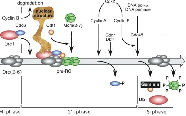
Figure 2. DNA replication in mammalian cells. ORC binds to DNA sequences recognised as
replication origins. ORC exists in M phase as a stable complex of Orc(2–5) subunits, then ORC1 subunit is selectively bound to ORC complex during the M to G1 transition; the remaining ORC subunits remain stably bound to chromatin throughout the cell cycle. Cdc6 associates with ORC1, and this ORC1-Cdc6 complex is present in mitotic cells. In G1 phase, then Cdt1 associated to Cdc6 and loads at least one Mcm(2–7) hexamer per replication fork. In mammals, only ORC1 is associated to nuclease-resistant nuclear structure during G1-phase and then is released from origin previous to DNA replication initiation. Behind pre-RC assembly, DNA replication is initiated by the sequential loading of Mcm10, Cdk2/cyclin A that phosphorylate and release Cdc6, Cdc7/Dbf4 and Cdk2/cyclin E that subsequently modify other members of pre-RC and finally allow Cdc45 to bring polymeraseα and DNA primase to the replication origin DNA, the later enzyme initiates synthesis of the first RNA-primed nascent DNA strands. This event marks the beginning of S-phase. Concomitant with DNA synthesis is taking place the inactivation of Cdt1 by Geminin and the phosphorylation of MCM proteins. Cdc6 and Cdt1 are then released from chromatin and eventually degraded. MCM proteins remain in the nucleus where they are weakly associated with chromatin.
1.2 Eukaryotic origins of DNA replication
1.2.1. Replication origins
In multicellular organisms the origins of DNA replication are still not well defined. The main difference between prokaryotic and eukaryotic replication origins resides in the genome became larger in eukaryotes and the replication of chromosomal DNA depends on the activity of many different origins, that have to be activated with
fine coordination in space and time35, 36. In fact, in higher eukaryotes, respect to the
yeast, the genome size is about 100 times larger and this requires a bigger number of origins to ensure complete DNA replication. The increased number of origins also makes their mapping more difficult.
The discovery of the hexameric ORC complex in all eukaryotes from budding yeast to man now provides a strategy to look for the common features of origins of DNA replication. Although sequences bound by ORC complex could define the origins, differently from what observed for yeast, in Drosophila and human, the ORC complex
have little or not sequence specificity binding activity37. The flexible sequence
specificity observed in multicellular eukaryotes, respect to bacteria and S. cerevisiae, probably allows more dynamic changes in the organization of the genome and its replication.
If ORC complex does not preferentially bind specific sequences, probably other elements drive ORC to the origins. One possibility is that other proteins interacting with ORC carry it to a specific sequence. This might occur through the direct interaction of ORC with chromatin remodelling complexes or proteins that alter the local topological state of DNA, as occur for Epstein-Barr virus (EBV). The EBV origin of plasmid replication (OriP) provides an interesting model to study ORC recruitment in human cells. EBV is a human herpesvirus that establishes latent infections in multiple cell types and this latent form exists as an episomal minichromosome that replicate once per cell
cycle and segregates similar to the cellular chromosome38. The EBNA1 protein of the
virus interacts with ORC and with other cellular factors to recruit ORC and contribute to
OriP replication activity39. By the same approach, it was found that not only the
interaction with other factors, but also several features of the chromosomes, influence the firing of replication origins, as observed for methylation of DNA, that blocks the
modifications that influence the activity of replication origins. The same was shown for
Drosophila ORC binding, that is inhibited by the DNA methylation on GC
nucleotides40. In addition, Drosophila ORC prefers negatively supercoiled DNA,
proving that the DNA topology could be a more important determinant than DNA
sequence for ORC binding8.
Thus, even if the proteins involved in eukaryotic DNA replication are conserved, the sequences on which they bound are highly divergent because metazoan ORC complex exhibit no sequence binding specificity.
Despite the inability of investigators to isolate an autonomously replicating sequence in mammalian systems, approximately 20 mammalian origins have been identified. Mammalian origins could be classified in two groups, one containing regions referred to as zone of initiation, where replication begins from one or several potential sites within a large region of DNA, and the other group including origins where replication initiates from a localized site in each cell cycle.
Many examples of initiation zones exist, including the human rRNA locus, the Chine hamster rhodopsin and the DHFR (dihydrofolate reductase) loci. The best characterized is the Chine hamster ovary DHFR locus, with at least three primary initiation site (ori β, β' and γ) that account for most of the initiation events in this
region41. Analysis of different DNA segments to drive initiation of replication when
placed in different chromosomal context, revealed that in high eukaryotes replication requires specific DNA sequences that are both close to and distant from the site of initiation. Therefore, local sequence alterations can either enhance or repress origin activity, but no single consensus DNA-sequence motif is necessary or sufficient for
replicator activity42. Moreover, by two-dimension gel mapping of a single copy of
DHFR locus indicated that replication begins from multiple sites spanning 55 Kb intergenic region between DHFR and the adjacent locus. Preference for initiation was
seen inside the central 35-40 Kb region, know to contain oriβ and oriγ43. This study
suggested that mammalian initiation zone is composed of a primary initiation site coupled with multiple lower frequency sites.
An example of mammalian origin where firing is restricted to a circumscribed site is represented by the human β-globin origin. Originally, initiation of replication from this locus was thought to start from a single bidirectional origin of replication. Thn, more detailed studies indeed revealed that the locus is actually composed of two non
overlapping genetic elements that have been described to behave as redundant
replicators when β-globin origin is assayed as an ectopic chromosomal site44.
An other example of mammalian replicators is the lamin B2 origin, the best characterized among all isolated human DNA replication origins. It was mapped on chromosome 19, in the 3' untraslated region of the lamin B2 gene and consists of an AT rich region of 70-100 bp that contains 11 bp directed repeats, in one of which a
bidirectional initiation site was precisely mapped45. Lamin B2 origin displays a cell
cycle dependent footprint that is longer at G1 and shrinks as cells progress into S
phase46. Inside this footprint area the transition between leading and lagging strands
synthesis has been mapped to a single nucleotide level45. Moreover, it was demonstrated
that cell cycle dependent footprints are in part due to the pre-RC binding. The chromatin surrounding the lamin B2 origin in G1 phase is bound by Cdc6, ORC1, ORC2 and MCM3, in S phase was found to bind by ORC2 and in M phase none of these pre-RC
members were detected47. This data suggests that mammalian origins are bound by the
same replication machinery as found in other eukaryotic systems, and this follow similar cell cycle regulation as in yeast. Additionally, this origin remains functional when transferred to other positions of the genome. Indeed, a DNA segment from lamin B2, comprising the larger footprint region, the start site of DNA replication and a CpG island, displays origin activity when moved to different chromosomal positions. This data confirm that this region contains a mammalian replicator and support the idea that sequence elements close to the replication start site play an important role in origin
activation48.
The ORC binding activity was also found in other replication regions like the TOP1 origin, which is localized in a GpC island usptream the human TOP1 gene on
chromosome 2049, and like the MCM4 origin that is located in a genomic region
overlapping the control elements of the divergently transcribed genes MCM4 and
PRKDC49.
Thus, from all these examples of eukaryotic replication origins emerge that alternative factors could be responsible for ORC complex recruitment to the origins, and most probably different mechanisms of regulation are involved in their activation.
1.2.2. Origins spatio-temporal organization
The density of the origins is variable, with a minimum of no origin in 500 kb and a maximum of one origin every 11 kb, as the case of HoxA locus. Assuming that most origins have similar efficiency, the conclusion is that the genome is replicated by extremely variable replicons. In some loci, several origins located close together may be
activated on the same DNA molecule at the same time50.
The isolated origins posses a wide range of time for the activation, and it seems to act a sort of temporal programme for timing of origin firing, in which the density in origin is important but not sufficient to determine the replication time and probably the
chromosomal environment plays a crucial role in this organization50.
It was also observed that DNA sequences encompassing multiple replicons result to be less sensitive to replicative stress, and important to maintain genome stability. On the contrary, those regions with less origins are most sensitive probably due to the lower probability to use a backup licensed origin, located in between of two collapsed replication forks.
There is a considerable plasticity in the process of origin activation, because in cells whit many potential replication origins, most of them are not systematically activated in all cell cycles and only a small percentage are used, while the other remain "dormant". The dormant replication origins can be activated when DNA replication is slowed down. The usage of replication origins in fact, can change under different circumstances, like changes in chromatin organization that directly affect the way in
which the origins are used in the subsequent cell cycle51. Also the presence of stalling
replication forks, due to the damaged bases on DNA, can lead to activation of dormant replication origins. The dormant origins are activated as consequence of rapid and transitory response to several changes, and not affect the long-term behavior of cells. Their firing is like an adaptation mechanism of the genome to ensure the efficient DNA replication.
This adaptation correlates both with changes in chromatin organization and
association of replication origins with the nuclear matrix51. Adjacent replicons are
replicated together in "factories" with all the DNA replicated in a single factory
co-localized within the nucleus52. This organization probably reflects the attachment of
specific DNA sequences to an insoluble nucleoskeleton or nuclear matrix, creating in this manner chromatin loops that delineate functional defined units of transcription and
replication. Moreover, it is known that there is a relationship between the size of DNA loop that appear to be tethered to the nuclear matrix and the average spacing between replication origins.
From further observations of discrete sites within the nucleus detected by BrdUrd incorporation in mammalian cells at the beginning of S phase appear globular and constant in size but then grow and appear to form ring structures, it was concluded that the replication takes place at discrete sites in the nucleus and many replicons in a cluster might replicate in close proximity. These sites are called replication foci and indeed
represent regions of DNA containing many replicons53. All studies to date demonstrate
that in early S-phase replication sites are distributed throughout the nucleoplasm, with the exception of regions occupied by heterochromatin or nucleoli, and appear as discrete sites of variable size and number. Moreover, foci replication patterns and size of foci, change during S phase. In fact, in early S the number of the foci is bigger and their size is smaller than those found in mid and late S phase. This suggests that origin firing is programmed by a different temporal activation during S phase. In budding yeast, for example, the origins are activated principally in mid S phase, but the activation is
visible throughout S phase54.
The decision point for the activation of specific origins in metazoan cells is the early G1 phase. Therefore, transcriptionally active euchromatin replicates earlier respect the inactive heterochromatin, probably because the transcription open the chromatin and allows easy access to replication factors.
1.2.3. Origin activity and transcription
The relationship between transcription and replication has been documented in viral DNA genomes, in which transcription factors participate in the initiation of viral
DNA synthesis by interacting with the viral initiator55, but the involvement of
transcription in replication and in regulating temporal activation of origins in eukaryotes is still unclear. Several lines of evidences indicate that transcription itself is not required for origin function in eukaryotes, but important is the competition between transcription and replication. In fact, during S phase in mammalian cells, there is an evident
separation of the active replication sites and active transcription sites56, indicating that
the β-globin origin is activated both in transcriptionally active and inactive cells, and some kind of interferences between transcription and replication can occur when these two processes are not in the same orientation. The participation of transcriptional elements in site specific initiation of DNA replication might be due to the fact that they
are part of the organization of chromatin domain competent for transcription57.
However, replication origins tend to be associated with coding genes and the known origins are frequently distributed close the gene promoters. Studies of both coding and non-coding DNA regions, have shown that origin density and gene density are strictly correlated and reflect the coordinate organisation of replication and transcription. This finding suggests that these two processes may have regulatory factors in common, indeed it was also observed a strong association between origins and CpG islands. CpG islands are genomic regions bound by many transcriptional factors and the role suggested for them is the direct or indirect recruitment of pre-RC members for regulation of origin selection. In mouse the origins associated with CpG islands result to be more efficient because recruitment of the pre-RC proteins is favoured at this sites. Moreover, the presence of transcriptional factors at origin sites appear to stimulates
replication also in others organisms58, 59. This stimulation may be a consequence of their
direct interaction with components of the replication machinery or recruitment of chromatin remodelling complexes that facilitate the access of the replication complexes to DNA. Nevertheless, not all active promoters are efficient sites for DNA replication, so the active origins associated with transcription regulatory elements have to contain additional information. In fact, the sequences containing known origins remain strong sites of DNA replication also when inserted ectopically, indicating that specific
replication starts point are recognized by replication machinery48, 60. Due most probably
by recognition of specific combinations of transcription factors.
The CG rich regions seem to be important also for define a timing program. Several studies showed that CG rich regions tend to replicate earlier than CG poor regions, revealing a strong correlation between regional CG content and density in origins. In fact, late-replicating DNA, coincide with AT-rich regions on metaphase chromosomes with low transcriptional activity, while early-replicating DNA coincided with GC-rich regions with high transcriptional activity. Regions lacking origins, typically with a low content of CG, are replicated passively and hence relatively late, contrary to regions with high density of origins, that are rich in CG and replicate earier. However, in same studies, other regions with high density of origins were not early
replicated. The strong correlation observed between origin density and GC richness is a consequence of the strong association between origins and promoters or more distal transcriptional regulatory elements. Thus, as already said, the CG content and the density in origins are not predictive of replication timing and a major role for determining a precise spatio-temporal program is probably to ascribe to the
chromosomal environment50, 61.
Replication origins are found also in non open chromatin regions, probably bound by transcriptional factors that do not alter the chromatin structure. So, as for the transcription, probably different combinations of transcriptional factors are involved in the regulation of replication initiation sites. In this context, the lack of a consensus
sequence in DNA replication origins could be explained62.
So, replication in higher eukaryotes is clearly initiated by the interaction of an initiator with a replicator, as the replicon model proposed, but the replicator is defined by several features, between which the DNA sequence constitutes only one of the elements that influence where DNA replication starts, and the initiator is composed by all elements involving in the formation of the replication complex.
1.2.4. Chromatin structure influence on origin activity
Since is not possible identify a consensus sequence for origins, it was considered the possibility that the chromatin environment and modifications may play a central role in DNA origin selection. In fact, multiple structures contribute to eukaryotic replicator activity, suggesting that both DNA sequence and chromatin packaging influence replication initiation.
The basic units of chromatin are the nucleosome, octamers of histones, composed by two copies of each H2A, H2B, H3 and H4, around which DNA spools with about 146 bp. They form nucleosomal structures that coiled up to form a fibre of 30 nm diameter stabilized by linker histone H1. Iterative folding of these fibres generates the mitotic chromosomes. The packaging of eukaryotic genomes into nucleosomes and higher order chromatin structure seem to limit the access of replication factors to DNA. Now, origins of DNA replication might be delineated by poorly defined epigenetic factors, therefore additional information regarding the genetic marks contained within the chromatin associated proteins related to origins, known as "epigenome", is
absolutely required63.
A connection between the replicons and the chromatin organization came by the observation that proteins involved in DNA replication could also be involved in the assembly of specific chromatin domains. In fact, the amino terminal portion of Orc1 in higher eukaryotes is able to associate with heterochromatin-associated protein HP1, which contains chromo-domains and is known to be involved in heterochromatin formation. This binding indicates that ORC might also interact with other chromo-domain proteins to allow the assembly of replicons in euchromatin. Members of the chromo-domain family are the Polycomb group proteins that are involved in chromatin organization. The amino terminal portion of Orc1 contains also a bromo-adjacent-homology domain, conserved from yeast to human, also present in proteins involved in
epigenetic regulation of transcription57.
The chromatin environment influences both replication timing and frequency of
origin activation64. Moreover, a close relationship has been observed between
replication timing, chromatin structure and transcriptional activity. It was shown that early replicating genes could be either expressed or silent, while late replicating genes were almost always silent. This correlation between early replication timing and transcription was validated in higher eukaryotes also using various types of microarray
approaches. Additionally, this approaches showed a correlation between the transcriptional activity and the chromatin state. Chromatin indeed, exists in a decondensed or condensed state, called euchromatin and heterochromatin respectively. The euchromatin contain either actively transcribing genes or potentially active ones, and the heterochromatin is transcriptionally silent.
A regulatory role of chromatin structure in DNA replication was suggested also by the effect of chromosomal position on origin activity in Drosophila. It was observed that euchromatic domains generally replicates early in S phase whereas heterochromatic
regions replicates later on65.
1.2.5. Histone modification and Trichostatin A
Regulation of chromatin structure occurs through post-translational modifications of the histones tails, including acetylation, methylation, phosphorylation, ubiquitylation and sumoylation. Post-translational modifications of histone tails are largely investigated and also multiple modifications in the structured globular domains of histones are recently analysed. These modifications can generate different interaction affinities for chromatin-associated proteins and enable a dynamic chromatin state in
which diverse nuclear processes can occur systematically66.
Two of the most studied modifications are the acetylation and deacetylation of lysines on the core histones, which are controlled by histone acetyltransferases HATs and histone deacetylases HDACs respectively. The acetylation state of a chromatin locus results from the activities of HATs and HDACs on the nucleosomes. The reversible acetylation of the N-terminal tails of histones is a prominent chromatin modification that is thought to alter the degree of chromatin compaction. Typically, histone acetylation correlates well with increased DNA access, while histone deacetylation, and also the histone metylation, correlate with the formation of transcriptional silent chromatin. Thus, the result of histone acetylation is a change in chromatin structure and a corresponding increase in the accessibility of the DNA by trans-acting factors. The enzymatic activities that are required to the access to chromosomal DNA then would take benefit from chromatin modification by histone acetylation. An example of this mechanism might be the case of the HAT that binds to pre-RC complex, HBO1 (histone acetyltransferase binding to ORC1),this enzyme is a
MYST domain protein, characterized by a highly conserved zinc finger and a putative histone acetyltransferase domain. Data showing association with pre-RC components such as ORC1 and MCM2, suggest a role for this histone acetyltransferase protein in DNA replication. The presence of this HAT around the origins opens the possibility that this factor is recruited to the pre-RC by multiple proteins interactions and propose an
active process in which chromatin is remodelled by replication initiators67, 68.
Alterations of HDACs activities were identified in tumor cells and contribute to the massive perturbations of gene expression in numerous tumours. HDAC inhibitors leads to differentiation, cell cycle arrest and apoptosis in tumour cells and in some cases, prevents tumour growth. The most know potent inhibitors of the HDACs is Tricostatin A (TSA), which belong to the group of hydroxamic acids and is a natural product isolated from Streptomyces platensis. Crystallographic analysis indicate that TSA interacts reversibly with the HDAC catalytic site preventing binding of the
substrate69.
Histone deacetylase inhibitors in general, represent a new class of targeted anti-cancer agents. Several of these compounds are in clinical trials with significant activity against a spectrum of both hematologic and solid tumors at doses that are well tolerated
by the patients70. One of the most effective and well studied HDAC inhibitor is the TSA
itself.
The importance of chromatin structure in spatially and temporally regulation of DNA replication initiation was analysed using TSA treatment itself. The level of histone H4 acetylation correlates with the frequency of replication initiation, as measured by the abundance of short nascent DNA strands, mostly in the human c-myc and lamin B2 origins, and quite less with the frequency of initiation across the β-globin locus. Cells treated with TSA result in a reversible increase of the acetylation level of histone H4, both globally and locally to initiation sites at origins. In all three origins, TSA treatment transiently promoted a more dispersive pattern of initiations, decreasing the abundance of nascent DNA at previously preferred initiation sites. When cells arrested in late G1 were released into TSA, they completed S phase more rapidly than untreated cells, possibly due to the earlier initiation from late-firing origins. Thus, histone deacetylation might modulate replication origin activity through its effects on chromatin structure, by changing the selection of initiation sites, and promoting DNA synthesis at dormant
Other published data regarding TSA cell treatment lead to the same observation that pattern of initiation site selection in a replication loci was altered and becoming more dispersive. Thus, preferred initiation sites become less active while the less frequently used initiation sites become more active after treatment with TSA. It was also shown that the β-globin origin was induced to initiate DNA synthesis earlier in S phase after treatment with TSA. Since that in c-myc origin, the redistribution of MCM proteins was altered after TSA treatment, it was suggested that histone acetylation is a temporally upstream event leading to pre-RC formation or that pre-RC formation
responds to other effects of TSA72.
Moreover, the observation that origin identity can change during development, strongly supports that epigenetic regulation is central in origin selection. Chorion genes of Drosophila have been employed to analyse the relationship between chromatin modification and origin activity in metazoan. In this organism the somatic follicle cells undergo a developmental transition from genomic replication to continuous re-replication at 4 different chorion locus, two of them were identified on the X and 3rd chromosomes. Nucleosomes at these chorion origins were found to be hyperacetylated. These epigenetic modifications of chromatin contribute to different origin usage during the development. In this particular case, widespread acetylation might allows DNA access to additional origin binding proteins thereby impaired origin activity, that then
results in a redistribution of the origins73. This observation is consistent with some
observation in yeast, indeed mutation in a histone deacetylase resulted in advanced activation of late firing origin, and interaction of HBO1 HAT with replication proteins.
Interestingly, another component of the pre-Rc complex, MCM3, is endogenously acetylated and the acetylated MCM3 form is strictly chromatin-bound in late G1 phase. Moreover, MCM3 associated protein (MCM3AP), a protein isolated by two-hybrid screening using MCM3 as bait, is a specific MCM3 acetyltransferase of the GNAT
superfamily74. The acetylase activity on MCM3 by MCM3AP is required to inhibit
initiation of DNA replication and the association of MCM3AP to chromatin alone is not sufficient for this inhibition. The interaction between MCM3 and MCM3AP is essential for nuclear localization and chromatin binding of MCM3AP. Hence, MCM3AP is a potent natural inhibitor of the initiation of DNA replication whose action is mediated by
interaction with MCM374.
Thus, chromatin acetylation seems to have an important effect on origin identity and activity, probably by relaxing chromatin and allowing proteins to gain access to the
origin binding sites. However, the precise step where this modification influences the origin activity remains still unclear. Nevertheless, in the chorion locus acetylation coincides with ORC binding to newly synthesied fibres and chromatin hyperacetylation determines redistribution of ORC2, suggesting that acetylation regulates DNA recognition by ORC.
1.2.6. Chromatin remodelling factors
In addition to histone modifications, nucleosome repositioning is involved in general chromatin remodelling events. Cell cycle changes in histone and in chromatin at eukaryotic origins are important regulatory feature controlling replication and access of licensing factors to DNA. Consistent with this, it was shown that a depletion of the ATP dependent chromatin remodelling complex ACF-ISWI, delayed progression of replication in late stage of S phase. ACF (ATP-utilizing chromatin assembly and remodeling factor) catalyzes the ATP-dependent assembly of periodic nucleosome arrays in vitro, and consists of ACF1 and the ISWI ATPase. ACF1 and ISWI are also subunits of CHRAC (chromatin accessibility complex), whose biochemical activities are similar to those of ACF. ACF1 forms a complex with the SNF2H isoform of ISWI and this complex is localized to pericentromeric heterochromatin during DNA replication. The depletion of this complex impairs the replication of pericentromeric region indicating the requirement of this complex to enables DNA replication through
highly condensed regions of heterochromatin in mammalian cells75. The replication
defect of ACF1 depleted cells was rescue by 5-aza-2-deoxycytidine treatments, which causes decondensation of heterochromatin by inhibition of DNA methylation. Although it is not known whether SNF2h alone plays a role in chromatin remodeling at replication origins or forks, it is worth noting that SNF2h is also recruited to remodel chromatin at
the Epstein-Barr virus origin where host cell initiation machinery is utilized76. Also in
Drosophila it was shown that chromatin-remodelling protein is implicated in promoting
DNA synthesis and chromatin formation. Biochemical experiments with ACF1-deficient embryo extracts further indicate that ACF/CHRAC is a major chromatin assembly factor in Drosophila. The phenotypes of flies lacking ACF1 suggest that
Nucleosome remodelling factors are known to regulate chromatin structure at nucleosomal level by ATP-dependent alteration of the interaction between histones and
DNA78. All these factors have ATPase subunits of the SWI-SNF superfamily. The
ATPase subunits are related to helicases and are the key to understand the mechanism of remodelling. Probably, the interaction of the ATPase with the nucleosome, combined with directional translocation of the DNA, leads to a shift of DNA segments relative to the histone surface. To induce the sliding of nucleosomes the ATPase requires the presence of the basic amino acids at histone 4 (H4) tail. The same residues are necessary for the folding of the nucleosomal fibre indicating that nucleosome-remodelling factors
directly interact with the histone determinants of fibres folding79.
1.2.7. Role of DNA topology
Also the topological state of the origin DNA play a fundamental role in regulation of DNA replication. It was precisely demonstrated that the topoisomerase I and II interact with sequences bound by the replication proteins and close to the start site in the
lamin B2 origin of replication80. Topoisomerases are enzymes that can modify the
tertiary structure of DNA without changing its primary structure. The need for these enzymes comes from the structure of DNA itself, because during the elongation the two fork complexes unwind the double helical accumulating positive supercoils and tortional stress. When two replication forks, from adjacent replicons, converge and finish replicating the DNA segment, termination occurs. At this stage the two daughter double helices need to de separated to ensure a proper sister chromatids segregation. Topoisomerases are therefore required to rapidly relax these supercoils and allow progression of the replication fork. Topoisomerases must also function to ensure that DNA strands are completely unlinked and replicated chromosomes can be segregated to daughter cells.
In the Lamin B2 origin, topoisomerases I and II selectively bind to ORC2 binding sites in a cell cycle dependent manner. Additionally, ORC2 interacts with topoisomerase II at G1 phase and with topoisomerase I at the G1/S transition. Moreover, these particular protein interactions occur when they are bound to DNA replication origin, indicating a close association with replication complex. Indeed, topoisomerase I was also shown to be essential for replication initiation. These two
topoisomerases are never acting in the pre-RC binding area at the same time, so that they seem to have specialized functions in the context of topology modulation for regulation of origin activity. Topoisomerase II seems to be especially involved in pre-RC assembly and Topoisomerase I in origin firing. The dynamic interplay between ORC topology-modifying enzymes and DNA replication origin throughout the cell cycle, demonstrate the importance of DNA topology for the origin regulation.
1.2.8. Cruciforms structures
Numerous studies have shown that also the cruciforms structures in DNA, that naturally occur as secondary structures, serves as recognition signals at or near origins
in yeast and mammas81. Stem-loops and cruciforms structures can be formed from DNA
inverted repeats. These structures are widely distributed in eukaryotes and may affect the supercoiling degree of DNA, nucleosome positioning and the formation of other DNA secondary structures, or directly interact with proteins. A dynamic distribution of DNA cruciforms in mammalian nuclei exists and their number becomes maximal at the G1/S boundary. A lot of evidences support the hypotesis that particular inverted repeats located in potential DNA replication origins give rise to cruciform structures that serves as attachment site for initiator proteins. Probably some cruciform-specific binding
proteins are involved in regulation of replication82. Thus, the mechanism proposed
involved cruciform stabilization and recognition by replication initiator proteins. CBP and 14-3-3 proteins are indicated as proteins involved in DNA replication through binding to cruciform structures. CBP (Cruciform-Binding Protein) was identify for his specificity for cruciform DNA regardless of its sequence and was then revealed as a member of 14-3-3 protein family. This family is highly conserved and widely distributed in eukaryotes, consisting of several isoforms in each species and with many functions ascribed. Among the 14-3-3 associated proteins were also identified some
1.3. Hox proteins
1.3.1. HOX genes
During the development, animals specify many different types of cells. Each cell must be developed in the correct context and at in the opportune time. For this purpose cells own the genetic information that then are translated into reproducible spatial and temporal signals.
All bilateral animals possess a common genetic mechanism for the regulation of the development along the body axis and HOX proteins are among the key regulators in
the specifying regional identities84, 85. These proteins are transcription factors that act
during normal embryo development.
The Hox genes are expressed in defined domains in the anterior-posterior axis and the importance of their function in assigning positional identities along the embryonic body is evident when an Hox gene function is disrupted, which usually results in
"homeotic transformations"84,86. The term "homeotic transformations" was introduced
by Bateson in 1894 and derived from the word "homeosis". It is used to describe the transformation of a structure or body segment, in form and shape, into another
homologous structure present in the body87.
The genes encoded for the HOX proteins were first identify in the fruit fly
Drosophila melanogaster. In this organism, mutations in such genes often resulted in
transformation of the body plan. Initially they were discovered by the observation of two dramatic alterations in the fruit fly body, in which after mutation of bithorax gene, the haltere balancing organ on the third thoracic segment is transformed into part of a wing whereas mutation in the antennapedia gene resulted in transformation of the antennae into legs.
Further studies in Drosophila showed that these genes were found to cluster together in the genome, forming two groups with a total of eight genes. A group, named
Antennapedia complex, regulates the development of the anterior body of the fly and is
comprised of five genes, whereas the other group, named Bithorax complex, regulates the development along the anterior-posterior axis and comprise three genes (Figure 3).
An important feature of these genes is the collinearity, indeed the gene order in the cluster mimics the order of expression of genes and their function along the anterior–posterior body axis: genes at the 5′ end of the cluster are expressed in, and
pattern the posterior part of the body, whereas genes at the 3′ end pattern the anterior end of the body. In some species, homeotic genes also exhibit temporal collinearity in addition to the spatial one: anterior genes are expressed first during development and posterior genes later.
The loss of function of any Hox gene allows the expression of other overlapped Hox gene, resulting in the transformation of a segment identity of the body towards the identity of the neighbouring segment. However, mutations in Hox genes do not always determine such strong phenotypes, but they can also cause very subtle defects, as frequently observed in organisms with multiple Hox clusters. This is due to the overlapping expression and functional redundancy of paralogous Hox genes of different clusters. Indeed, individual Hox genes, at least in higher animals, show a degree of functional redundancy, possibly caused by the duplication events of the Hox cluster described below. Thus, morphologic specification by HOX proteins may also be partially determined by the concentration of the single Hox member in a dynamic
interaction with other HOX proteins88. However, even in organisms with a single
cluster, as in Drosophila, homeotic transformation are primarily observed after mutations in those Hox genes that either have overlapping expression domains or are
engaged in a negative cross-regulation with other Hox genes85, 89.
Although first described in Drosophila melanogaster, homeotic transformations are found in many other organisms, which led to the conclusion that Hox proteins act as principle regulators of morphogenesis.
The characteristic of these proteins is the homeobox, a highly conserved domain in HOX proteins, constituted by a 60 aminoacids motif. The tertiary structure of this domain consist of three alpha helices that form an helix-turn-helix motif and an additional domain know as the aminoterminal arm, located just adjacent to the first helix. DNA contacts are formed primarily by residues 47, 50, 51, and 54 in third alpha-elices, the so called "recognition helix", and by an arginine in position 5 of the
amino-terminal arm90. This domain enables the homeobox proteins to bind to DNA at specific
binding sites and activate the transcription of their target genes. The rest of the protein may be very different, but this homeobox motif is crucial for its function. In some cases, however, the specificity also required non homeodomain residues, in particular, those
1.3.2. Hox cluster in Drosophila and Human
The Drosophila species contain eight homeotic genes distributed in two groups: the Antennapedia cluster is comprise of the five genes lab, pb, Dfd, Scr and Antp, whereas the Bithorax cluster comprise the three genes Ubx, Abd-A and Abd-B. The name of the genes was given based on the phenotype. If the first mutant isolated was dominant the symbol of the gene begins with capital letter as for Dfd, Scr, Antp, Ubx, AbdB and if the first mutant isolated was recessive the small letter as for lab, pb adbA. Actually, several other genes containing the homeodomain have been isolated from the
Drosophila genome at later stage and are more commonly reffered to as homeobox
genes.
The mammalian Hox genes family consists of 39 genes organised in four clusters labelled A, B, C and D, located on four different chromosome in the genome, respectively the chromosome 7, 17, 12 and 2 and numbered from 1 to 13, although no cluster contains a full set. They are structural and functional homologues of the homeotic complex of Drosophila and are thus thought to have arisen by two separate duplication events. For their homology with Drosophila homeotic genes, group 1 to 8 are considered analogous to the antennapedia (Antp-like) and group 9 to 13 to the
abdominal-B (Abd-B like) ones. It is believed that the evolutionary amplification of the
Hox genes started with a cis-amplification of a primordial Hox gene, producing 13 members, which was followed by a trans-duplication of most of the Hox cluster. The trans-duplication is further believed to have occurred twice, leading to the four Hox cluster present in mammalian genome.
These duplication events have had a direct consequence on the striking homology shown by the 39 Hox genes. The genes occupying the same relative position along the 5' to 3' chromosomal coordinate share a higher degree of sequence similarities than the genes occupying adjacent positions on the same chromosome. Indeed, for example, the comparison of the homeodomain sequence of HOXA1 with HOXB1, that have the same coordinate on different chromosomes, and both HOXA2 and HOXA13, distant but on the same chromosome, shows that HOXA1 presents an ~88% of homology with HOXB1, the ~67% of homology with HOXA2 and only ~48% with HOXA13.
The homeobox genes were first identified as developmental regulators and much attention was given to their functional role in embryogenesis and early development. Then, it has became clear that these regulatory genes are also active in normal adult
cells, where the expression of homeobox genes is somehow related to controlling cellular identity and regulation of genes necessary for cell division, cell adhesion and migration, morphological differentiation and apoptosis in metazoan cells. Several downstream targets of the HOX proteins in fact, play multiple roles in several pathways, mainly acting as transcription factors and regulating their own subset of genes. HOX proteins also participate in the regulation of expression of some binding cofactors that influence HOX genes expression.
As during the development, adult cells also possess a tissue-specific Hox gene expression characteristic of a specific organ tissue. This tissue-specificity expression might be responsible for the organ specific phenotype and for the positioned in the biaxial enviroment. The maintenance of HOX expression in adult stages depends on the action of Polycomb Group proteins, which are believed to restrict HOX expression to lineages in which the genes were initially activated. The Polycomb Group proteins act through epigenetic effects on chromatin structure. Another regulator of the activity of HOX proteins is Geminin. The ability of Geminin to block HOX function is linked to the interactions with HOX proteins themselves as well with Polycomb group proteins, wherewith forms multi-protein complexes that regulate chromatin structure to repress gene expression.
There are also a number of divergent homeobox genes present in both invertebrates and vertebrates. Their common feature is the presence of the homeodomain and the involvement in development and cellular differentiation, but differently to the Hox genes, they are located randomly at various chromosomal loci.
1.3.3. HOX and its cofactors
All the homeodomain-containing proteins seemingly recognise and bind to DNA without a stringent sequence specificity. All the HOX protein family bind to a very similar set of AT-rich DNA-binding sites. This poorly specificity in sequence recognition exhibited most in vitro, is probably due to the high conservation of the homeodomain. As a consequence of an almost invariant three-dimensional structure of the homeodomain, the majority of HOX proteins preferentially recognize a conserved,
but fairly unspecific, ATTA motif92. Clearly, the presence of this rather common
bound also by many non-HOX proteins, thus raising the question of how specificity is achieved.
Structural analyses revealed the mechanism used by HOX proteins to select specific binding sequences in vivo showing that HOX proteins recognize generic HOX-binding sites through the helix-turn-helix that bind DNA major groove, with an
additional contact in the minor groove through the aminoterminal arm90. The selection
among sites is critically dependent on minor groove interactions determined by positively charged aminoacid residues located in the aminoterminal arm of HOX
proteins93.
This low DNA-binding specificity however, strongly contrasts with the highly specific effects showed for HOX transcription factors. One well established way of HOX proteins to achieve specificity in vivo is to bind DNA cooperatively with other DNA binding cofactors. These other factors influence and determine the specificity of the HOX proteins. Many data, in fact, suggested that multiple domains within the HOX
proteins are essential for the in vivo specific DNA-binding94, 95. Thus, the emerging idea
is that HOX proteins would heterodimerize with many other factors and from these interactions result their sequence selectivity and specificity in DNA-binding. Thus, the more stringent selectivity in DNA binding observed in vivo, respect the relaxed one showed in vitro, is probably due to the cooperation with other proteins.
The factors involved in the specificity of the HOX proteins are called cofactors. These cofactors may reveal intrinsic latent specificities to the individual HOX
proteins96. This would also partially explain the increase in binding affinity by HOX
proteins bound to DNA in conjunction with cofactors compared to binding affinities of HOX monomer. The cooperative binding of HOX proteins with cofactors induces conformational changes revealing novel, specific binding properties of the complexes to their DNA targets.
The first cofactor identified in Drosophila was Extradenticle Exd. Mutations in the exd gene were originally identified as causing homeotic transformations of specific body segments in the fly, without altering the expression patterns of the HOX genes
themselves97. Thus, Exd function together with HOX proteins to alters the
morphological consequences of their activities. Exd encodes a divergent homeodomain protein related to a vertebrate protein Pbx1, that was independently identified by
mutations that leads to human preB cell acute lymphoblastic leukemia98. Also some