SAPIENZA Università di Roma
Dipartimento di Scienze Medico-Chirurgiche e Medicina Traslazionale
PH. D. IN ONCOLOGY Curriculum digestive oncology
Cycle: XXXII (A.Y.: 2019/2020)
TITLE
Targeting RNA metabolism to sensitize Pancreatic Ductal Adenocarcinoma to
therapeutic treatments.
Ph.D. Student
Valentina Panzeri
Tutor Prof. Emanuela Pilozzi Coordinator Prof. Bruno Annibale
2
SUMMARY
Chapter I. Pancreatic Ductal Adenocarcinoma (PDAC) ... 4
I. 1 Incidence and risk factors ... 4
I. 2 Pathogenesis and molecular characterization of PDAC. ... 5
I. 3 Diagnosis ... 7
I. 4 Therapy ... 8
I. 5 New approaches of therapy ... 10
Chapter II. Resistance tochemotherapy treatment in PDAC: looking for new targets and novel combination therapies ... 13
II.1 Resistance tochemotherapy treatment ... 13
II.2 "Co-treatment with gemcitabine and nab-paclitaxel exerts additive effects on pancreatic cancer cell death" ... 14
II.3 Paper "Co-treatment with gemcitabine and nab-paclitaxel exerts additive effects on pancreatic cancer cell death" published on Oncology Reports 2018 Apr;39(4):1984-1990 ... 15
Chapter III. RNA metabolism in cancer ... 23
III.1 RNA metabolism ... 23
III.2 RNA stability or RNA decay ... 24
III.3 The splicing mechanism and its role in cancer ... 26
III.4 Cleavage and polyadenylation mechanism and alternative polyadenylation ... 30
III.5 Role of ZEB1 in PDAC and genotoxic stress response ... 33
III.5.1 Structure and function ... 33
III.5.2 Role of ZEB1 in response to chemotherapy treatment ... 34
III.5.3 "Alternative polyadenylation of ZEB1 promotes its translation during genotoxic stress in pancreatic cancer cells" ... 35
III.5.4 Paper "Alternative polyadenylation of ZEB1 promotes its translation during genotoxic stress in pancreatic cancer cells" published on Cell Death and Disease (2017) 8, e3168. ... 36
Chapter IV. RNA Binding Proteins: structure and functions ... 45
IV.1 Role of RNA Binding Proteins: structure and functions ... 45
IV.2. The role of RBPs in cancer and novel therapeutic strategies targeting their activity. ... 46
IV.3. "Identification of the RNA binding protein MEX3A as a prognostic factor and chemosensitivity regulator in Pancreatic Ductal Adenocarcinoma(PDAC)" ... 51
IV.4. Paper: "Identification of the RNA binding protein MEX3A as a prognostic factor and chemosensitivity regulator in Pancreatic Ductal Adenocarcinoma (PDAC)" ... 52
IV.4.1 Abstract ... 52
3
IV.4.3 Material and methods ... 54
IV.4.4 Results ... 58
IV.4.5 Discussion ... 60
IV.4.6 Figures ... 63
IV.4.7 Figure legends ... 69
IV.4.8 Supplementary data ... 71
IV.4.9 References ... 72
Chapter V. Role of cyclin-dipendent kinase (CDK) in RNA processing dysregulation ... 74
V.1 Role of cyclin-dipendent kinase (CDK) in RNA metabolism ... 74
V.2 RNA processing dysregulation as tool to sensitize PDAC cells to therapeutic treatments. ... 76
V.2.1 Background ... 76
V.2.2 Results ... 76
Appendix... 80
1. Paper "The Splicing Factor PTBP1 Promotes Expression of Oncogenic Splice Variants and Predicts Poor Prognosis in Patients with Non-muscle-Invasive Bladder Cancer" ... 81
2. c-MYC empowers transcription and productive splicing of the oncogenic splicing factor Sam68 in cancer ... 93
4
Chapter I. Pancreatic Ductal Adenocarcinoma (PDAC)
I. 1 Incidence and risk factors
Pancreatic ductal adenocarcinoma (PDAC) is the third most common cause of cancer-related death, with a 5 years survival rate of <5% (Jemal et al. 2011)(Miller et al. 2016). Despite advances in surgery and chemotherapy, the overall prognosis has remained practically unchanged for many decades. Due to lack of early symptoms and accurate diagnostic markers, most patients are diagnosed with disease at late stages and primary metastases in liver, abdomen and lungs. Only the 10-15% of patients have up-front resectable disease (Winter et al. 2012), the only potentially curative therapy for PDAC to date. However, even after radical resection, many patients develop recurrence and die of their disease.
The incidence rate of PDAC varies significantly between countries: in 2018 the highest age-standardized rate (ASR) incidence was registered in Europe (7.7 per 100.000 people) and North America (7.6 per 100,000 people), the lowest in Africa (2.2 per 100.000 people)(Fig.1). (McGuigan et al. 2018)(Bray et al. 2018). The incidence of PDAC is increasing in developed countries and, in this regard, Saad and colleagues have recently reported that, between 1973 and 2014, the incidence rates has a increase od 1% per year (Saad et al. 2018).
Since the PDAC incidence is very different between countries suggest that environmental factors have a important role in this pathology. Cigarette smoking is most important modifiable risk factor in PDAC and several studies have demonstrated this positive association (Bosetti et al. 2012). Alcohol consumption has been investigated through many studies, but results have been discordant so far. However, abuse alcohol consumption cause of chronic pancreatitis, which is a known risk factor for PDAC. Thus, alcohol consumption can be regarded as a risk factor for this cancer (Samokhvalov, Rehm, and Roerecke 2015).
5
Figure 1. Map shows estimated age-standardized incidence rates (ASR) for pancreatic cancer worldwide in 2018,
including bothsexes and all ages(Rawla, Sunkara, and Gaduputi 2019).
Chronic pancreatitis is a long-standing inflammation of the pancreas, about 5% of patients with this disease will develop PDAC throughout their life (Raimondi et al. 2010). An additional risk factor for PDAC is obesity; a study by the World Cancer Research Fund has reported an association between increased body mass index (BMI) and PDAC: a meta-analysis of 19 studies revealed a 10% increased risk of PDAC for every 5 BMI units, without difference between males and females (McGuigan et al. 2018).
Diabetes is another well-established risk factor for PDAC; a meta-analysis showed a double risk in patients with type one diabetes with respect to control patients (Stevens, Roddam, and Beral 2007). Notably, PDAC can also cause diabetes, raising the interest for Glycated Hemoglobin 1c (HbA1c) as a potential biomarker of early detection of the disease (Grote et al. 2011) .
I. 2 Pathogenesis and molecular characterization of PDAC.
Most PDACs arise in the region of the head of the pancreas and exhibits a glandular pattern. Clinical and histological studies identified three different types of precursor lesions leading to PDAC: pancreatic intra epithelial neoplasia (PanIN), mucinous cystic neoplasm (MCN), and intraductal papillary mucinous neoplasm (IPMN). PanIN, whose precursor lesions can be microscopic, is graded into stages 1 to 3. PanIN stage 1 is characterized by columnar mucinous epithelium with soft nuclear atypia, while PanIN stage 2 and 3 have more disorganized
6
structural and nuclear atypia (Fig. 2). MCN and IPMN are macroscopic cystic precursor lesions and they are less common. MCNs are large mucin producing columnar epithelial cystic lesions, supported by ovarian type stroma, while the IPMN derive in the main pancreatic duct or its major branches and grows into large cystic structures (Distler et al. 2014).
Figure 2. The PanIN progression model (Hackeng et al. 2016)
Molecular pathology studies and extensive genomic analyses have identified a model of the progression of pancreatic adenocarcinoma. More than 90% of cases of PanIN of all grades have KRAS mutations (Kanda et al. 2012). The mutational inactivation of the tumor suppressors CDKN2A, p53, and SMAD family member 4 (SMAD4) is detected with increasing frequency in type 2 - 3 lesions of PanIN, suggesting that KRAS mutations contribute to their initiation and that subsequent mutations are important for tumor progression (Roberts et al. 2016). The IPMS have GNAS (~40/80%), RNF42 (~50%) and KRAS (~40/65%) mutation (Fig.3) (Ryan, Hong, and Bardeesy 2014).
7
Figure 3. Approximate Frequencies of Mutations in Patients with Pancreatic Ductal Adenocarcinomas and
Intraductal Papillary Mucinous Neoplasms(Ryan, Hong, and Bardeesy 2014).
Interestingly, a recent genomic analysis in PDAC identified 32 recurrently mutated genes that can be attribute into 10 pathways: KRAS, TGF-β, WNT, NOTCH, ROBO/SLIT signaling, G1/S transition, chromatin modification, SWI-SNF, DNA repair and RNA processing (Bailey et al. 2016). Interestingly, four PDAC sub-types are defined by expression analysis: squamous, pancreatic progenitor, immunogenic and aberrantly differentiated endocrine exocrine, each of which has a characteristic transcriptional signature (Bailey et al. 2016).
The recent evidence of genetic alteration in PDAC underline a large heterogeneity, which may be the basis of differences in progression and response to chemotherapeutic treatment.
I. 3 Diagnosis
Pancreatic cancer is mostly diagnosed in an advanced stage; usually early-stage pancreatic cancer is clinically silent and present symptoms that are non-specific like anorexia, abdominal pain, jaundice, weight loss, early satiety, dyspepsia and nausea (Krech and Walsh 1991). Thus, in the majority of cases the diagnosis occurs too late, when patients have already metastatic disease (Michl and Gress 2013). Almost all patients who of patients who have receive this
8
diagnosis die from the disease: ~70% of these die from extensive metastatic disease and ~30% have bulky primary tumors.
To date, the diagnostic modalities for suspected pancreatic cancer and high-risk screening include computed tomography (CT), magnetic resonance imaging (MRI), and endoscopic ultrasound-guided fine-needle aspiration for cytological diagnosis (EUS). The test of serum cancer antigen 19-9 (CA 19-9) is the only additional marker approved by the United States Food and Drug Administration for PDAC (McGuigan et al. 2018)(Kim et al. 2004).
To standardize clinical classification it was necessary to introduce standard criteria to to assess the severity of the disease. The Eastern Cooperative Oncology Group (ECOG) Performance Status, published in 1982, determines the quality of life of the patient. The scale goes from 0 to 5, with 0 denoting person fully active, without limitation, and 5 death (Oken et al. 1982).
Another method of staging is TNM (tumor/node/metastasis) classification, which is largely used for PDAC: it describes tumors for size, presence of lymph nodes invasion, metastases and the resectability of primarytumor(Cong et al. 2018). On this basis, PDAC is classified in four stages:
Stage I: tumor is localized in pancreas end resectable.
Stage II: tumor is spread locally without invasion of other organism or lymphonodes and it is resectable.
Stage III: tumor spread to lymph nodes and invaded the pancreatic duct, only a fraction of cancers is resectable.
Stage IV: tumor is spread to distal organs and the disease at this stage is defined as metastatic.
I. 4 Therapy
Pancreatic cancer therapy remains a challenge; surgical resection is currently the only potential cure for PDAC. Unfortunately, ~60% of patients are diagnosed when they display metastases and are not eligible to surgery ("unresectable"). Of the remaining 30-40% of PDAC cases that do not present with metastases, 15-20% is amenable to surgery ("resectable"), while the majority presents with locally advanced disease and undergoes non-surgical treatments ("borderline resectabale").
An emerging strategy for pancreatic cancer, especially for borderline resectable, is neoadjuvant or preoperative therapy. The use of FOLFIRINOX, that is a combination of fluorouracil, irinotecan, oxaliplatin and leucovorin, followed by accelerated short course radiation therapy is
9
currently under clinical trial (NCT01591733, ClinicalTrials.gov) and now it is in phase 3 trial (Murphy et al. 2018). At the moment, studies for use of gemcitabine plus albumin bound paclitaxel particles (nab-paclitaxel) like neoadjuvant therapy are in progress (NCT01560949, ClinicalTrials.gov.) The purpose is to increase patients who may benefit of surgery, that is the only potential cure for this pathology (Fig. 4)(Neoptolemos et al. 2018).
Figure 4. Suggested treatment algorithm for patients with pancreatic cancer (Neoptolemos et al. 2018).
The palliative chemotherapy is usually reserved for patients with distant metastases and/or local irresectability. Until 2011, monotherapy with gemcitabine was the first line chemotherapy ultil 2011, when the PRODIGE 4/ACCORD 11 trial demonstrated that the use of FOLFIRINOX is more benefical than in patients with metastatic pancreatic cancer (11.1 vs 6.8 months median overall survival)(Conroy et al. 2011). In 2013, the phase III MPACT trial reporting good results for another combination therapy: nab paclitaxel with gemcitabine in patients with metastatic pancreatic cancer (Von Hoff et al. 2011). Nowadays, both therapies are available for metastatic pancreatic cancer first line therapy: FOLFIRINOX is mainly used for patients with an ECOG status of 0 or 1, while the combination of Nab paclitaxel–Gemcitabine is reserved for patients with ECOG 2.
10
Local therapies for the unresectable tumours are considered an option for tumor control and/or symptom relief. Some methods for loco-regional therapy are: irreversible electroporation (IRE), stereotactic body radiation (SBRT), radiofrequency ablation (RFA), high intensity focused ultrasound (HIFU) and others. Studies analyzing local therapies have reported good results with tumour regression and prolonged survival in a number of patients. However, conclusive data are scarce, owing to the lack of RCTs (Neoptolemos et al. 2018).
Since many patients develop resistance or do not show benefits at first-line palliative chemotherapy, performing a second line chemotherapy acquires clinical importance for many patients. At the moment, nanoliposomal irinotecan (or oxaliplatin) and/or 5-FU–FA seems to be the best option in patients treated with first-line gemcitabine based therapy, while for patients who have used FOLFIRINOX in first line is recommended a gemcitabine treatment; but unfortunately today there are still no trials that confirm this (Neoptolemos et al. 2018).
I. 5 New approaches of therapy
Numerous targeted agents have been evaluated alone or in combination with chemotherapy in metastatic pancreatic cancer, unfortunately, most agents have so far failed to improve patient survival. Thus, finding of new therapies represents a clinical priority.
The PDAC is characterized by plentiful fibrotic stroma, causing stiffness and a poorly vascularization in tumor, which creates a barrier to drug uptake. Several approaches have been tested to reduce this stromal barrier and enhance drug delivery. Systemic administration of the modified hyaluronidase (HA) molecule PEGPH20 established decreased intratumoral HA and remodels the stroma, increasing the number of functioning tumor blood vessels. Now clinical trials to study PEGPH20 in combination with nab- paclitaxel and gemcitabine or with FOLFIRINOX are underway (Hingorani et al. 2018). Another drug used to act against the stroma is the IPI-926 inhibitor, it inhibits sonic hedgehog pathway signaling; but further studies are needed to characterize the action of this inhibitor (Olive et al. 2009) (Rhim et al. 2014). Moreover, the stroma creates an environment that impairs drug delivery to the tumor, but at the same time, seems to protect against tumor progression; more research is thus required to understand the role of stroma in PDAC.
A key mechanism by which cancer cells survive and establish tumours is suppression of the immune response, either directly or via other cells in the tumor microenvironment.
11
Mechanisms of immune-suppresion operated by cancer cells include activation of regulatory T cells (Treg cells) or myeloid- derived suppressor cells (MDSCs), inhibition of effectors T cells (Teff cells) or antigen presenting cells (APCs) and modulation of macrophage populations within the tumor.
T cell response is controlled by receptors with inhibitory functions, now known as immune checkpoints, which include CTLA-4, PD-1 and BTLA. Agents blocking these molecules are able to promotes endogenous anti-tumor T cell responses, in order to limit tumor growth and they are showing enormous promise in a number of cancer types (Seidel, Otsuka, and Kabashima 2018). However, to date, pancreatic cancer has proved refractory to such therapeutic approaches, probably reflecting the immunosuppressive nature of the pancreatic cancer microenvironment. An example is an anti-CTLA-4 antibody (Ipilimumab), which has been previously effective in the treatment of melanoma, renal cell carcinoma, and prostate cancer, but has shown poor results in PDAC treatment (Silva and Long 2017)(Paniccia et al. 2015). Another immunotherapeutic approach is a cancer vaccine using a tumour- specific antigen. Cancer vaccines are designed to augment antigen presentation and activate antigen-specific effectors and memory T cells, which are then primed to kill tumor cells expressing these antigens (Soares et al. 2012). The most common cellular targets utilized in recent clinical trials of PDAC cancer vaccines, including: telomerase, Wilms tumor gene, KIF20A, alpha-galactosyl (α-Gal), survivin, mutated Ras protein, human mucin MUC1 protein, and vascular endothelial growth factor receptor 2 (VEGFR2). Despite the positive immune response of patientes and good tolerability to this type of therapy, the impact on overall survival (OS) is minimal in PDAC (Paniccia et al. 2015).
The typical tumor microenviroment of PDAC that inhibits a good influx of T cells might justify the failure of immunotherapy. Moreover, the progression time of the PDAC is rapid and prevents a good response of the immune system, which requires months to develop (Paniccia et al. 2015).
Another goal of research in oncologyis the development of precision medicine, whose main purpose is to select the best therapy or combination therapy for a single patient. The diversity of genetic mutations and the tumor heterogeneity are some of obstacles that can be found in PDAC for the precision medicine. Ongoing clinical trials that investigate by genome sequencing (WGS) and RNA sequencing (RNASeq) to detect actionable drug target (NCT02750657, ClinicalTrials.gov)(Aung et al. 2018) or immunohistochemistry analysis before and after treatment to identify biomarkers that characterize patients who respond to a specific
12
treatment compared to another (Torres and Grippo 2018). Further studies are needed to discover the key molecules that characterize the drug response of each patient. The personal medicine may potentially demonstrate higher efficacy compared to a single regimen for all subtypes of PDAC.
13
Chapter II. Resistance tochemotherapy treatment in PDAC: looking for new targets and novel combination therapies
II.1 Resistance tochemotherapy treatment
Gemcitabine (2,2-difluoro 2-deoxycytidine, dFdC) has been thestandard of care for PDAC from 1997 to 2011, until the introduction of FOLFIRINOX (combination of 5-fluorouracil, folinic acid, irinotecan plus oxaliplatin). Emerging of drug resistance unfortunately limits the efficacy of these chemotherapeutic. Chemoresistance can be classified into two categories: intrinsic resistance (de novo or innate) and acquired resistance. Intrinsic resistance refers to a condition of chemotherapy inefficacy since the beginning of the treatment, probably due to genetic factors. On the other hand, acquired resistance develops only after the chemotherapy treatment, probably in this period the cancer cells undergo a series of alterations that cause the ultimately refractoriness to chemotherapy. Most patients develop resistance within weeks of treatment initiation, leading to poor survival (Amrutkar and Gladhaug 2017). For this reason, finding a new combination of drugs or other possible targets is a clinical urgency.
Pancreatic tumor chemosensitivity can be modulated by different signaling pathways regulating growth, proliferation, differentiation, apoptosis, invasion and angiogenesis, such as Akt, epidermal growthfactor receptor (EGFR), Notch, mitogen-activated protein kinases (MAPK). Furthermore, also epithelial-mesenchymal transition (EMT) is implicated in chemoresistance in pancreatic cancer (Z. Wang et al. 2011).
An example of the involvement of the MNK pathway in drug-resistance acquisition by PDAC was described by Adesso L. and collaborators. In their study they showed that gemcitabine induces activation of the MNK pathway and up-regulation of splicing factor SRSF1, that promotes splicing of the MNK2b transcript variant, which is able to bypass up stream regulatory pathways, thus confering increased resistance to gemcitabine. Suppression of this process enhanced the cytotoxic effects of gemcitabine, suggesting that this pathway might represent a promising therapeutic target for PDAC (Adesso et al. 2013). Another splice-variant promoting drug-resistance in PDAC cells is the PKM2 isoform of the pyruvate kinase gene (PKM), that is involved in cellular metabolism promoting the Warburg Effect (Warburg 1956). Chronic exposure to gemcitabine was found to promote PKM2 isoform and promoting PKM1 isoform rescues PDAC sensitivity to gemcitabine, this suggest the role of PKM2 in this process
14
Considering the pathology of PDAC, studies are underway to look for more effective treatments able to bypass the problem of chemoresistance. Promising treatment modalities include targeting of pathways that induce chemoresistance, reshaping the desmoplastic stroma, enhancing immune checkpoint therapies and finding new combinations of chemotherapy coupled also to radiotherapy (Chandana, Babiker, and Mahadevan 2019). To conclude, the drug resistance in pancreatic cancer is multifaceted aspect and future studies targeting different pathways are required to understand and successfully treat pancreatic cancer.
II.2 "Co-treatment with gemcitabine and nab-paclitaxel exerts additive effects on
pancreatic cancer cell death"
Since PDAC patients develop early Gemcitabine resistance, we have investigate on the effect of combined treatment of Gemcitabine with Nab-Paclitaxel. We have analyzed the cell proliferation, death, apoptosis and cell cycle distribution in PDAC cell lines. We found that the combined treatment exerted additive effects on cell death, even at lower doses of the drugs. Here I report the relative paper, in which I have collaborated.
15
II.3 Paper "Co-treatment with gemcitabine and nab-paclitaxel exerts additive effects on
pancreatic cancer cell death" published on Oncology Reports 2018 Apr;39(4):1984-1990
Ilaria Passacantilli, Valentina Panzeri, Francesca Terracciano, Gianfranco Delle Fave, Claudio Sette and Gabriele Capurso
ONCOLOGY REPORTS
Abstract. Pancreatic ductal adenocarcinoma (PDAC) is a highly aggressive cancer and current treatments exert small effects on life expectancy. The most common adjuvant treat-ment for PDAC is gemcitabine. However, relapse almost invariably occurs and most patients develop metastatic, incur-able disease. The aim of the present study was to assess the activity of nanoparticle albumin-bound paclitaxel (nab-pacli-taxel) alone or in combination with gemcitabine in PDAC cell lines displaying different degrees of sensitivity to gemcitabine treatment. We evaluated the effects of gemcitabine and nab-paclitaxel and their combination on cell proliferation, death, apoptosis and cell cycle distribution in PDAC cell lines either sensitive to gemcitabine, or with primary or secondary resistance to gemcitabine. Our results indicated that the dose-response of PDAC cell lines to nab-paclitaxel was similar, regardless of their sensitivity to gemcitabine. In addition, nab-paclitaxel elicited similar cytotoxic effects on a PDAC cell line highly resistant to gemcitabine that was selected after prolonged exposure to the drug. Notably, we found that combined treatment with gemcitabine and nab-paclitaxel exerted additive effects on cell death, even at lower doses of the drugs. The combined treatment caused an increase in cell death by apoptosis and in cell cycle blockage in S phase, as assessed by flow cytometry and western blot analysis of the PARP-1 cleavage. These results revealed that a combined treatment with nab-paclitaxel may overcome resis-tance to gemcitabine and may represent a valuable therapeutic approach for PDAC.
Introduction
Pancreatic ductal adenocarcinoma (PDAC) is the fourth leading cause of cancer-related mortality (1) and it is esti-mated to become the secondby 2030 (2). Less than 20% of PDAC patients are eligible for surgical resection (3) and, since chemotherapy and radiotherapy only marginally improve survival (4), the 5-year survival rate for patients is approxi-mately 5% (1).
Since its approval by the FDA in 1996, the standard treat-ment for advanced PDAC in the past two decades has been chemotherapy with gemcitabine, a nucleoside analogue of deoxycytidine that has been also extensively used for the treat-ment of other solid tumors. Gemcitabine, however, offers only a small improvement of survival to patients with advanced PDAC compared to 5-fluorouracil (5-FU) (5). The relative lack of response to gemcitabine treatments is attributed to mecha-nisms of either primary or acquired resistance, many of which have been investigated extensively (6). For instance, resistance to gemcitabine can be acquired through mechanisms related to its transport, cellular uptake and metabolism within tumor cells. Furthermore, the activation of pro-survival signaling pathways and the expression of specific microRNAs can also influence the response to this drug (6). Recently, we have highlighted the impact of alternative splicing on both short-term and long-short-term resistance to gemcitabine. Upon brief exposure to the drug, upregulation of the oncogenic splicing factor SRSF1 induces splicing of the MNK2b protein kinase variant and phosphorylation of the translation factor eIF4E, which promote PDAC cell survival under genotoxic stress (7). Conversely, selection of gemcitabine-resistant PDAC clones after chronic exposure to the drug, correlated with increased expression of the polypyrimidine-tract binding protein (PTBP1) and alternative splicing of the pyruvate kinase gene (PKM) resulting in the promotion of the PKM2 isoform (8). The expression of PKM2 was required for the maintenance of gemcitabine-resistance in PDAC cell lines and correlated with worse recurrence-free survival in operated patients treated with adjuvant gemcitabine (8).
Recently, the standard treatment for patients with advanced PDAC has improved due to the positive results of trials with the
Co-treatment with gemcitabine and nab-paclitaxel exerts
additive effects on pancreatic cancer cell death
ILARIA PASSACANTILLI2,3, VALENTINA PANZERI1,2, FRANCESCA TERRACCIANO1,2, GIANFRANCO DELLE FAVE1, CLAUDIO SETTE2,3 and GABRIELE CAPURSO1
1Medical and Surgical Department of Clinical Sciences and Translational Medicine, Digestive and Liver Disease Unit, Sant'Andrea Hospital, ‘Sapienza’ University, I-00199 Rome; 2Department of Biomedicine and Prevention, University of Rome ‘Tor Vergata’, I-00133 Rome;
3Laboratory of Neuroembriology, Fondazione Santa Lucia, I-00142 Rome, Italy Received July 31, 2017; Accepted January 19, 2018
DOI: 10.3892/or.2018.6233
Correspondence to: Dr Gabriele Capurso, Digestive and Liver Disease Unit, Sant'Andrea Hospital, Via di Grottarossa 1035, I-00189 Rome, Italy
E-mail: [email protected]
Key words: nab-paclitaxel, gemcitabine, drug resistance, pancreatic adenocarcinoma, cell cycle, cell death
PASSACANTILLI et al: NAB-PACLITAXEL ENHANCES THE CYTOTOXICITY OF GEMCITABINE
2
combination of fluorouracil-leucovorin-irinotecan-oxaliplatin (FOLFIRINOX) (9)and the addition of nanoparticle albumin-bound paclitaxel (nab-paclitaxel) to gemcitabine (10,11). These combined regimens are now considered the standard care for patients with advanced PDAC. However, toxicity limits FOLFIRINOX use to patients with a good performance status, while the combination of gemcitabine and nab-paclitaxel is usually more tolerable. Single-agent gemcitabine therapy is still an acceptable treatment in patients with advanced disease and reduced performance status, as well as in the adjuvant setting after surgical resection (12).
Nab-paclitaxel (Abraxane®; Celgene Inc., Odenton, MD,
USA) is a specific formulation of paclitaxel that was developed to improve its solubility and to overcome resistance due to the desmoplastic stroma surrounding PDAC cells (13). Paclitaxel is a taxane and acts by reversibly binding to tubulin, causing defects in mitotic functions that lead to blockage of the cell cycle and eventually to apoptosis, with mechanisms that differ from those of gemcitabine. While the clinical use of nab-paclitaxel and gemcitabine has been investigated exten-sively (14), the available data on the activity of nab-paclitaxel as a single-agent therapy in PDAC both in clinical trials and preclinical models are poor. Therefore, the aim of the present study was to assess the activity of nab-paclitaxel alone or in combination with gemcitabine in PDAC cell lines displaying different degrees of sensitivity to gemcitabine treatment. Materials and methods
Cell cultures and drugs. All cell lines were obtained from the Centre for Molecular Oncology, Barts Cancer Institute (London, UK) in 2004 and authenticated in 2012. The HPAF-II, Pt45P1, PANC-1 and PANC-1 DR cell lines were cultured in RPMI-1640 (Lonza, Basel, Switzerland) and MiaPaCa-2 cell line was cultured in Dulbecco's modified Eagle's medium (DMEM; Lonza). All media were supplemented with 10% fetal bovine serum (FBS; Gibco, Gaithersburg, MD, USA), gentamycin, penicillin, streptomycin and non-essential amino acids and the cells were maintained at 37˚C with 5% CO2.
Nab-paclitaxel (Abraxane®; kindly provided by Celgene
Inc.) was dissolved in physiological solution. Gemcitabine (Eli Lilly and Company, Clinton, IN, USA) was dissolved in water. The cells were plated at 50% confluency. Twenty-four hours after plating, the cells were treated with nab-paclitaxel and/or gemcitabine at the indicated concentrations for 24, 48 and 72 h before being collected for further analyses.
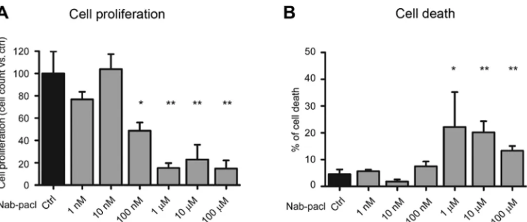
Cell viability assays. The cells were plated at 50% confluency in 96 wells and, after 24 h, treated with nab-paclitaxel at the concentrations indicated in Fig. 1. After 72 h of treatment, the cell viability was evaluated by MTS assay (Promega, Madison, WI, USA) following the manufacturer's instructions and by assessing the optical density (OD) at 490 nm. The results are represented as the mean ± standard deviation (SD) of three experiments.
For cell death, the cells were plated at 70% confluency and, after 24 h, treated with gemcitabine and/or nab-paclitaxel at the indicated doses. After an additional 48 h, the cells were washed in phosphate-buffered saline (PBS), trypsinized and incubated with 0.4% Trypan Blue stain (Sigma-Aldrich, St. Louis, MO, USA). Blue positive cells were then counted
using the Countess II Automated Cell Counter (Invitrogen Life Technologies, Carlsbad, CA, USA) and the percentage of cell death was determined. The results are represented as the mean ± SD of three experiments.
BrdU-PI staining and cell cycle analysis. For the cell cycle analysis, the cells were treated with 10 µM BrdU (Sigma-Aldrich) in the final 30 min of treatments. Subsequently, the cells were trypsinized, washed in chilled PBS and resuspended in PBS/ethanol 70%. The samples were incubated at -20˚C until use. The cells were then centrifuged at 2,000 rpm for 5 min, washed with PBS and incubated with 2 N HCl/0.5% Triton X-100 at room temperature (RT) for 30 min. The cells were centrifuged at 2,000 rpm for 5 min and then resuspended with 0.1 M NaB4O. After incubation for 2 min at
RT, the cells were washed with PBS/1% BSA and incubated for 1 h at RT in a solution of 0.5% Tween-20/1% BSA in PBS containing 10 µl of anti-BrdU 1 mM (Becton-Dickinson and Company, Franklin Lakes, NJ, USA). Subsequently, the cells were washed with PBS/1% BSA and incubated in a solution of PBS/0.5% Tween-20/1% BSA containing 5 µl of Alexa Flour 488 anti-mouse IgG-FITC (polyclonal; cat. no. A-11001; Thermo Fisher Scientific, Waltham, MA, USA) for 30 min at RT. The cells were washed with PBS/1% BSA and incubated with PBS containing 1 mg/ml RNAse A (Roche, Basel, Switzerland) and 20 µg/ml propidium iodide (PI; Sigma-Aldrich) for 30 min at 37˚C. Subsequently the cells stained with BrDU-PI were analyzed by FACS.
Cell extracts and western blot analysis. MiaPaCa-2 cells were resuspended in lysis buffer (50 mM HEPES pH 7,4, 10% glyc-erol, 15 mM MgCl2, 150 mM NaCl; 15 mM EGTA; 20 mM β-glycerophosphate; 1 mM dithiothreitol, 0.5 mM NaVO4,
1 mM NaF and protease inhibitor cocktail) supplemented with 1% Triton X-100, sonicated for 5 sec and centrifuged for 10 min at 13,000 rpm at 4˚C. Supernatants were collected, diluted in sodium dodecyl sulphate (SDS) sample buffer and boiled for 5 min. The proteins were separated on 8 or 12% SDS-PAGE gel and transferred onto PVDF blotting membranes (Amersham Hybond; GE Healthcare, Little Chalfont, UK). The membranes were saturated in 5% non-fat dry milk in PBS plus 0.1% Tween-20 for 1 h at RT and incubated overnight at 4˚C with the following primary antibodies: Rabbit anti-PARP1 (1:500; Cell Signaling Technology, Inc., Danvers, MA, USA), mouse anti-actin (1:1,000; Santa Cruz Biotechnology, Dallas, TX, USA), rabbit anti-cyclin E2 (1:1,000; Cell Signaling Technology), rabbit cyclin A2 (1:1,000), rabbit anti-cyclin B1 (1:1,000), mouse anti-anti-cyclin D1 (1,000; cat. no. A-12) (all from Santa Cruz Biotechnology).
Results
Nab-paclitaxel exerts cytotoxic effects in PDAC cells displaying different primary sensitivity to gemcitabine. In order to assess the efficacy of nab-paclitaxel on cell proliferation and viability, we analyzed the dose-response to nab-paclitaxel of the PDAC cells displaying different sensitivity to gemcitabine, with the MiaPaCa-2 and Panc-1 cells demonstrating the highest resistance to gemcitabine, thus offering in vitro models of primary resistance to this drug (7,8,15).
ONCOLOGY REPORTS 3
Nab-paclitaxel induced a significant reduction of cell prolif-eration (60-65%) starting from the dose of 100 nM compared to controls in all PDAC cells (Fig. 1A). Furthermore, at this
dose, nab-paclitaxel induced a significant increase of cell death in all cell lines with the exception of Panc-1 cells (Fig. 1B). Notably, the increase of cell death at 100 nM was modest in
Figure 1. Nab-paclitaxel exerts a similar cytotoxic effect in PDAC cells with different sensitivity to gemcitabine. Histograms display the analysis of (A) cell proliferation performed by an MTS assay (HPAF-II and PT45P1) or by cell count (MiaPaCa-2 and Panc-1) and (B) cell death performed by Trypan blue after 72 h of treatment with the indicated doses of nab-paclitaxel. The results represent the mean ± SD of three experiments. Significance vs. control, was determined by Student's t-test: **P<0.01.
PASSACANTILLI et al: NAB-PACLITAXEL ENHANCES THE CYTOTOXICITY OF GEMCITABINE
4
HPAF-II (6%) and Pt45P1 (7%) cells, whereas it was very high in MiaPaCa-2 cells (54%) (Fig. 1B), which displayed higher resistance to gemcitabine (15). Conversely, nab-paclitaxel significantly reduced Panc-1 cell proliferation at this dose without inducing cell death, whereas cell viability was affected only at micromolar doses of the drug (Fig. 1A and B). At these higher doses (1-100 µM), nab-paclitaxel led to substantial induction of cell death in the HPAF-II, MiaPaCa-2 and Panc-1 cell lines, while cell death remained at 20% in Pt45P1 even at the highest dose (Fig. 1B).
These results revealed that, regardless of their sensitivity to gemcitabine, the PDAC cells demonstrated similar sensitivity to nab-paclitaxel in terms of inhibition of cell proliferation, however, different response in terms of cell death.
Nab-paclitaxel exerts a cytotoxic effect in PDAC cells with secondary gemcitabine resistance. As aforementioned we selected PDAC cells which acquired resistance to gemcitabine after chronic exposure to the drug (8). Notably, these cells were also more resistant to cisplatin (8), another drug exerting geno-toxic stress. To examine whether these drug-resistant (DR) cells were still sensitive to nab-paclitaxel, a dose-response study was performed. We found that DR-Panc-1 cells maintained the same sensitivity to nab-paclitaxel as the parental cell line, with significant inhibition of cell proliferation starting at the dose of 100 nM, while cell death increased significantly at the dose of 1 µM (Fig. 2A and B). These results confirmed that nab-paclitaxel sensitivity did not correlate with gemcitabine sensitivity and suggested that nab-paclitaxel may overcome acquired resistance to gemcitabine in PDAC cells.
Combined treatment with Nab-paclitaxel and gemcitabine exerts additive effects on the inhibition of cell proliferation. In order to understand whether nab-paclitaxel in combina-tion with gemcitabine enhances the cytostatic and cytotoxic effects of the chemotherapeutic treatment, we tested their combined action on cell proliferation and death in MiaPaCa-2 cells, a cell line demonstrating relatively high resistance to gemcitabine (15). The combination of gemcitabine (100 nM) and nab-paclitaxel (10 nM) exerted a significant increase in cell death compared to gemcitabine alone (56 vs. 37%) (Fig. 3A). Notably, the effect of the combined treatment was similar to
that exerted by gemcitabine alone at a dose 100 times higher (i.e. 10 µM; Fig. 3A). The combination of gemcitabine (30 nM) and nab-paclitaxel (10 nM) elicited a significant additive effect even when used at a suboptimal dosage (Fig. 3B). Furthermore, similar effects were also obtained with the highly resistant DR-Panc-1 cells, albeit at considerably higher doses (Fig. 3C). These data indicated that combined treatment with nab-pacli-taxel and gemcitabine enhanced the cytotoxic effects of both drugs, allowing to lower their doses, thus possibly limiting adverse effects.
Combination of suboptimal doses of nab-paclitaxel and gemcitabine induces a significant increase in apoptosis. To investigate the nature of the additive effect of gemcitabine and nab-paclitaxel on PDAC cell death, we analyzed cell cycle progression and cell death in more detail in MiaPaCa-2 cells. Flow cytometry analysis with propidium iodide (PI) of cells treated with suboptimal doses of gemcitabine (30 nM) and nab-paclitaxel (10 nM) for 48 h indicated that gemcitabine strongly affected the cell cycle progression, leading to cell accumulation in S phase, whereas nab-paclitaxel elicited very mild effects. Notably, however, the addition of nab-paclitaxel to gemcitabine led to the appearance of a defined peak in the sub-G1 population of MiaPaCa-2 cells (Fig. 4A), indicating cell death by apoptosis. To confirm the effect of the combined treatment on cell apoptosis, we monitored cleavage of poly(ADP-ribose) polymerase (PARP1) by western blot analysis. Consistent with the appearance of the sub-G1 peak, PARP1 cleavage was noticeably increased in MiaPaCa-2 cells treated with the combination of the two drugs (Fig. 4B). Addition of nab-paclitaxel to gemcitabine induces a stronger cell cycle blockage in S phase. The PI profile indicated that co-treatment with nab-paclitaxel enhanced the accumulation of cells in the S phase of the cycle compared to gemcitabine alone (Fig. 4A). To further investigate this possibility, we analyzed the incorporation of BrdU as a precise marker of DNA duplication in S phase. A short pulse of BrdU was administered to MiaPaCa-2 cells 30 min before harvesting, following 24 h of incubation with the drugs. We observed that both gemcitabine and nab-paclitaxel, used as single agents, caused an accumulation of cells in the S phase (from 45,93
Figure 2. Nab-paclitaxel exerts cytotoxic effect in PANC-1 DR cell line with secondary resistance to gemcitabine. Histograms reveal the analysis of (A) cell proliferation performed by cell count and (B) cell death performed by Trypan blue cell count after 72 h of treatment with the indicated doses of nab-paclitaxel. The results represent the mean ± SD of three experiments. Significance vs. control was determined by Student's t-test: *P<0.05 and **P<0.01.
ONCOLOGY REPORTS 5
to 78,30 and 74,37%) (Fig. 5A). Notably, the combination of both drugs resulted in an additive effect on the accumulation of cells in S phase, which reached 84.84%. As a consequence of this blockage in cell cycle progression, co-treatment with gemcitabine and nab-paclitaxel resulted in a sharp reduction of cells transiting in the G2 phase (Fig. 5A).
In addition, we checked the changes in cell cycle progression by monitoring the expression levels of phase-specific cyclins. As dislplayed in Fig. 5B, cyclin D1 levels were not affected by treatments, whereas cyclin A2 and E2 levels increased after gemcitabine administration either alone or in combina-tion with nab-Paclitaxel, confirming that the cells are mainly
blocked in the S phase. Treatment with nab-paclitaxel alone did not cause accumulation of S phase cyclins (Fig. 5B), even though the cells were blocked at this stage of the cycle. Since we noticed that nab-paclitaxel caused accumulation of cells in the left-most region of S phase (Fig. 5A), indicating very little duplication of DNA, it is probable that this drug blocks cells before the accumulation of cyclins E2 and A2. Additionally, we observed that the combined treatment with both drugs reduced the expression of cyclin B1 compared to gemcitabine alone. Since this cyclin is involved in the S-G2 cell cycle transition, its levels reflect the reduction of cells in G2 phase, which was observed in flow cytometric analyses (Figs. 4A and 5A).
Figure 3. Combined treatment of gemcitabine and nab-paclitaxel exert an additive effect on the inhibition of cell proliferation. Histograms display the analysis of cell death performed by Trypan blue on MiaPaCa-2 cells after (A) 72 h of treatments with the indicated doses of nab-paclitaxel and gemcitabine. Significance vs. control, was determined by Student's t-test: *P<0.05. (B) Analysis of cell death after 48 h of treatments with the indicated suboptimal doses of
nab-paclitaxel and gemcitabine. Statistical analysis was performed by one-way analysis of variance by ANOVA test followed by Tukey's Multiple Comparison post-test (*P<0.05). (C) Analysis of cell death performed by Trypan blue on Panc-1 DR cells after 72 h of treatments with the indicated doses of nab-paclitaxel
and gemcitabine. Significance vs. control, was determined by Student's t-test: *P<0.05. All results represent the mean ± SD of three experiments.
Figure 4. Combination of suboptimal dose of nab-paclitaxel and gemcitabine induces a significant increase of cell death. (A) Panels display the percentage of cells stained for propidium iodide (PI) in Sub-G1, G1, S and G2 phases in MiaPaCa-2 cells treated with nab-paclitaxel, gemcitabine or both drugs for 48 h. (B) Western blot analysis of cleaved protein PARP-1 in MiaPaCa-2 cells treated with nab-paclitaxel, gemcitabine or both drugs for 48 h.
PASSACANTILLI et al: NAB-PACLITAXEL ENHANCES THE CYTOTOXICITY OF GEMCITABINE
6
Discussion
The aim of the present study was to examine the activity of nab-paclitaxel alone or in combination with gemcitabine in PDAC cell lines displaying different degree of primary resis-tance to gemcitabine and in a previously described model of secondary resistance to the drug (7,8).
The results of the present study revealed that nab-paclitaxel is effective in PDAC cells irrespective of their sensitivity to gemcitabine and to the status of primary or secondary (acquired) resistance (Figs. 1 and 2). Notably, both drugs demonstrated an addictive effect at suboptimal doses in cell lines with primary or secondary resistance to gemcitabine (Fig. 3).
To investigate the underlying mechanisms of the observed efficacy of nab-paclitaxel, we explored the changes occurring in the cell cycle (Figs. 4 and 5). Our results indicated that nab-paclitaxel blocked cell proliferation in a different manner compared to gemcitabine. Although both drugs caused an arrest in S phase, the cells treated with gemcitabine exhibited a different extent of DNA duplication, whereas the peak of cells treated with nab-paclitaxel is present in the left region of the graph, indicating that cells arrest as soon as they start duplicating their DNA. This difference is also illustrated by the accumulation of S phase cyclins, which is evident in gemcitabine- but not in nab-paclitaxel-treated cells. While the blockage in S phase is expected after gemcitabine exposure, due to depletion of the nucleotide pool required for DNA duplication, cells treated with nab-paclitaxel were expected to arrest in mitosis or late G2 phase due to defects in spindle elon-gation. However, recent data have revealed that cells treated with paclitaxel often proceeded through mitosis into the next interphase, where the majority of cell deaths occurred (16). In
particular, nab-paclitaxel seemed to interfere with the very early stages of the S phase in PDAC cells (Fig. 5A). This may explain why it was previously found that the interference with the DNA replication origin activity enhanced the response of cells to paclitaxel (16).
The different mechanism of S phase blockage by the two drugs may explain the additive affect observed in the combined treatment. Markedly, such additive effect was observed both at the cell cycle and the cell death level, indicating a causal relationship between the two events. Although the molecular mechanisms involved in such effect need further investigation, our results indicated that nab-paclitaxel strongly enhances the cytotoxicity of gemcitabine and may help to overcome both primary and acquired resistance to this drug.
The in vitro results of the present study revealed that nab-paclitaxel, alone or in combination with gemcitabine, is an active drug in preclinical models of gemcitabine-resistant PDAC. Hence, our observations indicated that, in certain clinical scenarios, nab-paclitaxel could be active in patients with PDAC that are not responding to gemcitabine, even in monotherapy. However, clinical data on the use of nab-paclitaxel as a single agent in patients with PDAC that were previously treated with gemcitabine, are limited. In a small phase II trial, 19 patients received nab-paclitaxel after progres-sion under gemcitabine-based therapy (17). One of them (5.3%) had a confirmed partial response and 6 (32%) exhibited stable disease as the best response. Another single-center retrospective study evaluated the use of nab-paclitaxel in 20 patients with advanced PDAC who previously exhibited progression under gemcitabine, 40% of whom also received FOLFIRINOX. Notably, about two thirds of patients had stable disease as best response, although the median OS was
Figure 5. Combination of suboptimal dose of nab-paclitaxel and gemcitabine induces a significant increase of cell death. (A) Panels display the percentage of cells stained for propidium iodide (PI) and anti-BrdU FITC antibody. They are detected in G1, S and G2 phases in MiaPaCa-2 cells treated with nab-paclitaxel, gemcitabine or both drugs for 24 h. (B) Western blot analysis of cyclin D1, A, E and B1 in MiaPaCa-2 cells treated with nab-paclitaxel, gemcitabine or both drugs for 24 h.
ONCOLOGY REPORTS 7
only of 5.2 months (18). Further studies are needed to elucidate whether this approach may be beneficial, possibly in patients with less advanced disease.
The present study is one of the few that aimed to evaluate the efficacy of nab-paclitaxel in preclinical settings, using cell lines that are models of both primary and secondary resis-tance to gemcitabine. As the investigation is limited to in vitro models, the results should be interpreted with caution and the mechanisms of the activity observed in the present study need further experiments to be elucidated. In particular, our data are useful to generate hypotheses that need to be confirmed in other models, such as animal ones, that may better recapitu-late the human pathology. Conversely, the additive effect of nab-paclitaxel and gemcitabine observed in these experiments cannot be due to factors that have been extensively investigated in animal models and that are related with tumor stroma and penetration of drugs. Another limitation of the present study concerns the lack of a defined mechanism for the observed effects. Following the revision process, we tested some common signal transduction pathways that could be involved in the response to these chemotherapeutic agents, such as the PI3K-mTOR and ERK pathways (data not shown). However, we did not find a direct correlation with the cytotoxic effect. Thus, further studies are needed to analyze the mechanisms underlying the observed effects. Considering the above-mentioned limitations, our results revealed that treatment with nab-paclitaxel may overcome resistance to gemcitabine and may represent a potentially valuable therapeutic approach for advanced PDAC.
Acknowledgements
The present study was supported by grants from the Associazione Italiana per la Ricerca sul Cancro (AIRC; IG18790 for C.S. and IG 17177 for G.C.).
Competing interests
The authors declare that they have no competing interests. References
1. Siegel RL, Miller KD and Jemal A: Cancer Statistics, 2017. CA Cancer J Clin 67: 7-30, 2017.
2. Rahib L, Smith BD, Aizenberg R, Rosenzweig AB, Fleshman JM and Matrisian LM: Projecting cancer incidence and deaths to 2030: The unexpected burden of thyroid, liver, and pancreas cancers in the United States. Cancer Res 74: 2913-2921, 2014. 3. Fogel EL, Shahda S, Sandrasegaran K, DeWitt J, Easler JJ,
Agarwal DM, Eagleson M, Zyromski NJ, House MG, Ellsworth S, et al: A multidisciplinary approach to pancreas cancer in 2016: A review. Am J Gastroenterol 112: 537-554, 2017.
4. Neuzillet C, Tijeras-Raballand A, Bourget P, Cros J, Couvelard A, Sauvanet A, Vullierme MP, Tournigand C and Hammel P: State of the art and future directions of pancreatic ductal adenocar-cinoma therapy. Pharmacol Ther 155: 80-104, 2015.
5. Burris HA III, Moore MJ, Andersen J, Green MR, Rothenberg ML, Modiano MR, Cripps MC, Portenoy RK, Storniolo AM, Tarassoff P, et al: Improvements in survival and clinical benefit with gemcitabine as first-line therapy for patients with advanced pancreas cancer: A randomized trial. J Clin Oncol 15: 2403-2413, 1997.
6. Binenbaum Y, Na'ara S and Gil Z: Gemcitabine resistance in pancreatic ductal adenocarcinoma. Drug Resist Updat 23: 55-68, 2015.
7. Adesso L, Calabretta S, Barbagallo F, Capurso G, Pilozzi E, Geremia R, Delle Fave G and Sette C: Gemcitabine triggers a pro-survival response in pancreatic cancer cells through acti-vation of the MNK2/eIF4E pathway. Oncogene 32: 2848-2857, 2013.
8. Calabretta S, Bielli P, Passacantilli I, Pilozzi E, Fendrich V, Capurso G, Fave GD and Sette C: Modulation of PKM alter-native splicing by PTBP1 promotes gemcitabine resistance in pancreatic cancer cells. Oncogene 35: 2031-2039, 2016.
9. Conroy T, Desseigne F, Ychou M, Bouché O, Guimbaud R, Bécouarn Y, Adenis A, Raoul JL, Gourgou-Bourgade S, de la Fouchardière C, et al: FOLFIRINOX versus gemcitabine for metastatic pancreatic cancer. N Engl J Med 364: 1817-1825, 2011.
10. Von Hoff DD, Ramanathan RK, Borad MJ, Laheru DA, Smith LS, Wood TE, Korn RL, Desai N, Trieu V, Iglesias JL, et al: Gemcitabine plus nab-paclitaxel is an active regimen in patients with advanced pancreatic cancer: A phase I/II trial. J Clin Oncol 29: 4548-4554, 2011.
11. Von Hoff DD, Ervin T, Arena FP, Chiorean EG, Infante J, Moore M, Seay T, Tjulandin SA, Ma WW, Saleh MN, et al: Increased survival in pancreatic cancer with nab-paclitaxel plus gemcitabine. N Engl J Med 369: 1691-1703, 2013.
12. Vaccaro V, Sperduti I, Vari S, Bria E, Melisi D, Garufi C, Nuzzo C, Scarpa A, Tortora G, Cognetti F, et al: Metastatic pancreatic cancer: Is there a light at the end of the tunnel? World J Gastroenterol 21: 4788-4801, 2015.
13. Lemstrova R, Melichar B and Mohelnikova-Duchonova B: Therapeutic potential of taxanes in the treatment of metastatic pancreatic cancer. Cancer Chemother Pharmacol 78: 1101-1111, 2016.
14. Neesse A, Algül H, Tuveson DA and Gress TM: Stromal biology and therapy in pancreatic cancer: A changing paradigm. Gut 64: 1476-1484, 2015.
15. Arumugam T, Ramachandran V, Fournier KF, Wang H, Marquis L, Abbruzzese JL, Gallick GE, Logsdon CD, McConkey DJ and Choi W: Epithelial to mesenchymal transition contributes to drug resistance in pancreatic cancer. Cancer Res 69: 5820-5828, 2009.
16. Koh SB, Mascalchi P, Rodriguez E, Lin Y, Jodrell DI, Richards FM and Lyons SK: A quantitative FastFUCCI assay defines cell cycle dynamics at a single-cell level. J Cell Sci 130: 512-520, 2017.
17. Hosein PJ, de Lima Lopes G Jr, Pastorini VH, Gomez C, Macintyre J, Zayas G, Reis I, Montero AJ, Merchan JR and Rocha Lima CM: A phase II trial of nab-Paclitaxel as second-line therapy in patients with advanced pancreatic cancer. Am J Clin Oncol 36: 151-156, 2013.
18. Peddi PF, Cho M, Wang J, Gao F and Wang-Gillam A: Nab-paclitaxel monotherapy in refractory pancreatic adenocar-cinoma. J Gastrointest Oncol 4: 370-373, 2013.
23
Chapter III. RNA metabolism in cancer
III.1 RNA metabolism
A majority of eukaryotic genes are regulated at transcriptional and post-transcriptional levels (Fig. 5). Cancer evolves through perturbations in these processesthat in turn modulate to the advantage of tumor cells cellular processes such as proliferation, differentiation, cell-cycle control, metabolism, apoptosis, motility, invasion, and angiogenesis.
The main steps of RNA processing occur co-transcriptionally and include:
• capping: the addition of a to 7- methylguanosine cap the 5' end of the pre- mRNA soon after Pol II initiates transcription, important for the RNA stability, involve in RNA quality control mechanism and contributes to nuclear export of mRNA.
• splicing: process that mediates removal of introns and the joining of exons and occur while Pol II transcribes DNA into RNA.
• polyadenylation: the addition of a poly(A) tail to the 3' end of the pre- mRNA before Pol II completes transcription.
Figure 5.Overview of RNA metabolism (readapted from
24
Other steps of RNA processing take place at the post-transcriptional level, the most important are:
• mRNA export: process that occur through the Nuclear Pore Complexes (NPC) by binding to the cap-binding proteins CBP20 and CBP80 and transcription/export complex (TREX). • mRNA stability regulation: it can happen through different process: decapping Dcp1/2
factors, deadenylase Ccr4/Pop2/Not complex, nonsense mediated decay (NMD), microRNA, long non coding RNA.
• translational regulation: mRNAs also can be sequestered in stress granules or P-bodies.
The investigation of molecular mechanisms underlying abnormal RNA processing in cancer cells is providing new opportunities of development of cancer therapeutics
III.2 RNA stability or RNA decay
The regulation of mRNA stability is a fine process for cell physiology because it permits transient expression of proteins and increases the flexibility of gene expression together with regulation rate of mRNA synthesis. Since mRNA stability is essential to determine the proteins that are produced, many factors control this event.
The addition of 7-methylguanosine-cap structure at 5' of transcript ( 5′ cap) and the tail of polyA at 3' protect mRNAs from decay by impeding the processing of exonucleases. Furthermore, the interaction between proteins that binds the cap (eIF4E) and the poly(A)-binding protein (PABP) promove the mRNA circularizes in the cytoplasm, this conformation lends mRNA more stable sequestering the 5′ and 3′ ends. The deadenylation induces the destabilization of transcript by releasing PABP from the 3′ and exposing mRNA to attack by exosome complex that is responsible for 3′→5′ decay. Another process is the deccaping by DCP1 and DCP2 enzymes that remove the 5'cap, thus making RNA susceptible to the decay through 5′→3′ exonuclease, XRN1 and XRN2 (Fig. 6)(Garneau, Wilusz, and Wilusz 2007)
25
Figure 6. Mechanisms of mRNA decay (Garneau, Wilusz, and Wilusz 2007)
Non sense-mediated decay (NMD) is an evolutionarily conserved mechanism that recognizes mRNAs with a premature termination codons (PTCs) and degrades them. The identification of these transcipts occur by a complex proteins called the exon complex (EJC) that is deposited approximately 20–25 bp upstream of every intron after RNA splicing processing. The presence of an EJC after a stop codon triggers NMD, whereas EJCs before the stop codon do not, thus this system selects the mRNA that undergoRNA decay(Kurosaki and Maquat 2016).
One of the locations where the RNA decay takes place are the P-bodies, cytoplasmic ribonucleoprotein (RNP) granules primarily composed of non translating mRNA and a core of proteins involved in mRNA decay and translation repression (decapping enzyme complex, 5′ to 3′ exonuclease, deadenylase complex) (Eulalio, Behm-Ansmant, and Izaurralde 2007). Moreover P-bodies contain proteins and microRNAs involved in the miRNA repression pathway; microRNAs are hairpin-derived RNAs 20–24 nucleotides long, that post-transcriptionally repress the expression of target genes by binding to the 3' UTR of mRNA. The repressed mRNAs remain in the P body in a "standby state", thus they are ready to be transcript; this system provides a way to transiently arrest translation, so the cell has developed a faster regulation mechanism than the de novo transcription of the target gene. (Decker and Parker 2012)(Di Leva, Garofalo, and Croce 2014).
26 III.3 The splicing mechanism and its role in cancer
The splicing mechanism is required for the maturation of almost all human transcripts. Notably, pre-mRNAs can be processed into different mature mRNAs through alternative splicing of multiple exons, thus enabling multiple potential protein products to be generated from a single gene. This process is guided by the spliceosome, a dynamic complex consisting of small nuclear RNAs (snRNAsU1, U2, U4, U5 and U6) and many other protein factors. During the splicing reaction this complex undergoes different conformational and compositional changes (Dvinge et al. 2016).
U1 snRNP recognizes and binds to the 5′ splice site, which is located at the start of the intron, whereas U2 snRNP pairs with the branch site region adjacent to the 3′ splice site, assisted by interactions with U2 auxiliary factors (U2AFs, which form the U2AF complex) that bind to the 3′ splice site, located at the end of the intron. Interactions between U1 and U2 snRNPs bring the 5′ss and 3′ss into close proximity. Following recruitment of the U4/U6-U5 tri-snRNP, the assembled spliceosome is in active conformation; U2 and U6 interact, causing dissociation of U4 from U6 snRNA and exposition of the 5′ of U6, which binds the 5′ss displacing U1 snRNA. The splicing process proceeds via two sequential trans-esterification reactions that join the exons and release an intron lariat that is subsequently degraded. Finally, the spliceosome components are recycled for subsequent reation of splicing (Fig.7 A)(Shi 2017)(Dvinge et al. 2016).
Alternative spling events can be classified based on how the mature transcript is formed; we can found events of constitutive spliced exon, exon cassette, alternative 5'/3' splice site, retained intron and mutualy exclusive exons (Fig. 7 B).
27
Figure 7. Schematic representation of splicing mechanism and model of constitutive and alternative splicing
(Dvinge et al. 2016).
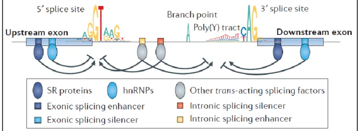
The activity of the spliceosome is regulated by both cis acting sequences on the pre-mRNA and trans acting factors,which may enhance or inhibit both recognition of the splice sites and splicing catalysis. The cis-acting regulatory elements are classified according to their location and activity: exonic and intronic splicing enhancers (ESEs and ISEs) or silencers (ESSs and ISSs). The trans acting RNA binding proteins (RBPs) recognize these elements and in turn promote or inhibit spliceosome assembly and activity. Two main classes of RBPs regulates this process: the Ser/Arg rich (SR) proteins, which mainly exert a positive regulation on the spliceosome, and the heterogeneous nuclear ribo nucleoproteins (hnRNPs), which often act antagonistically (Fig. 8). SR and hnRNPs regulate splicing in diverse ways, including facilitating recruitment of snRNP or RBPs, mask a splice site, ‘looping out’ an exon and other mechanisms (Dvinge et al. 2016).
28
Figure 8. Spliceosome regulatory elements (Dvinge et al. 2016).
Interestingly, the activities of RBPs often depend on their relative locations within pre-mRNAs. For example hnRNPI, primarily known as polypyrimidine tract binding protein 1 (PTBP1), modulates splicing in a context-dependent manner: it promotes exon inclusion when it binds in the downstream intron and exon skipping when it binds in the upstream intron (Llorian et al. 2010). In addition to the SR proteins and hnRNPs, many other RBPs regulate splicing, including CELF, MBNL, RBFOX, STAR, NOVA and ESRP proteins. Thus, alteration of the expression of these RBPs can impact alternative splicing (Dvinge et al. 2016).
Regulation of alternative splicing has essential roles in cancer cell biology, including proliferation, differentiation, cell-cycle control, metabolism, apoptosis, motility, invasion and angiogenesis. Deregulated splicing can lead to generate oncogenic isoform that contribute to tumor establishment, progression and resistance to therapy. Analysis of data from 8,705 patients with one of 32 types of cancer revealed that tumors have up to 30% more alternative splicing events than normal tissues (Kahles et al. 2018). Many studies have found links between the altered expression and/or activity of splicing factors and cancer (Pagliarini, Naro, and Sette 2015)(J. Zhang and Manley 2013). Below I report some examples of peculiar cancer isoforms (Fig. 9).
29
Figure 9. Examples of alternative splicing patterns in cancer.
The RON gene encodes a tyrosine kinase receptor involved in cell mobility and invasion in response to MSP binding. The isoform ΔRON, which lacks exon 11, is over-expressed in a number of cancers, this exon encoding for an extracellular domain, that remains constitutively active in the truncated ΔRON promoting cancer invasiveness (Collesi et al. 1996).
BRAF is a proto-oncogene encoding the serine/threonine protein kinase BRAF, which regulates the MAPK/ERK signaling pathway. The skipping of exons 4–8 cause a the truncated enzyme (BRAFV600E) that is insensitive to the BRAF inhibitors (vemurafenib), often is utilized in cancer, and confers melanoma cell resistance to the drugs (Poulikakos et al. 2011).
BCL-X belongs to the BCL-2 protein family, that is involved in anti- or pro- apoptotic regulators. BCL2L1 produces two splice isoforms by the alternative use of two competing 5′ splice sites in exon 2. The longer isoform BCL-XL has antiapoptotic effects and is over-expressed in various
30
cancer types, while the shorter isoform BCL-XS is proapoptotic and is downregulated in cancer
(Olopade et al. 1997)(Trisciuoglio et al. 2017).
VEGF stimulates angiogenesis required for tumor growth. Selection of the proximal 3′splice sites produces isoforms VEGF, that has a pro-angiogenic role and are over-expressed in a number of tumors. Instead the VEGFb isoform is formed when the distal 3′splice sites are used, it has a anti-angiogenic role and it is down-regulated in tumors. These opposing functions are caused by the different C-terminal produced, VEGFb fails to bind to its receptor, the neurophilin 1, which is required for activation of VEGF signal transduction (Nowak et al. 2008).
In tumor the isorform PKM2 is predominant and performs a critical role for tumor metabolism, like previus descripts (Dayton, Jacks, and Vander Heiden 2016).
CD44 is a trans-membrane glycoprotein that mediates the response of cells to their cellular microenvironment: lymphocyte homing, adhesion, migration, and regulation of cell growth. This variety of roles is favoured by the existence of multiple CD44 splice variants, the CD44 gene is composed of 10 constitutively spliced exons and 9 variable exons, residing between constitutive exons 5 and 6 (Ponta, Sherman, and Herrlich 2003). For example, CD44 molecules contain v6–7 were expressed specifically in a metastasizing pancreatic carcinoma cell line, but not in the parental tumor, suggesting that these variants was sufficient to render parental tumor cells metastatic (David and Manley 2010).
These cancer isoforms are due, largely, to alteration of growth pathway, that are able to influence splicing through their control of RBP and/or splicing factor. For example C-Myc regulates up-regulation of PTB, hnRNPA1 and hnRNPA2; ERK controls Sam68 activity and AKT mediates phosphorylation of SR proteins (J. Zhang and Manley 2013).
III.4 Cleavage and polyadenylation mechanism and alternative polyadenylation
The 3′ end of majority of eukaryotic mRNAs and some long non-coding RNAs is cleaved and polyadenylated. It is a post-transcriptional process that involve endonucleolytic cleavage of the transcript followed by the addition of the poly(A) tail. This process, in which they are involved cis and trans elements, occurs a relevant role in stability, nuclear export and translation regulation of mRNAs. The key cis-element is located 15–30 nt upstream of the cleavage site and it is a hexameric consensus motif called the poly(A) signal (PAS), the canonical form is AAUAAA, but it can assume different variants and are mostly. Moreover, U- or GU-rich downstream
31
sequence elements (DSEs) and less well-defined upstream sequence elements (USEs) are nearby of PAS and they enhance cleavage efficiency. The PAS hexamer is recognized by cleavage and polyadenylation specification factor (CPSF), which is recruited co-transcriptionally and it composed of six polypeptides, named CPSF4, CPSF2, CPSF3, FIP1L1, WDR33 and CPSF1, although only the latter is the subunit that recognizes the PAS. Cleavage stimulation factor (CSTF), which is composed of three subunits (CSTF1,CSTF2, CSTF3), binds the DSE, while the cleavage factor Im (CFIm) and CFIIm interact with the USE. The poly(A) polymerase (PAP), the scaffold protein simplekin and polyadenylated-binding nuclear protein 1 (PABPN1) are additional factors that were required for conclusion of process (Fig. 10) (Elkon, Ugalde, and Agami 2013)(Gruber and Zavolan 2019).
Figure 10. Core players involved in cleavage and polyadenylation mechanism (Elkon, Ugalde, and Agami 2013).
By transcriptome-wide sequencing emerges that most human genes have more than one poly (A) site, indicating the existence of alternative polyadenylation (APA). APA events can be classified into four general classes: tandem 3′UTR APA which are the most frequent APA forms and generates different isoforms changing only in the length of their 3'UTR; alternative terminal exon APA, in which the transcript ends in another exon; alternative intronic polyadenylation (IPA), in which results from the recognition of cleavage sites within introns (Fig. 11).
32
Figure 11. The four different APA types (Elkon, Ugalde, and Agami 2013)
3'UTR is a important regulative region of transcripts, it interacts with microRNAs(miRNAs) and RNA-binding proteins (RBPs). For example, in some cases oncogene activation in cancer cells is led by the generation of more stable isoforms with shorter 3'UTR, that compared to their counterparts with long 3'UTRs escape from repressive effect of miRNAs (Mayr and Bartel 2009).
33
Various multifunctional proteins that regulate splicing also participate in poly(A) site selection: PTBP1, ELAVL1, hnRNPC, PCBP1 and PABN1 (Gruber and Zavolan 2019). For example it was demonstrated that U1snRNP protects pre-mRNAs from premature cleavage (Fig.12) and its knock down promove induction of intronic poly(A) sites usage, independently of the role of U1 in splicing (Kaida et al. 2010). Physiologically, the level of U1 is not known to be regulated, thus suggest that its transient knowdown occurs in conditions in which overall transcription is upregulated. This decrease of U1 should cause a global 3′UTR shortening, similarly to the phenomenon observed in proliferation and cancer cells (Elkon, Ugalde, and Agami 2013)(Mayr and Bartel 2009).
III.5 Role of ZEB1 in PDAC and genotoxic stress response III.5.1 Structure and function
ZEB1 is a protein encoded by the ZEB1 gene which is located on chromosome 10p11.2 in humans. It is a key factor of Epithelial–mesenchymal transition (EMT), a reversible cellular program that transiently places epithelial cells into mesenchymal cell states, involved in specific steps of embryogenesis, such as gastrulation and tissue morphogenesis during development. Moreover, it has a relevant role also in cancer progression, cancer stem cell maintenance, metastasis settlement and resistance to chemotherapy (Dongre and Weinberg 2019). ZEB1 is a transciptional factor and regulates the transcription by binding to E-promoter DNA sequences (5′-CANNTG-3′) of its targets. In humans ZEB1 contains more two zinc-finger clusters at N- and C-terminal ends of the protein. The middle region consists of a homeodomain, a Smad interaction domain and a C-terminal binding protein (CtBP) domain (Fig. 13). CtBP cannot directly bind to DNA and participates in the regulation of ZEB1 function by interacting with other regulatory factors.