Seconda Facoltà di Medicina e Chirurgia
CORSO DI LAUREA SPECIALISTICA IN MEDICINA E CHIRURGIA
Tesi di Dottorato
Dottorato di ricerca in TECNOLOGIE BIOMEDICHE IN MEDICINA
CLINICA (31° ciclo)
PULMONARY HYPERTENSION AND CARDIAC FUNCTION IN
CONGENITAL DIAPHRAGMATIC HERNIA: RELATIONSHIP TO DISEASE
SEVERITY AND OUTCOMES
Relatore
Candidata
Chiar.mo Prof.
Anna Claudia Massolo
Bruno Marino Taussig de Bodonia
Matricola n°368060
Keywords (not in title) Systolic Diastolic Right ventricle Left ventricle Pulmonary hypertension Abbreviations
AV, atrio-ventricular valve
CDH, congenital diaphragmatic hernia DINT, duration of intubation
ECMO, extracorporeal membrane oxygenation FAC, fractional area change
GCS, global circumferential strain GLS, global longitudinal strain GRS, global radial strain LOS, length of stay LV, left ventricle miRNA, microRNA
O:E TFLV, observed-to-expected total fetal lung volume OI: oxygenation index
TDI, tissue Doppler imaging PAP, pulmonary artery pressure RV, right ventricle
Abstract Objective
To assess patterns of postnatal biventricular function and their relationship to prenatal and postnatal markers of disease severity in infants with congenital diaphragmatic hernia (CDH). To investigate clusters of micro RNA as 17-92 expressed in pulmonary hypoplasia in patients with CDH.
Study Design
Observational case-control study of cardiac function in infants with CDH in the first 5 days of life. Systolic and diastolic function in the right (RV) and left (LV) ventricles were assessed using speckle tracking-derived global strain and tissue Doppler imaging of myocardial velocities. In another cohort of CDH, Bronchoalveolar aspiration (BAL) fluid was obtained and metabolomic analysis was run for micro RNA 17-92 cluster. Specifically miR17, miR18a, miR19a, miR19b, miR20a, miR92 were analyzed. Correlation between cardiac function and prenatal observed:expected total fetal lung volume (TFLV), oxygenation index (OI), duration of intubation and length of stay were assessed.
Results
All measures of systolic and diastolic function were significantly reduced in the CDH group (n=25) compared to controls (n=20) at <48 hours, and improved by 72-96 hours. LV global systolic longitudinal strain (LV GLS) correlated with prenatal TFLV (r2 0.32, p 0.03), OI (r2 0.35, P<0.001), duration of intubation (r2 0.24, p 0.04),
CDH patients who did not survive and/or required ECMO compared to those who did not; -11.5 (5.3) % vs -16.9 (5.3) %, p 0.02. For the metabolomic analysis we found miR17, miE18a, miR19b, miR20a were found significantly higher in CDH (p<0.05) and miR19a lower (p<0.05). No significant correlation was found between miR92 expression and clinical outcomes.
Conclusions
Right and left ventricular function are impaired in the transitional period in infants with CDH. Early left ventricular systolic function correlates with prenatal and postnatal markers of clinical disease severity and may be an important determinant of disease severity and therapeutic target in CDH. These findings support regular assessment of cardiac function in CDH and investigational trials of targeted cardiovascular therapies. miR 17-92 cluster may represent a potential hallmark of PH in CDH however from our result doesn’t predict disease severity.
Introduction
Congenital diaphragmatic hernia (CDH) affects 1 in 6000 live births and is characterized by herniation of the abdominal contents into the fetal thorax. CDH remains a challenging condition for affected individuals and treating clinical teams. Despite advances in intensive care and surgical management over the past half-century, CDH continues to cause significant mortality and morbidity (1,2).
Pulmonary hypoplasia and pulmonary hypertension, due to abnormal pulmonary vascular development, play central roles in CDH pathophysiology (3–5). There is also increasing recognition that the function of the heart may be a key determinant of CDH severity (6).
The role of cardiac function in CDH pathophysiology
Recent international treatment guidelines highlight the importance of assessing and treating cardiac function in CDH and randomized controlled trials are currently in progress to assess cardiovascular therapies (7–9). However, a missing piece of the CDH puzzle has been limited investigation of the timing, nature, and frequency of postnatal cardiac dysfunction in CDH and its relationship to clinical markers of severity.
The advent of modern echocardiographic techniques, including strain analysis using speckle tracking echocardiography and tissue Doppler imaging (TDI), allows improved quantitative assessment of global and regional systolic and diastolic function (10,11). Application of these techniques in infants with CDH may help reveal the mechanics of cardiac dysfunction and have implications for improving clinical management and outcomes.
The role of “omics” and pulmonary hypertension in CDH pathophysiology
PH contributes to high mortality in CDH. A better understanding of the regulatory mechanisms underlying the pathology in CDH might allow the identification of prognostic biomarkers and potential therapeutic targets (miR CDH).
Recent studies provide evidence of abnormal patterns in proliferation, apoptosis and cell differentiation, and altered expression of growth and transcription factors and genes reported to be involved in the etiology of pulmonary hypoplasia in CDH (12). These mechanisms seem to regulated from clusters of mirco RNA. MicroRNAs (miRNAs, miRs) are a large group of noncoding small RNAs about 22 nucleotides in length. miRs mainly act accelerating the degradation of messenger RNAs or to repress the translation, thus, negatively regulating the expression of target genes (13). Regulating up to around one third of all miRs in human, miRs have been identified to play important regulatory roles in a wide array of pathological processes and embryonic organ morphogenesis (14–16). The functions of miRs in lung development are subjects of many investigations (13,15,17,18). For example, the miR-17-92 family has been reported to regulate early lung development by promoting proliferation and inhibiting differentiation of lung epithelium. The deletion of miR-17- 92 cluster in mice resulted in immediate postnatal death with smaller embryos and hypoplastic lungs (19,20), while overexpression of this cluster leads to severe developmental defects with high proliferation and undifferentiated phenotype of lung epithelial progenitor cells (21).
This study aimed to assess patterns of ventricular function in infants with CDH in the first week of life and to investigate their relationship with pre-and post-natal measures of disease severity.
For the purpose of this thesis, we investigated in a separate group of neonates with CDH, the miR 17-92 cluster in the lungs to explore possible predictors of pulmonary hypertension severity in CDH.
Methods
Study design and setting
The study included two centers. For the “ventricular function” evaluation patients were recruited at the Royal Hospital for Children, Glasgow and for the genetic analysis of miRNA patients were enrolled at the Bambino Gesù Hospital. Both are a regional neonatal surgical center and national extra-corporeal membrane oxygenation (ECMO) center.
Demographic data for both groups
Sex, gestation, birth weight, and mode of delivery were collected from the electronic patient record. In the CDH group additional characteristics, treatment, and outcome data were obtained; side and size of defect (22), age at repair, use of patch, stomach and liver position at time of surgery, cardiovascular therapies and ECMO, duration of intubation (DINT) and neonatal length of stay (LOS) in
CDH cases were managed by a multi-disciplinary team in both centers according to an institutional treatment protocol, in accordance with international guidelines (23). Briefly, this incorporates a “gentle” ventilation strategy minimizing volutrauma and barotrauma, targeted use of pulmonary vasodilators (inhaled nitric oxide and intravenous sildenafil) based on clinical and echocardiographic evidence of clinically significant pulmonary hypertension, and selective use of cardiotropes (milrinone, dopamine, and epinephrine) based on clinical and echocardiographic evidence of impaired cardiac function and systemic blood flow. Prostaglandin E1 is used to maintain ductal patency in the presence of systemic/suprasystemic pulmonary artery pressures, right ventricular dysfunction and a closing arterial duct. ECMO is employed in selected cases to treat respiratory and cardiovascular compromise resistant to optimized medical therapy which is considered reversible.
Study population, analysis and results will be presented describing firstly the “ventricular function group” (Section 1) which represents the main topic of this study. Separately, in a second section, we will describe the “omics group” (Section 2).
Section 1: Ventricular function group
This retrospective case-control study was conducted at the Royal Hospital for Children, Glasgow.
CDH group: Infants with CDH, both inborn and outborn, admitted to the study center between January 2015 and January 2018 were considered for inclusion. Cases with significant associated congenital anomalies (significant cardiac anomalies other than ventricular septal defect, major chromosomal anomalies e.g. chromosomal trisomies), were excluded. Prenatally diagnosed CDH cases underwent routine magnetic resonance imaging (MR) at 25-28 weeks gestation to measure observed-to-expected ratio of total fetal lung volume (O:E TFLV), as previously described (24,25). MR data was used in preference to ultrasound lung-head ratio based on prior reports of improved predictive value of MR lung volume assessment in CDH (26).
Control group
A control group of infants with esophageal atresia treated during the study period was identified from electronic records. These infants routinely have echocardiographic assessment in the first 48 hours of life and formed a convenience sample for comparison with CDH cases. Only infants who were breathing spontaneously in air, without clinical or echocardiographic evidence of pulmonary hypertension or congenital cardiac anomaly (including ventricular septal defect), were included.
Echocardiography data
Only for the ventricular function group, echocardiographic data were collected by the study researchers (NP, EA) according to an institutional protocol using a GE Vivid E9 (GE Medical Systems, Milwaukee, WI) with a 6 MHz probe and analyzed using EchoPac software incorporating QLab (EchoPAC version 202; GE Medical
Systems, Milwaukee, WI). Post-acquisition analysis was performed by a single observer (ACM). The first echocardiogram (<48 hours of age) was analyzed in all CDH cases and controls. Follow-up echocardiograms at 72-120 hours was also analyzed in CDH cases, but were not available for controls.
Cardiac function in the right ventricle (RV) and left ventricle (LV) were assessed using pulsed wave tissue Doppler imaging (TDI), myocardial strain derived from speckle tracking echocardiography, fractional area change (FAC), and Doppler flow across the atrioventricular valves (AV).
TDI velocities in systole (s’) and diastole (e’) were measured in the basal myocardium of the LV and RV lateral walls and the interventricular septum from an apical 4-chamber view, as previously described (27).
STE analysis was used to assess systolic strain (myocardial shortening) from gray-scale 2-dimensional (2D) cine-loops, acquired in the 4-chamber apical view (for longitudinal strain) and 2-chamber short axis at mid-ventricle level (for radial and circumferential strain, LV only). Images with an optimized frame rate:heart rate ratio were selected (28). Regions of interest were traced within the endocardial border and adjusted manually to optimize tissue-tracking. Peak global longitudinal (GLS, in the LV and RV) circumferential (GCS, LV only) and radial strain (GRS, LV only) were obtained.
FAC, a global measure of ventricular function was calculated as the percentage change in ventricular area using end systolic area (ESA) and end diastolic area (EDA) measurements obtained by manually tracing the internal contours of the endocardial border from apical 4-chamber images (29). AV valve velocities in early (E) and late (A) diastole were measured from pulsed wave Doppler waveforms across the mitral and tricuspid valve, as measures of diastolic function.
Pulmonary artery pressure (PAP) was assessed using patent ductus arteriosus (PDA) flow, and tricuspid regurgitation velocity (TR) when present. A pulsed wave Doppler waveform was obtained in the PDA from which the velocity-time integer of left-to-right and right-to-left flow components was manually traced and a ratio calculated (VTIL-R) to quantify PAP. Maximal velocity of TR was assessed by
pulsed-wave Doppler and inserted into a modified Bernoulli equation to derive estimated peak RV systolic pressure (RVSPEST) (30). Using these measures PAP was also
classified as <2/3rd systemic blood pressure, 2/3rd systemic-to-systemic blood
pressure, or > systemic blood pressure (31). All Doppler-derived data were averaged over five consecutive cardiac cycles. Oxygenation index (OI) at the time of initial echocardiogram was calculated using the formula :(% inspired oxygen x mean airway pressure x 100) / Arterial PO2. Arterial PO2 was obtained from arterial
blood gases obtained from right radial or umbilical artery.
Section 2: Omics group
Neonates were enrolled at Bambino Gesù Children’s Hospital and patients were selected prospectively and case-control.
Study population
CDH group: Infants with CDH, both inborn and outborn, admitted to the study center between January 2015 and January 2018 were considered for inclusion. Same inclusion and exclusion criteria of the ventricular function group were applied.
Control group
A control group of infants with esophageal atresia treated during the study period was identified.
Omics Analysis
All patients, intubated, underwent bronchoalveolar lavage (BAL) fluid suction within the first 48 hours of life. 1ml/kg of saline solution was administered through the endotracheal tube and after three ventilation cycles was extracted. BAL were stored at -80°C and then analyzed for the study of the mi-RNA 17-92 cluster. The RNA extraction were vortexed and centrifuged for 5 minutes at 1250 r/min. Adequate dosage was assessed through spectrophotometry. RNA was extracted with the miRNeasy Mini Kit (Qiagen), for the Real-Time PCR. QuantiTect Reverse Transcription Kit (Qiagen) was used to obtain RNA transcription, and amplified with SYBR Green PCR Master Mix (Qiagen). From the 17-92 cluster we analyzed the following miR: mi-RNA 16, mi-RNA 17, mi-RNA 18, mi-RNA 19A, mi-RNA 19b, mi-RNA 20a e mi-RNA 92. U6 snRNA was used as control. REAL-time PCR was performed through the StepOnePlus TM real-time machine (Applied Biosystems).
Data were summarized as median (range) or mean (standard deviation). Global strain and FAC data from CDH cases and controls at <48 hours were compared using unpaired T test. TDI and AV valve Doppler data were not available for controls, instead comparison was made to existing normative published data (32). CDH data at <48 hours and 72-120 hours were compared using paired T tests. Correlation was assessed between measures of cardiac function at <48 hrs and O:E TFLV, OI, DINT, LOS, and LV size using LV EDA. Chi-square or Fisher’s exact tests
were used to compare categorical data. Cardiac function parameters in CDH group at < 48hrs were compared between survivors vs. non-survivors, ECMO vs. no ECMO, and the combination of these (non-survival+/-ECMO). miRs were compared between the two groups using unpaired T test. Correlation was assessed between measures of miRs and LHR, OI, DINT, LOS, survivals or death and
need of ECMO or not. Analysis was performed using Prism 7 (GraphPad Software, La Jolla, CA, USA). A p value < 0.05 was considered significant. Statistical support was provided by an experienced statistician. Institutional ethical approval was provided for data analysis and publication.
Results
Section 1: Ventricular function group
Demographic and treatment data
Twenty-seven CDH cases and 20 controls were included. Two CDH cases were excluded due to insufficient echocardiographic images for analysis. There were no significant differences in demographic data between CDH cases and controls, Table 1.
CDH characteristics, treatment, and outcome data are provided in Table 1. Prenatal MRI data at weeks gestational age was available for 15 (60%) CDH cases, mean MRI O:E TFLV, was 38 (14)%. Twenty-two CDH cases (81%) underwent surgical repair at a median (range) of 9 (2-20) days. Of these 14 (63%) required patch repair. Seventeen CDH cases (68%) survived to discharge. For survivors, median (range) duration of intubation was 23 (4-72) days, and length of stay was 41 (17-164) days.
Eight (32%) cases received ECMO. Twenty-three CDH cases (92%) received cardiotropes. The same proportion received inhaled nitric oxide, of whom 16 (64%) also received intravenous sildenafil.
Cardiac function and PAP data at <48 hours and 72-120 hours
STE-derived global strain analysis was performed in all cases at <48 hours, but only for GLS parameters at 120 hours (insufficient images for GCS and GRS at 72-120 hours). TDI velocities were measurable in 18 CDH cases, and AV Doppler velocities in 11 cases, at both time points.
In the first 48 hours of life, all global strain parameters were significantly reduced in the LV (GLS, GCS and GRS) and RV (GLS) compared to controls, Table 2. TDI systolic (s’) and diastolic (e’) velocities in the RV, septum and LV, as well Doppler inflow velocities (TV E, MV E) were also significantly reduced compared to existing normative data. PFO shunt was left-to-right in 16 (59%) CDH cases, right-to-left or bidirectional in five (19%) cases, or no shunt seen in 6 (22%) cases. There were no
significant differences in RV or LV strain or FAC parameters between infants with left-to-right PFO shunt versus those with bidirectional or right-to-left shunt.
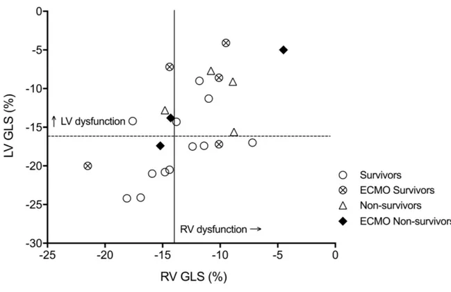
Cut off values for abnormal RV and LV GLS at < 48 hours of life were determined as one standard deviation below the mean from control data; RV GLS cut off ≤-14%, LV GLS cut off ≤=-16%. According to these definitions, seven CDH cases (28%) had normal RV and LV GLS, four CDH cases (16%) had abnormal RV GLS only, five (20%) had abnormal LV GLS only, and nine (36%) had abnormal LV and RV GLS, Figure 1.
At 72-120 hours of age, RV-GLS and LV-GLS increased significantly, compared to <48 hours of age. LV FAC and LV EDA were significantly reduced in the CDH group at <48 hours compared to controls and to CDH cases at 72-120 hours, Table 2. LV EDA at <48 hours did not correlate with STE measures of LV function.
TR was measurable in 16 (64%) of cases. Twenty-one CDH cases (84%) had a PDA at <48 hours of age, in which flow was bidirectional in 17 (68%) cases, right-to-left in three cases (12%), and left-to-right in one case (4%). Mean (SD) RVSPEST was 50
(18) mmHg, and mean (SD) PDA VTIL:R was 1.35 (2.1). Measures of PAP were not
significantly different at <48 vs 72-120 hours of age. PAP at <48 hours was classified as <2/3rd systemic blood pressure in 3 cases, 2/3rd systemic-to-systemic
blood pressure in 12 cases, and > systemic blood pressure in 11 cases.
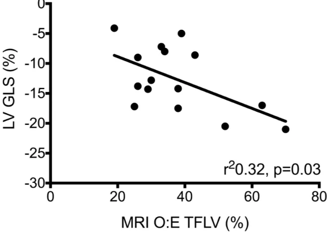
MRI O:E TFLV correlated significantly with LV GLS (r2 0.32, p=0.03, Figure 2a) and
TDI LV E’ (r2=0.45, p=0.02) at <48 hours, but not with measures of RV function,
RVSPEST or PDA VTIL:R. There were no significant differences in cardiac function
parameters (FAC and global strain measures) between cases based on PAP category.
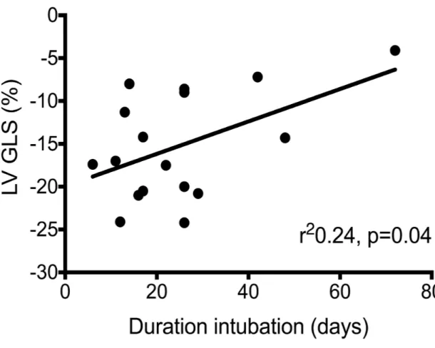
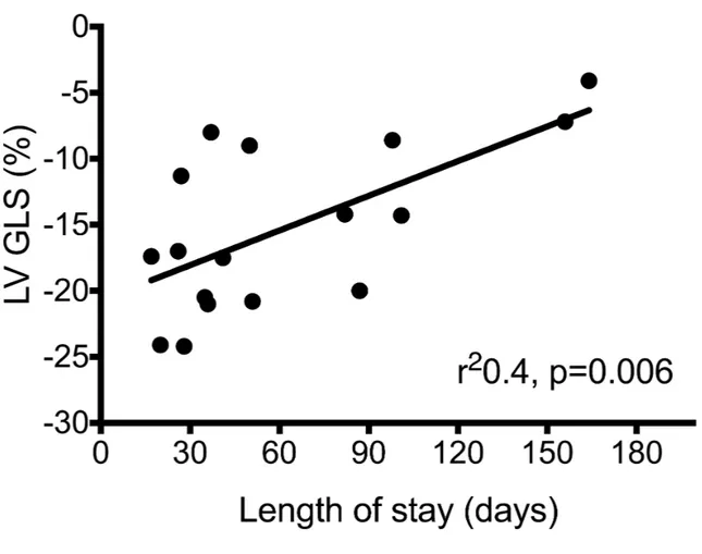
LV GLS at < 48 hours of age correlated significantly with OI (r2 0.35, P<0.001), DINT
(r2 0.24, p=0.04) and LOS (r2 0.4, p=0.006), Figures 2b and 2c.
LV GLS at < 48hours of age was significantly lower in CDH cases with the combined outcome of death+/-ECMO versus survivors/no ECMO: -11.5 (5.3) % vs. -16.9 (5.3) %, p=0.02. LV GLS in CDH survivors vs. non-survivors was 15.4 (6.1) % vs. -11.6 (4.5) %, p=0.11, and in non-ECMO vs ECMO CDH cases was -15.6 (5.4) % vs. -11.7 (6.1) %, p=0.15.
Section 2: Omics group
20 neonates with CDH were enrolled, 3 were excluded for insufficient BAL fluid. 17 controls with OA were included too. No statistical differences were found between the two groups for GA, weight, and sex (GA 37,4 +2 vs 38,2 +1,8, p=0,23; weight at birth 2.770 + 599 vs 2.965 + 470, p=0,33). Of these 20 patients, 16 survived, 6 underwent ECMO. Of 4 non survivors, 2 underwent ECMO.
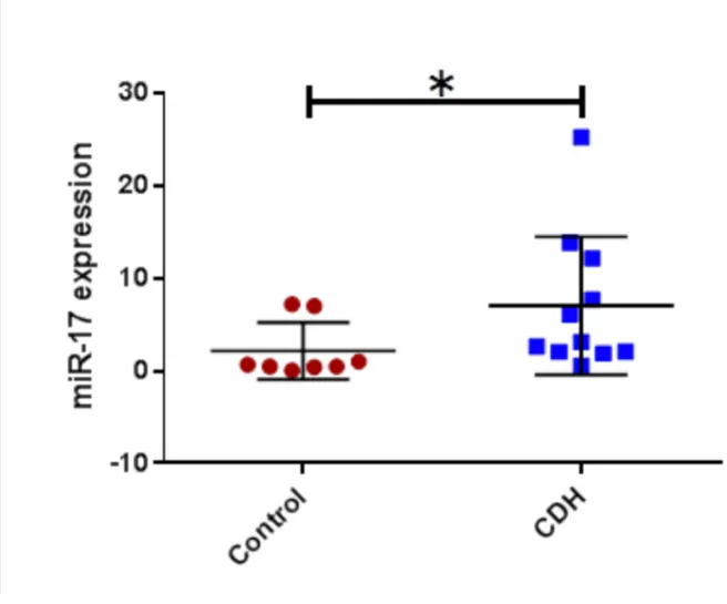
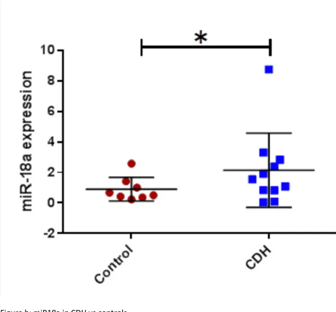
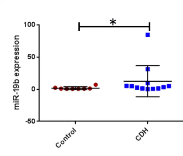
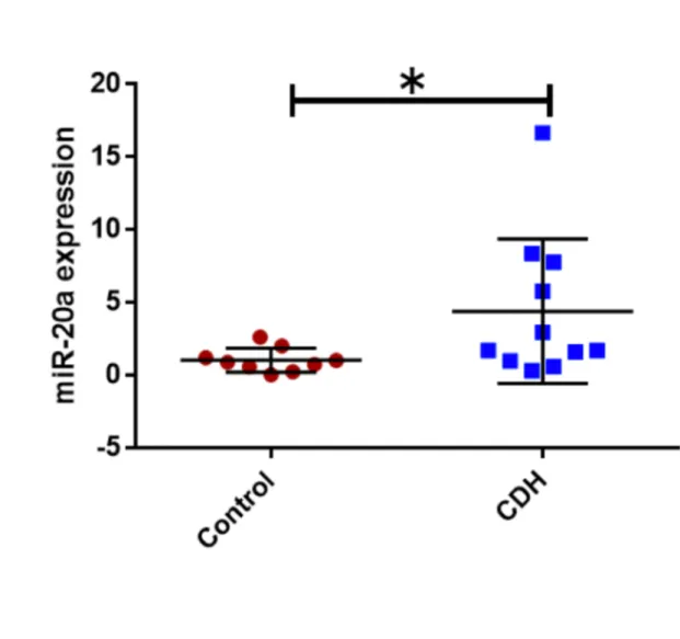
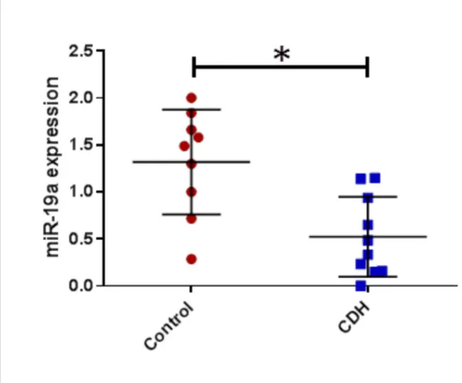
For the analysis of the 17-92 cluster in the BAL fluid in CDH patients compared to controls we found a significant increase of miR16, miR18a, miR17, miR19b, miR20a (t-test ; CDH vs control; p< 0,05) (Figure 4 a,b,c, and d) and a significant decrease
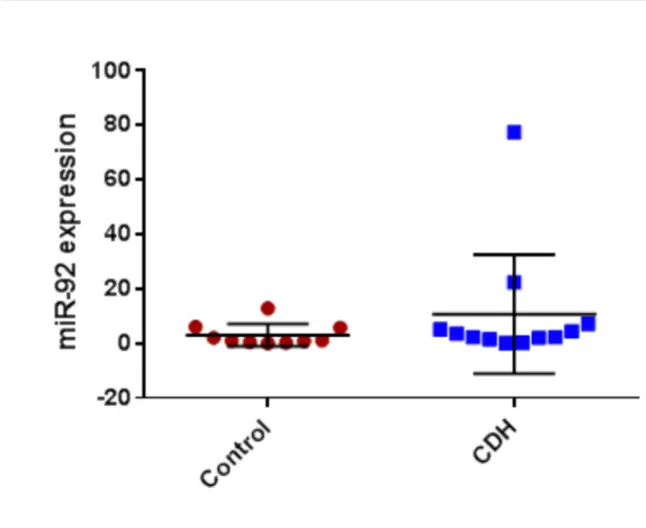
of miR19a in the CDH group (Figure 4 e) and no statistical difference was found for miR92.
Discussion
We assessed cardiac function in the early postnatal period in infants with CDH and a control population using a panel of echocardiographic techniques including strain analysis and tissue Doppler imaging. There is growing experience of these techniques in newborn infants and recognition of their benefits over traditional geometric measures of cardiac function (33). Our findings provide new insights into patterns of cardiac dysfunction in CDH, their relationship to disease severity, and may inform improvements in clinical management and outcomes.
Patterns of ventricular dysfunction after birth in CDH
RV and LV dysfunction were present in CDH patients in the first 48 hours of life. RV dysfunction was predominantly diastolic in nature, consistent with previous reports (34,35). RV diastolic dysfunction likely results from the acute and prolonged increase in afterload (PVR) after birth, due to functional and structural abnormalities of the pulmonary vasculature characteristic of CDH (36).
Postnatal LV dysfunction was characterized by impairment of both systolic function (in longitudinal, circumferential and radial dimensions) and diastolic relaxation. This global LV dysfunction during the transition from fetal to postnatal life may have four etiological factors; First, fetal LV hypoplasia due to mechanical compression and reduced LV blood flow, the latter resulting from reduced
pulmonary venous return and altered ductus venosus flow (37–39). Second, reduced postnatal LV filling due to persistently elevated pulmonary vascular resistance and reduced pulmonary blood flow. Third, transmission of RV dysfunction to the LV, via mechanisms of ventricular interdependence including the shared septum, muscle fibres, and pericardium (40). Fourth, the acute increase in afterload at birth as the LV takes over as the systemic ventricle and the placenta is removed from the circulation.
Early cardiac dysfunction appears to be common in CDH. Using cut-off values for longitudinal global strain derived from control group data, we observed dysfunction in over two-thirds of cases and biventricular dysfunction in one-third of cases in this cohort.
RV and LV function improved significantly within the first week of life, as we have previously observed (41,42). It was not possible within this study to unravel the relative contributions of time, endogenous pathways and exogenous therapies, however the potential for improvement in function may be important when making therapeutic decisions, including continuing intensive care support, cardiovascular therapies, and timing of surgery, as discussed later.
Ventricular dysfunction and CDH severity
LV systolic function correlated with fetal lung volumes, an established marker of pulmonary hypoplasia and predictor of illness severity (43,44). Fetal left ventricular hypoplasia in CDH is also more severe in infants with smaller prenatal
lung volumes (45). Taken together these findings support the theory that postnatal LV dysfunction has prenatal origins linked to the severity of pulmonary and LV hypoplasia.
We also observed a relationship between early LV systolic function and clinically relevant postnatal outcomes. LV function was worse in infants who died and/or received ECMO, and demonstrated a significant negative association with oxygenation index, duration of intubation and length of stay in survivors. Similarly, Altit et al observed reduced LV longitudinal strain in infants with CDH who received ECMO compared to those who did not (46). These consistent findings suggest that LV function may be a primary determinant of disease severity in CDH, rather than simply a secondary consequence. Furthermore, early LV function may be an important parameter for future inclusion in early predictive algorithms in CDH (47).
LV dysfunction may plausibly destabilize infants at the transition to postnatal life by reducing systemic blood flow and oxygen delivery, contributing to tissue hypoxia and acidosis. This in turn may lead to a vicious cycle of worsening cardiac function and pulmonary vasoconstriction. Further investigation of the relationship between early cardiac function and outcome in CDH in larger cohorts, such as CDH registries, is now warranted (48).
Early RV function did not correlate with clinical outcomes, in contrast to a prior investigation (41). One possible explanation may be a change in management
approach in the current study cohort toward earlier use of pulmonary vasodilators and cardiotropic support. The potential of this early, targeted treatment approach to alter outcome merits further investigation.
Of note, PAP in the first 48 hours did not correlate with fetal lung volume or clinical outcomes, consistent with previous reports (41,46). This may be due to the fact that most normal infants have raised PAP after birth, but also highlights the challenge of measuring PAP accurately using echocardiography, and the importance of assessing cardiac function directly in addition to PAP (49).
An additional observation was the variable patterns of inter-atrial flow. Though shunt direction was not associated with cardiac function in this limited cohort, our findings highlight the importance of assessing intra-cardiac shunts in CDH patients to appreciate their potential influence on gas exchange and systemic oxygenation.
Implications for assessment and treatment of CDH
Recent CDH treatment guidelines from North America and Europe note the role for echocardiographic assessment of routine PAP and cardiac function (7,8). Based on our findings, both RV and LV function should be assessed regularly starting from the first days of life to detect early dysfunction and monitor response to therapy. Future consensus guidelines should include recommendations for the timing and techniques to assess cardiac function in CDH.
Identifying infants at greatest risk of cardiac dysfunction may allow preventative or anticipatory management. Our results suggest that MR fetal lung volumes might be useful to stratify infants most likely to develop LV dysfunction. Kinsella et al have recently proposed that routine measurement of fetal LV size may predict postnatal LV function (6).
A possible approach to minimize postnatal cardiac dysfunction may be delayed umbilical cord clamping combined with lung recruitment to maintain circulating volume, improve pulmonary blood flow and optimize left ventricular function (50). Further investigation is needed to assess whether this approach is feasible and beneficial in CDH.
Improved assessment and understanding of RV and LV function in CDH can guide rational, targeted selection of cardiovascular therapies. RV diastolic dysfunction is an indication for pulmonary vasodilatation to reduce afterload, combined with lusitropic support to improve diastolic function. LV dysfunction, meanwhile, should benefit from afterload reduction by systemic vasodilatation and inotropic support. One agent with these combined properties is milrinone, a phospho-diesterase 3 inhibitor (51). Our findings add further support to a randomized controlled trial of milrinone in CDH which is now in progress (9).
Current treatment guidelines advocate fluid bolus as first line therapy for systemic hypotension in CDH. However, if low blood pressure is due to LV dysfunction then inotropes may be more appropriate, such as low dose epinephrine administered
in our cohort. Systemic vasoconstrictors increase LV afterload and may exacerbate LV dysfunction, this may explain the variable response to vasopressin in CDH (52).
The role of pulmonary vasodilators in the setting of LV dysfunction in CDH is unclear. There is a theoretical concern that by increasing pulmonary blood flow and LV preload they may exacerbate LV diastolic dysfunction and pulmonary venous hypertension (53–56). Conversely, by improving RV function they may indirectly improve LV performance via mechanism of ventricular interdependence. The CoDiNOS trial, a current multi-center randomized controlled trial of intravenous sildenafil and iNO, incorporates assessment of cardiac function and may help to resolve this question (57).
Cardiac function assessment might also help guide ECMO use. In severe LV dysfunction and veno-arterial ECMO is appropriate to provide cardiac support and time for function to improve in the first days of life. In severe ventilatory and oxygenation failure, where LV function is preserved, veno-venous (VV) ECMO may suffice.
Cardiac function assessment may also inform timing of surgery in CDH. Our observation that cardiac function improved at 72-120 hours in unrepaired CDH, supports a strategy of delayed repair to optimize cardiac function and pulmonary artery pressure pre-operatively (58). Conversely, Tanaka et al have observed improved LV diastolic wall strain after early CDH repair on day 1 of life, suggesting
that early reduction of the hernia may improve LV function (59). Comparison of these approaches merits further investigation.
miRNA
The miR17-92 cluster showed an abnormal expression in patients with CDH. miRNAs has been implicated in the development of PH particularly cluster miR17-92(60).
One of the pathogenetic hallmark of PH is the dysregulation expression of bone morphogenetic protein receptor type II (BMPR2)(61). BMPR2 regulates growth, differentiation, and apoptosis in a diverse number of cell lines, including mesenchymal and epithelial cells, acting as instructive signals during embryogenesis and contributing to the maintenance and repair of adult tissues(16,17). Mutations in the BMPR2 gene have been found in approximately 70% of families with idiopathic PH(62,63). In the lung, BMPR-II is highly expressed on the vascular endothelium of the pulmonary arteries(62). Morrell et al. demonstrated that expression of BMPR-II is markedly reduced in the pulmonary vasculature of patients with mutations in the BMPR-II gene (64). BMPR-II expression was significantly reduced in the pulmonary vasculature of patients with PH in whom no mutation in the BMPR2 gene was identified suggesting that a critical reduction in the expression of BMPR-II may be important to the pathogenesis of PH, whether or not there is a mutation in the gene. Several miRNAs encoded by the miR17-92 were retrieved as potential regulators of it. Brock et al. reported that an overexpression resulted in a strong reduction of the BMPR2 protein (61). Some studies reported by stimulation experiments, that the
miR17-92 cluster is also modulated by interleukin (IL)-6, a cytokine involved in the pathogenesis of pulmonary hypertension. IL-6 signaling is mainly mediated by STAT3 (signal transducer and activator of transcription 3), the expression of STAT3 was knocked down by small interfering RNA, which abolished the IL-6–mediated expression of miR17-92 (61). STAT 3 has been reported to promote lung branching morphogenesis, bronchiolar epithelial cell proliferation (65). STAT 3 through IL-6 stimulates miR17-92, particularly miR17 and 20. Also, miR17 and 20 can bind BMPR2 and down regulate it. In our CDH population miR17 was significantly over-expressed compared to controls. Chen et al. found that smooth muscle cell (SMC)-specific knockout of miR17-92 attenuated hypoxia-induced PH in mice, whereas reconstitution of miR17-92 restored hypoxia-induced PH in these mice (60). Inhibition of miR17, a member of the miR17-92 cluster, induces p21 and prevents hypoxia-induced pulmonary hypertension (PH) (60). Our findings add further support to the hypothesis that miRNA 17-92 cluster is a contributor of pulmonary hypertension in CDH although no correlation with clinical outcomes were found. This suggests that miRs may represent an hallmark of PH in CDH patients but don’t represent a predictor of disease severity.
Limitations
CDH is a relatively rare condition limiting the sample size. This may have contributed to the absence of statistically significant relationship between cardiac function and survival or ECMO as isolated outcomes. Although echocardiographic data was collected according to an institutional protocol not all parameters could be analyzed in each subject, a challenge of retrospective investigation and the
need for optimized echocardiographic images for advanced functional analysis. Measurement of cardiac dimensions is particularly prone to error, which may have contributed to the lack of association between and left ventricular area and postnatal function.
The site of arterial blood gas sampling for arterial PO2 measurement is an important consideration (66). In the presence or right-to-left PDA shunting samples obtained from a post-ductal position such as an umbilical arterial catheter (nineteen cases in this cohort, 76%) may yield a higher OI than those obtained pre-duct from the right radial artery (6 cases, 24%).
We were unable to control for or distinguish the individual effect of therapies such as cardiotropes, pulmonary vasodilators and ECMO on cardiac function. However, most therapies were commenced after the first echocardiograms which were therefore considered to reflect native function. Longitudinal analysis of cardiac function, beyond the fifth day of life, was not included in this study and is an important area of ongoing investigation.
No sufficient echocardiography evaluation was available for the patients from the miRNA group, thus no correlation between the miRNA and severity of PH and cardiac function were explored. Further studies are warranted to investigate the relation between it.
Conclusions
Infants with CDH are at risk of global RV and LV dysfunction in the early postnatal period. LV systolic function correlates with prenatal lung volume and clinical outcome measures. miRs may be considered hallmarks of PH, although they don’t
represent predictors of disease severity. These findings support routine, early and regular assessment of cardiac function in CDH to guide targeted therapies. Current and future trials incorporating this approach may lead to improved outcomes in CDH.
Acknowledgements
I am very grateful to Dr Neil Patel for his support with technical aspects of echocardiographic data collection and analysis, to Professor Bruno Marino for his support and assistance with data interpretation.
Figure legends:
Figure 1: RV and LV global longitudinal strain in CDH patients in the first 48hrs of life
LV, left ventricle; RV, right ventricle; GLS global longitudinal strain; dashed line indicates cut off values for abnormal LV ≤ - 16% and solid line indicates cut-off for abnormal RV GLS ≤ -14%
Figure 2: LV GLS and outcomes (a) MR O:E TFLV (b) duration of intubation; (c) length of stay
Figure 2a: MRI O:E TFLV vs. LV GLS
Figure 2b: Duration of intubation vs. LV GLS Figure 2c: Length of stay vs. LV GLS
Figure 3: miRNA 17-92 cluster in CDH vs controls Figure a: miR17 in CDH vs controls
Figure b: miR18a in CDH vs controls Figure c: miR19b in CDH vs controls Figure d: miR20a in CDH vs controls Figure e: miR19a in CDH vs controls
References
1. Harting MT, Lally KP. The Congenital Diaphragmatic Hernia Study Group registry update. Vol. 19, Seminars in Fetal and Neonatal Medicine. 2014. p. 370–5.
2. Long A-M, Bunch KJ, Knight M, Kurinczuk JJ, Losty PD, BAPS-CASS. Early population-based outcomes of infants born with congenital diaphragmatic hernia. Arch Dis Child Fetal Neonatal Ed [Internet]. 2018;fetalneonatal-2017-313933. Available from: http://www.ncbi.nlm.nih.gov/pubmed/29305406
3. Harting MT. Congenital diaphragmatic hernia-associated pulmonary hypertension. Semin Pediatr Surg. 2017;26(3):147–53.
4. Kinsella JP, Ivy DD, Abman SH. Pulmonary vasodilator therapy in congenital diaphragmatic hernia: Acute, late, and chronic pulmonary hypertension. Semin Perinatol. 2005;29(2):123–8.
5. Kipfmueller F, Heindel K, Schroeder L, Berg C, Dewald O, Reutter H, et al. Early postnatal echocardiographic assessment of pulmonary blood flow in newborns with congenital diaphragmatic hernia. J Perinat Med [Internet]. 2017;0(0). Available from: http://www.degruyter.com/view/j/jpme.ahead-of-print/jpm-2017-0031/jpm-2017-0031.xml
6. Kinsella JP, Steinhorn RH, Mullen MP, Hopper RK, Keller RL, Ivy DD, et al. The Left Ventricle in Congenital Diaphragmatic Hernia: Implications for the Management of Pulmonary Hypertension. J Pediatr [Internet]. 2018 Apr 5 [cited 2018 May
22];197:17–22. Available from: http://www.ncbi.nlm.nih.gov/pubmed/29628412 7. Canadian T, Diaphragmatic C, Collaborative H. Diagnosis and management of
congenital diaphragmatic hernia: a clinical practice guideline. Can Med Assoc J. 2018;190(4):E103–12.
8. Snoek KG, Reiss IKM, Greenough A, Capolupo I, Urlesberger B, Wessel L, et al.
Standardized postnatal management of infants with congenital diaphragmatic hernia in Europe: The CDH EURO Consortium Consensus - 2015 Update. Neonatology. 2016;110(1).
9. Lakshminrusimha S, Keszler M, Kirpalani H, Van Meurs K, Chess P, Ambalavanan N, et al. Milrinone in congenital diaphragmatic hernia – a randomized pilot trial: study protocol, review of literature and survey of current practices. Matern Heal Neonatol Perinatol [Internet]. 2017;3(1):27. Available from:
https://mhnpjournal.biomedcentral.com/articles/10.1186/s40748-017-0066-9 10. Breatnach CR, Levy PT, James AT, Franklin O, El-Khuffash A. Novel echocardiography
methods in the functional assessment of the newborn heart. Vol. 110, Neonatology. 2016. p. 248–60.
11. Forsey, J., Friedberg, M.K. & Mertens, L., 2013. Speckle tracking echocardiography in pediatric and congenital heart disease. Echocardiography, 30(4), pp.447–459.Forsey J, Friedberg MK, Mertens L. Speckle tracking echocardiography in pediatric and
congenital heart disease. Echocardiography. 2013;30(4):447–59.
12. Zhu S, He Q, Zhang R, Wang Y, Zhong W, Xia H, et al. Decreased expression of miR-33 in fetal lungs of nitrofen-induced congenital diaphragmatic hernia rat model. J Pediatr Surg [Internet]. 2016;51(7):1096–100. Available from:
http://dx.doi.org/10.1016/j.jpedsurg.2016.02.083
13. Dermitzakis ET. From gene expression to disease risk. Nature Genetics. 2008. 14. Petricoin EF, Zoon KC, Kohn EC, Barrett JC, Liotta LA. Clinical proteomics: Translating
benchside promise into bedside reality. Nature Reviews Drug Discovery. 2002. 15. Vlahou A, Fountoulakis M. Proteomic approaches in the search for disease
biomarkers. Journal of Chromatography B: Analytical Technologies in the Biomedical and Life Sciences. 2005.
16. Rifai N, Gillette MA, Carr SA. Protein biomarker discovery and validation: The long and uncertain path to clinical utility. Nature Biotechnology. 2006.
17. Kell DB. Systems biology, metabolic modelling and metabolomics in drug discovery and development. Drug Discovery Today. 2006.
18. Wikoff WR, Gangoiti JA, Barshop BA, Siuzdak G. Metabolomics identifies
perturbations in human disorders of propionate metabolism. Clin Chem. 2007; 19. Urbanczyk-Wochniak E, Leudemann A, Kopka J, Selbig J, Roessner-Tunali U, Willmitzer
L, et al. Parallel analysis of transcript and metabolic profiles: A new approch in systems biology. EMBO Rep. 2003;
20. Dessì A, Cesare Marincola F, Masili A, Gazzolo D, Fanos V. Clinical Metabolomics and Nutrition: The New Frontier in Neonatology and Pediatrics. BioMed Research
International. 2014.
21. Dunn WB, Broadhurst DI, Atherton HJ, Goodacre R, Griffin JL. Systems level studies of mammalian metabolomes: The roles of mass spectrometry and nuclear magnetic resonance spectroscopy. Chemical Society Reviews. 2011.
22. Putnam LR, Harting MT, Tsao K, Morini F, Yoder BA, Luco M, et al. Congenital Diaphragmatic Hernia Defect Size and Infant Morbidity at Discharge. Pediatrics. 2016;138(5):e20162043–e20162043.
23. Snoek KG, Reiss IKM, Greenough A, Capolupo I, Urlesberger B, Wessel L, et al.
Standardized postnatal management of infants with congenital diaphragmatic hernia in Europe: The CDH EURO Consortium Consensus - 2015 Update. Neonatology. 2016;110(1):66–74.
24. Rypens F, Metens T, Rocourt N, Sonigo P, Brunelle F, Quere MP, et al. Fetal lung volume: Estimation at MR imaging - Initial results. Radiology. 2001;219(1):236–41. 25. Gorincour G, Bouvenot J, Mourot MG, Sonigo P, Chaumoitre K, Garel C, et al. Prenatal
prognosis of congenital diaphragmatic hernia using magnetic resonance imaging measurement of fetal lung volume. Ultrasound Obstet Gynecol. 2005;26(7):738–44. 26. Bebbington M, Victoria T, Danzer E, Moldenhauer J, Khalek N, Johnson M, et al.
Comparison of ultrasound and magnetic resonance imaging parameters in predicting survival in isolated left-sided congenital diaphragmatic hernia. Ultrasound Obstet Gynecol. 2013;(November 2013):670–4.
27. Patel N, Mills JF, Cheung MMH. Assessment of right ventricular function using tissue doppler imaging in infants with pulmonary hypertension. Neonatology.
2009;96(3):193–9.
28. Sanchez AA, Levy PT, Sekarski TJ, Hamvas A, Holland MR, Singh GK. Effects of frame rate on two-dimensional speckle tracking-derived measurements of myocardial deformation in premature infants. Echocardiography. 2015;32(5):839–47.
29. Levy PT, Dioneda B, Holland MR, Sekarski TJ, Lee CK, Mathur A, et al. Right ventricular function in preterm and term neonates: Reference values for right ventricle areas and fractional area of change. J Am Soc Echocardiogr. 2015;28(5):559–69.
30. Skinner JR, Boys RJ, Hunter S, Hey EN. Non-invasive assessment of pulmonary arterial pressure in healthy neonates. Arch Dis Child. 1991;66(4 SPEC NO):386–90.
31. Lusk LA, Wai KC, Moon-Grady AJ, Steurer MA, Keller RL. Persistence of pulmonary hypertension by echocardiography predicts short-term outcomes in congenital diaphragmatic hernia. J Pediatr [Internet]. 2015;166(2):251–6. Available from: http://dx.doi.org/10.1016/j.jpeds.2014.10.024
32. Mori K, Nakagawa R, Nii M, Edagawa T, Takehara Y, Inoue M, et al. Pulsed wave Doppler tissue echocardiography assessment of the long axis function of the right and left ventricles during the early neonatal period. Heart. 2004;90(October 2007):175– 80.
33. Jain A, Mohamed A, El-Khuffash A, Connelly KA, Dallaire F, Jankov RP, et al. A comprehensive echocardiographic protocol for assessing neonatal right ventricular dimensions and function in the transitional period: Normative data and z scores. J Am Soc Echocardiogr [Internet]. 2014;27(12):1293–304. Available from:
http://dx.doi.org/10.1016/j.echo.2014.08.018
34. Patel N, Mills JF, Cheung MMH. Assessment of right ventricular function using tissue doppler imaging in infants with pulmonary hypertension. Neonatology. 2009;96(3). 35. Aggarwal S, Stockman PT, Klein MD, Natarajan G. The right ventricular systolic to
diastolic duration ratio: a simple prognostic marker in congenital diaphragmatic hernia? Acta Paediatr [Internet]. 2011;100(10):1315–8. Available from:
http://doi.wiley.com/10.1111/j.1651-2227.2011.02302.x
36. Yamataka T, Puri P. Pulmonary artery structural changes in pulmonary hypertension complicating congenital diaphragmatic hernia. J Pediatr Surg. 1997;32(3):387–90. 37. Siebert JR, Haas JE, Beckwith JB. Left ventricular hypoplasia in congenital
diaphragmatic hernia. J Pediatr Surg. 1984;19(5):567–71.
38. Schwartz SM, Vermilion RP, Hirschl RB. Evaluation of left ventricular mass in children with left-sided congenital diaphragmatic hernia. J Pediatr. 1994;125(3):447–51. 39. Baumgart S, Paul JJ, Huhta JC, Katz AL, Paul KE, Spettell C, et al. Cardiac malposition,
redistribution of fetal cardiac output, and left heart hypoplasia reduce survival in neonates with congenital diaphragmatic hernia requiring extracorporeal membrane
oxygenation. J Pediatr. 1998;133(1):57–62.
40. Buckberg GD, Hoffman JIE, Cecil Coghlan H, Nanda NC. Ventricular structure-function relations in health and disease: Part I. The normal heart. Eur J Cardio-thoracic Surg. 2015;47(4):587–601.
41. Moenkemeyer F, Patel N. Right ventricular diastolic function measured by tissue doppler imaging predicts early outcome in congenital diaphragmatic hernia. Pediatr Crit Care Med. 2014;15(1):49–55.
42. Patel N, Kipfmueller F. Cardiac dysfunction in congenital diaphragmatic hernia: Pathophysiology, clinical assessment, and management. Semin Pediatr Surg. 2017;26(3).
43. Büsing KA, Kilian AK, Schaible T, Endler C, Schaffelder R, Neff KW. MR Relative Fetal Lung Volume in Congenital Diaphragmatic Hernia: Survival and Need for
Extracorporeal Membrane Oxygenation. Radiology. 2008;248(1):240–6.
44. Jani J, Cannie M, Sonigo P, Robert Y, Moreno O, Benachi a., et al. Value of prenatal magnetic resonance imaging in the prediction of postnatal outcome in fetuses with diaphragmatic hernia. Ultrasound Obstet Gynecol. 2008;32(6):793–9.
45. Byrne FA, Keller RL, Meadows J, Miniati D, Brook MM, Silverman NH, et al. Severe left diaphragmatic hernia limits size of fetal left heart more than does right diaphragmatic hernia. Ultrasound Obstet Gynecol. 2015;46(6):688–94.
46. Altit G, Bhombal S, Van Meurs K, Tacy TA. Ventricular Performance is Associated with Need for Extracorporeal Membrane Oxygenation in Newborns with Congenital Diaphragmatic Hernia. J Pediatr [Internet]. 2017;1–8. Available from:
http://linkinghub.elsevier.com/retrieve/pii/S0022347617311411
Pediatr Surg. 2017;26(3):136–9.
48. Lally PA, Skarsgard ED. Congenital diaphragmatic hernia: The role of
multi-institutional collaboration and patient registries in supporting best practice. Semin Pediatr Surg. 2017;26(3):129–35.
49. Emmanouilides GC, Moss AJ, Duffie ER, Adams FH. Pulmonary arterial pressure changes in human newborn infants from birth to 3 days of age. J Pediatr [Internet]. 1964;65(3):327–33. Available from:
http://www.sciencedirect.com/science/article/pii/S0022347664803954
50. Hooper SB, Binder-Heschl C, Polglase GR, Gill AW, Kluckow M, Wallace EM, et al. The timing of umbilical cord clamping at birth: physiological considerations. Matern Heal Neonatol Perinatol. 2016;2(1):4.
51. Patel N. Use of milrinone to treat cardiac dysfunction in infants with pulmonary hypertension secondary to congenital diaphragmatic hernia: A review of six patients. Neonatology. 2012;102(2):130–6.
52. Acker SN, Kinsella JP, Abman SH, Gien J. Vasopressin improves hemodynamic status in infants with congenital diaphragmatic hernia. J Pediatr [Internet]. 2014;165(1):53– 58.e1. Available from: http://dx.doi.org/10.1016/j.jpeds.2014.03.059
53. Campbell BT, Herbst KW, Briden KE, Neff S, Ruscher K a., Hagadorn JI. Inhaled Nitric Oxide Use in Neonates With Congenital Diaphragmatic Hernia. Pediatrics.
2014;134(2):e420–6.
54. Gien J, Kinsella JP. Management of pulmonary hypertension in infants with congenital diaphragmatic hernia. J Perinatol [Internet]. 2016;36(s2):S28–31. Available from: http://dx.doi.org/10.1038/jp.2016.46
pulmonary hypertension. Vol. 132, Circulation. 2015. 2037-2099 p.
56. Loh E, Stamler JS, Hare JM, Loscalzo J, Colucci WS. Cardiovascular effects of inhaled nitric oxide in patients with left ventricular dysfunction. Circulation. 1994;90(6):2780– 5.
57. Newborns with Congenital Diaphragmatic hernia: inhaled Nitric Oxide versus intravenous Sildenafil: an international randomized controlled trial (CoDiNOS). 58. Deeney S, Howley LW, Hodges M, Liechty KW, Marwan AI, Gien J, et al. Impact of
Objective Echocardiographic Criteria for Timing of Congenital Diaphragmatic Hernia Repair. J Pediatr [Internet]. 2017; Available from:
https://doi.org/10.1016/j.jpeds.2017.09.004
59. Tanaka T, Inamura N, Ishii R, Kayatani F, Yoneda A, Tazuke Y, et al. The evaluation of diastolic function using the diastolic wall strain (DWS) before and after radical surgery for congenital diaphragmatic hernia. Pediatr Surg Int. 2015;31(10):905–10.
60. Pan S, Chen R, Aebersold R, Brentnall TA. Mass Spectrometry Based
Glycoproteomics—From a Proteomics Perspective. Mol Cell Proteomics. 2011; 61. Brock M, Trenkmann M, Gay RE, Michel BA, Gay S, Fischler M, et al. Interleukin-6
modulates the expression of the bone morphogenic protein receptor type ii through a novel STAT3-microRNA cluster 17/92 pathway. Circ Res. 2009;104(10):1184–91. 62. Aldred MA, Vijayakrishnan J, James V, Soubrier F, Gomez-Sanchez MA, Martensson G,
et al. BMPR2 gene rearrangements account for a significant proportion of mutations in familial and idiopathic pulmonary arterial hypertension. Hum Mutat. 2006; 63. Germain M, Eyries M, Montani D, Poirier O, Girerd B, Dorfmüller P, et al.
Genome-wide association analysis identifies a susceptibility locus for pulmonary arterial hypertension. Nat Genet. 2013;
64. Morrell NW. Pulmonary Hypertension Due to BMPR2 Mutation: A New Paradigm for Tissue Remodeling? Proc Am Thorac Soc. 2006;
65. Kameyama H, Kudoh S, Hatakeyama J, Matuo A, Ito T. Significance of Stat3 Signaling in Epithelial Cell Differentiation of Fetal Mouse Lungs. 2017;50(1):1–9.
66. Gien J, Kinsella JP. Differences in preductal and postductal arterial blood gas
measurements in infants with severe congenital diaphragmatic hernia. Arch Dis Child Fetal Neonatal Ed. 2016;101(4):F314–8.
Figure 1: RV and LV global longitudinal strain in CDH patients in the first 48hrs of life
LV, left ventricle; RV, right ventricle; GLS global longitudinal strain; dashed line indicates cut off values for abnormal LV ≤ - 16% and solid line indicates cut-off for abnormal RV GLS ≤ -14%
Figure 2: LV GLS and outcomes (a) MR O:E TFLV (b) duration of intubation; (c) length of stay
Figure 2c: Length of stay vs. LV GLS