UNIVERSITÀ
POLITECNICA
DELLE
MARCHE
SCUOLA
DI
DOTTORATO
DI
RICERCA
DELLA
FACOLTÀ
DI
MEDICINA
E
CHIRURGIA
C
ORSO
DI
DOTTORATO
IN
SALUTE
DELL’UOMO
XXXI
CICLO
ADVANCED TECHNOLOGIES ENHANCE EXERCISE EFFECTIVENESS
IN
NEURODEGENERATIVE
DISORDERS:
EVIDENCE
FROM
NEUROPLASTICITY STUDIES
PhD Supervisor:
Prof. Maria Gabriella CERAVOLO
PhD Candidate:
Dr. Elisa ANDRENELLI
ABSTRACTS ... 4 ABSTRACT ... 4 ABSTRACT (ITALIANO) ... 5 NEUROINFLAMMATION AND NEUROPLASTICITY IN NEURODEGENERATIVE DISORDERS ... 6 PARKINSON’S DISEASE ... 7 MULTIPLE SCLEROSIS ... 8 NEUROINFLAMMATION, NEUROPLASTICITY AND PHYSICAL EXERCISE ... 9 SHAPING SENSORIMOTOR PLASTICITY THROUGH ROBOTIC GAIT TRAINING WITH G-EO SYSTEM IN PARKINSON’S DISEASE. ... 11 INTRODUCTION ... 11 METHODS ... 12 Trial design ... 12 Participants ... 12 Intervention ... 13 Ethics ... 13 Data and Outcome Measures ... 14 Sample size ... 16 Statistical Analysis ... 16 RESULTS ... 17 DISCUSSION ... 18 CONCLUSION ... 23 TABLES ... 24 FIGURES ... 27 IMPACT OF EXOSKELETON-ASSISTED GAIT TRAINING ON WALKING AND BRAIN PLASTICITY IN PEOPLE WITH MULTIPLE SCLEROSIS. ... 30 INTRODUCTION ... 30
METHODS ... 30 Trial design ... 30 Participants ... 31 Intervention ... 31 Ethics ... 32 Data and Outcome measures ... 32 Sample size ... 34 Statistical Analysis ... 34 RESULTS ... 35 DISCUSSION ... 36 Limitations ... 40 CONCLUSION ... 40 TABLES ... 41 FIGURES ... 43 REFERENCES ... 45
Abstracts
Abstract
Neurodegenerative diseases such as Parkinson’s disease and multiple sclerosis are characterised by the appearance of reactive microglial and astroglial cells, a process referred to as neuroinflammation. Activation of glia cells can induce an increase in the levels of pro- and anti-inflammatory cytokines and reactive oxygen species, which can lead to the modulation of neuronal function and neurotoxicity observed in several brain pathologies. There is no conclusive evidence that can classify the inflammation as a cause or a consequence of the disease onset. However, therapeutic approaches specifically targeting neuroinflammation and neuroplasticity may represent an effective strategy to interfere with the disease progression and consequently for preventing or treating the related symptoms. Exercise is known to effectively modulate inflammation and has been reported to change the inflammatory state to become anti-inflammatory or neuroprotective. Moreover, exercise increases synaptic plasticity by directly affecting synaptic structure and potentiating synaptic strength, and indirectly by strengthening the underlying systems that support plasticity including neurogenesis, metabolism and vascular function. More studies are needed to elucidate the likely range of intensity, duration, frequency, and type (aerobic or task oriented) of exercise that is required to induce such important target responses. In the following studies we showed how specific gait training could improve symptoms that are unresponsive or that poorly respond to pharmacological treatment in two common neurodegenerative disorders, Parkinson's disease and multiple sclerosis. At baseline assessment, all patients enrolled, either suffering from Parkinson's disease or multiple sclerosis, showed an impaired neuroplasticity that recovered only after robot gait training. This neurophysiological result was correlated to clinical improvement of the freezing of gait in Parkinson's disease and gait and balance in multiple sclerosis.
Abstract (Italiano)
Le malattie neurodegenerative come la Malattia di Parkinson e la Sclerosi Multipla sono caratterizzate dalla comparsa di cellule microgliali e astrogliali reattive, un processo noto come neuroinfiammazione. L'attivazione delle cellule gliali può indurre un aumento dei livelli di citochine pro- e antinfiammatorie e di ossigeno reattivo, che può portare alla modulazione della funzione neuronale e della neurotossicità osservata in diverse patologie neurologiche. Non ci sono prove conclusive che possano classificare l'infiammazione come una causa o una conseguenza dell'insorgenza della malattia. Tuttavia, approcci terapeutici specificamente mirati alla neuroinfiammazione e alla neuroplasticità possono rappresentare una strategia efficace per interferire con la progressione della malattia e di conseguenza per prevenire o trattare i sintomi correlati. È noto che l'esercizio fisico moduli efficacemente l'infiammazione e aumenti la plasticità sinaptica influenzando direttamente la struttura sinaptica e indirettamente rafforzando i sistemi sottostanti di supporto ad essa tra cui la neurogenesi, il metabolismo e la funzione vascolare. Sono necessari ulteriori studi per chiarire i parametri dell’esercizio necessari per modulare infiammazione e neuroplasticità, come l‘intensità, la durata, la frequenza e il tipo (aerobico o task oriented) di esercizio. Negli studi seguenti abbiamo dimostrato come un allenamento specifico dell'andatura potrebbe migliorare i sintomi che non rispondono o che rispondono scarsamente al trattamento farmacologico in due comuni disordini neurodegenerativi, la malattia di Parkinson e la sclerosi multipla. Tutti i pazienti arruolati, sia affetti da malattia di Parkinson che da sclerosi multipla, alla valutazione basale hanno mostrato un’alterata neuroplasticità che in entrambi gli studi si è ripristinata solo dopo training del cammino con robot. Questo dato neurofisiologico si è tradotto nel miglioramento clinico del freezing del cammino nella malattia di Parkinson e del cammino ed equilibrio nella sclerosi multipla.
Neuroinflammation and Neuroplasticity in Neurodegenerative
Disorders
The Neuroinflammation represents a common phenomenon in neurodegenerative disorders. In the central nervous system, microglia, the resident innate immune cells, play major role in the inflammatory process and their sustained overactivation could trigger a self-damaging process. There is no conclusive evidence that can classify the inflammation as a cause or a consequence of the disease onset. However, after a primary insult of environmental or genetic origin the microglial reaction may enhance and perpetuate the neuronal degeneration 1
. The mechanism behind this self-propelling degeneration cycles is not well known, though the inflammatory cytokines, including TNFα, IL1β, IL6 and IFNγ, NO, PGE2 and superoxide or an early direct phagocytosis against normal neurons are very likely to be involved 2
.
Consequently, microglial cells can influence synaptic plasticity by secreting above-mentioned factors that affect synaptic responses. The best-documented role of microglia in controlling neural network functions, however, comes from the analysis of TNFα effects on synaptic connectivity. TNFα is a proinflammatory cytokine, which is released by microglial cells. Rise in TNFα is a hallmark of acute and chronic neuroinflammation as well as various neuropathological developments including Parkinson’s diseases and multiple sclerosis 3
. Under physiological conditions, the low levels of TNFα is a potent effector of synaptic scaling 4, 5
a uniform adjustment in the strength of all synapses formed on a given neuron in response to prolonged changes in the electrical activity. Accordingly, an imbalance in the mechanisms that mediate inflammation can lead to impairment of plasticity and progressive neuronal degeneration. These data, on one hand, suggest that activated microglia is implicated in the pathogenesis of various neurodegenerative disorders 2
diseases. Among available therapeutic approaches, physical exercises could be an effective and safe treatment.
Parkinson’s Disease
Chronic neuroinflammation has been considered a key neuropathological aspect of Parkinson’s disease (PD) from the first description of clusters of reactive microglia surrounding degenerating neurons in the substantia nigra of PD brains 6
to the most recent findings in the putamen, hippocampus and cortex regions 7 8, 9
. Various stimuli initiating dopamine degeneration result in microglial activation through stimulatory signalling molecules such as active matrix metallo-proteinase-3 (aMMP-3), α-synuclein and neuromelanin leakage. Activated microglia cause dopamine neuronal degeneration either by superoxide, NO and other proinflammatory cytokines or by direct phagocytosis against normal neurons. This self-propelling degeneration cycles sustain chronic inflammatory condition and eventually induce progressive degeneration 1, 2
. Both pro- and anti-inflammatory cytokines have been described in the brain, cerebrospinal fluid and blood of PD patients, and gene regulation of cytokines and mediators of the immune response seem to be region and stage-dependent in neurodegenerative diseases 8, 10-12
. So, cytokines production may have a protective role in the early disease stage by means of anti-inflammatory cytokines, to become harmful by means of neurotoxic and pro-inflammatory mediators as disease progresses 13
. To confirm this hypothesis there are the studies of animal models of PD showing that dopaminergic degeneration was associated with a gradual microglia polarization to the inflammatory over the anti-inflammatory phenotype 14
. Based on these considerations, the potential toxic action of levodopa to dopaminergic neurons has been highly debated over the decades 15
and altogether the evidences suggest that levodopa might not hasten the clinical progression of PD in terms of dopaminergic neurons degeneration 16. However, its contribution to oxidative stress and neuroinflammation in the dopaminergic areas seem to be well-defined 13, 16
. Moreover, recent studies have evidenced an exacerbated neuroinflammatory reaction in the striatum of parkinsonian
rats that developed dyskinetic responses following levodopa administration (LID) 17, 18
. So, besides the classical pathophysiological mechanisms of LID that included abnormal corticostriatal neurotransmission and maladaptive changes in striatal medium spiny neurons 19-23, recent studies support a role of levodopa-induced inflammatory responses, involving the glia cells and microglia-secreted cytokines 13. Cytokines may contribute to the altered neuronal responses occurring in LID via several mechanisms, by targeting receptor trafficking and function in medium spiny neurons, but also dopamine synthesis in preserved dopaminergic terminals and 5-HT metabolism in serotonergic neurons 13. Moreover, in PD, there seems to be a correlation between cortical microglial activation and cognitive impairment and cerebral hypometabolism 24
. Hence, therapeutic approaches specifically targeting neuroinflammation may represent an effective strategy to modulate neuroplasticity and consequently for preventing or treating the related symptoms. The link between neuroinflammation and neuroplasticity in PD was also demonstrated in studies that assess the brain-derived neurotrophic factor (BDNF) level, a neurotrophin involved in brain plasticity. Aggregated α-synuclein can induce an acute, local neuroinflammatory process in PD- associated brain structures, which suppresses BDNF expression and reduces BDNF protein levels. Serum BDNF levels are directly correlated with degeneration of striatum in PD and low serum levels of BDNF is correlated with decreased cognitive function in early PD patients 25, 26. Moreover BDNF decrease in serum is associated with the progression of motor symptoms 27
.
Multiple Sclerosis
Multiple sclerosis (MS) is the prototype inflammatory autoimmune disorder of central nervous system 28
. Disease activity is defined by clinical relapses and/or lesion activity in CNS imaging and is related to episodes of tissue injury associated with inflammation. Chronic inflammation and neurodegeneration are interlinked in MS from early stages of disease course 29 30, 31. Studies have shown an increase of Th1 pro-inflammatory cytokines such as interleukin (IL)-1, IL-6 and tumor necrosis factor (TNF-α) and a decrease of Th2 anti-inflammatory cytokines, such as IL-10. The
higher concentration of most pro-inflammatory cytokines is associated with central nervous system (CNS) inflammation that is observed in MS pathogenesis and can intensify demyelination processes in the CNS 32. Consequently, regulation of the balance between Th1 and Th2 cytokines, and the reduction of inflammation, is accompanied with the control and improvement of MS pathogenic processes involving axonal demyelination and transection 33 32 34. Commonly, neurodegeneration has been regarded as a result of inflammatory demyelination due to peripheral immune system activation (the outside-in hypothesis). Recently an explanation of disease progression suggests that inflammatory demyelinating processes in early MS trigger a cascade of events (among others microglia activation, chronic oxidative injury, mitochondrial damage in axons) that lead to neurodegeneration and are amplified by pathogenic mechanisms related to brain ageing and accumulated disease burden 35 36
. Alternatively, MS can be regarded as a primary degenerative condition, which initiates in the myelinating unit (oligodendroglia, their processes and myelin) and results in neuroinflammation (the inside-out hypothesis)37, 38
. It is highly likely that immune-triggered inflammation in turn drives further damage and degeneration of CNS elements, creating a vicious circle 39.
Neuroinflammation, neuroplasticity and physical exercise
It is only recently that data are beginning to emerge on how exercise may affect the neuroinflammation that occurs in aging and neurodegenerative conditions, and indeed if inflammation can deter or impede the potential beneficial effects of exercise, including cognition 40
. An increased level of inflammation has been observed in many patients with neurodegenerative diseases like PD 41. According to observational studies, nonsteroidal anti-inflammatory drugs are suggested to reduce the risk to develop neurodegenerative diseases, in particular, ibuprofen could be beneficial and reduce the risk to develop PD by up to 27%42. However, it is important to note that
no overall effect of reducing the risk to develop PD by all NSAIDs together was detected and presently no recommendations to use NSAIDs in preventing PD can be made42
. Exercise is known to effectively modulate inflammation and has been reported to change the inflammatory state to become anti- inflammatory or neuroprotective. From preclinical studies, the positive effects of exercise have been related to increased levels of neurotrophic factors, elevated expression of anti-inflammatory cytokines, and reduced levels of pro-anti-inflammatory cytokines and activated microglia
43
. Exercise accelerates cellular and molecular cascades 44
induces the expression of genes associated with plasticity 45, promotes neurogenesis 46, changes glutamate receptors and their subunits 47
and increases vascularization and brain metabolism48
. As a result, these changes promote improvements in learning, memory and plasticity of the central nervous system 49
. In addition, exercise reduces peripheral risk factors for cognitive decline such as hypertension and insulin resistance, components of the metabolic syndrome that converge to increase the risk for brain dysfunction and neurodegeneration. A common mechanism underlying the central and peripheral effects of exercise might be related to inflammation, which can impair growth factor signaling both systemically and in the brain. Thus, through regulation of growth factors and reduction of peripheral and central risk factors, exercise ensures successful brain function 50
. Overall, exercise improves brain health and function, and delays the onset or and slow the decline in neurodegenerative diseases including PD or Multiple Sclerosis 50
. However, more studies are needed to elucidate the likely range of intensity, duration, frequency, and type (aerobic or anaerobic, exercise or physiotherapy) of exercise that is required to induce such important target responses.
Shaping sensorimotor plasticity through robotic gait training with
G-EO system in Parkinson’s Disease.
Introduction
Freezing of gait (FOG) is a disabling symptom of advanced Parkinson’s Disease (PD) resulting in an increased risk of falls and decreased autonomy and quality of life 51
. The pathogenesis is unclear, especially for the medication-resistant FOG. Multiple factors are involved, impairment in internal drivers of movement, disregulation of different external aspect, dopaminergic mechanisms, selective cognitive dysfunction 52. Likewise, multiple brain areas are involved, in fact FOG seems to be related in part to disruptions in the executive-attention network along with regional tissue loss including the premotor area, inferior frontal gyrus, precentral gyrus, and the parietal lobe, the caudate nucleus and the locomotor centers in the brainstem. 53, 54
. Maladaptive neural compensation may present transiently in the presence of acute conflicting motor, cognitive or emotional stimulus processing, thus causing acute network overload and resulting in episodic impairment of stepping 54. Therefore, FOG remains a difficult rehabilitation problem to manage. Although frequently reported in the literature, applying cues is not self-evident for the treatment of resistant-FOG and it is poorly reproducible 55
. In order for cueing to be effective, it needs careful matching to the specific motor correlates of FOG, the provoking circumstances, medication status, and the cognitive profile of each patient 55. Moreover, to be effective the treatment should aim to modify the neuronal network while trying to restore a more physiological gait and avoid FOG. Robotic rehabilitation seems to be a promising therapy to reduce FOG events and improve gait parameters 56 probably because based on all those parameters necessary to enhance motor learning and therefore modulate brain plasticity in all levels. The present study compares the effects of a robotic gait training using G-EO system with respect to conventional physical therapy on medication-resistant FOG using clinical and neurophysiological parameters recorded both in OFF- and ON-LevoDopa state.
Methods
Trial design
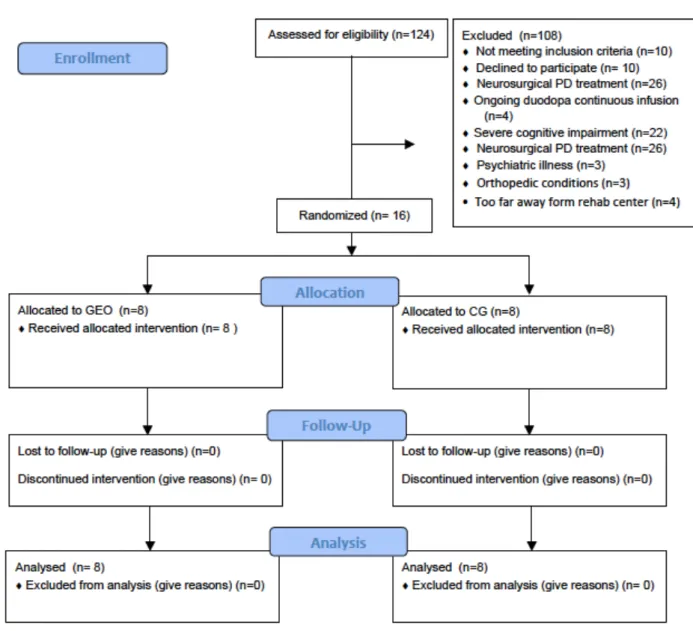
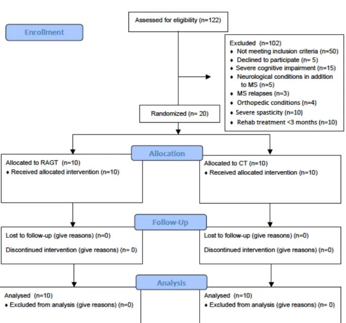
This was a parallel-group, 1:1 allocation ratio, randomized trial. The flow of subjects through the study (from enrolment to intervention allocation, follow-up and data analysis) is displayed in figure 1. The outcome assessors and data analysts were blinded to the group allocation of the participants until statistical analysis.
Participants
We studied PD subjects with history of resistant freezing of gait (FOG) who were consecutively referred to Neurorehabilitation Clinic of Ancona in the period between December 2016 and March 2018. Inclusion criteria were: confirmed idiopathic PD diagnosis according to the UK Brain Bank Criteria57
; history of FOG resistant to levodopa regardless of the dose, Hoehn and Yahr (H&Y)58
stage ≤ 4 as determined in the “off” phase; ability to adhere to the visit schedule and protocol requirements, and willingness to complete the study; ability to understand and give informed consent. Exclusion criteria were: atypical Parkinsonism (subjects with Parkinsonian features caused by disorder such as multiple system atrophy, progressive supranuclear palsy, dementia with Lewy bodies or multiple brain infarcts); severe cognitive impairment (MMSE<18); change of PD medication during the month before the enrolment; presence of dyskinesias; previous neurosurgical treatment for PD (e.g., procedures including ablation or deep brain stimulation); on-going duodopa continuous infusion; deficits of somatic sensation involving the lower limbs; vestibular disorders or paroxysmal vertigo; other neurological or orthopedic conditions involving the lower limbs (musculoskeletal diseases, severe osteoarthritis, peripheral neuropathy, joint replacement); clinically significant psychiatric illness; significant history of cardiac, pulmonary, hepatic or renal disease or other condition or any major complication/illness which, in the opinion of the investigator, contraindicates his/her participation; any type of rehabilitation treatment performed in
Subjects were randomly allocated into two groups of equal size:
Experimental Group (GEO group): the subjects underwent robot-assisted gait training based on the end-effector principle, the G-EO System (Reha Tecnology AG, Olten, Switzerland)
Control Group (CG): the subjects performed treadmill and over-ground walking training according to conventional physical therapy.
Intervention
Both GEO and CG received 12 training sessions of 45 minutes each (including rest and stretching), 3 days/week for four consecutive weeks. GEO treatment consisted in 5 minutes of lower limb muscle stretching, 20-30 minutes continuous robot gait training at variable speeds (from 0,9 km/h to 2,2 km/h) based on subject tolerance, and 5 minutes of free walk overground, at the end. The first 2-3 sessions were used to set and personalize robot parameters, let patients familiarize with the device and increase the exercise tolerance. During the following sessions, the support was gradually reduced and the speed increased. Disregarding patients’ complaints of fatigue or tiredness, all subjects were encouraged to carry on the training, without interruptions, except during the first session, when the therapist could decide to stop the device for a while giving the subjects a short rest. The CG underwent conventional physical therapy including muscle strengthening, proprioceptive exercises, balance and walking training performed both overground (10-15 minutes) and over a treadmill (10-15 minutes) using visual cueing strategies.
Ethics
The study protocol was conducted according to Good Clinical Practice requirements and conformed to Helsinki Declaration. All participants gave written informed consent.
Data and Outcome Measures
Demographic and clinical data at baseline
The following variables were recorded at baseline and regarded as independent factors of outcome: age at enrolment, gender, Body Mass Index, education, disease duration, FOG duration, Hoehn & Yahr stage in ON and OFF medication states, levodopa equivalent daily dose 59 60
, cognitive functions Montreal Cognitive Assessment (MoCA) 61.
Outcome measures
• Number of FOG episodes detected during videotaped Timed Up and Go (TUG)62 tests, total FOG time and total task time (simple test version and dual task variants-motor and cognitive). TUG time and FOG time are expressed in seconds. The standard TUG test records the time that a subject takes to rise from a chair, walk five meters, turn around, walk back to the chair, and sit down. In TUG cognitive test the subjects are asked to complete the test while counting backward by three from a randomly selected number between 20 and 100. In TUG manual test the patients have to complete the test holding a tray with two cups filled with water. The videotapes are coded and scored by a trained physician blinded to time point of assessment. Subjects performed three different examinations for each test (simple, dual motor task and dual cognitive task), and final scores represent the averages of the single examinations scores. Moreover, subjects wore a smartphone-based system for gait and FOG assessment 63
during TUG execution in order to quantify FOG episodes and increase data accuracy.
• Number of FOG episodes at home: subjects wore a smartphone-based system for gait and FOG assessment 63 at home for three consecutive days in order to monitor freezing ecologically during the daily life. This device gives information about FOG frequency expressed as a relationship between freezing numbers of events per minutes and between duration time and total gait time.
• The total score of the Gait and Falls Questionnaire (GFQ), Total score of New Freezing of Gait Questionnaire (NFOG-Q) 64: The tests were selected in order to assess the patients’ perception of FOG and gait. The questionnaires were administered to patients under medical supervision.
• Home diary of falls: subjects were encouraged to report the number of falls occurring during last week.
• Balance Evaluation Systems Test (Mini-BESTest)59: the test was used to provide information on the postural control systems underlying balance impairments and as a fall prediction tool.
• The motor score of the MDS-Unified Parkinson’s Disease Rating Scale (UPDRS) 65: to assess PD motor impairment.
• Six-Minute Walking Test (6MWT)66
during ON state was used to evaluate gait endurance. • Montreal Cognitive Assessment (MoCA): was used to check global cognitive function. • Total score Parkinson's Disease Questionnaire – 39 (PDQ 39) 67: to assess quality of life. • Neurophysiological parameters. Neuroplasticity was determined applying transcranial magnetic stimulation (TMS) with the rapid-rate Paired Associative Stimulation protocol (rPAS) developed by Quartarone68. This plasticity-inducing protocol involves median nerve stimulation at the wrist (constant current square wave pulses with of 500 ms pulse width) and TMS over the most affected primary motor cortex (M1) at 25 ms interstimulus interval (ISI), at a rate of 5 Hz to provide 600 pairs of stimuli in 2 minutes. TMS was delivered with a 7-cm figure-of-eight coil connected to a Magstim Rapid stimulator (The Magstim Company, Whitland, UK). The coil was held with the handle pointing backwards and laterally at about 45 degrees to the mid-sagittal plane to induce the first current in the cortex in the posterior-to-anterior direction over the optimal position for eliciting motor evoked responses in the Abductor Pollicis Brevis (APB) muscle. Motor evoked potentials (MEPs) were recorded at baseline (beforePAS_T0) and for up to 15 minutes (at 5 minutes _T1, at 10 minutes _T2 and at 15 minutes_T3) after rPAS. MEPs were recorded from APB with 20 stimuli
at 0.1 Hz over the contralateral M1. The stimulus intensity was adjusted to produce MEPs of 1 mV in the relaxed target muscles at baseline and was kept constant at the different times of assessment during the experiment (from T0 to T3). MEP amplitudes were measured peak to peak and then averaged.
Time of assessment
Patients were evaluated before (T0) and immediately (less than 3 days) after treatment (T1). They were tested approximately 1 h after drug intake in a stable ON condition in the morning, and in OFF medication state, after last 12 hours of pharmacological therapy washout. Mini Best test, motor UPDRS, TUG test and neurophysiological study were performed in both ON and OFF medication states; clinical questionnaire, MoCA and 6MWT in ON state.
Sample size
For sample size calculation we based on the findings from previous controlled trials where similar intervention protocols for robot-assisted gait training and conventional physical therapy involved PD patients 69
. Based on such evidence, we estimated that the proportion of patients with resistant FOG, who would have shown an improvement in the NFOG-Q score after conventional physical therapy would be proxy to 0; at the same time, the proportion of subjects expected to show a positive change in the NFOG-Q score of at least 1 point after GEO training would have been more than 70%. Therefore, in order to demonstrate a significant impact of GEO training on NFOG-Q with a 90% statistical power and 0.05 alpha error, we needed 8 subjects per group.
Statistical Analysis
The distribution of clinical and demographic variables was studied using descriptive statistics. The Mann-Whitney test and Chi-square Test were used to compare the distribution of continuous or nominal variables, respectively, in the two groups. The effect of the rehabilitation strategy on each clinical variable considered was assessed by a two-factor analysis of variance: group (robotic
rehabilitation versus traditional treatment) and time (end of treatment versus baseline), with repeated measures in the time factor. Spearman correlation test was used to evaluate the correlations between variables; the multiple regression test was used to explore the independent predictive value of several independent variables that showed significant correlations with the dependent variables. Significance level was set at p ≤ 0.05. Statistical analysis was performed by means of the Statview Statistics, version 5.
Results
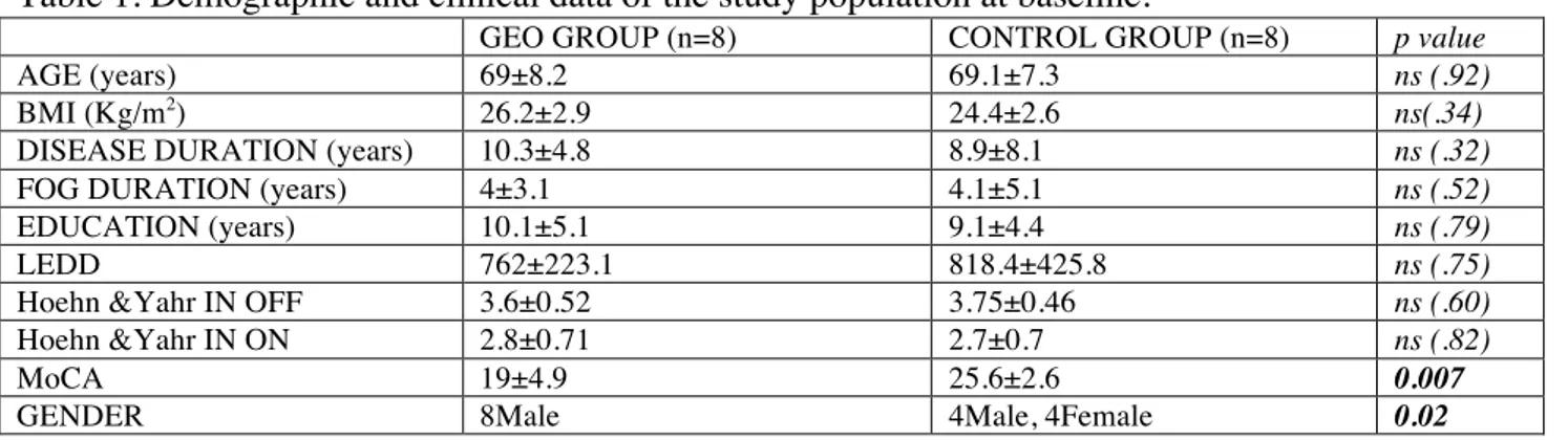
We studied 16 subjects with idiopathic PD who were allocated randomly to two groups of 8. All patients completed the treatment without adverse events and there were no dropouts at the end of the study. The demographic and clinical features of patients are summarized in table 1. Both groups were comparable at baseline in age, BMI, disease duration, FOG duration, education, LEDD and disease stage. The patients belonging to the GEO group showed lower cognitive functions than those belonging to the control group (p=.007). The males were 8 in the GEO group and 4 in the CG (p=.02). After GEO treatment an increased speed was observed in most subjects when requested to complete the simple TUG test in both medication state (p=.02), the manual TUG test and the cognitive TUG test in OFF state (p=.04 and p=.02, respectively). The walking distance during 6MWT increased after GEO training (p=.01). Regarding FOG events there were a reduction of duration time during simple TUG test and cognitive TUG test in OFF medication state (p=.03 and p=.04, respectively) and a reduction of FOG events number during cognitive TUG test in OFF state(p=.04). Moreover, the following measures improved after GEO treatment: NFGQ (p=.03), GFQ (p=.02), MiniBESTest in OFF and in ON states (p=.048 and p=.05, respectively), PDQ39 (p=.03) (Table 2) Finally, when we compared some of gait features recorded by the GEO system on the first and last rehabilitation session, respectively, we could appreciate a striking difference, signalling how intense was the training and how far the subjects improved their motor performances and endurance (Tables 3, 4).
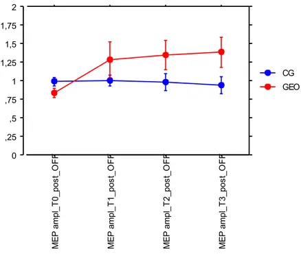
In the control group there were a reduction in the number of FOG episodes during cognitive TUG test in ON (p=.04), in the meters performed in the 6MWT (p=.04). Moreover, the smartphone app recorded a reduction in the FOG duration time and number during manual TUG test (p=.03) in ON state after conventional treatment (Table 2). FOG data recorded through smartphone app and clinical overlapped, showing no statistically significant differences. Results from repeated measures analysis of variance showed significant time x treatment effect in favour of the GEO group in the following variables: speed to complete simple TUG test in OFF state (p=.04), GFQ total score(p=.05), cognitive functions (p=.047), UPDRS III OFF subscore (p=.04), FOG frequency during daily living as detected by smartphone app at home (p=.025) (Table 2). With respect to the neuroplasticity assessment, all subjects reacted to rPAS protocol without change in MEP amplitudes before rehabilitation in both medication states (Figures 2, 3). Conversely, only after robotic treatment, a significant progressive increase in MEP amplitude was observed following the TMS stimulation protocol (from T0 to T3) suggesting that the GEO group recovered brain plasticity after training in both drug states (p<.0001)(Figures 4, 5).
The Spearman correlation analysis showed a relationship between the recovery of neuroplasticity (Δ MEP amplitude between T0 and T3) after GEO in OFF state and the reduction of FOG events at home (p=.05; Rho=-.786), Moreover there was a correlation between the recovery of neuroplasticity after GEO in OFF state and the improvement in UPDRS III in OFF state (p=.05; Rho=-.731). The recovery of plasticity correlated with the type of treatment (p=<.0001; Chi2
=16).
Discussion
The aim of this study was to test whether robotic rehabilitation based on GEO system is more effective than conventional physical therapy in improving gait, in particular resistant FOG, and to investigate the effects of treatment on neuroplasticity. The main observations were represented by the improvement of gait performance and the reduction of FOG events after GEO. The episodic nature of FOG and the influence of behaviour and mood on its appearance make it a complex
symptom to study, especially in laboratory setting where the data are poorly reproducible and do not correspond to the daily living, and this is why we chose to get an objective measurement even in the home environment by means of smartphone app. FOG occurrence and duration appeared to decrease especially in the home environment, more than in the laboratory setting and this findings corresponded with the perceptions of the subjects as shown by the improvement of their answers in GFQ and PDQ39. In outpatient clinic, the greater improvement after GEO treatment was appreciated in the OFF condition during simple and cognitive TUG tests and MiniBESTest. Moreover the GEO group showed a greater reduction in UPDRS III score and an improvement in cognitive functions as shown by the greater increase on MoCA score. It has been largely described how subjects with PD can provide near to optimal performances especially when they are requested to execute single tasks in the ON condition. Although we decided to expose them to dual tasks with the manual and cognitive TUG tests, none of these tricks allowed enhancing the sensitivity of the test towards GEO rehabilitation efficacy. Conversely, the opportunity of monitoring FOG in the patients home environment, exploiting a wearable device, allowed us to demonstrate the effectiveness of robotic training: the algorithm embedded in the smartphone quantified the number of FOG episodes and the total FOG duration during each minute walking at home revealing that both parameters were cut by almost one half after the end of training. As expected, the robotic gait training allowed increasing endurance, measured by the 6 minutes walking test; however, the increase was detected also after conventional training. This is not surprising, in fact both gait trainings are based on aerobic exercise and it was already shown that such exercise lead to endurance increase. The main difference between the two treatments is the ability of the robotic treatment to determine a motor learning even in subjects with impaired neuroplasticity. All the subjects showed impaired mechanisms of neuroplasticity before treatment in both medication states as shown by rPAS protocol. Many studies have demonstrated altered plasticity in Basal Ganglia related subcortical structures and in the primary motor cortex (M1) in various stages of PD 70
. The aberrant plasticity in PD may be directly responsible for the decreased motor skill learning observed
in patients and plays an essential role in the development of Parkinsonian symptoms 71 72
. To date, it is clear that the alteration of neuroplasticity in PD is tightly linked to the dopamine and accordingly to dopaminergic therapy and the motor complications of levodopa 73-75. However, whether the impairment in plastic response of motor cortex is a cause or a consequence of the motor complications remains an open question. Kishore A et al., showed in their study that if the motor response to levodopa was stable, the motor cortex was responsive to plasticity-induction protocols but if the motor response was complicated, the motor cortex was less responsive or unresponsive to plasticity- induction protocols 74. In fact, PD patients with levodopa-induced dyskinesias (LIDs) showed absent or poor plastic responses in the M1 and these were not restored by levodopa like as in PD patients without LIDs 19, 76
. Concerning levodopa-resistant FOG, the pathophysiology is unclear. However, at least two distinct pathophysiological pathways seem to be involved: the first one, involving impairment of the frontal lobes, appears to be a general mechanism also at work in other pathologies and even in ‘normal’ ageing; the second one, independent on the frontal lobes and possibly involving lower centers such as the mesencephalic locomotor area, could be more specific to advanced PD 77. Our results showed an abnormal plasticity in PD patients with resistant FOG that is not restored by levodopa, a response similar to that found in patients with dyskinesias. It is therefore tempting to speculate that the pathophysiology of the two motor complications is similar and so that there is a possible closed link between the onset of resistant FOG and the alteration of neuroplasticity. Moreover the restoration of plasticity after robotic gait training and the closed relationship shown between this finding and the reduction of FOG events at home reinforces this hypothesis. PD animal models allowed demonstrating the neurorestorative effects of exercise through a modulation of dopamine and glutamatergic neurotransmissions. The exercise can increase post-lesion dopamine neurotransmission as follows: 1) enhancement of vesicular release of dopamine; 2) increase of synaptic occupancy; 3) decrease of dopamine clearance through the reduction of DAT expression and the reversing of dopamine D2 receptors in the dorsal striatum,
neurotransmission that has an important role in the learning process. Studies in the PD mouse model have shown that intensive exercise can restore aspects of glutamate receptor expression, including the expression of AMPA receptors 79. In addition to its effects on glutamate receptors, exercise can also alter the storage and release of glutamate in presynaptic terminals, which might also improve circuit function and diminish the increased inhibitory drive of the dopamine-depleted striatum 80, 81. There is strong evidence from the literature that goal-based and aerobic exercise 82 might strengthen and improve motor circuitry through mechanisms that include increased synaptic strength resulting from raised dopamine and glutamate neurotransmission within the basal ganglia accompanied by increased dendritic spine formation. Exercise leads to improved generalised brain health including increased expression of neurotrophic factors, increased blood flow, altered immune response, increased neurogenesis (especially within the hippocampus), and altered metabolism (ie, improved mitochondrial health) 83. Such changes might lead to anti-inflammatory effect and an enhanced neuronal circuitry between the basal ganglia and its cortical and thalamic connections, which ultimately result in improved motor, non-motor, and cognitive behaviour in patients with Parkinson’s disease 84. Recent studies have focused on identifying exercise parameters that are essential for promoting activity-dependent neuroplasticity and ensuring the efficacy and the effectiveness lasting over time. Both basic research and clinical studies suggest that an aerobic exercise with its anti-inflammatory effect 43, 85
along with a goal based training are essentials to modulate brain plasticity 84
. In particular, high intensity (ie, high repetition, velocity, complexity)82
, specificity, difficulty, relevance and complexity of practice are parameters unavoidable 86 84
. This study showed that GEO system unlike conventional training was able to restore plasticity and this finding could be attributed to the following mechanisms of action: provide continuous proprioceptive cues; enhance the spinal control of locomotion (central pattern generators); improve postural control; promote aerobic recondition and muscle strengthening of lower limbs; force to provide an alternate gait pattern with a physiological joint amplitude and steady spatio-temporal features, like step length, symmetry and cadence, that represent key elements of FOG generation;
give the novelty effect, able to capture the patient’s attention to a greater extent than the widely known treadmill devices 56, 87, 88 89 69, 90-92
. Both implicit and explicit learning are involved during training with G-EO system: in fact, while implicit learning is ensured by the continuous repetition of lower limb movement, imposed by the robotic device, explicit learning is promoted thanks to the continuous feedback given by the device and the physiotherapist, who oversees the training and helps the patient to be aware of his own movements and harmonize them with the robot. The recovery of neuroplasticity induced by GEO system correlated with the reduction of FOG events at home and with the improvement in PD motor impairment. The small sample size is the main limitation of the study, partially offset by the use of instrumental measures of neurophysiological changes and motor behaviour: such measures strengthen the clinical observations also characterising the originality of the research protocol. Another limitation of the study is the presence of a different number of male and female individuals in the two groups since biological sex may be an important factor that moderates the relationship between exercise and neuroplasticity. However, a large gap exists in the current knowledge as few studies of exercise and brain health have directly examined this potential sex difference and most of them are on animal samples 93
. It is also currently not known whether the proposed mechanisms underlying exercise effects on the brain differ by sex 93. Moreover sex differences could be related to age-related cognitive and brain volume decline and in this study no differences were seen in age and cognitive functions between males and females and severe cognitive impairment was excluded before enrolment. In the end, both clinical and neurophysiological findings support the same conclusion: robot gait training is effective training is effective in improving drug-resistant gait disorders in the advanced phase of PD, likely through enhancing motor learning, as revealed by the recovery of brain plasticity and cognitive functions.
Conclusion
Robotic gait training with G-EO System is an effective rehabilitation approach able to improve gait performance and reduce the FOG in patients suffering from Parkinson's disease in the advanced phase, complicated by drug resistant axial symptoms. The training has been proven to be able to restore brain plasticity and promote motor learning, thus providing the biological bases for the improvement of gait function.
Compared to the more widely used treadmill training, the G-EO system is definitely a more expensive and cumbersome system; therefore, to date, only few rehabilitation facilities are equipped with it. Although the implementation of our rehabilitation protocol on a large scale is currently impractical, an accurate patient selection, based on the clinical features, like disease duration and absence of dyskinesias, would allow to refer for treatment only those subjects who are expected to obtain the greatest benefits.
Tables
Table 1. Demographic and clinical data of the study population at baseline.
GEO GROUP (n=8) CONTROL GROUP (n=8) p value
AGE (years) 69±8.2 69.1±7.3 ns (.92)
BMI (Kg/m2) 26.2±2.9 24.4±2.6 ns(.34)
DISEASE DURATION (years) 10.3±4.8 8.9±8.1 ns (.32)
FOG DURATION (years) 4±3.1 4.1±5.1 ns (.52)
EDUCATION (years) 10.1±5.1 9.1±4.4 ns (.79)
LEDD 762±223.1 818.4±425.8 ns (.75)
Hoehn &Yahr IN OFF 3.6±0.52 3.75±0.46 ns (.60)
Hoehn &Yahr IN ON 2.8±0.71 2.7±0.7 ns (.82)
MoCA 19±4.9 25.6±2.6 0.007
GENDER 8Male 4Male, 4Female 0.02
BMI: Body Max Index; F: female; FOG: freezing of gait; LEDD: levodopa equivalent daily dose; M: male; m: meters; MoCA: Montreal Cognitive Assessment; n: number; ns: not significant; OFF: OFF medication state; ON: ON medication state; Kg: kilograms.
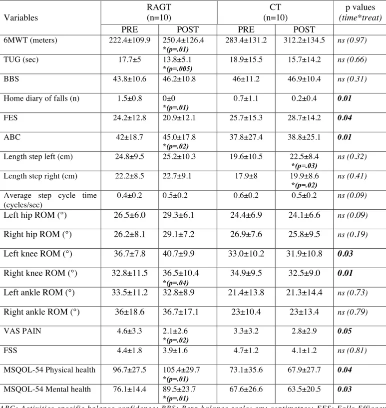
Table 2. Clinical variables before(pre) and after (post) rehabilitation in the two groups.
Variables GEO GROUP (n=8) CONTROL GROUP (n=8) p values
(time*group)
PRE POST PRE POST
TUG simple (sec) OFF 54.7±37.6 30.5±25.2
*(p=0.02)
121.1±234 142±245.9 0.04
TUG manual (sec) OFF 49.7±49.6 25±13.7
*(p=0.04)
77.7±92.5 121.5±129 ns (0.09)
TUG cognitive (sec) OFF 118±115 69.5±90.6
*(p=0.02)
65.5±59.3 41.4±18.5 ns (0.51)
TUG simple (sec) ON 28.2±17.4 24.3±16
*(p=0.02)
22.2±7 18.3±5 ns (0.99)
TUG manual (sec) ON 28.4±14.7 26.4±17.9 35±23.3 23.2±6.2 ns (0.27)
TUG cognitive (sec) ON 40±36 34±29 30.2±13.4 21.7±7.4 ns (0.70)
(clin) N FOG in TUG simple OFF 2±1.8 0.9±1 12.2±25.9 18.9±28.7 ns (0.26)
(clin) T FOG in TUG simple (sec) OFF 20.5±21.5 9.1±16.2 *(p=0.03)
86±197.3 108±209 ns (0.10)
(s.a.)N FOG in TUG simple OFF 3.4±3.5 1.5±2.1 19.1±40.3 23.5±32.5 ns (0.58)
(s.a.)T FOG in TUG simple (sec) OFF 15.8±18.8 7.3±11.7 85±193 116.4±239 ns (0.12)
(clin) N FOG in TUG manual OFF 1.8±2.2 0.6±0.9 17.9±8 6.2±7.1 ns (0.38)
(clin) T FOG in TUG manual (sec) OFF 24.1±39.4 4.7±9.7 47.9±93 79.9±111 ns (0.19)
(s.a.)N FOG in TUG manual OFF 2.7±4.2 1±1.2 24.4±6.9 20.9±41 ns (0.37)
(s.a.)T FOG in TUG manual (sec) OFF 9.7±14.1 4.7±9.3 50.1±97.5 64.1±86.7 ns (0.74)
(clin) N FOG in TUG cognitive OFF 4.8±5.3 2.8±4.7 *(p=0.04)
33.0±10.2 6.2±5.9 ns (0.92)
(clin) T FOG in TUG cognitive (sec) OFF 61.3±75.3 29.3±63.6 *(p=0.04)
37.9±64.2 37.4±69.1 ns (0.38)
(s.a.)N FOG in TUG cognitive OFF 4.6±8 4.6±8 8.6±7.2 9.4±11.7 ns (0.20)
(clin) N FOG in TUG simple ON 0.9±0.9 0.7±0.9 1.2±1.6 0.6±0.5 ns (0.33)
(clin) T FOG in TUG simple (sec) ON 2.9±3.7 2.6±4 2.4±3.4 0.6±0.6 ns (0.25)
(s.a.)N FOG in TUG simple ON 1.3±1.4 1±1.3 1.7±2.2 0.9±0.9 ns (0.37)
(s.a.)T FOG in TUG simple (sec) ON 2.4±3.2 3±4.7 2.6±3.5 0.8±1 ns (0.06)
(clin) N FOG in TUG manual ON 0.9±0.8 0.5±0.7 2.7±3.5 0.9±1.3 ns (0.21)
(clin) T FOG in TUG manual (sec) ON 2.6±3.5 2.2±4.8 5.6±9.5 1.5±2.6 ns (0.25)
(s.a.)N FOG in TUG manual ON 1.6±1.5 1.2±1.7 3.4±4.5 1±1.7
*(p=0.03)
ns (0.12)
(s.a.)T FOG in TUG manual (sec) ON 2.8±3.7 2.7±5.7 5.9±10.1 1.9±3.9 *(p=0.03)
ns (0.23)
(clin) N FOG in TUG cognitive ON 1.2±1.7 0.7±0.9 2.8±2.9 1.2±1.3 *(p=0.04)
ns (0.20)
(clin) T FOG in TUG cognitive (sec) ON 7.6±13.6 6.6±12.4 6.4±8.2 2±3 ns (0.31)
(s.a.)N FOG in TUG cognitive ON 1.8±3 1.4±2 3.9±3.8 2±2.3 ns (0.14)
(s.a.)T FOG in TUG cognitive (sec) ON 7.5±14.4 5.6±9.6 7.3±9 2.4±3.4 ns (0.47)
n FOG/min at HOME 5.3±3.2 3.5±2.8
*(p=0.04)
5.2±1.2 4.7±2.0 ns (0.28)
T FOG/min at HOME (sec/min) 11.1±7.7 5.6±5.3 *(p=0.03) 12.6±6.1 14.1±10.3 0.03 GFQ 22.4±9.6 20.3±8.6 *(p=0.02) 24.4±10.3 24.5±9.1 0.05 NFOG-Q 18.3±6.5 17.1±6.7 *(p=0.03) 17.5±4.6 17.9±3.8 ns (0.08)
Home diary of falls (n) 0.4±1.1 0.1±0.4 0.6±1.4 0 ns (0.51)
MiniBESTest OFF 13±9.5 16.5±7.2 *(p=0.04) 16±5.4 15.5±4.4 0.03 MiniBESTest ON 17.6±7.6 19.4±6.2 *(p=0.04) 20.3±5.9 21±6.1 ns (0.37) 6MWT (meters) 294±129.2 328±136 *(p=0.01) 308±111.4 346±113.7 *(p=.04) ns (0.78)
UPDRS III OFF 32.8±12.7 29.8±10.4
*(p=0.026) 32.5±6 33±5.6 0.04 UPDRS III ON 24.4±8.7 24.1±9.5 18.3±5.4 20.3±6 ns (0.16) MoCA 19±4.9 21.5±6 *(p=0.04) 25.6±2.6 25.6±3 0.05 PDQ39 37±16 34.1±14.1 *(p=0.03) 30.8±9.8 31±10.9 0.02
Clin: clinician assessment; GFQ: gait and falls questionnaire; N: number; sec: seconds; min: minutes; MoCA: Montreal Cognitive Assessment; NFOG-Q: New Freezing of gait questionnaire; ns: not significant; OFF: OFF medication state; ON: ON medication state; PDQ 39: Parkinson’s Disease Questionnaire-39; sec: seconds; s.a: smartphone app assessment; T: duration time; TUG: timed up and go test; UPDRS: Unified Parkinson’s Disease rating Scale; 6MWT: six minutes walking test. The data are expressed as mean ± standard deviation. Statistically significant intra-group
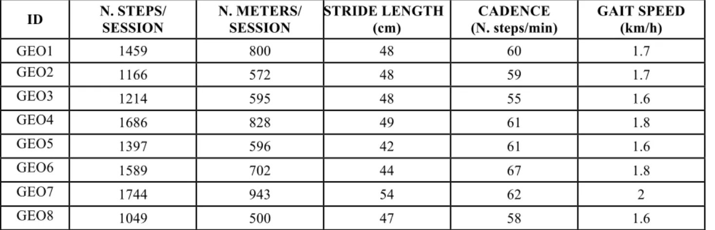
Table 3. Gait parameters exhibited by each subject during Robotic gait training: average values were computed across all training sessions.
ID N. STEPS/ SESSION N. METERS/ SESSION STRIDE LENGTH (cm) CADENCE (N. steps/min) GAIT SPEED (km/h) GEO1 1459 800 48 60 1.7 GEO2 1166 572 48 59 1.7 GEO3 1214 595 48 55 1.6 GEO4 1686 828 49 61 1.8 GEO5 1397 596 42 61 1.6 GEO6 1589 702 44 67 1.8 GEO7 1744 943 54 62 2 GEO8 1049 500 47 58 1.6
cm: centimetres; h: hour; Km: Kilometres; min: minutes; N: number.
Table 4. Gait parameters exhibited by each subject during Robotic gait training on the first and last rehabilitation session.
ID STEPS f STEPS l GAIT SPEED f GAIT SPEED l BWS f BWS l
GEO1 582 1822 1.4 1.9 7 5 GEO2 438 1812 1.3 1.8 15 5 GEO3 478 1808 1.2 1.8 27 22 GEO4 822 1830 1.5 1.9 3 0 GEO5 648 2004 1.3 1.7 0 0 GEO6 780 1288 1.6 1.7 5 0 GEO7 1158 1326 1.6 2.2 9 0 GEO8 508 1432 1.6 1.8 0 0 p value 0.01 0.01 0.03
Figures
Figure 2. rPAS effect on MEPs amplitude before rehabilitation in OFF medication state
Figure 3. rPAS effect on MEPs amplitude before rehabilitation in ON medication state 0 ,25 ,5 ,75 1 1,25 1,5 1,75 2 ME P a mp l_ T 0 _ p re _ O F F ME P a mp l_ T 1 _ p re _ O F F ME P a mp l_ T 2 _ p re _ O F F ME P a mp l_ T 3 _ p re O F F GEO CG 0 ,25 ,5 ,75 1 1,25 1,5 1,75 2 M E P a m p l_ T 0 _ p re _ O N M E P a m p l_ T 1 _ p re _ O N M E P a m p l_ T 2 _ p re _ O N M E P a m p l_ T 3 _ p re O N GEO CG
Figure 4. rPAS effect on MEPs amplitude after rehabilitation in OFF medication state
Figure 5. rPAS effect on MEPs amplitude after rehabilitation in ON medication state 0 ,25 ,5 ,75 1 1,25 1,5 1,75 2 ME P a mp l_ T 0 _ p o s t_ O F F ME P a mp l_ T 1 _ p o s t_ O F F ME P a mp l_ T 2 _ p o s t_ O F F ME P a mp l_ T 3 _ p o s t_ O F F GEO CG 0 ,25 ,5 ,75 1 1,25 1,5 1,75 2 ME P a mp l_ T 0 _ p o s t_ O N ME P a mp l_ T 1 _ p o s t_ O N ME P a mp l_ T 2 _ p o s t_ O N ME P a mp l_ T 3 _ p o s t_ O N GEO CG
Impact of exoskeleton-assisted gait training on walking and brain
plasticity in people with Multiple Sclerosis.
Introduction
Multiple sclerosis (MS) is an inflammatory immune-mediate disorder of the central nervous system and, with a lifetime risk of one in 400, potentially the most common cause of neurological disability in young adults 28. About 85% of patients with MS experience gait disturbances 94 that affect their daily activities, social life, emotional health and socioeconomic status 95
even at early phases 96
. Unfortunately, more than 66% do not retain the ability to walk 20 years after the diagnosis 97. The clinical recovery after a relapse and the disease course are influenced by neuroinflammation that profoundly subverts both brain plasticity 98
and the physiological processes of learning, memory and clinical recovery after neural damage. Although rehabilitation was shown to improve walking 99
, the superiority of one approach over another 100,101,102,103
or the effects of exercise on brain plasticity have not yet been fully clarified. The aim of this randomized controlled study is to compare the effects of robotic gait training with EKSO versus conventional gait training, in terms of both function improvement and recovery of neuroplasticity response.
Methods
Trial design
This was a parallel-group, 1:1 allocation ratio, randomized trial. The flow of subjects through the study (from enrolment to intervention allocation, follow-up and data analysis) is displayed in figure 6. The outcome assessors and data analysts were blinded to the group allocations of the participants until statistical analysis.
Participants
We studied subjects with confirmed MS diagnosis 104
, who were consecutively referred to “Santo Stefano” Rehabilitation Institute of Ancona in the period between September 2017 and January 2018. The inclusion criteria were: age range from 18 to 65 years; any level of walking disability, as determined by an Expanded Disability Status Scale (EDSS)105 score > 3.0 < 6.5; ability to stand upright unassisted; compliance with the anthropometric requirements needed to wear the exoskeleton, namely, height between 1.55 and 1.88 m, weight <100 kg and pelvic width <46 cm; ability to understand and give informed consent.
Exclusion criteria were: cognitive impairment as determined by a Mini Mental State Examination (MMSE) 106
score <24/30; neurological conditions in addition to MS; orthopaedic disorders involving the lower limbs (musculoskeletal diseases, severe osteoarthritis, osteoporosis, previous fractures); cardiovascular co-morbidity (recent myocardial infarction, heart failure, uncontrolled hypertension, orthostatic hypotension); moderate-severe spasticity, as defined by a Modified Ashworth Scale score 107 ≥ 3 or contractures that may severely restrict the lower limbs’ range of motion; MS relapses or changes in drug therapy or any other confounding factor during the study; rehabilitation treatment within 3 months before the study enrolment.
Subjects were randomly allocated into two groups of equal size:
Experimental Group: the subjects underwent robot-assisted gait training with an exoskeleton device (Robotic Assisted Gait Training group_RAGT).
Control Group: the subjects performed treadmill and over-ground walking training according to conventional physical therapy (Conventional therapy group_CT).
Intervention
Both RAGT and CT groups received 12 training sessions of 45 minutes each (including rest and stretching), 3 days/week for four consecutive weeks.
The RAGT group underwent gait training with a robotic exoskeleton (Ekso GTTM
Richmond CA, USA). During the first session, the first step program was used to provide all the power required to stand up, sit down, and walk. At first the therapist gently pushed the patient's body in order to stimulate step ignition. During the following sessions, the support was gradually reduced and the pro-step program was set so that the patient started stepping by himself transferring the body weight forward and laterally. The assistance provided by the robot during walking could be maximal or adaptive depending on the patient's abilities. Therefore, the robot could offer a total support or a mild assistance according to the gait parameters recorded after each step, thus providing only the power required to complete the step.
The CT group underwent conventional physical therapy including active joint mobilization, muscle strengthening, proprioceptive exercises, balance and walking training performed both overground (10-15 minutes) and over a treadmill (10-15 minutes).
Ethics
The study protocol was conducted according to Good Clinical Practice requirements and conformed to Helsinki Declaration. All participants gave written informed consent.
Data and Outcome measures
Demographic and clinical data at baseline
The following variables were recorded at baseline and regarded as independent factors of outcome: age at enrolment, gender, disease duration, disability status (EDSS), lower limb motor impairment (motricity index), cognitive functions (Mini Mental State Examination_MMSE and Frontal Assessment Battery__FAB) and mood disturbances (Beck Depression Inventory version II_BDI II).
Outcome measures Motor function
• The 6 minutes walking test (6MWT) 108 to assess aerobic capacity/endurance • Timed up and go test (TUG) 109
• Berg balance scale (BBS) 110
to evaluate balance and fall risk.
• Home diary of falls: subjects were encouraged to report the number of falls occurring during last week.
• Falls Efficacy Scale (FES) 111
to measure fear of falling and its possible impact on physical performance.
• Activities-specific balance confidence (ABC score) 112
to measure balance confidence in performing several functional activities.
• Spatio-temporal parameters of gait recorded through the Walker view system (a high-tech treadmill that can simultaneously provide computerized Gait analysis and movement analysis for all segments of the body): speed, step cycle time, step length, range of motion of the trunk, hips, knees and ankles.
Pain, fatigue and quality of life
• The Visual Analogue Scale (VAS) 113
was used to quantify the patient perception of pain • The Fatigue Severity Scale 114114
was administered to assess fatigue perception • Multiple Sclerosis Quality of Life-54(MSQOL-54) 115
was used to measure quality of life related to physical health (PHC) and mental health (MHC)
Neurophysiological parameters
Neuroplasticity was determined applying transcranial magnetic stimulation (TMS) with the rapid-rate Paired Associative Stimulation protocol (rPAS) developed by Quartarone 68. This plasticity-inducing protocol involves median nerve stimulation at the wrist (constant current square wave pulses with of 500 ms pulse width) and TMS over the most affected primary motor cortex (M1) at 25 ms interstimulus interval (ISI), at a rate of 5 Hz to provide 600 pairs of stimuli in 2 minutes 68
. TMS was delivered with a 7-cm figure-of-eight coil connected to a Magstim Rapid stimulator (The Magstim Company, Whitland, UK). The coil was held with the handle pointing backwards and laterally at about 45 degrees to the mid-sagittal plane to induce the first current in the cortex in the
posterior-to-anterior direction over the optimal position for eliciting motor evoked responses in the Abductor Pollicis Brevis (APB) muscle. Motor evoked potentials (MEPs) were recorded at baseline (beforePAS_T0) and for up to 15 minutes (at 5 minutes _T1, at 10 minutes _T2 and at 15 minutes_T3) after rPAS. MEPs were recorded from APB with 20 stimuli at 0.1 Hz over the contralateral M1. The stimulus intensity was adjusted to produce MEPs of 1 mV in the relaxed target muscles at baseline and was kept constant at the different times of assessment during the experiment (from T0 to T3). MEP amplitudes were measured peak to peak and then averaged.
Time of assessment
The whole clinical and neurophysiological assessment was conducted twice: before (pre) and after (post) twelve treatment sessions.
Sample size
For sample size calculation we focused on the risk for falling, assuming that reducing such risk would have represented a clinically relevant outcome for subjects with multiple sclerosis.
Based on previous reports, we assumed that being exposed to a conventional training would have decreased the risk for falling by 20%, whereas exercising with Ekso would have reduced the risk by more than 80%.
Therefore, in order to demonstrate a significant impact of RAGT on the risk for falling with a 90% statistical power and 0.05 alpha error, we needed 10 subjects per group.
Statistical Analysis
The distribution of clinical and demographic variables was studied using descriptive statistics. The Mann-Whitney test and Chi-square Test were used to compare the distribution of continuous or nominal variables, respectively, in the two groups. The effect of the rehabilitation strategy on each clinical variable considered was assessed by a two-factor analysis of variance: group (robotic
rehabilitation versus traditional treatment) and time (end of treatment versus baseline), with repeated measures in the time factor. The simple regression test was used to evaluate the correlations between changes in the variables; the multiple regression test was used to explore the independent predictive value of several independent variables that showed significant correlations with the dependent variables. Significance level was set at p ≤ 0.05. Statistical analysis was performed by means of the Statview Statistics, version 5.
Results
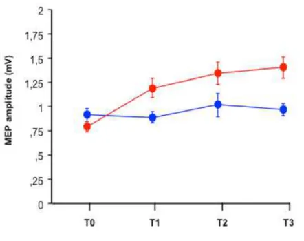
We studied 20 patients with Secondary Progressive MS who were allocated randomly to two groups of 10. All patients completed the treatment without adverse events and there were no dropouts at the end of the study. The demographic and clinical features of MS patients are summarized in table 5. Both groups were comparable at baseline in age, disease-related disability (EDSS), limb strength (motricity index) and cognitive functions. No subject was suffering from depression. The patients belonging to the RAGT group showed a longer, although not statistically significant, disease duration than those belonging to the control group. The gait performance of both groups of patients improved significantly by the end of the training program as shown by the positive change in the 6MWT (p= 0.02), BBS (p= 0.03) and TUG test (p= 0.0002) values. Moreover, both treatments were associated with a significant small reduction in fatigue (p=0.015). Results from repeated measures analysis of variance showed significant time x treatment effect in favour of the RAGT group in the following variables: number of falls and FES, ROM of the right knee, ROM of the left knee, pain VAS, ABC score, MSQOL-54 Mental Health, MSQOL-54 Physical Health (Table 6). With respect to the neuroplasticity assessment, all subjects reacted to rPAS protocol without change in MEP amplitudes before rehabilitation (Figure 7). Conversely, only after robotic treatment, a significant progressive increase in MEP amplitude was observed following the stimulation protocol (from T0 to T3) suggesting that the RAGT group recovered brain plasticity after training (p<0.0001) (Figure 8). A significant direct correlation was found between Δ MEP amplitude and the improvement
after rehabilitation of the following outcome measures: MSQOL Mental Health-54(R⌃2=.392p=0.002) and VAS pain(R⌃2=.231; p=0.023). Moreover, the restoration of neuroplasticity correlated with a longer disease duration (R⌃2=.249; p=0.015). No significant correlation was found between Δ MEP amplitude and the neurocognitive assessment at baseline. In the multiple regression analysis, treatment type (RAGT) was the only independent variable correlated to plasticity recovery (p=0.02, Chi square 5.1).
Discussion
In this study of patients with MS who underwent two different types of rehabilitation programs, both groups of patients showed a significant improvement of gait performance and fatigue after the rehabilitation treatment. Nevertheless, patients receiving robotic training had better results at the end of treatment. In particular, patients in RAGT group showed statistically significant greater improvements in pain perception and quality of life together with a greater reduction of falls. Furthermore, only the patients undergoing robotic training showed a recovery of brain plasticity. Mobility loss is common in people with MS and represents a major contributor to decreased quality of life, disruption to employment and increased financial burden116
. Congruently, walking impairment is a good index of MS-related physical disability progression, as assessed by the EDSS. Several neurological deficits such as muscle weakness, spasticity, ataxia and sensory disturbance lead to significant impairment of gait even in the early stage of the disease 117,118. Common abnormalities of spatiotemporal parameters of gait have been widely reported and include decreased gait velocity, decreased step and stride length, increased double support time, decreased swing phase, and increased step variability 96,119,120. This study showed that both gait-training protocols were able to improve gait performance with fatigue reduction. These results are in agreement with those of previous studies on the use of rehabilitation protocols for gait 95,121, 122,123,124. Different rehabilitation approaches might be useful in improving gait performance such as whole-body vibration 125, end-effector system training and sensory integration balance training 126
treadmill training 127, body weight supported treadmill training 99
, bicycle ergometer with balance exercise, home-based lower-limb strengthening and balance exercise 122
, vestibular rehabilitation 123 , robot assisted training 99,103, 128,129
. To date, the published papers were unable to provide any suggestions for clinical practice, regarding which is the most effective treatment to improve gait and balance functions, maybe for the extreme variability of clinical manifestations, for the heterogeneity of outcome measures or for the small sample size. In fact, only few RCT studies 128,99,103,100,129 evaluated whether robotic rehabilitation may be superior to conventional walking training in terms of gait performance and most of them are pilot studies 127-133
or study protocols 134
. Our results confirm the same effect on gait performance of the different approaches; moreover, at variance to other studies, in which most benefits were observed in individuals with mild-to-moderate disability, we report the efficacy of treatments, especially of the robotic gait training, in patients with a wide range of gait disability [3.0-6.5] and long disease duration. A recent systematic review conducted in individuals with MS and severe mobility disability showed limited evidence for the benefits of exercise training, conventional or adapted exercise, because of conflicting results 135
. Maybe, the missing element to get a clearer picture of the effectiveness or superiority of a specific treatment is the study of neuroplasticity. Measures of plasticity can provide insights into disease pathogenesis, improve treatment strategies and help identify substrates of treatment effects 136
.Neuroplasticity can be broadly defined as “the ability of the nervous system to respond to intrinsic and extrinsic stimuli by reorganizing its structure, function and connections; it can be described at many levels, from molecular to cellular to system and to behaviour; and can occur during development, in response to the environment, in support of learning, in response to disease, or in relation to therapy” 136
. Different forms of plasticity have been described and different instruments are available to assess it. Long-term potentiation (LTP) and long-term depression (LTD) are two forms of synaptic plasticity that can be induced by different TMS protocols, with the effects being measured as changes of MEPs amplitude after stimulation in comparison to the baseline amplitude 137. PAS has been first