A Role for Human SP
␣ as a Pattern Recognition Receptor
*
Received for publication, May 9, 2005, and in revised form, July 15, 2005 Published, JBC Papers in Press, July 19, 2005, DOI 10.1074/jbc.M505042200Maria-Rosa Sarrias
‡, Sandra Rosello´
‡, Fernando Sa´nchez-Barbero
§, Josep M. Sierra
¶, Jordi Vila
¶,
Jose´ Ye´lamos
储1, Jordi Vives
‡, Cristina Casals
§, and Francisco Lozano
‡2From the
‡Servei d
⬘Immunologia,
¶Servei de Microbiologia, Hospital Clı´nic i Provincial de Barcelona, Institut de Investigacions
Biome`diques August Pi i Sunyer, Barcelona 08036, the
储Departamento de Bioquı´mica, Biologı´a Molecular e Inmunologı´a,
Universidad de Murcia, Murcia 30100, and the
§Department of Biochemistry and Molecular Biology I, Faculty of Biology,
Complutense University of Madrid, Madrid 28040, Spain
Human Sp␣ is a soluble protein belonging to group B of the scav-enger receptor cysteine-rich (SRCR) superfamily for which little functional information is available. It is expressed by macrophages present in lymphoid tissues (spleen, lymph node, thymus, and bone marrow), and it binds to myelomonocytic and lymphoid cells, which suggests that it may play an important role in the regulation of the innate and adaptive immune systems. In the present study we show that recombinant human Sp␣ (rSp␣) binds to the surface of several Gram-positive and Gram-negative bacterial strains. Competition studies indicated that such binding is mediated by the recognition of lipoteichoic acid (LTA) and lipopolysaccharide (LPS), respec-tively, through nonoverlapping sites on the Sp␣ molecule. The most conserved part of LPS (2-keto-3-deoxyoctulosonic acid and lipid A) was shown to be involved in the recognition by Sp␣. Bacterial bind-ing studies usbind-ing the SRCR domain 1 of Sp␣ showed that this domain retains both the LPS and LTA binding activities, indicating that both bacterial interacting sites are retained in a single SRCR domain. Furthermore, rSp␣ induced aggregation of Gram-positive and Gram-negative bacteria strains. On the other hand, rSp␣ inhib-ited tumor necrosis factor-␣ secretion by human monocytes stimu-lated with LPS or LTA. Binding of Sp␣ to conserved components of bacterial surfaces and modulation of the monocyte response indi-cate that this molecule is an active constituent of the innate immune response of the host.
Macrophages play a major role in host innate defense. They are found in many tissues, particularly in those that function in the filtration of blood or lymph fluids, such as liver, spleen, lung, and lymph nodes. They recognize, internalize, and destroy harmful endogenous and foreign substances. Such recognition occurs through the direct binding to macrophage receptors or upon antibody or complement coating. Macrophage receptors are fixed germ line-encoded proteins that can directly recognize pathogen-associated common structures, hence their name, pattern recognition receptors (1, 2). These include the mannose receptor, CD14, Toll-like receptors, and scavenger receptors among others. The structures being recognized are conserved products of microbial pathogens, not shared by the host, that are essential for their survival (1). Among them, the best studied is the bacterial
Gram-nega-tive cell wall lipopolysaccharide (LPS),3a very potent activator of innate
immune responses. In its structure, LPS is composed of a membrane-anchored lipophilic component, the lipid A, covalently linked to a highly variable polysaccharide chain (O-antigen) through the inner and outer cores. The inner core consists of two or more 2-keto-3-deoxyoctu-losonic acid (KDO) sugars (linked to the lipid A) and two or three hep-tose sugars, which are in turn linked to the outer core (made of common sugars). Other examples of conserved pathogenic structures recognized by the innate immune system include lipoteichoic acid (LTA) from Gram-positive bacteria, lipoarabinomannan from mycobacteria, and mannan from fungi.
A vast number of structurally diverse cell surface or soluble glycopro-teins have been defined as scavenger receptors (SR) (3). Their common feature is that they are involved in the recognition and endocytosis of polyanionic molecules, such as oxidized or acetylated low density lipoproteins. Other SR ligands include proteins, polyribonucleotides, polysaccharides, and lipids. SR are thus able to recognize a wide range of structures. They include host components such as collagen and throm-bospondin by CD36, and altered self-like oxidized low density lipopro-tein by most SR (3), as well as pathogen-associated structures such as LPS by the class A macrophage scavenger receptor type I (SR-AI) (4), and the macrophage receptor with collagenous structure (MARCO) (5–7).
Some SR belong to the scavenger receptor cysteine-rich superfamily (SRCR-SF), which is an ancient and highly conserved family of receptors characterized by the presence of one or several repeats of a cysteine-rich extracellular domain named SRCR (8). This domain consists of⬃100 residues containing 6 – 8 cysteines with a well conserved disulfide bond pattern. The SRCR-SF has been divided into two groups based on the position and number of cysteine residues in each SRCR domain, as well as on its intron-exon organization. Thus, group A includes members possessing 6 cysteines in their SRCR domains, each of which is encoded by at least two exons. In contrast, the SRCR domains of group B proteins mostly contain 8 cysteine residues and are encoded by a single exon. Members of either group can contain the SRCR as a single domain, as tandem repeats or as part of multidomain mosaic proteins (in combi-nation with e.g. epidermal growth factor domains, serine protease domains, collagenous regions, or other domains) (9).
Despite the high conservation of the SRCR domains throughout evo-lution, there is no evidence for common functions and/or ligands *This work was supported by Ministerio de Ciencia y Tecnologı´a Grants SAF 2001-18321
and 2004-13251 and Fondo de Investigacio´n Sanitaria Grant 02/1890. The costs of publication of this article were defrayed in part by the payment of page charges. This article must therefore be hereby marked “advertisement” in accordance with 18 U.S.C. Section 1734 solely to indicate this fact.
1Investigator from the Ramo´n y Cajal Program.
2To whom correspondence should be addressed: Servei d’Immunologia, Hospital Clı´nic,
Villarroel 170, 08036 Barcelona, Spain. Tel.: 34-93-2275488; Fax: 34-93-4518038; E-mail: [email protected].
3The abbreviations used are: LPS, lipopolysaccharide; SRCR, scavenger receptor
cys-teine-rich; SF, superfamily; rSp␣, recombinant human Sp␣; LTA, lipoteichoic acid; KDO, 2-keto-3-deoxyoctulosonic acid; TNF␣, tumor necrosis factor-␣; ELISA, enzyme-linked immunosorbent assay; FCS, fetal calf serum; PBS, phosphate-buffered saline; FITC, fluorescein 5-isothiocyanate; HRP, horseradish peroxidase; mAb, monoclonal antibody; hSA, human serum albumin; SR, scavenger receptors; MARCO, macrophage receptor with collagenous structure; LXR, liver X receptor; TBS, Tris-buffered saline; mSp␣, mouse Sp␣; rSp␣D1, recombinant form of Sp␣ SRCR domain 1; CRP, cAMP receptor protein.
among the members of the SRCR-SF. This protein family currently includes about 25 members expressed on cells associated with the immune system such as macrophages (e.g. SR-AI, MARCO, CD163, and Mac2-binding protein) (5, 8, 10, 11), lymphocytes (e.g. CD5, CD6, and T19/WT1) (12–15), and on nonimmune cells such as epithelia from the digestive, respiratory, and urinary tracts (e.g. DMBT1 and S4D-SRCRB) (16, 17). Some of them have been implicated in the development of the immune system and in the regulation of innate and adaptive immune responses (18).
Human Sp␣ (hSp␣) is an SRCR-SF group B protein for which limited functional information is available. It is a soluble 38-kDa glycoprotein composed of three SRCR domains, and it is expressed by macrophages present in lymphoid tissues (spleen, lymph node, thymus, bone marrow, and fetal liver). Cell binding studies showed the existence of putative receptors for Sp␣ on peripheral monocytes but not on T and B lympho-cytes (19). However, hSp␣ bound to certain myelomonocytic (THP-1 and K562) cell lines as well as to some T (HUT78) and B (Raji) cell lines. The expression of hSp␣ in lymphoid organs involved in the develop-ment and maintenance of the lymphoid compartdevelop-ment as well as in immune surveillance further suggests that this protein may play an important role in regulating the immune system (19). Furthermore, Sp␣ may also play a role in the homeostasis of IgM, because it has been found present in IgM but not IgG or IgA fractions from human serum (20).
A mouse protein that shares 73% amino acid identity with hSp␣ has been described (21, 22). This protein is named AIM (for apoptosis inhib-itory factor secreted by macrophages) and is considered the mouse homologue of hSp␣ (mSp␣), although discrepancies on the expression pattern of both proteins have been reported (22). AIM function has been analyzed using two different mouse models. Work with mice defi-cient for AIM (AIM⫺/⫺) shows that this protein prevents apoptosis of double-positive thymocytes induced by dexamethasone and ␥-irradia-tion (22), as well as apoptosis of T and NKT cells from Corynebacterium parvum-induced liver granulomas (23). Additionally, results from transgenic mice overexpressing AIM indicate that this molecule sup-ports the survival and the phagocytic activity of macrophages in liver inflammatory (hepatitis) lesions (24). Recently, a study on mice lacking the nuclear liver X␣ receptor (LXR␣⫺/⫺) has shown that AIM is responsible for the resistance to infection with the intracellular bacteria Listeria monocytogenes(25). It was shown that LXR␣ up-regulates the expression of AIM, which in turn supports macrophage survival and bacterial clearance in the setting of L. monocytogenes infection (25).
In the present study we have used a previously characterized recom-binant form of human Sp␣ (rSp␣) (26) to show that it is able to bind to the surface of Gram-positive and Gram-negative bacteria through the recognition of pathogen-associated molecular patterns (namely, LPS and LTA).
MATERIALS AND METHODS
Reagents and Buffers—Unless indicated, all cells used in this study were grown in RPMI 1640 medium (Invitrogen) supplemented with 100 units/ml penicillin, 100g/ml streptomycin, and 10% FCS (Invitrogen) (referred to as culture medium below). The human embryonic kidney epithelial cell line HEK 293-EBNA (Invitrogen) was grown in Dulbec-co’s modified Eagle’s medium/F-12 (Invitrogen), supplemented with 100 units/ml penicillin, 100g/ml streptomycin, 250 g/ml geneticin (G418), and 10% FCS. Purified LPS, LTA, KDO, and lipid A were pur-chased from Sigma. PBS (Roche Diagnostics) is 137 mMNaCl, 3 mM
KCl, 8 mMNa2HPO4, 1.5 mMKH2PO4, pH 7.4; TBS is 140 mMNaCl,
50 mMTris-HCl, pH 7.4. hSA was purchased from Grı´fols (Grı´fols,
Barcelona, Spain). Re-LPS is a Re595 mutant of lipopolysaccharide
from Salmonella minnesota, purchased from Sigma. Fluorescein and fluorescein 5-isothiocyanate (FITC, isomer I) were purchased from Molecular Probes (Eugene, OR). Methanol and chloroform used to dissolve Re-LPS were high pressure liquid chromatography grade (Scharlau, Barcelona, Spain).
Bacterial Strains—The Escherichia coli strains serovar O26:K60(B6): H11 (CECT 351), serovar:O rough H48 (CECT 433), and serovar O26: K60(B6): H⫺ (CECT 732) were purchased from the Spanish Collection of Type Cultures (CECT, University of Valencia, Valencia, Spain). Staphylococcus epidermidiswas purchased from the American Type Culture Collection (ATCC, Manassas, VA). The rest of the bacterial strains used in this study are clinical strains isolated from patients of the Hospital Clinic of Barcelona, Spain. They were characterized by the Department of Microbiology of the Hospital Clinic of Barcelona using standard biochemical procedures.
Bacteria were grown in Luria Bertoni broth (LB) at 37 °C with aera-tion, except for Streptococcus pneumoniae, which was grown in Todd Hewitt broth at 37 °C. Bacterial strains were harvested by centrifugation at 3500⫻ g for 10 min and resuspended in TBS to a final density of 1010
bacteria per ml. Quantification was done by plating bacteria dilutions on agar plates.
Proteins and Antibodies—The expression and purification of a rSp␣ has been reported elsewhere (26). The same expression system was used to generate the recombinant form of Sp␣ SRCR domain 1 (rSp␣D1). The latter protein was generated as follows. Specific cDNA encompass-ing amino acids Ser-1 to Gly-137 (mature protein numberencompass-ing) was amplified from the pCEP-Pu vector carrying the Sp␣ full-length cDNA, by PCR amplification with the 5⬘-GCCCGGCTAGCGTCTCCATCT-GGAGTGCG-3⬘ (forward) and 5⬘-CGCGGATCCTAACCCTCTGG-GACTG-3⬘ (reverse) Sp␣-specific primers, as described previously (26). The forward primer incorporated the NheI restriction site (underlined), whereas the reverse primer incorporated a stop codon followed by the BamHI restriction site (underlined). The amplified PCR product was NheI/BamHI-restricted and cloned into appropriately digested pCEP-Pu vector. As a consequence of the cloning process, the natural signal peptide was replaced by the BM-40 leader sequence, which resulted in the addition of amino acids Ala-Pro-Leu-Ala to its N-termi-nal sequence (26 –28). Then, semi-confluent HEK 293-EBNA cells grown in 6-well culture dishes were transfected with 4g of the result-ing plasmid construct usresult-ing Lipofectamine 2000 (Invitrogen), followresult-ing the manufacturer’s instructions. Transfectants were selected with 1 g/ml puromycin (Sigma) in the culture medium. Cell transfectants expressing rSp␣ D1 were grown to confluence in Dulbecco’s modified Eagle’s medium/FCS. The medium was collected every 48 –72 h over a period of 15 days.
The recombinant proteins rSp␣ and rSp␣D1 were affinity-purified over two different CNBr-activated Sepharose 4B columns (Amersham Biosciences) covalently coupled to the anti-Sp␣ mAb 3B1a (26). Unbound protein was washed off with PBS containing 0.5MNaCl and 1% Nonidet P-40. rSp␣ or rSp␣D1 were eluted with PBS containing 3.5
MMgCl2and exchanged to PBS, using HiTrapTMdesalting columns
(Amersham Biosciences). The purified proteins were separated on SDS-PAGE under reducing conditions, and their purity was assessed by staining with Coomassie Blue. The yield was approximately of 2 mg of purified rSp␣ and 500 g of rSp␣D1 per liter of culture medium.
Protein Biotinylation—EZ-Link PEO-maleimide-activated biotin (Pierce) was dissolved in PBS at a concentration of 10 mMand then
added to rSp␣ or rSp␣D1 solution at a 5:1 molar ratio. The reaction mixture was incubated for 30 min at room temperature and then desalted and exchanged into PBS over a HiTrapTMdesalting column to
remove the free biotin. Monitoring of the biotinylation reactions was performed by SDS-PAGE and Western blotting analysis.
ELISAs—12g of LPS purified from E. coli O55:B5, O111:B4, or O26:B6 (Sigma) was used to coat 96-well microtiter plates (Nunc, Roskilde, Denmark) in PBS, overnight at 4 °C. Nonspecific binding to the wells was prevented by the addition of PBS containing 1% bovine serum albumin for 1 h at room temperature. Several concentrations of rSp␣ were then added to the wells and incubated for 1 h at room tem-perature. Bound rSp␣ was detected with the addition of biotinylated mAb 3B1a (1.8g), for 1 h at room temperature, followed by a 30-min incubation with a 1:1000 dilution of HRP-labeled streptavidin (DAKO, Glostrup, Denmark). Between each incubation step, unbound protein, antibody, or HRP-streptavidin was washed off three times with PBS, 0.01% Tween 20. Color was developed by adding 3,3⬘,5,5⬘-tetramethyl-benzidine liquid substrate (Sigma), and the absorbance was read at 405 nm. The assay was repeated twice with similar results.
Bacterial Binding Studies—Binding of Sp␣ to bacteria was studied following a method described previously (4) although with slight mod-ifications. For direct binding studies,⬃5 ⫻ 107bacteria (in 30l) were
incubated with 1g of biotinylated rSp␣, 1 g of cold rSp␣, or 0.7 g of rSp␣D1, in 400 l of TBS containing 1% bovine serum albumin and 5 mMCa2⫹(binding buffer), by gentle orbital rotation for 1 h at 4 °C. In some experiments 5 mM Ca2⫹was replaced by 5 mMEDTA in the binding and washing steps. For competition studies, 1g of biotinylated rSp␣ was incubated for 30 min on ice with several amounts of LPS, LTA, KDO, or lipid A in 100l of binding buffer. Then a suspension contain-ing 5⫻ 107bacteria (30l) was added and incubated on an orbital
rotator for 1 h at 4 °C. In all cases, bacteria were pelleted and washed three times with 0.5 ml of binding buffer and twice with 0.5 ml of TBS, 5 mMCa2⫹. The washed pellets were then resuspended in SDS-PAGE reducing sample buffer and denatured by heating at 100 °C for 15 min. Protein binding was analyzed by Western blotting, as follows. Proteins were fractionated by electrophoresis through 10 or 15% SDS-PAGE and electrotransferred to a nitrocellulose membrane (Bio-Rad). Nonspecific binding to the membrane was blocked with PBS, 10% nonfat milk pow-der for 1 h at room temperature. For rSp␣ or rSp␣D1 detection, a 2 g/ml solution of mAb 3B1a in blocking buffer was added and incu-bated for 1 h. Then a 1:1000 solution of HRP-labeled protein G (DAKO), for bound mAb detection, or HRP-labeled streptavidin (DAKO), for biotinylated protein detection, were added and incubated for 30 min at room temperature. The membranes were washed three times for 15 min with PBS, 0.01% Tween 20 between incubation steps. Finally, the blots were developed by chemiluminescence (Supersignal West Dura Extended Duration Substrate, Pierce).
Determination of the Apparent Dissociation Constant of FITC-Re-LPS/rSp␣ Complexes—Re-LPS was labeled with FITC by binding the phosphoethanolamine groups of Re-LPS to FITC as described previ-ously (29, 30). The Re-LPS concentration of the fluorescent derivative was determined by quantification of the KDO, and the content of fluo-rescein was determined spectrophotometrically at 493 nm, using a molar absorption coefficient of 77,000M⫺1cm⫺1(29, 30). The deriva-tization procedure did not alter the biological activity of the unsubsti-tuted molecule (29, 30).
Fluorescence measurements were carried out using an SLM-Aminco AB-2 spectrofluorimeter with a thermostated cuvette holder (⫾0.1 °C), using 5⫻ 5 mm path length quartz cuvettes. Fluorescent emission spec-tra of FITC-Re-LPS (0.5g/ml) were measured in the absence or pres-ence of rSp␣ in 5 mMTris, pH 8, 100 mMNaCl, 2 nMEDTA at 25 °C. The
blanks (protein alone) and FITC-Re-LPS samples (with and without protein) were excited at 470 nm, and emission spectra were recorded
from 500 to 650 nm. The apparent dissociation constant (KD) for
FITC-Re-LPS/rSp␣ complexes was obtained by analyzing the time depend-ence of the fluorescdepend-ence change when 0.5 g/ml M FITC-Re-LPS reacted with various concentrations of rSp␣ at 25 °C. Fluorescence emission was monitored at 520 nm for 30 min. These experiments were performed twice and in duplicate, exactly as described previously for KD
determination for Re-LPS interaction with LBP, CD14, and surfactant protein A (29, 30).
Bacteria Aggregation Assays—A fresh overnight culture of bacteria was harvested by centrifugation at 3500⫻ g for 10 min and resuspended in PBS to a final density of 1010bacteria per ml. FITC (Sigma) was
dissolved in PBS at a concentration of 10 mMand then added to the bacterial suspensions to a final concentration of 2 mM. The reaction mixtures were incubated for 30 min at room temperature, and excess unbound FITC was removed by extensive washing with PBS. 10l of FITC-labeled bacterial suspension (5⫻ 108bacteria) in TBS containing 5 mMCa2⫹or 5 mMEDTA were mixed with 10l of protein solution
(rSp␣, hSA, or rSp␣D1) or peptide SRCR2P (QGRVEVLYRG-SWGTVC) (31) at a final concentration of 2Mand incubated
over-night at room temperature. The peptide SRCR2P was kindly provided by Dr. Floris J. Bikker, from the Vrije Universiteit (Amsterdam, The Netherlands). Then the samples were transferred to a microscopic slide and examined by fluorescence microscopy.
Measurement of TNF␣ Production—Peripheral blood mononuclear cells were isolated from buffy coat samples of healthy volunteer blood donors of the Blood Bank of the Hospital Clinic of Barcelona by stand-ard density gradient centrifugation over Ficoll (LinfoSep, Biomedics, Madrid, Spain). The peripheral blood mononuclear cell-containing interface was harvested, and cells were washed twice in PBS before the addition of culture medium. The number of monocytes was determined in an ABX Minipack LMG cell counter (HoribaABX Diagnostics, Mont-pellier, France). Peripheral blood mononuclear cells were plated at 106 monocytes/well in 24-well tissue culture plates in RPMI medium con-taining 10% FCS. The monocyte population was enriched by adherence to the cell culture plate for 1–2 h at 37 °C, at which time nonadherent cells were removed. Monocytes were cultured in RPMI medium con-taining 10% FCS for 1–2 days and preincubated with culture superna-tants from rSp␣-expressing or untransfected HEK 293 cells (control), for 30 min at 37 °C. The amount of rSp␣ in the supernatant (5 g/ml) was determined by sandwich ELISA, as described before (26). The monocytes were then stimulated for 4 h with LPS (100 ng/ml) or LTA (5 g/ml) in the presence of 5% FCS at 37 °C. Cell viability was confirmed by trypan blue exclusion.
Culture supernatants were collected and assayed for TNF␣ produc-tion with the OPTEIA ELISA kit, following the manufacturer’s instruc-tions (Pharmingen). Monocytes from three donors were analyzed, and the assays were performed in duplicate.
RESULTS
AIM/mSp␣ has been shown to be important for macrophage immune function and has been implicated in the regulation of immune responses during bacterial infection (22–24). In our search for a putative function of human Sp␣, we focused our interest on its bacteria binding abilities. Such ability has been reported only for a few members of the SRCR-SF, namely SR-AI/II (4), MARCO (5–7), and gp-340/DMBT1 (32) as well as its mouse homologue CRP-ductin (33). To determine whether human Sp␣ could directly bind to the surface of whole bacteria, we used an approach that was employed previously for the study of SRAI binding to bacteria (4). An array of Gram-positive and Gram-negative bacterial strains was incubated with biotinylated affinity-purified rSp␣.
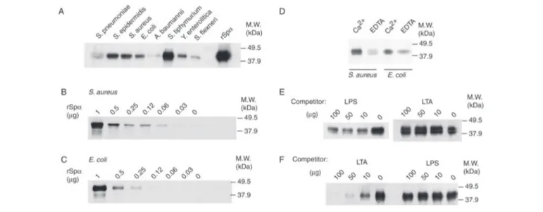
The production and characterization of rSp␣ have been reported recently (26), and it shares antigenic and structural properties with the circulating form of Sp␣ present in human serum. The bacterial pellet and any bound protein were solubilized with SDS-PAGE sample buffer and subjected to gel electrophoresis under reducing conditions. The presence of biotinylated rSp␣ in bacterial pellets was assessed by West-ern blot using HRP-labeled streptavidin. Fig. 1A shows the presence of a band of 38 kDa, the molecular weight of biotinylated rSp␣ on all lanes of the Western blot. This indicates that biotinylated rSp␣ bound to con-served structures present on the surface of both Gram-positive (S. pneu-moniae, S. epidermidis, and Staphylococcus aureus) and Gram-negative (E. coli, Acinetobacter baumannii, Salmonella typhimurium, Yersinia enterolı´tica, and Shigella flexneri) whole bacteria. The variable intensity of the band found among the different bacterial strains used may indi-cate differences in either binding affinity or bacterial inoculum, although the inoculum was always quantified (5⫻ 107cells/sample).
Binding of rSp␣ to Gram-positive (S. aureus) and Gram-negative (E. coli) bacteria was dose-dependent, as observed in Fig. 1, B and C, respectively.
To assess whether the binding of rSp␣ to bacteria was influenced by calcium, we performed similar in vitro bacteria binding assays in the presence of Ca2⫹or EDTA in the binding and washing buffers. As illus-trated by Fig. 1D, the presence of biotinylated rSp␣ was greatly reduced in E. coli and S. aureus bacterial cell pellets in the absence of calcium. This indicates that calcium facilitates the Sp␣ recognition of cell wall components from Gram-positive and Gram-negative bacteria.
We next sought to determine whether the observed binding of rSp␣ to bacteria was specific and to identify which bacterial cell surface struc-tures were being recognized. To answer these questions, competition experiments were designed, in which biotinylated rSp␣ was incubated with increasing concentrations of purified LPS, or LTA, before the addi-tion of a suspension of either E. coli or S. aureus (5⫻ 107cells). LPS and
LTA were assayed because they are ubiquitous cell surface components of these microorganisms. As illustrated by Fig. 1E, binding of biotin-ylated rSp␣ to E. coli was competed in a dose-dependent manner by LPS (from E. coli), but not by LTA (from S. aureus), indicating that this inhibition was specific. On the contrary, when the binding of rSp␣ to
S. aureuswas studied, LPS did not affect such an interaction. However, this binding was inhibited in a dose-dependent manner by LTA from S. aureus(Fig. 1F), as well as from Bacillus subtilis (data not shown). The inhibitory effect of LPS and LTA indicates that binding of rSp␣ to the surface of Gram-negative and Gram-positive bacteria is specific and is mediated through the recognition of LPS and LTA, respectively. The fact that soluble LPS failed to compete rSp␣ binding to Gram-positive bacteria, and that LTA failed to compete binding to Gram-negative bacteria, indicates Sp␣ interacts with LPS and LTA through nonover-lapping binding sites.
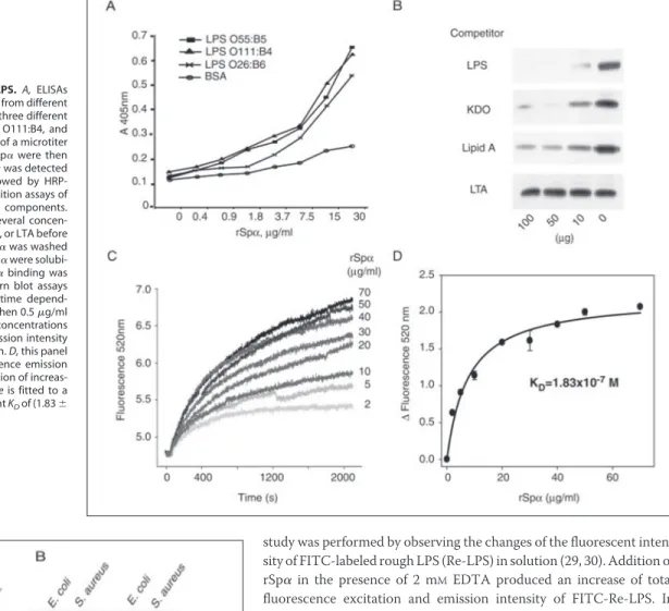
Further experiments were designed to study the rSp␣-LPS interac-tion. First, we sought to dissect the part of the LPS molecule that inter-acts with Sp␣. A direct binding ELISA was performed in which plates were coated with LPS purified from three different E. coli strains, namely LPS O55:B5, O111:B4, and O26:B6. These strains mainly differ in the composition of their O-antigen. Plates were then incubated with unlabeled rSp␣, and its binding was assessed with biotinylated anti-rSp␣ mAb 3B1a (26). The results presented in Fig. 2A show that anti-rSp␣ bound to all LPS preparations in a dose-dependent fashion.
Then, in vitro bacteria binding assays were performed by using three different strains of E. coli: serovar O26:K60 (B6):H11 (CECT 351), sero-var O rough: H48 (CECT 433), and serosero-var O26:K60(B6): H⫺ (CECT 732). These E. coli strains differ by the presence or absence of O-antigen (O26 versus rough), of capsule (K-antigen), and of flagella (H-antigen). Biotinylated rSp␣ bound to all three E. coli strains studied (data not shown). All these data suggested that the Sp␣-LPS interaction might occur through the most conserved part of LPS, namely the KDO and/or lipid A moieties. Therefore, to dissect further the part of the LPS mol-ecule to which Sp␣ binds, competition binding experiments were per-formed, in which biotinylated rSp␣ was incubated with several concen-trations of purified LPS, KDO, or lipid A, as well as LTA as negative control, before the addition of rough E. coli. Fig. 2B shows the dose-de-pendent inhibitory effect of LPS and KDO on the binding of rSp␣ to rough E. coli. Most interestingly, preincubation with lipid A had an inhibitory effect as well, although this effect was not as notorious (Fig. 2B). As expected, the addition of LTA did not affect binding of rSp␣ to rough E. coli (Fig. 2B). These results suggest that rSp␣ may mostly be
FIGURE 1. Binding of rSp␣ to Gram-positive and Gram-negative bacteria. A, binding of rSp␣-biotin to an array of Gram-positive and -negative bacteria. B, dose-dependent binding of rSp␣-biotin to S. aureus. C, dose-dependent binding of rSp␣-biotin to E. coli. D, binding of biotinylated rSp␣ to S. aureus and E. coli in the presence of 5 mMCa2⫹or 5 mMEDTA. E, competition binding assays of rSp␣-biotin to E. coli in the presence of LPS or LTA. F, competition binding assays of rSp␣-biotin to S. aureus in the presence of LPS or LTA. In all cases, rSp␣-biotin was incubated with a suspension of 5 ⫻ 107bacteria. Unbound Sp␣ was washed off, and then bacteria and bound Sp␣ were solubilized with SDS sample buffer and
recognizing the KDO part of LPS, but it may as well bind to the lipid A component with less affinity. However, the presence of traces of KDO on the lipid A preparation cannot be disregarded.
We next sought to determine the affinity of rSp␣ binding to LPS. This
study was performed by observing the changes of the fluorescent inten-sity of FITC-labeled rough LPS (Re-LPS) in solution (29, 30). Addition of rSp␣ in the presence of 2 mMEDTA produced an increase of total
fluorescence excitation and emission intensity of FITC-Re-LPS. In order to confirm that the changes observed in the fluorescence proper-ties of FITC-Re-LPS were because of the presence of rSp␣, but not to the fluorescence moiety of the labeled LPS, we studied the potential inter-action between rSp␣ and fluorescein. Addition of rSp␣ to fluorescein did not affect the fluorescence spectrum of the dye (data not shown). To calculate the apparent equilibrium dissociation constant (KD) of the
FITC-Re-LPS/rSp␣ complexes, we measured the time dependence of the fluorescent change when 0.5g/ml FITC-Re-LPS reacted with increasing amounts of rSp␣ (Fig. 2C). This change increased as a func-tion of rSp␣ concentrafunc-tion and was saturable (Fig. 2D). The KDfor the
FITC-Re-LPS-rSp␣ interaction was 1.83 (⫾0.07) ⫻ 10⫺7M, considering
a molecular mass of 38 kDa for rSp␣.
To test whether the bacterial binding activity of Sp␣ is retained within a single SRCR domain, we expressed a truncated form of Sp␣, rSp␣D1, which spans from Ser-1 to Gly-137 (mature protein numbering) and includes the most N-terminal SRCR domain of the protein (D1) and the Pro-Ser-Thr (PST)-rich region that separates it from D2. rSp␣D1 was expressed in an episomal mammalian expression system that was used previously to produce efficiently the full-length rSp␣ (26). rSp␣D1 was purified from the culture supernatants by affinity chromatography with mAb 3B1a covalently coupled to CNBr-activated Sepharose. Such puri-fication yielded two protein products of⬃12 and ⬃10 kDa, as observed by SDS-PAGE and Coomassie Blue staining (Fig. 3A). Similar differ-ences in molecular weight were observed in full-length rSp␣, as well as in Sp␣ from human serum, which are due to differences in sialic acid content (26).
FIGURE 3. Sp␣ SRCR domain 1 binds to bacteria. A, expression and purification of rSp␣ and rSp␣D1. Both proteins were affinity-purified from culture supernatants with mAb 3B1a. The purified proteins were resolved on a 15% SDS-PAGE under reducing conditions and stained with Coomassie Blue. B, binding of rSp␣-biotin and rSp␣D1-biotin to E. coli and S. aureus. C, effect of unlabeled rSp␣ and rSp␣D1on the binding of biotinylated rSp␣ to E. coli. Several amounts of unlabeled proteins were incubated with
E. coli (indicated as fold of molar excess), before the addition of biotinylated rSp␣. B, and C, after incubation, unbound protein was washed off. The bacteria and bound proteins
were solubilized with SDS sample buffer, and binding of biotinylated proteins was assessed by SDS-PAGE and Western blot assays with HRP-labeled streptavidin. FIGURE 2. Binding of rSp␣ to LPS. A, ELISAs
showing direct binding rSp␣ to LPS from different bacterial strains. LPS purified from three different
E. coli strains, namely LPS O55:B5, O111:B4, and
O26:B6, were coated into the wells of a microtiter plate. Several concentrations of rSp␣ were then added to the wells, and bound Sp␣ was detected with biotinylated mAb 3B1a, followed by HRP-conjugated streptavidin. B, competition assays of rSp␣ binding with different LPS components. rSp␣-biotin was incubated with several concen-trations of purified LPS, KDO, lipid A, or LTA before the addition of E. coli. Unbound Sp␣ was washed off, and then bacteria and bound Sp␣ were solubi-lized with SDS sample buffer. rSp␣ binding was assessed by SDS-PAGE and Western blot assays with HRP-labeled streptavidin. C, time depend-ence of the fluorescdepend-ence change when 0.5g/ml FITC-Re-LPS reacts with increasing concentrations of purified rSp␣. Fluorescence emission intensity was monitored at 520 nm for 35 min. D, this panel shows the net change in fluorescence emission intensity of FITC-Re-LPS upon addition of increas-ing amounts of rSp␣. The solid line is fitted to a rectangular hyperbola with apparent KDof (1.83⫾
The affinity-purified rSp␣D1 molecule was biotin-labeled and tested for binding to bacteria as was done for the full-length rSp␣. Thus, bioti-nylated rSp␣D1 was incubated with cell suspensions (5 ⫻ 107cells) of
Gram-positive (S. aureus) and Gram-negative (E. coli) bacteria. There-after, the presence of associated biotin-labeled protein in bacterial pel-lets was assessed by SDS-PAGE and Western blot with HRP-labeled streptavidin. Fig. 3B shows that rSp␣D1 retained the bacterial binding activity, indicating that this single SRCR domain contains all the resi-dues necessary for its interaction with Gram-positive and Gram-nega-tive bacteria. Furthermore, competition assays showed that unlabeled rSp␣D1 was able to compete binding of full-length biotinylated rSp␣ to both E. coli (Fig. 3C) and S. aureus (data not shown). To be able to compare the inhibitory effect of cold rSp␣ with that of rSp␣D1, the results are plotted as their molar excess with respect to the moles of biotinylated rSp␣. Most interestingly, a higher molar excess of rSp␣D1 than of cold rSp␣ was required to achieve inhibition (Fig. 3C), suggest-ing that other regions in the molecule (i.e. SRCR domains 2 and 3) may be interacting with the bacteria as well.
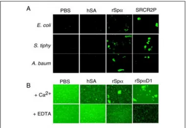
We hypothesized that the existence of multiple bacterial binding domains on the Sp␣ molecule would lead to bacterial aggregation
phe-nomena. To examine this possibility, several strains of bacteria were labeled with FITC and incubated overnight at room temperature with the recombinant proteins, as well as with hSA, used as a negative con-trol. Equimolar concentrations of the synthetic peptide SCRC2P were used as positive control in this assay. This peptide represents a consen-sus sequence (QGRVEVLYRGSWGTVC) from the loop on gp-340/ DMBT1 that runs between Cys-1 and Cys-2 (Fig. 4) and has been described previously to bind to bacteria and to aggregate them (34). In the present study, aggregation was observed by direct examination on a fluorescence microscope. Fig. 5 shows that the presence of rSp␣ induced aggregation of Gram-negative (E. coli, A. baumannii, and S. typhimurium) (Fig. 5A) as well as Gram-positive (S. aureus) (Fig. 5B) bacteria, to a similar extent as the positive control SCRC2P peptide. This effect was not observed in the presence of the negative control hSA. Because binding of rSp␣ to bacteria was influenced by the presence of calcium (see Fig. 1D), we further explored whether this was also the case for its bacterial aggregation properties. As illustrated in Fig. 5B, the presence of EDTA interfered with the aggregation capabilities of rSp␣. Bacterial aggregation assays were also performed with the rSp␣D1 domain. Intriguingly, rSp␣D1 induced bacterial aggregation in the pres-ence of calcium as well (Fig. 5B).
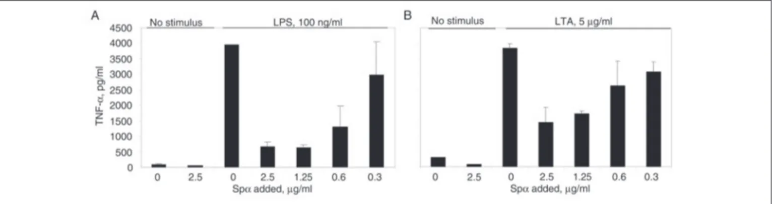
To determine whether Sp␣ influences the monocyte inflammatory response to bacterial surface components, plastic-adherent peripheral blood mononuclear cells (monocytes) from healthy blood donors were incubated at 37 °C with LPS for 4 h in the presence of culture superna-tants containing or not increasing amounts of rSp␣ (Fig. 6). rSp␣ or medium alone had no effect on TNF␣ production by monocytes (Fig. 6, Aand B). On the contrary, upon LPS addition, rSp␣ showed a dose-de-pendent inhibitory effect on TNF␣ secretion (Fig. 6A). Because LTA is also a ligand for Sp␣, we tested whether rSp␣ was able to inhibit the monocyte responses to this bacterial cell wall component. As shown in Fig. 6B, rSp␣ also inhibited LTA-induced TNF␣ secretion by mono-cytes in a dose-dependent fashion.
DISCUSSION
The present report shows that Sp␣, a soluble molecule secreted by macrophages, is involved in the recognition of pathogen-associated molecular patterns, namely LPS and LTA. This finding is of relevance because Sp␣ is a protein exclusively composed of (three) SRCR domains, which are very ancient and highly conserved protein modules whose function is still poorly understood. The SRCR domain was first reported on the mouse type I class A macrophage scavenger receptor
FIGURE 5. A, rSp␣ induces bacterial aggregation. The indicated strains of bacteria were labeled with FITC and incubated with the recombinant proteins, as well as the negative control hSA, overnight at room temperature. Equimolar concentrations of the synthetic peptide SCRC2P were used as positive control. B, effect of Ca2⫹on the aggre-gation of S. aureus by rSp␣. Aggregation experiments were performed in the presence of Ca2⫹or EDTA in the binding buffer. Aggregation was observed by direct examination on a fluorescence microscope.
FIGURE 4. A, schematic illustration of a group B SRCR domain. The eight cysteine residues and the disulfide bond pattern are shown (dotted line, disulfide bond absent in group A SRCR domains).
Numbers indicate amount of residues between
cysteines. The black line below Cys-1 indicates the region where the bacteria binding domains have been described. B, alignment of DMBT1pbs1 pep-tide with the homologous regions on Sp␣ domains. C, alignment of the region in MARCO where the RXR motif was identified. Residues that constitute the bacterial binding motifs are indi-cated in boldface.
(SR-AI) (8), and it is the molecular signature of the so-called SRCR superfamily (SRCR-SF), which includes membrane-bound and -soluble proteins reported as representatives of diverse animal phyla, from low invertebrates to high vertebrates (9). In fact, the SRCR domain is one of the few protein modules from which evolution has settled and has built a myriad of different proteins (35). It has a characteristic fold consisting of␣-helices and -strands assembled onto each other in a fashion that is predictable and stable in evolution (36). However, to date, the biological function of most SRCR domains has not been determined with cer-tainty, and there are no unifying functions and/or ligands among the members of the SRCR-SF. Some members have been involved in pro-tein-protein interactions (9), and the best-studied example of it is the interaction of CD6 with CD166/activated leukocyte-cell adhesion mol-ecule (37). Most intriguingly, three SRCR-SF members have been shown to bind to bacterial surface components, namely SR-AI (4), MARCO (5–7), and gp-340/DMBT1 (32) as well as its mouse homo-logue CRP-ductin (33). Although binding of SR-AI to bacteria is medi-ated through its collagenous domain (4), binding of MARCO, gp340/ DMBT1, and CRP-ductin is mediated through their SRCR domains (5–7, 33, 34).
Mutagenesis studies using recombinant forms of the human and mouse SRCR domain of MARCO demonstrated a crucial role for an RXR motif for its high affinity binding to bacteria (7) (Fig. 4). On the other hand, using the approach of chemical fragmentation, peptide mapping, and alanine substitution scan (31, 34), an 11-mer peptide, GRVEVLYRGSW, was identified as the minimal bacteria-binding site on the SRCR domains of DMBT1. Within this peptide, residues VELV and W were critical for this function (Fig. 4). Both motifs (RXR and VEVLXXXXW) are located within the same protein loop, which is highly conserved throughout the SRCR family, suggesting that a con-served bacterial binding domain/function may be shared by many pro-teins of this family.
Comparison of the primary sequence of Sp␣ with that of MARCO and gp-340/DMBT1 revealed a high level of amino acid similarity within the first loop of their SRCR domains (Fig. 4). However, the bacterial binding motif of MARCO (RXR) is disrupted in Sp␣ by the insertion of a Cys residue. On the other hand, none of the SRCR domains of Sp␣ fully contain all the critical residues (VEVL and W) determined for gp-340/ DMBT1 binding to bacteria. Moreover, Bikker et al. (34) found that consensus sequences of other SRCR proteins that contain the gp-340/ DMBT1-binding motif did not bind to bacteria. In fact, only those of DMBT1 and its orthologues showed significant bacterial binding activ-ity (34). The authors of this study proposed that other residues may be affecting the three-dimensional structure of the motif, and
there-fore whole proteins should be tested for conclusive results on bacteria binding.
Despite the apparent absence of a bacterial binding motif in Sp␣, we tested the possibility that Sp␣ may bind to bacteria. In a series of in vitro studies using recombinant, biotinylated Sp␣, we show that this protein can bind to a variety of Gram-positive and Gram-negative bacteria in a dose-dependent fashion. Furthermore, this binding is facilitated by the presence of divalent ions such as Ca2⫹. Most interestingly, the binding of rSp␣ to Gram-negative bacteria (E. coli) was inhibited by soluble LPS but not by soluble LTA, whereas its binding to Gram-positive bacteria (S. aureus) was inhibited by soluble LTA and not by soluble LPS. These results indicate, first, that the observed interaction of rSp␣ with the bacterial surface is specific. Second, and more important, they indicate that rSp␣ binds to LTA and LPS through independent and nonoverlap-ping sites of the molecule. This fact stresses the existence of qualitative differences between Sp␣ and gp-340/DMBT1 regarding their bacterial binding properties, because the VEVLXXXXW motif mediates binding to both Gram-negative and Gram-positive bacteria. Furthermore, from the experiments performed with the most N-terminal SRCR domain of Sp␣ (rSp␣D1), it can be concluded that the bacterial binding activity of Sp␣ is retained within a single SRCR domain. rSp␣D1 was able to bind to both Gram-positive and Gram-negative bacteria, indicating that this domain contained binding sites for both LPS and LTA. The fact that rSp␣D1 inhibited binding of biotinylated rSp␣ to a lesser extent than full-length rSp␣ further suggests that SRCR domain 1 of Sp␣ may not be the only bacteria-interacting site. Therefore, it is possible that SRCR domains 2 and/or 3 play a role in such interaction. The bacterial aggre-gation properties displayed by rSp␣ are in complete agreement with this assumption. The analysis by PAGE under nondenaturing conditions showed that full-length rSp␣ presents as a monomer (data not shown). However, the same analysis showed that rSp␣D1 forms a considerable amount of dimers. Therefore, we cannot exclude the possibility that bacterial aggregation in the presence of rSp␣D1 is because of dimeriza-tion of the protein. As suggested before, multiple copies of a binding site on the different domains of an SRCR-SF protein may lead to enhanced bacterial binding, as well as bacterial aggregation. Taken together, our results indicate that the previously proposed SRCR bacterial binding motifs (RXR and VELVXXXW) are not the only structures within an SRCR domain able to interact with bacterial components. It is therefore possible that other SRCR-SF members not containing any of these motifs might bind as well to bacteria, which would suggest a common function for the members of this ancient family of proteins.
To our knowledge, this is the first time an SRCR-SF group B protein has been reported to interact with the cell wall constituents LPS and
FIGURE 6. rSp␣ inhibits monocyte TNF␣ production upon LPS and LTA stimulation. Plastic-adherent peripheral blood mononuclear cells (monocytes) from healthy blood donors were incubated at 37 °C with LPS (A) or LTA (B) for 4 h in the presence or absence of culture supernatants containing increasing amounts of rSp␣. Culture supernatants were collected and assayed for TNF␣ production with an ELISA.
LTA. From the bacteria binding experiments we could observe that rSp␣ bound to smooth as well as rough strains of E. coli (data not shown), as well as to several Gram-negative strains. This indicates that rSp␣ is able to bind to the surface of Gram-negative bacteria carrying the antigen O component of LPS, but the latter is not necessary for the rSp␣-bacteria interaction. We hypothesized that rSp␣ might be recog-nizing a more internal and conserved part of LPS, i.e. the KDO or the lipid A. The competition experiments with soluble KDO and lipid A corroborated our hypothesis. We could observe that preincubation with both KDO and lipid A inhibited binding of rSp␣ to E. coli. This inhibi-tion was remarkably higher in the presence of KDO, indicating a higher affinity for this part of the LPS molecule. Alternatively, the limited inhi-bition observed by the lipid A component of LPS could be due to traces of KDO in this preparation.
rSp␣ binding to rough LPS (Re-LPS) has an apparent KDof 1.83
(⫾0.07) ⫻ 10⫺7M, as determined by studying the interaction of rSp␣
with FITC-labeled Re-LPS in solution. A comparison with the KDvalues
for FITC-Re-LPS䡠LBP, FITC-Re-LPS䡠CD14, and FITC-Re-LPS-surfac-tant protein A complexes, determined under the same conditions and methodology (29, 30), indicates that the affinity of rSp␣ for Re-LPS appears to be 10-fold less than that of soluble CD14 and surfactant protein A and 100-fold less than that of LBP (29, 30). Because the bac-terial binding studies indicated that Ca2⫹might facilitate binding of Sp␣ to LPS, it is possible that the presence of divalent ions increases the apparent affinity of the Sp␣-LPS interaction. Studies are under way to answer these questions.
In accordance with our data, it has been suggested recently (25) that AIM may have antimicrobial functions. This was suggested by a study on mice lacking the liver X␣-receptor (LXR␣). These mice became highly susceptible to infection with the intracellular bacteria L. mono-cytogenes, which was mainly because of altered macrophage function, accelerated apoptosis and defective bacterial clearance. Most interest-ingly, upon L. monocytogenes infection, the LXR␣⫺/⫺ mice showed a loss of AIM expression, which led to the observed enhanced macro-phage apoptosis. But the authors of this study suggested that, independ-ent of its ability to inhibit apoptosis, AIM could also have antimicrobial functions. These functions are demonstrated in the present study for human Sp␣.
Previous cell binding studies and our own observation (data not shown) demonstrated the existence of putative receptors for Sp␣ on peripheral monocytes (19). We have found that human Sp␣ inhibited activation of monocytes by LPS and LTA in a dose-dependent fashion. Our data on TNF␣ secretion suggest that Sp␣ is involved in modulating the inflammatory processes induced by these pathogen-associated molecular patterns. Further studies are in progress to determine the molecular basis for the anti-inflammatory effects of Sp␣ reported here. Finally, to our knowledge, this is the first report on the function of human Sp␣. Binding of Sp␣ to conserved components of bacterial sur-faces and modulation of the monocyte response indicate that this mol-ecule is an active constituent of the innate immune response of the host.
Acknowledgments—We thank Drs. Arie van Nieuw Amerongen and Floris J. Bikker, from the Department of Oral Biochemistry, Academic Centre for Den-tistry Amsterdam, Vrije Universiteit en Universiteit van Amsterdam, The Netherlands, for kindly providing reagents and sharing helpful information.
REFERENCES
1. Janeway, C. A., and Medzhitov, R. (2002) Annu. Rev. Immunol. 20, 197–216 2. Gordon, S. (2002) Cell 111, 927–930
3. Brown, M. S., and Goldstein, J. L. (1983) Annu. Rev. Biochem. 52, 223–261 4. Dunne, D. W., Resnick, D., Greenberg, J., Krieger, M., and Joiner, K. A. (1994) Proc.
Natl. Acad. Sci. U. S. A. 91,1863–1867
5. Elomaa, O., Kangas, M., Sahlberg, C., Tuukkanen, J., Sormunen, R., Liakka, A., Thesleff, I., Kraal, G., and Tryggvason, K. (1995) Cell 80, 603– 609
6. Elomaa, O., Sankala, M., Pikkarainen, T., Bergmann, U., Tuuttila, A., Raatikainen-Ahokas, A., Sariola, H., and Tryggvason, K. (1998) J. Biol. Chem. 273, 4530 – 4538 7. Brannstrom, A., Sankala, M., Tryggvason, K., and Pikkarainen, T. (2002) Biochem.
Biophys. Res. Commun. 290,1462–1469
8. Freeman, M., Ashkenas, J., Rees, D. J., Kingsley, D. M., Copeland, N. G., Jenkins, N. A., and Krieger, M. (1990) Proc. Natl. Acad. Sci. U. S. A. 87, 8810 – 8814
9. Sarrias, M. R., Gronlund, J., Padilla, O., Madsen, J., Holmskov, U., and Lozano, F. (2004) Crit. Rev. Immunol. 24, 1–37
10. Law, S. K., Micklem, K. J., Shaw, J. M., Zhang, X. P., Dong, Y., Willis, A. C., and Mason, D. Y. (1993) Eur. J. Immunol. 23, 2320 –2325
11. Koths, K., Taylor, E., Halenbeck, R., Casipit, C., and Wang, A. (1993) J. Biol. Chem.
268,14245–14249
12. Aruffo, A., Melnick, M. B., Linsley, P. S., and Seed, B. (1991) J. Exp. Med. 174, 949 –952 13. Jones, N. H., Clabby, M. L., Dialynas, D. P., Huang, H. J., Herzenberg, L. A., and
Strominger, J. L. (1986) Nature 323, 346 –349
14. Gimferrer, I., Farnos, M., Calvo, M., Mittelbrunn, M., Enrich, C., Sanchez-Madrid, F., Vives, J., and Lozano, F. (2002) J. Biol. Chem. 278, 8564 – 8571
15. Wijngaard, P. L., Metzelaar, M. J., MacHugh, N. D., Morrison, W. I., and Clevers, H. C. (1992) J. Immunol. 149, 3273–3277
16. Holmskov, U., Mollenhauer, J., Madsen, J., Vitved, L., Gronlund, J., Tornoe, I., Kliem, A., Reid, K. B., Poustka, A., and Skjodt, K. (1999) Proc. Natl. Acad. Sci. U. S. A. 96, 10794 –10799
17. Padilla, O., Pujana, M. A., Lopez-de la Iglesia, A., Gimferrer, I., Arman, M., Vila, J. M., Places, L., Vives, J., Estivill, X., and Lozano, F. (2002) Immunogenetics 54, 621– 634 18. Aruffo, A., Bowen, M. A., Patel, D. D., Haynes, B. F., Starling, G. C., Gebe, J. A., and
Bajorath, J. (1997) Immunol. Today 18, 498 –504
19. Gebe, J. A., Kiener, P. A., Ring, H. Z., Li, X., Francke, U., and Aruffo, A. (1997) J. Biol.
Chem. 272,6151– 6158
20. Tissot, J. D., Sanchez, J. C., Vuadens, F., Scherl, A., Schifferli, J. A., Hochstrasser, D. F., Schneider, P., and Duchosal, M. A. (2002) Electrophoresis 23, 1203–1206 21. Gebe, J. A., Llewellyn, M., Hoggatt, H., and Aruffo, A. (2000) Immunology 99, 78 – 86 22. Miyazaki, T., Hirokami, Y., Matsuhashi, N., Takatsuka, H., and Naito, M. (1999) J.
Exp. Med. 189,413– 422
23. Kuwata, K., Watanabe, H., Yamamoto, T., Miyazaki, T., and Naito, M. (2004) Comp.
Hepatol. 3,Suppl. 1, S44
24. Haruta, I., Kato, Y., Hashimoto, E., Minjares, C., Kennedy, S., Uto, H., Yamauchi, K., Kobayashi, M., Yusa, S., Muller, U., Hayashi, N., and Miyazaki, T. (2001) J. Biol. Chem.
276,22910 –22914
25. Joseph, S. B., Bradley, M. N., Castrillo, A., Bruhn, K. W., Mak, P. A., Pei, L., Hogenesch, J., O’Connell, R. M., Cheng, G., Saez, E., Miller, J. F., and Tontonoz, P. (2004) Cell 119, 299 –309
26. Sarrias, M. R., Padilla, O., Monreal, Y., Carrascal, M., Abian, J., Vives, J., Yelamos, J., and Lozano, F. (2004) Tissue Antigens 63, 335–344
27. Nischt, R., Pottgiesser, J., Krieg, T., Mayer, U., Aumailley, M., and Timpl, R. (1991)
Eur. J. Biochem. 200,529 –536
28. Kohfeldt, E., Maurer, P., Vannahme, C., and Timpl, R. (1997) FEBS Lett. 414, 557–561 29. Tobias, P. S., Soldau, K., Gegner, J. A., Mintz, D., and Ulevitch, R. J. (1995) J. Biol.
Chem. 270,10482–10488
30. Garcia-Verdugo, I., Sanchez-Barbero, F., Soldau, K., Tobias, P. S., and Casals, C. (2005) Biochem. J., in press
31. Bikker, F. J., Ligtenberg, A. J., Nazmi, K., Veerman, E. C., van’t Hof, W., Bolscher, J. G., Poustka, A., Nieuw Amerongen, A. V., and Mollenhauer, J. (2002) J. Biol. Chem. 277, 32109 –32115
32. Carlen, A., Bratt, P., Stenudd, C., Olsson, J., and Stromberg, N. (1998) J. Dent. Res. 77, 81–90
33. Madsen, J., Tornoe, I., Nielsen, O., Lausen, M., Krebs, I., Mollenhauer, J., Kollender, G., Poustka, A., Skjodt, K., and Holmskov, U. (2003) Eur. J. Immunol. 33, 2327–2336 34. Bikker, F. J., Ligtenberg, A. J., End, C., Renner, M., Blaich, S., Lyer, S., Wittig, R., van’t Hof, W., Veerman, E. C., Nazmi, K., de Blieck-Hogervorst, J. M., Kioschis, P., Nieuw Amerongen, A. V., Poustka, A., and Mollenhauer, J. (2004) J. Biol. Chem. 279, 47699 – 47703
35. Liu, Y., and Shaw, S. (2001) Trends Immunol. 22, 227–229
36. Hohenester, E., Sasaki, T., and Timpl, R. (1999) Nat. Struct. Biol. 6, 228 –232 37. Patel, D. D., Wee, S. F., Whichard, L. P., Bowen, M. A., Pesando, J. M., Aruffo, A., and