https://doi.org/10.1007/s12016-020-08814-5
Predictive Features and Clinical Presentation of Interstitial Lung
Disease in Inflammatory Myositis
Tamara Vojinovic1 · Ilaria Cavazzana1 · Paolo Ceruti2 · Micaela Fredi1,3 · Denise Modina2 · Marialma Berlendis2 · Franco Franceschini1,3
Accepted: 21 October 2020 © The Author(s) 2020
Abstract
Interstitial lung disease (ILD) represents one of the most severe extra-muscular features of idiopathic inflammatory myositis (IIM). We aimed to identify any clinical and serological predictors of ILD in a monocentric cohort of 165 IIM patients. ILD+ patients were defined as having restrictive impairment in lung function tests and signs of ILD at chest high-resolution computed tomography (HRCT). Available HRCT images were centralized and classified in different ILD patterns: non-specific interstitial pneumonia (NSIP), organizing pneumonia (OP), usual interstitial pneumonia-like (UIP), indeterminate for UIP, and interstitial lung abnormalities (ILA). Lung function test data were recorded at onset, at 1 and 5 years after ILD diagnosis.
ILD was found in 52 IIM patients (31.5%): 46.2% was affected by anti-synthetase syndrome (ARS), 21% by polymyositis (PM), 19% by dermatomyositis (DM), and 13.5% by overlap myositis. Most of ILD+ showed NSIP (31.9%), OP (19%), inde-terminate for UIP (19%), and UIP (12.8%) patterns. At multivariate analysis, ILD was predicted by anti-Ro52 (p: 0.0026) and dyspnea (p: 0.015) at IIM onset. Most of ILD onset within is 12 months after IIM. In five cases, ILD occurs after 12 months since IIM diagnosis: these patients more frequently show dry cough and anti-Ku antibodies. Anti-Ro52 + ILD patients showed a significant increase of DLCO at 1 and 5 years of follow-up, compared with anti-Ro52 negative cases.
ILD occurs in about one third of IIM and was predicted by dyspnea at onset and anti-Ro52 antibodies. Anti-Ro52 defines a subgroup of ILD showing a significant improvement of DLCO during follow-up.
This retrospective study has been approved by local ethic committee (ASST-Spedali Civili of Brescia, Italy); protocol num-ber: NP3511
Keywords Idiopathic inflammatory myositis (IIM) · Interstitial lung disease (ILD) · Anti-Ro52 antibodies · Anti-ku
antibodies · Anti-synthetase syndrome
Introduction
Interstitial lung disease (ILD) represents one of the most frequent complaints in idiopathic inflammatory myositis (IIM) and could deeply mark the disease course, in terms of functional limitation, treatment strategy, and mortality [1]. The rate of ILD widely ranges from 20 to 86%, depending on the screening technique used and the composition of dif-ferent types of IIM considered [2]. ILD is one of the clinical hallmarks of anti-synthetase syndrome (ARS) [3], charac-terized by the triad of myositis, arthritis, and ILD, in which ILD occurs in about 60–80% of cases [4]. In non-Jo-1+ ARS, ILD is diagnosed in up to 80% of cases and could even represent the first symptom, at disease onset, defining a group of patients with a worse prognosis compared with
Tamara Vojinovic and Ilaria Cavazzana equally contributed to this work
Electronic supplementary material The online version of this article (https ://doi.org/10.1007/s1201 6-020-08814 -5) contains supplementary material, which is available to authorized users. * Franco Franceschini
1 Rheumatology and Clinical Immunology Unit, ASST Spedali Civili, Piazzale Spedali Civili 1, 25123 Brescia, Italy 2 Pulmonology Unit, ASST Spedali Civili, Piazzale Spedali
Civili 1, 25123 Brescia, Italy
3 Clinical and Experimental Science Department, University of Brescia, Piazza del Mercato 15, 25121 Brescia, Italy
anti-Jo-1+ cases [4]. Nevertheless, the prognosis of ILD-associated ARS is usually favorable [5], with improvement of lung function tests and a good response to treatment [6]. By contrast, a severe ILD, named rapidly progressive ILD (RPILD), was described, especially in Asian cohorts, in hypo- or amyopathic dermatomyositis (DM), associated with anti-MDA-5 antibodies, characterized by a rapid decline of functional status and a high mortality rate [6–11].
Although many papers define the rate and the radiologi-cal patterns of ILD associated with IIM [1, 4], few data are published regarding predictive factors of ILD development and its prognosis [1, 6, 7, 12, 13], especially in Caucasian cohorts.
The aim of this retrospective study was to identify clinical and serological features of IIM patients occurring at onset, able to predict the early or late development of ILD during follow-up.
Patients
We retrospectively analyzed 165 consecutive adult patients affected by IIM, followed up in Rheumatology and Clini-cal Immunology Unit of Brescia, Italy, in order to identify patients with ILD. Most of IIM were represented by poly-myositis (PM) (52 cases), DM (55 cases), or overlap IIM (24 cases), according to the Bohan and Peter criteria [14]; ARS [15] were found in 31 patients, while inclusion body myositis (IBM) [16] and necrotizing autoimmune myositis (NAM) [17] were diagnosed in 2 and 1 patients, respectively. A diagnosis of IIM in overlap with other systemic autoim-mune diseases was made when recent criteria for systemic sclerosis (SSc) [18], systemic lupus erythematosus (SLE) [19], Sjogren’s syndrome (SS) [20], and rheumatoid arthritis (RA) [21] were achieved.
Clinical data were obtained from clinical charts. For all the patients, we collected demographic and epidemiologi-cal data; extramuscular findings, including skin manifesta-tions (DM rash, mechanic’s hands, sclerodactyly, ischemic ulcers), calcinosis, arthritis, Raynaud’s phenomenon, dys-phagia, myocarditis; myositis, defined as loss of muscle strength, myopathic electromyographic alterations, eleva-tions of CK, transaminases and LDH values at onset, and histological findings in muscle biopsy, when performed.
Lung involvement was clinically defined as dyspnea and/or dry cough or occasional finding of ILD at thoracic imaging. According to clinical practice, at IIM onset, every patient performed chest X-ray and lung function test (LFT): if ILD was suspected, basing on symptoms, X-ray or LFT, a chest high-resolution computed tomogra-phy (HRCT) was performed to identify patients with ILD (i.e., ILD+). ILD+ patients were defined as having restric-tive impairment in lung function tests (total lung capacity
(TLC), forced vital capacity (FVC), and diffusing capacity of the lung for carbon monoxide (DLCO) < 80% of pre-dicted) and signs of ILD at HRCT [22]. Isolated reduction of respiratory muscle strength, without any radiological finding of ILD, was excluded from ILD+ group. Basing on these data, ILD was diagnosed in 52 patients.
According to our clinical practice, ILD+ patients per-formed LFTs every year. In ILD+ group, we collected lung function test data at onset, at 1 year, and 5 years after ILD diagnosis. Clinical ILD onset was classified according to other authors [2, 5] in subacute presentation, chronic pres-entation, or asymptomatic ILD. An acute presentation (or RPILD) was defined as a worsening of radiologic inter-stitial change with progressive dyspnea and hypoxemia within 1 month of the onset of respiratory symptoms [10,
11].
Methods
At our first observation, every IIM patient underwent chest X-ray and lung function tests, including TLC, FVC, DLCO, maximal inspiratory, and expiratory pressure (MIP and MEP) evaluation, and HRCT was performed in cases suspected for ILD. Available HRCT images were central-ized and evaluated by an expert pulmonologist (C.P.), in order to confirm and/or recognize different ILD patterns. The radiological findings were grouped into six main HRCT patterns: non-specific interstitial pneumonia pattern (NSIP), organizing pneumonia pattern (OP), NSIP/OP overlap pat-tern, usual interstitial pneumonia-like pattern (UIP), inde-terminate for UIP pattern, and interstitial lung abnormalities pattern (ILA), according to the current definitions in the literature [23–25].
Autoantibodies’ positivity was retrieved from clinical charts. Briefly, anti-nuclear were analyzed in all sera by IIF on HEp-2 cells, while anti-extractable nuclear antigen (ENA) antibodies were searched by ELISA (ANA screen 9 EliSA, Euroimmun, Lubeck, Germany) and immunoblot (ANA-PROFILE3, Eurommun, Lubeck, Germany). Myosi-tis-specific and myositis-associated autoantibodies (MSA and MAA) were detected, at onset, in every IIM patient, when anti-ENA resulted negative, using commercial Line-blot (Euroline Autoimmune Inflammatory Myopathies 15 Ag (IgG) Euroimmun, Lubeck, Germany) including recom-binant human proteins for Mi-2 alpha, TIF1γ, MDA-5, NXP-2, SAE, Ku, PM-Scl75/100, SRP, Jo-1, PL-7, PL-1NXP-2, EJ, and OJ. Anti-HMGCR antibodies were tested with QUANTA Lite HMGCR ELISA in 57 available samples of myositis patients [26, 27].
This retrospective study was performed according to the principles of the Declaration of Helsinki.
Statistical Analysis
The variables were expressed as mean ± standard deviation (SD). The difference between groups was detected by Fisher exact test and Student’s t test or Mann-Whitney test, when appropriated, using Statview Software (5.0.1.0). Significant values were defined for p < 0.05. Survival rate was analyzed with Kaplan-Meier curve and Log-rank (Mantel Cox) test.
Results
Demographic, Clinical, and Laboratory Features
ILD was found in 52 patients among 165 IIM cases (31.5%), with a mean age at onset of 54.13 years (SD: 14.7) and a mean follow-up of 92.4 months (SD: 66.4). The prevalence of ILD in different IIMs is shown in Table 1: ILD was signif-icantly more frequent in ARS (77.4%) compared with other IIMs (21.37%; p < 0.0001; OR: 12.6; 95CI: 4.9–32.28), while less frequently occurred in DM (p: 0.008) and in PM (p: 0.048).
Among 24 cases of overlap IIM (13 SSc, 6 SLE, 3 Sjogren’s syndrome and 2 RA), 7 patients showed ILD: 4 IIM/SSc, 2 IIM/SLE, and 1 IIM/RA.
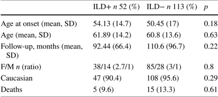
Demographic data of 52 ILD+ and 113 ILD− patients are summarized in Table 2. We recorded five deaths in ILD cases (9.6%), due to respiratory failure only in two: one case affected by PM, deceased after 13 years from onset, one case affected by overlap PM/SSc, deceased after 4 years from onset. No difference in survival rate was recorded between ILD+ and ILD− groups (p: 0.76; 100% at 1 year and 96% at 5 years in ILD+; 98% at 1 year and 95.5% at 5 years in ILD−).
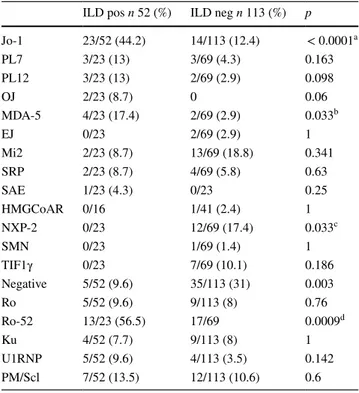
ILD+ patients were more frequently affected by ARS (p < 0.0001; OR: 12.9) and less frequently by DM (p: 0.012) (Table 3). According to these data, ILD+ group showed more frequently anti-Jo-1 (p < 0.0001; OR: 5.6) and
anti-MDA-5 antibodies (p: 0.033; OR: 7.05). On the con-trary, anti-NXP2 are found only in ILD− cases (p: 0.033). Among MAA, anti-Ro52 antibodies were significantly asso-ciated with ILD (p: 0.0009; OR: 3.97) (Table 4). Among autoantibodies’ study, we registered 5 and 35 patients negative for MSA and/or MAA, respectively, in ILD+ and ILD− groups: these patients showed isolated ANA positiv-ity without other autoantibodies’ specificpositiv-ity, including SSc-specific or SLE-SSc-specific serological markers.
No differences were detected in clinical features at IIM onset between groups, except for mechanic’s hands (p < 0.0001), arthritis (p: 0.01), polyarthritis (p: 0.001), and dyspnea (p < 0.0001) more frequently associated with ILD. By contrast, facial rash (p: 0.003), myositis (p < 0.0001), as well as higher levels of CK and AST, were more frequently found in ILD− IIM at onset, as shown in Table 5.
Multivariate analysis confirmed that dyspnea at IIM onset as well as anti-52 kD Ro antibodies predict the development of ILD (p: 0.0155 and p: 0.0026, respectively), while facial rash and anti-NXP-2 antibodies were associated with no ILD development during follow-up (p: 0.055 and p: 0.0003, respectively).
Analyzing lung function test at baseline, at 1 and 5 years after ILD onset, we registered a significant improvement of FVC values between baseline and 1 year in a total of
Table 1 ILD prevalence in different IIMs
Not included 2 patients with inclusion body myositis (IBM) and 1 patient with necrotizing autoimmune myositis (NAM)
a ARS vs all b DM vs all c PM vs all IIM-ILD (%) p OR ARS 24/31 (77.4)a < 0.0001 12.6 (4.9–32.28 DM 10/55 (18.2)b 0.008 0.34 (0.15–0.65) PM 11/52 (21.1)c 0.048 0.45 (0.2–0.97) Overlap IIM 7/24 (29.2) 0.8
Table 2 Demographic data of 52 IIM patients with ILD (ILD+) and 113 IIM patients without ILD (ILD−)
ILD+ n 52 (%) ILD− n 113 (%) p Age at onset (mean, SD) 54.13 (14.7) 50.45 (17) 0.18
Age (mean, SD) 61.89 (14.2) 60.8 (13.6) 0.63
Follow-up, months (mean,
SD) 92.44 (66.4) 110.6 (96.7) 0.22
F/M n (ratio) 38/14 (2.7/1) 85/28 (3/1) 0.8
Caucasian 47 (90.4) 108 (95.6) 0.29
Deaths 5 (9.6) 15 (13.3) 0.61
Table 3 IIM distribution in 52 IIM patients with ILD (ILD+) and 113 IIM patients without ILD (ILD−)
a OR: 12.98 (5.074–33.204) b OR: 0.36 (0.164–0.789)
ILD pos n 52 (%) ILD neg n 113
(%) p ARS 24 (46.1) 7 (6.2) < 0.0001a DM 10 (19.2) 45 (39.8) 0.012b PM 11 (21.1) 41 (36.3) 0.071 Overlap myositis 7 (13.5) 17 (15) 1 Necrotizing myositis 0 2 (1.8) 1 IBM 0 1 (0.9) 1
52 ILD+ patients (p: 0.034) (Table 6). No difference was detected in DLCO, DLCO/VA, and FVC at baseline com-paring different subgroups, namely, anti-Jo-1+, anti-Ro52+, anti-MDA5+, or anti-Ku+ patients. As shown in Table 6, 13 anti-Ro-52+ showed higher DLCO values at 1 and 5 years (p: 0.04 and 0.005, respectively) compared with 38 Ro52− patients. Anti-Jo-1 cases showed the same trend, with a significantly higher DLCO at 1 and 5 years (p: 0.049 and p: 0.021, respectively). When considering only anti-Jo-1 positive and negative cases without anti-Ro52 or Ro/SSA,
this difference is not evident: isolated anti-Jo-1-positive and anti-Jo-1-negative groups showed the same values of FVC, DLCO, and DLCO/VA at baseline, at 1 and 5 years of follow-up.
ILD Features
Analyzing 52 patients with ILD, smoking habit was found in 8 patients (15.4%), while 44 patients (88.6%) were non-smokers. Clinical respiratory presentation was dyspnea (69.2%) and cough (21%), while no symptoms were reported in 21% of cases. A chronic ILD presentation was found in 30 patients (57.7%), subacute presentation in 5 (9.6%), RPILD in 10 (19.2%), and asymptomatic onset in 6 (11.5%). In one case, ILD onset was before the diagnosis of DM. We recorded five deaths among ILD+, in only two cases related to respiratory failure (PM and overlap PM/SSc): no differ-ence between dead and alive patients was found, except for anti-OJ antibodies more frequently detected in dead (2/5:40%) than in alive cases (0%; p: 0.012).
We encountered ten cases of RPILD, four of which showed significant desaturation since the onset (affected by ARS in two, DM and PM in one case each). HRCT patterns were represented by BOOP (five cases), NSIP (two cases), and UIP-like (three cases). No differences in terms of other clinical features as well as autoantibody distribution, includ-ing anti-MDA-5 antibodies, were found between RPILD and non-RPILD.
Chest CT images were available in 47 out of 52 patients. The assessment of CT images led to the following pattern classification: NSIP pattern in 15 (31.9%), OP pattern in 9 (19.1%), NSIP/OP pattern in 4 (8.5%), UIP-like pattern in 6 (12.8%), indeterminate for UIP pattern in 9 (19.1%), and ILA pattern in 4 (8.5%).
No significant differences in demographic data, diag-noses’ distribution, clinical features at IIM onset, or MSA occurrence were found between the patterns. Significant fea-tures associated with CT patterns are summarized in Table 1
(Supplemental material): at onset, NSIP cases showed a lower FVC value (p: 0.02) and significantly lower DLCO Table 4 Myositis-specific (MSA) and myositis-associated (MAA)
autoantibodies in 52 IIM patients with ILD (ILD+) and 113 IIM patients without ILD (ILD−)
a OR: 5.6 (2.56–12.26) b OR: 7.05 (1.19–41.49) c OR: 0.000 (inf) dOR: 3.97 (1.48–10.7)
ILD pos n 52 (%) ILD neg n 113 (%) p
Jo-1 23/52 (44.2) 14/113 (12.4) < 0.0001a PL7 3/23 (13) 3/69 (4.3) 0.163 PL12 3/23 (13) 2/69 (2.9) 0.098 OJ 2/23 (8.7) 0 0.06 MDA-5 4/23 (17.4) 2/69 (2.9) 0.033b EJ 0/23 2/69 (2.9) 1 Mi2 2/23 (8.7) 13/69 (18.8) 0.341 SRP 2/23 (8.7) 4/69 (5.8) 0.63 SAE 1/23 (4.3) 0/23 0.25 HMGCoAR 0/16 1/41 (2.4) 1 NXP-2 0/23 12/69 (17.4) 0.033c SMN 0/23 1/69 (1.4) 1 TIF1γ 0/23 7/69 (10.1) 0.186 Negative 5/52 (9.6) 35/113 (31) 0.003 Ro 5/52 (9.6) 9/113 (8) 0.76 Ro-52 13/23 (56.5) 17/69 0.0009d Ku 4/52 (7.7) 9/113 (8) 1 U1RNP 5/52 (9.6) 4/113 (3.5) 0.142 PM/Scl 7/52 (13.5) 12/113 (10.6) 0.6
Table 5 Onset clinical features
positively and negatively associated with ILD IIM at univariate analysis
ILD pos n 52 (%) ILD neg n 113 (%) p OR
Mechanic’s hands 23 (44.2) 8 (7.1) < 0.0001 10.4 (4.22–25.69) Arthritis 22 (42.3) 25 (22.1) 0.01 2.58 (1.27–5.235) Polyarthritis 16 (30.8) 10 (8.8) 0.001 4.58 (1.905–11) Myocarditis 4 (7.7) 2 (1.8) 0.079 Dyspnea 46 (88.5) 50 (44.2) < 0.0001 9.66 (3.8–24.4) Facial rash 10 (19.2) 49 (43.4) 0.003 0.311 (0.142–0.681) Myositis 32 (61.5) 102 (90.3) < 0.0001 0.173 (0.075–0.398) CPK levels 1375.3 (1703.5) 4186 (6360.1) 0.0001 AST levels 89.5 (85.7) 178.4 (250.1) 0.014
value (p: 0.0004) than UIP-like cases. During the time, no significant variations of DLCO, FVC, or DLCO/VA values were recorded both in NSIP and UIP-like cases both at 1 and 5 years since onset.
Most of ILD onset occurred within 12 months from the IIM diagnosis (47/52 patients: 90.38%). No differences in terms of diagnoses, smoking habit (past or current), clinical features at onset, or clinical presentation of ILD were found between patients with ILD onset before or after 12 months since IIM. Cough was more frequently reported in patients with ILD onset after 12 months (p: 0.057). MSA were equally distributed in two groups, while anti-Ku antibodies were more frequently detected in patients with ILD onset later (40%) compared with patients with ILD onset earlier (4.2%) (p: 0.048; OR: 13; 95%CI: 1.3–130).
Discussion
ILD is a frequent clinical feature of IIM [1] with a wide vari-ation of clinical presentvari-ation and prognosis, especially for the rapid progressive ILD variant [3, 5–8]. The heterogene-ity of ILD behavior during the time and the clinical hetero-geneity of IIM itself make this feature difficult to manage. Moreover, the absence of ILD in IIM classification criteria [28] and the lack of clinical predictive factors of ILD devel-opment and progression make ILD a clinical challenge.
The pathogenesis of different IIMs is a complex picture, where different actors are involved, including genetic pre-disposition, environmental triggers, dysregulated innate and adaptive immune response, variable expression of alpha interferon inducible genes [2, 29], and inflammatory cytokines and chemokines [29]. It is widely known that PM and DM show different expression of lymphocytes, with a predominant CD8+ T cell-mediated myofiber infiltration in PM, while a B and CD4+ T cell infiltration in perivascular areas of muscle and skin is prevalent in DM [2].
T cell activation appears to play a key role also in ini-tiating lung damage in both PM and DM-associated ILD, demonstrated by a predominance of T lymphocytes in bron-choalveolar lavage with a decreased CD4+/CD8+ ratio [30] and high inflammatory cytokine expression in ILD-IIM, in particular TNF alpha in ILD+ PM and DM. Different authors reported the high expression of IL-8 [31] and other inflammatory cytokines (namely, IL-15) in lung tissue of in MDA-5+ RPILD [32]. By contrast, although ARS represents the IIM most frequently associated with ILD, a specific link with a peculiar inflammatory chemokine expression remains speculative [2].
This study aimed to identify any clinical and immunolog-ical features predicting the development of ILD at IIM onset. We recorded ILD in 31% out of 165 IIM, based on HRCT scans, reviewed by an expert pulmonologist, and lung func-tion tests. ILD rate is higher in ARS, confirming previous data, reporting ILD in about 80% of ARS [1, 3, 5]. We found ILD in 19% of DM patients and this rate did not change when only CADM is considered: this data is partially in contrast with most of the published data reporting up to 80% of ILD in CADM in Asian cohorts [6].
Anti-MDA5 is usually associated with RPILD, also in some European and American cohorts [7, 8, 33]. In our case series, anti-MDA-5 antibodies were found in similar rate in both ILD+ and ILD− cases, as well as in acute and chronic presentation ILD+ cases. Anti-MDA-5 antibodies are considered the hallmark of RPILD with a poor outcome in Asian cohorts [6, 7]. In American cohorts, anti-MDA-5 antibodies were more rarely associated with ILD, and when occurred, ILD is usually responsive to immunosuppression [34], suggesting a peculiar behavior according to different ethic/genetic backgrounds. In our cohort, anti-MDA-5 did not represent a laboratory feature predicting the develop-ment of ILD: this discrepancy could be explained by dif-ferent ethnic cohorts or by difdif-ferent laboratory methods used for detection, namely line blot in our case series, Table 6 Variation of DLCO and FVC values during time in different autoantibodies’ groups in 52 ILD+
a FVC baseline-1 year: p 0.034
b DLCO at 1 year: all Jo-1 vs other ILD: p 0.049
c DLCO 1 year: Ro52+ (73.5 ± 20.9) vs other ILD (59.5 ± 16.7): p 0.04 d DLCO 5 years: Ro52 (87 ± 19.6) vs other ILD (61.1 ± 11.4): p 0.0005
DLCO baseline
mean (SD) DLCO 1 year mean (SD) DLCO 5 years mean (SD) FVC baseline mean (SD) FVC 1 year mean (SD) FVC 5 years mean (SD)
All ILD+ (n 52) 62.8 (22.9) 63 (18.4) 67 (17.5) 83.6 (21.4) 94.2 (23.8)a 92.7 (29) all Jo-1+ (n 23) 58.3 (20.8) 56.8 (15.6)b 63.9 (20) 78.9 (20.9) 88.7 (26.8) 89.7 (31.9 Jo-1+ (Ro52−) (n 16) 55.1 (18.4) 55.58 (11.6) 57.5 (9.6) 75.9 (17) 90 (25.3) 89.55 (31.8) MDA-5+ 76.5 (16.38) 81.2 (22.4) 81.5 (27.5) 86.5 (10.5) 110 (23.6) 110 (12) Ku+ 78.7 (21.2) 78.5 (20.9) 61.3 (2.1) 93.7 (26) 105 (23.8) 83.3 (31.7) Ro52+ (n 13) 70.7 (20.6) 73.5 (20.9)c 87 (19.6)d 89.8 (23.7) 80.4 (16.6) 92.6 (14.6)
immunoprecipitation in Baltimora’s groups [34], and ELISA in other reports [6, 33]. Nevertheless, our data mirror those of a collaborative Italian study where ELISA, immunopre-cipitation, and Western blot were used [35].
In our study, dyspnea at IIM onset and anti-Ro52 antibod-ies could predict the development of ILD (p: 0.0155 and p: 0.0026) by multivariate analysis. The finding that anti-Ro52 could be strictly associated to the development of ILD is well known. Anti-Ro52 antibodies are known to represent the myositis-associated antibody more frequently found in IIM, particularly in anti-Jo-1+ patients. Recently, different authors described that anti-Ro52 [36] or anti-Ro/SSA [37] represent a negative prognostic value due to their association with severe ILD picture, at onset. Nevertheless, both groups reported a progressive stabilization or improvement in ILD anti-Ro associated [36, 37], as reported in our cohort, where anti-Ro-52 is the only serological marker of functional tests’ improvement at 1 and 5 years after ILD onset. By contrast, other authors reported a higher rate of RPILD in anti-Ro52+ patients with ARS [38], while in juvenile DM, anti-Ro52 is a predictive factor of non-remission due to active associated-ILD [39].
The clinical presentation of ILD is variable in our cohort, spreading from asymptomatic to dyspnea and cough, as pre-viously described [38]. Published data confirm that the dif-ferent clinical presentations of IIM did not confer a predic-tive value of ILD severity [38]. Noteworthy, in our hands, the onset with severe myositis, characterized by higher CPK and AST levels, represented a protective clinical pattern from ILD, although by univariate analysis.
Most of ILD cases are onset early, namely, within 12 months from IIM: although anti-MDA-5 are found only in patients with early onset ILD, in our cohort, this autoan-tibody does not represent a marker of RPILD. In contrast, cough and anti-Ku antibodies were more frequently found in late onset ILD. Anti-Ku antibodies are a known marker of overlap disease, including IIM and systemic sclerosis (SSc), strictly associated with ILD [40]. Moreover, some authors defined a clinical picture characterized by anti-Ku positivity and elevated CPK, predictive of ILD development [41]. In our study, CK elevation and myositis are not associ-ated with ILD; however, anti-Ku antibodies are associassoci-ated with late onset ILD, namely after at least 12 months from IIM onset. This could suggest that a nonspecific symptom, such as cough, and a rare autoantibody, namely anti-Ku, are found in IIM patients with a late and slow onset of ILD. To date, it is unknown if anti-Ku could really mark ILD with a slow rate progression or if anti-Ku-positive patients could be misdiagnosed and achieve an ILD-IIM diagnosis definition later. Anyway, in our study, anti-Ku+ cases did not show difference in lung functional tests compared with anti-Ku-negative group, at baseline and at different timepoints during follow-up.
Different radiological HRCT patterns have been recorded in our IIM patients, according to other cohorts [1]: NSIP pattern represents the prevalent finding, while UIP-like and OP patterns were found in about 12% and 20% of cases. NSIP pattern is the prevalent CT pattern also in SSc, while UIP pattern covers about 10% of cases [42]. In SSc, UIP pattern defines patients with worse prognosis [43], as well as in ARS cases [44].
Patients with NSIP pattern onset with lower DLCO and FVC values compared with UIP-like cases. However, both groups remained stable during time, without significant reduction of functional parameters both at 1 and 5 years from onset.
Only four cases of ILA pattern were identified (8.5%). The exact meaning of this finding in autoimmune back-grounds is not yet known. However, recent data seem to sug-gest that these alterations share common genetically driven biologic pathways with idiopathic pulmonary fibrosis, which is a specific type of chronic, progressive, fibrosing inter-stitial pneumonia of unknown cause, with an unfavorable prognosis [45].
The present paper has some limitations, the most impor-tant of which is the retrospective design. Nevertheless, the long follow-up, the exhaustive search for autoantibodies, and the review of each HRCT available by an expert pulmonolo-gist represent strength points of the paper.
Conclusion
We report here a cohort of largely prevalent Caucasian patients affected from IIM from a single center. A third of patients developed ILD. ARS was significantly associated with ILD, while DM was negatively associated. Although dyspnea and anti-Ro52 antibodies are associated with ILD at multivariate analysis, anti-Ro52 antibodies define a sub-group of ILD-IIM with significant improvement during follow-up.
Authors’ Contributions T.V. collected and analyzed patients’ clinical data and radiological images; I.C. analyzed patients’ data, performed statistical analysis, and wrote the original draft of the paper; P.C. ana-lyzed the radiological images; M.F., D.M., and M.B. actively partici-pated in paper’s draft correction; F.F. conceived the original idea of the project and participated to the paper’s writing process.
Funding Open access funding provided by Università degli Studi di Brescia within the CRUI-CARE Agreement.
Compliance with Ethical Standards
Ethical Approval This retrospective study has been approved by local ethic committee (ASST-Spedali Civili of Brescia, Italy); protocol num-ber: NP3511.
Informed Consent Data were obtained from medical charts: every patient signed our institutional consent for the management of per-sonal data.
Data Availability The datasets used and/or analyzed during the cur-rent study are available from the corresponding author on reasonable request.
Open Access This article is licensed under a Creative Commons Attri-bution 4.0 International License, which permits use, sharing, adapta-tion, distribution and reproduction in any medium or format, as long as you give appropriate credit to the original author(s) and the source, provide a link to the Creative Commons licence, and indicate if changes were made. The images or other third party material in this article are included in the article’s Creative Commons licence, unless indicated otherwise in a credit line to the material. If material is not included in the article’s Creative Commons licence and your intended use is not permitted by statutory regulation or exceeds the permitted use, you will need to obtain permission directly from the copyright holder. To view a copy of this licence, visit http://creat iveco mmons .org/licen ses/by/4.0/.
References
1. Harvier B, Uzunhan Y (2020) Inflammatory myopathy-related interstitial lung disease: From pathophysiology to treatment. Front Med 6:326
2. Saketkoo LA, Ascherman DP, Cottin V, Christopher-Stine L, Danoff SK, Oddis CV (2010) Interstitial lung disease in idiopathic inflammatory myopathy. Curr Rheumatol Rev 6:108–119 3. Cavagna L, Nuno L, Scire’ CA, Govoni M, Lopez-Longo FJ,
Franceschini F et al (2015) Clinical spectrum time course in anti Jo-1 positive antisynthetase syndrome results from an interna-tional retrospective multicenter study. Medicine 94:e1144 4. Cavagna L, Trallero-Araguas E, Meloni F, Cavazzana I,
Rojas-Serrano J, Feist E et al (2019) Influence of antisynthetase antibod-ies specificitantibod-ies on antisynthetase syndrome clinical spectrum time course. J Clin Med 8:2013
5. Hu JW, Kim DS, Lee CK, Yoo B, Bum SJ, Kitaichi M, Colby TV (2007) Two distinct clinical types of interstitial lung dis-ease associated with polymyositis-dermatomyositis. Respir Med 101:1761–1769
6. Yoshida N, Okamoto M, Kaieda S, Fujimoto K, Ebata T, Tajiri M et al (2017) Association of anti-aminoacyl-transfer RNA syn-thetase antibody and anti-melanoma differentiation-associated gene 5 antibody with the therapeutic response of polymyositis/ dermatomyositis-associated interstitial lung disease. Respir Invest 55:24–32
7. Gan YZ, Zhang LH, Ma L, Sun F, Li YH, An Y, Li ZG, Yi H (2020) Risk factors of interstitial lung diseases in clinically amyo-pathic dermatomyositis. Chinese Med J 133:644–649
8. Labrador-Horrillo M, Martinez MA, Selva-O’Callaghan A, Tral-lero-Araguas E, Balada E, Vilardell-Tarres M, et al. (2014) Anti-MDA5 antibodies in a large Mediterranean population of adults with dermatomyositis. J Immunol Re 290797
9. Moghadam-Kia S, Oddis CV, Sato S, Kuwana M, Aggarwal R (2017) Antimelanoma differentiation-associated Gene 5 antibody: Expanding the clinical spectrum in North American patients with Dermatomyositis. J Rheumatol 44:319–325
10. Chen Z, Cao M, PlanaZ, Liang J, Cai H, Kuwana M, Sun L. MN (2013) Utility of anti-melanoma differentiation-associated gene 5 antibody measurement in identifying patients with dermatomyosi-tis and a high risk for developing rapidly progressive interstitial
lung disease: a review of the literature and a meta-analysis. Arthr Care Res 65:1316–1324
11. Sato S, Hoshino K, Satoh T, Fujita T, Kawakami Y, Fujita T, Kuwana M (2009) RNA helicase encoded by melanoma differ-entiation-associated gene 5 is a major autoantigen in patients with clinically amyopathic dermatomyositis: Association with rapidly progressive interstitial lung disease. Arthritis Rheum 60:2193–2200
12. Zuo Y, Yie L, Liu M, Li S, Liu W, Chen F, et al (2020) Clinical significance of radiological patterns of HRCT and their associa-tion with macrophage activaassocia-tion in dermatomyositis. Rheumatol-ogy. https ://doi.org/10.1093/rheum atolo gy/keaa0 34
13. Fathi M, Barbasso Helmers S, Lundberg IE (2012) KL-6: A serological biomarker for interstitial lung disease in patients with polymyositis and dermatomyositis. J Intern Med 271:589–597 14. Bohan A, Peter JB (1975) Polymyositis and dermatomyositis (first
of two parts). N Engl J Med 292:344–347
15. Imbert-Masseau A, Hamidou M, Agard C, Groulleau JY, Cherin P (2003) Antisynthetase syndrome. Joint Bone Spine 70:161–168 16. Dimachkie MM, Barohn RJ (2012) Inclusion body myositis.
Semin Neurol 32:237–245
17. Allenbach Y, Mammen AL, Benveniste O, Stenzel W (2018) Immune-Mediated Necrotizing Myopathies Working Group. 224th ENMC International Workshop: Clinico-sero-pathological classification of immune-mediated necrotizing myopathies Zand-voort, The Netherlands, 14–16 October 2016. Neuromuscul Dis-ord 28:87–99
18. van den Hoogen F, Khanna D, Fransen J, Johnson SR, Baron M, Tyndall A et al (2013) Classification criteria for systemic scle-rosis: An ACR-EULAR collaborative initiative. Arthr Rheum 65:2737–2747
19. Aringer M, Costenbaden K, Daikh D, Brinks R, Mosca M, Ram-sey-Goldman R et al (2019) European League against rheumatism/ American College of Rheumatology classification criteria for Sys-temic Lupus Erythematosus 71:1400–1412
20. Vitali C, Bombardieri S, Jonsson R, Moutsopoulos HM, Alex-ander EL, Carsons SE et al (2002) Classification criteria for Sjogren’s syndrome: A revised version of the European criteria proposed by the American-European Consensus Group. Ann Rheum Dis 61:554–558
21. Aletaha D, Teogi T, Silman AJ, Funovitis J, Felson DT, Bingham CO III et al (2010) 2010 Rheumatoid arthritis classification crite-ria. Arhritis Rheum 62:2569–2581
22. Bradley B, Branley HM, Egan JJ, Greaves MS, Hansell DM, Harrison NK et al (2008) Interstitial Lung Disease Guideline. Interstitial lung disease guideline: The British Thoracic Society in collaboration with the Thoracic Society of Australia and New Zea-land and the Irish Thoracic Society. Thorax 63(Suppl V):v1–v58 23. Travis WD, Costabel U, Hansell DM, King TE, Lynch DA,
Nicholson AG et al (2013) An official American Thoracic Society/ European Respiratory Society statement: Update of the interna-tional multidisciplinary classification of the idiopathic interstitial pneumonias. Am J Respir Crit Care Med 188:733–748
24. Sverzellati N, Lynch DA, Hansell DM, Johkoh T, King TE Jr, Travis WD (2015) American Thoracic Society – European Respiratory Society Classification of the Idiopathic Interstitial Pneumonias: Advances in Knowledge since 2002. Radiographics 35:1849–1871
25. Raghu G, Remy-Jardin M, Myers JL, Richeldi L, Ryerson CJ, Lederer DJ et al (2018) Diagnosis of idiopathic pulmonary fibro-sis. An Official ATS/ERS/JRS/ALAT Clinical Practice Guideline. Am J Respir Crit Care Med 198:e44–e68
26. Musset L, Miyara M, Benveniste O, Charuel JL, Shikhman A, Boyer O et al (2014) Analysis of autoantibodies to 3-hydroxy-3-methylglutaryl-coenzyme A reductase using different technolo-gies. J Immunol Res 2014:405956
27. Musset L, Allenbach Y, Benveniste O, Boyer O, Bossuyt X, Ben-tow C et al (2016) Anti-HMGCR antibodies as a biomarker for immune-mediated necrotizing myopathies: A history of statins and experience from a large international multi-center study. Autoimmun Rev 15:983–993
28. Bottai M, Triamlund A, Santoni G, Werth VP, Pilkington C, de Visser M et al (2017) EULAR/ACR classification criteria for adult and juvenile idiopathic inflammatory myopathies and their major subgroups: A methodology report. RMD Open 14:e000507 29. Ceribelli A, De Santis M, Isailovic N, Gershwin NE, Selmi C
(2017) The immune response and the pathogenesis of idiopathic inflammatory myositis: A critical review. Clin Rev Allergy Immu-nol 52:58–70
30. Englund P, Wahlström J, Fathi M, Rasmussen E, Grunewald J, Tornling G et al (2007) Restricted T cell receptor BV gene usage in the lungs and muscles of patients with idiopathic inflammatory myopathies. Arthritis Rheum 56:372–383
31. Gono T, Kaneko H, Kawaguchi Y, Hanaoka M, Kataoka S, Kuwana M et al (2014) Cytokine profiles in polymyositis and dermatomyositis complicated by rapidly progressive or chronic interstitial lung disease. Rheumatology 56:2196–2203
32. Shimizu T, Koga T, Furukawa K, Horai Y (2020) IL-15 is a bio-marker involved in the development of rapidly progressive intersti-tial lung disease complicated with polymyositis/dermatomyositis. J Intern med. https ://doi.org/10.1111/joim.13154
33. Moghadam-Kia S, Oddis CV, Sato S, Kuwana M, Aggarwal R (2016) Anti-melanoma differentiation–associated gene 5 is associ-ated with rapidly progressive lung disease and poor survival in US patients with amyopathic and myopathic dermatomyositis. Arthr Care Res 68:689–694
34. Hall JC, Casciola-Rosen L, Samedy L-A et al (2013) Anti-mel-anoma differentiation-associated protein 5-associated dermato-myositis: Expanding the clinical spectrum. Arthritis Care Res 65:1307–1315
35. Ceribelli A, Fredi M, Taraborelli M, Cavazzana I, Tincani A, Selmi C et al (2014) Prevalence and clinical significance of anti-MDA5 antibodies in European patients with polymyositis/der-matomyositis. Clin Exp Rheum 32:891–897
36. Marie I, Hatron PY, Dominique S, Cherin P, Mouthon L, Menard J-F et al (2012) Short-term and long-term outcome of anti-Jo1-positive patients with anti-Ro52 antibody. Semin Arthritis Rheum 41:890–899
37. La Corte R, Lo Monaco A, Locaputo A, Dolzani F, Trotta F (2006) In patients with antisynthetase syndrome the occurrence of anti-Ro/SSA antibodies causes a more severe interstitial lung disease. Autoimmunity 39:249–253
38. Shi J, Li S, Yang H, Zhang Y, Peng Q, Lu X, Wang G (2017) Clinical profiles and prognosis of patients with distinct antisyn-thetase autoantibodies. J Rheumatology 44:1051–1057
39. Sabbagh S, Pinal-Fernandez I, Kishi T, Targoff IN, Miller FW, Rider LG et al (2019) Anti-Ro52 autoantibodies are associated with interstitial lung disease and more severe disease in patients with juvenile myositis. Ann Rheum Dis 78:988–995
40. Betteridge Z, Tansley S, Shaddick G, Chinoy H, Cooper RG, New RP et al (2019) Frequency, mutual exclusivity and clinical associations of myositis autoantibodies in a combined European cohort of idiopathic inflammatory myopathy patients. J Autoim-mun 101:48–55
41. Spielmann L, Nespola B, Severac F, Andres E, Kessler R, Guffroy A et al (2019) Anti-Ku syndrome with elevated CK and anti-Ku syndrome with anti-dsDNA are two distinct entities with different outcomes. Ann Rheum Dis 78:1101–1106
42. Shappley C, Paik JJ, Saketkoo LA (2019) Myositis-related inter-stitial lung diseases: diagnostic features, treatment, and complica-tions. Curr Treatm Opt Rheumatol 5:56–83
43. Perelas A, Silver RM, Arrossi AV, Highland KB (2020) Systemic sclerosis-associated interstitial lung disease. Lancet Respir Med 8:304–320
44. Marie I, Josse S, Hatron PY, Dominique S, Hachulla E, Janvresse A et al (2013) Interstitial lung disease in anti-Jo-1 patients with antisynthetase syndrome. Arthritis Care Res 65:800–808 45. Hobbs BD, Putman RK, Araki T, Nishino M, Gudmunsson G,
Gudnason V et al (2019) Overlap of genetic risk between intersti-tial lung abnormalities and idiopathic pulmonary fibrosis. Am J Respir Crit Care Med 200:1402–1413
Publisher’s Note Springer Nature remains neutral with regard to jurisdictional claims in published maps and institutional affiliations.