Case report
Membranous lipodystrophy
Case report and review of the literature
Manuel Fernández Prada
a,*, Santiago Muñoz-Fernández
a, Enrique Gil-Garay
b,
Fernando López-Barea
c, Emilio Martín-Mola
aa Department of Rheumatology, Hospital Universitario La Paz, Universidad Autónoma de Madrid, Madrid, Spain b Department of Orthopedic Surgery, Hospital Universitario La Paz, Universidad Autónoma de Madrid, Madrid, Spain
c Department of Pathology, Hospital Universitario La Paz, Universidad Autónoma de Madrid, Madrid, Spain
Received 22 October 2001; accepted 6 June 2002
Abstract
Membranous lipodystrophy (ML) is a rare hereditary disorder of adipose tissue characterized by polycystic bone lesions and progressive dementia. We describe the case of a 36-year-old woman with mechanical bone pain. Routine laboratory analyses revealed only a type IV hyperlipoproteinemia and hyperexcretion of urinary calcium. Roentgenograms of short and long bones showed symmetrical, well-defined, non-expansile cystic lesions. Bone biopsy found a yellow lipid-like substance in the osteolytic lesions and histopathological studies were non-specific. Neuropsychiatric examination, including cranial computerized tomography (CT), was found to be normal. According to clinical, analytical, radiological and histological findings ML was the diagnosis. No previous cases of ML have been reported in our country as we review the literature concerning this disease.
© 2003 Published by Éditions scientifiques et médicales Elsevier SAS.
Keywords: Membranous lipodystrophy; Nasu-Hakola disease; Polycystic lipomembranous osteodysplasia; Presenile dementia
1. Introduction
Membranous lipodystrophy (ML) (or polycystic li-pomembranous osteodysplasia with sclerosing leukoen-cephalopathy [1] or Nasu-Hakola disease [2]) was first de-scribed in 1968 by Jarvi et al. in Finland. ML is a rare disease that affects young adults of either sex, who present with psychiatric symptoms and multiple lytic bone lesions [3]. The pathogenesis and genetic defect of this disease are still unknown. It was first described in 1968 by Jarvi et al. [4] in Finland and was initially given the name “polycystic li-pomembranous osteodysplasia combined with progressive dementia”. In 1973 it was described by Nasu et al. as a form of regressive degeneration or destruction of the adipose tis-sue [2].
There are cases reported only in a few countries, mainly in Japan [2,5–7], and sporadic cases in United States [8], Scan-dinavian countries [1,9–11,17], Italy, England, South Africa,
Turkey, France and Belgium [12–16]. An autosomal reces-sive mode of inheritance has been suggested [10,17,18].
The disease usually affects young adults of either sex, and is characteristic of the presence of multiple lytic bone lesions and progressive dementia whose initial symptoms appear in the fourth or fifth decade of life [3].
In this article we show the first case of ML reported in our country and we review the literature concerning this unusual disease.
2. Case report
A 36-year-old Caucasian woman with a 4-year history of bilateral mechanical knee pain without joint effusion was first seen in our hospital in December 1995. The patient did not have any other symptom. The physical examination did not show any relevant sign. Knee X-ray plains showed sym-metrical, well-defined, non-expansile cystic lesions with non-sclerotic margins in the distal ends of the femur and tibia of both knees. An extensive radiologic study was done show-ing similar symmetric lesions in metaphyseal and epiphyseal
* Corresponding author. Avda Menorca No. 2, Portal 2-H, piso 2°B, 28290 Las Rozas, Madrid, Spain.
E-mail address: [email protected] (M. Fernández Prada).
www.elsevier.com/locate/bonsoi
© 2003 Published by Éditions scientifiques et médicales Elsevier SAS. doi:10.1016/S1297-319X(03)00073-3
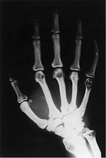
locations of tubular bones of ankles (tibia and fibula), hu-merus, ulna, radius, carpus, metacarpals and proximal pha-langes of hands (Figs. 1–4).
Laboratory tests showed only a type IV hyperlipopro-teinemia (with predominance of triglycerides and VLDL)
and hyperexcretion of urinary calcium (694 mg in the 24 h collection). Parathyroid and kidney functions were investi-gated extensively and found to be normal. We did not find any other significant laboratory finding.
Bone densitometry was done by dual X-ray absorptiom-etry and was found to be normal.
In order to rule out a neoplasia with bone extension an open bone biopsy was obtained from two locations: distal epiphyseal of right femur and distal epiphyseal of right ra-dius. The same findings were seen into the two locations: a gross appearance subcutaneous fat, a very thin and eburneal cortical bone tissue surrounding the osteolytic lesion that contained a yellow lipid-like substance. The histopathologi-cal studies showed a cortihistopathologi-cal bone and endosteal fat tissues of normal characteristics without any infiltrative lesion in both samples (Fig. 5). A normal synovial tissue was found from a sample obtained from the right knee.
Taking all of these radiological, analytical and histopatho-logical features ML was diagnosed. Underlying diseases (including neoplasic disorders, etc.) were ruled out. Abnor-malities in brain, kidney and liver have been described in this disorder. For this reason a computerized tomography (CT) was obtained from the cranium without any pathological
Fig. 1.Roentgenograms of the knees show symmetrical, well-defined, non-expansile cystic lesions in the distal ends of femur and tibia of both knees.
Fig. 2.Roentgenograms of the right hand show the same kind of lytic lesions in the distal ends of radius and ulna, carpal bones, metacarpals and proximal phalanges.
Fig. 3.Roentgenograms of the left hand show the same kind of lytic lesions in the distal ends of radius and ulna, carpal bones, metacarpals and proximal phalanges.
finding, and an abdominal ultrasonography was done that was normal. Neuropsychiatric and neurologic examination revealed no pathologic findings.
It has been suggested that this condition has no effective treatment. However, we started a treatment for the hyperlipo-proteinemia and the hypercalciuria with bezafibrato and hy-droclorotiazidic diuretic with improvement of triglycerides
levels from 551 to 239 mg/dl (normal range in our laboratory from 35 to 165) and the calciuria from 694 mg in 24 h to 274 mg (normal values: 4 mg/kg of body weight; body weight of the patient: 62.9 kg).
After 3 years of follow-up the patient has not developed any neuropsychiatric symptom, any fracture and any other symptom. When the treatment has been stopped the triglyc-erid level and the calciuria worsened with improvement im-mediately after restarting the treatment.
3. Discussion
ML is an uncommon disorder that usually appears in young adults of either sex. The initial symptoms arise in the skeleton between the second or third decade of life, with pain, tenderness and fracture after minor injury. Arthritis is not a predominant feature of the disease [3]. In the X-ray examination multiple, multilocular, symmetric and non-expansile cystic characteristic lesions without sclerotic mar-gins can be found in the distal ends of long bones, carpal and tarsal bones, and the short bones of the hands and feet.
Neuropsychiatric symptoms appear later, in the fourth or fifth decade of life, and vary from non-specific symptoms such as memory loss or personality changes to other more important conditions like seizures, decline in mental function and profound dementia, usually in more advanced state of the disease. Patients usually die young, and sometimes the dis-ease resembles Alzheimer’s disdis-ease [3,18–20]. There appear to be three clinical types of ML: a bone-dominant type, a brain-dominant type and an intermediate one [20]. Our pa-tient could be in the first type.
The neuro-radiologic findings are non-specific and CT shows mild to moderate cerebral cortical atrophy with en-larged lateral ventricles and cerebral sulci. Calcification in the basal ganglia can also be seen. Magnetic resonance (MR) findings in T1-weighted images reveal dilatation of ven-tricles and cerebral cortical sulci in addition to decreased volume of the cerebral white matter. T2-weighted MR im-ages show a signal intensity of the white matter that is equal to or greater than that of the cortex and a reduced signal density in the putamen and thalamus [21]. There are no prominent patchy or confluent hyperintense areas. It is useful to differentiate between this disease and multi-infarct de-mentia [20].
There are few comments in the literature of the disease about the analytical abnormalities that can be found in these patients. In the majority of papers, the study was found to be normal. However, a type IV hyperlipoproteinemia has been reported as a characteristic feature of the acquired and con-genital lipodystrophies [22].
The pathogenesis of ML is unclear. Some authors believe that the disease is a multisystem genetic disorder of lipid metabolism with autosomal recessive mode of inheritance, but no abnormality of lipid metabolic enzymes has been found [2,5]. Other authors propose that severe chronic va-sogenic edema is the main pathogenetic mechanism of severe
Fig. 4.Roentgenograms of the left ankle show multiloculated lytic lesions in the distal ends of tibia and fibula.
Fig. 5.The histopathological study showed a cortical bone and endosteal fat tissues of normal characteristics without any infiltrative lesion.
leukoencephalopathy development, and that these vascular changes are also present in bone lesions [23].
On the other hand, there are authors who suggest that it represents a metaplastic disorder leading to the replacement of bone for abnormal lipid material [24] and another opinion is that the material is derived from degenerated cells of adjacent stroma [25].
Histopathological ML-like changes are non-specific. A yellow, lipid-like substance is found in the cystic lesions and microscopic examination reveals the tissue to be composed of a lipomatous organization of mature fat cells. The cystic spaces are covered with thick hyaline eosinophilic mem-branes that are positive for periodic acid-Schiff staining.
For the diagnosis of ML the histopathological study is useful, mainly to exclude neoplasm with bone extension. The ML-like changes found in the osteolytic lesions of these patients are non-specific and these changes of fat tissue have also been found in other diseases [14]. In fact, some authors have established ML diagnosis with unique MR findings, without cystic lesions in bones or histological features [21]. Differential diagnosis of ML is not easy, because these peculiar changes in fat tissue have been associated with other local and systemic diseases including lupus erythematosus, eythema nodosum, fat tissue granuloma, hemodialysis pa-tients with carpal tunnel syndrome, progressive systemic sclerosis, diabetes mellitus, cases of limb ischemic necrosis caused by arteriosclerotic obstruction, thromboamgiitis obliterans, statis dermatitis, etc. [26–31].
Features similar to those found in the osseous tissue have been found in the brain, regarding neuropsychiatric manifes-tations characteristic of this disease. These cardinal features are the destruction of white matter, loss of both myelin and nerve fibers with intensive reactive gliosis, cortical atrophy and presence of multiple calcifications of the basal ganglia. Some authors proposed that brain and bone lesions may be assumed to share the same pathogenesis [25].
We present in this paper the first case of ML reported in our country. In the few laboratory data described of these patients, we have not found hypercalciuria. This condition could represent the fat metaplastic disorder previously pro-posed in which normal bone is replaced by lipidic material. In addition, we have not found any specific treatment in the literature of the disease and it has been suggested that this is a fatal illness. However, we have treated and controlled the hyperlipemia and the hypercalciuria. The first could be im-portant in the development of the disease and for this reason its control could delay the development of the fatal neuro-logic affectation. Moreover, the treatment of the hypercalci-uria found in our case, could contribute to delay the develop-ment of new osteolytic lesions.
In conclusion, ML is a rare and progressive disease with a possible genetic etiology and with a poor prognosis when it affects the brain. For this, to reach an early diagnosis as well as giving genetic counseling is necessary. It is unknown if the treatment of the metabolic disturbs could delay the fatal
evolution to the neurologic disease or if it could prevent bone fractures.
References
[1] Järvi O, Hakola P, Sourander P, Kormano M, Nevalainen T, Kalimo H. Polycystic lipomembranous osteodysplasia with sclerosing leukoen-cephalopathy (PLO-SL). In: Eriksson AW, et al., editors. Population structure and genetic disorders, Seventh Sigrid Juselius Symposium. London: Academic Press; 1980. p. 656–64.
[2] Nasu T, Tsukahara Y, Teryama K. A lipid metabolic disease—membranous lipodystrophy: an autopsy case demonstrating numerous peculiar membrane structures composed of compound lipid in bone marrow and various adipose tissues. Acta Pathol Jpn 1973;23: 539–58.
[3] Adolfsson R, Forsell A, Johansson G. Hereditary polycystic osteod-ysplasia with progressive dementia in Sweden. Lancet 1978;1: 1.209–10.
[4] Jarvi OH, Hakola HPA, Lauttamus LL. Cystic capillary-necrotic osteodysplasia: a systemic bone disease probably caused by arteriolar and capillary necrosis. Relation to brain affections. State University of Milan; 1968 Seventh International Congress of International Acad-emy of Pathology, Abstracts.
[5] Akai M, Tateishi A, Cheng CH, Morii K, Abe M, Ohno T, et al. Membranous lipodystrophy. J Bone Jt Surg 1977;59A:802–9. [6] Matsuo T, Suetsugu M, Eguchi M, Sasaki M, Tsuneyoshi M.
Mem-branous lipodystrophy. A case report. Arch Psychiatr Nervenkr 1982; 231:123–30.
[7] Yagishita S, Ito Y, Ikezaki R. Lipomembranous polycystic osteodys-plasia. Virchows Arch A Pathol Anat Histol 1976;372:245–51. [8] Wood C. Membranous lipodystrophy of bone. Arch Pathol Lab Med
1978;102:22–7.
[9] Edvardsen P, Halvorsen TB, Nesse O. Lipomembranous osteodysplasia: a case report. Int Orthop 1983;7:99–103.
[10] Hakola HPA. Neuropsychiatric and genetic aspects of a new heredi-tary disease characterized by progressive dementia and lipomembra-nous polycystic osteodysplasia. Acta Psychiatr Scand 1972; 232(Suppl):1–173.
[11] Sourander P. A new entity of phacomatosis. B. Brain lesions (scleros-ing leukoencephalopathy). Acta Pathol Microbiol Immunol Scand 1970;215(Suppl):44.
[12] Iannaccone S, Ferini SL, Nemni R, Marchettini P, Corbo M, Pinto P, et al. Peripheral motor-sensory neuropathy in membranous lipodystrophy (Nasu’s disease): a case report. Clin Neuropathol 1992; 11:49–53.
[13] Preziusso L, Muncibi F, Aglietti FB. A case of membranous lipodys-trophy with skeletal involvement. Chir Organi Mov 1992;77:205–11. [14] Koçer N, Dervisoglu S, Ersavasti G, Altug A, Çokyüksel O. Case report 867. Membranous lipodystrophy. Skelet Radiol 1994;23:577–9.
[15] Forest M, Fontanet A, Swargot M, Tomeno B, Kahn MF. Lipodystro-phie membranaire, 1er cas français. Rev Rhum Mal Osteoart 1991;58: 644 (abstract).
[16] Steinfelds F, Cuvelier B, Stallenberg B, Appelboom P. Maladie de Nasu-Hakola. Rev Rhum Mal Osteoart 1997;64:825 (abstract). [17] Hakola HPA. Polycystic lipomembranous osteodysplasia with
scle-rosing leukoencephalopathy (membranous lipodystrophy). A neurop-sychiatric follow-up study. Monogr Pneurop-sychiatrica Fennica 1990;17. [18] Bird TD, Koerker RM, Leaird BJ, Vlcek BW, Thorning DR.
Lipomembranous polycystic osteodysplasia (brain, bone and fat disease): a genetic cause of presenile dementia. Neurology 1983;33: 81–6.
[19] Shibata K, Uchiyama S, Takeuchi M, Kobayashi I, Maruyama S. A case of membranous lipodystrophy (Nasu) with diffuse cerebral white matter involvement and cerebellar atrophy on brain CT and MRI. Rinsho Shinkeigaku 1990;30:1232–7.
[20] Araki T, Ohba H, Monzawa S, Sakuyama K, Hachiya J, Seki T, et al. Membranous lipodystrophy: MR imaging appearance of the brain. Radiology 1983;180:793–7.
[21] Motohashi N, Shinohara M, Shioe K, Fukuzawa H, Akiyama Y, Kariya T. A case of membranous lipodystrophy (Nasu-Hakola disease) with unique MRI findings. Neuroradiology 1995;37:549–50. [22] Foster DW. The lipodystrophies and other rare disorders of adipose tissue. Harrison’s Principles of Internal Medicine. 13th ed. New York: McGraw-Hill, Inc; 1994.
[23] Kalimo H, Sourander P, Järvi O, Hakola P. Vascular changes and blood-brain barrier damage in the pathogenesis of polycystic lipomembranous osteodysplasia with sclerosing leukoencephalopa-thy (membranous lipodystrophy). Acta Neurol Scand 1994;89: 353–61.
[24] Resnick D, Niwayama G. Diagnosis of bone and joint disorders. 2nd ed. Philadelphia: Saunders; 1988.
[25] Pazzaglia UE, Benazzo F, Byers P, Riboni L, Ceciliani L. Pathogen-esis of membranous lipodystrophy. Case report and review of the literature. Clin Orthop Rel Res 1987;225:279–87.
[26] Jorizzo JL, White WL, Zanolli MD, Greer KE, Solomon AR, Jet-ton RL. Sclerosing panniculitis. Arch Dermatol 1991;127:554–8. [27] Sueki H, Shinmura Y, Fujisawa R, Jitsukawa K, Sato S. Ultrastructural
study of the histogenesis of membranocystic lesions (Nasu) in diabet-ics. J Cutaneous Pathol 1986;13:390–401.
[28] Machinami R. Incidence of membranous lipodystrophy-like change among patients with limb necrosis caused by chronic arterial obstruc-tion. Arch Pathol Lab Med 1984;108:823–6.
[29] Arnold HL. Lupus erythematosus profundus. AMA Arch Dermatol 1956;73:15–32.
[30] Kuwabara H, Uda H, Saito K. A light and electron microscopical study of membranocystic lesions in a case of lupus erythematosus profundus. Acta Pathol Jpn 1991;41:286–90.
[31] Horita S, Nitta K, Yamaguchi Y, Ozasa H, Yumura W, Ota K, et al. Membranous-lipodystrophy-like changes among hemodialysis patients with carpal tunnel syndrome. Nephron 1995;70:116–7.