AperTO - Archivio Istituzionale Open Access dell'Università di Torino
Original Citation:
Experimental pain processing in individuals with cognitive impairment: current state of the science
Published version:
DOI:10.1097/j.pain.0000000000000195 Terms of use:
Open Access
(Article begins on next page)
Anyone can freely access the full text of works made available as "Open Access". Works made available under a Creative Commons license can be used according to the terms and conditions of said license. Use of all other works requires consent of the right holder (author or publisher) if not exempted from copyright protection by the applicable law. Availability:
This is the author's manuscript
PAIN Publish Ahead of Print
DOI: 10.1097/j.pain.0000000000000195
Experimental pain processing in individuals with cognitive impairment:
Current state of the science
Ruth Defrin1, Martina Amanzio2, Marina de Tommaso3, Violeta Dimova4, Sasa Filipovic5, David P. Finn6, Lydia Gimenez-Llort7, Sara Invitto3, Christina Jensen-Dahm8, Stefan Lautenbacher9, Joukje M. Oosterman10, Laura Petrini11, Chaim G. Pick12, Gisele Pickering13,
Lene Vase14, Miriam Kunz9 1
Department of Physical Therapy and Sagol School of Neuroscience, Tel Aviv University, Israel 2
Department of Psychology, University of Torino, Italy 3
Departmentof Basic Medical Science, Neuroscience and Sense Organs, Bari University, Italy 4
Institute of Clinical Pharmacology & Department of Anesthesiology, Intensive Care Medicine and Pain Therapy, University of Frankfurt, Germany
5
Institute for Medical Research, University of Belgrade, Serbia 6
Pharmacology and Therapeutics, School of Medicine, NCBES Centre for Pain Research and Galway Neuroscience Centre, National University of Ireland, Galway, Ireland
7
Institute of Neuroscience & Department of Psychiatry and Forensic Medicine, Universitat Autònoma de Barcelona, Spain
8
Danish Dementia Research Centre, Department of Neurology, Rigshospitalet, University of Copenhagen, Denmark
9
Department of Physiological Psychology, University of Bamberg, Germany 10
Radboud University Nijmegen, Donders Institute for Brain, Cognition and Behaviour, Nijmegen, The Netherlands
11
Center for Sensory-Motor Interaction (SMI), Aalborg University, Denmark 12
Departement of Anatomy and Sagol School of Neuroscience, Tel-Aviv University, Israel 13
Departmentof Pharmacology, Faculty of Medicine, Clermont University, France 14
Department of Psychology and behavioral Sciences, Aarhus University, Aarhus, Denmark
Number of text pages (excluding references): 29 Number of figures: 0
Number of tables: 3 Corresponding author:
Ruth Defrin, PhD
Department of Physical Therapy Sackler Faculty of Medicine Tel-Aviv University Tel-Aviv, 69978 Israel E-mail: [email protected] Tel: +972-3-6405431
ACCEPTED
Abstract
Cognitive impairment (CI) can develop during the course of ageing and is a feature of many neurological and neurodegenerative diseases. Many individuals with CI have
substantial, sustained and complex healthcare needs which frequently include pain. However, individuals with CI can have difficulty communicating the features of their pain to others, which in turn presents a significant challenge for effective diagnosis and treatment of their pain. Herein, we review the literature on responsivity of individuals with CI to experimental pain stimuli. We discuss pain responding across a large number of neurological and
neurodegenerative disorders in which CI is typically present.
Overall, the existing data suggest that pain processing is altered in most individuals with CI compared to cognitively intact matched controls. The precise nature of these alterations varies with the type of CI (or associated clinical condition) and may also depend on the type of pain stimulation used and the type of pain responses assessed. Nevertheless, it is clear that regardless of the etiology of CI, patients do feel noxious stimuli; with more evidence for hypersensitivity than hyposensitivity to these stimuli compared to cognitively unimpaired individuals. Our current understanding of the neurobiological mechanisms underpinning these alterations is limited, but may be enhanced through the use of animal models of CI which also exhibit alterations in nociceptive responding. Further research employing additional
behavioural indices of pain is warranted. Increased understanding of altered experimental pain processing in CI will facilitate the development of improved diagnostic and therapeutic
approaches for pain in individuals with CI.
Key words: cognitive impairment, experimental pain, dementia, neurodegenerative
disorders; developmental disorders, pain perception
1. Introduction
Cognitive impairment (CI) refers to deficits or impairment in cognitive function. The term may describe deficits in global intellectual performance, but may also refer to deficits in specific cognitive domains including memory, language, attention, perception, reasoning and executive function. CI can be found in various patient groups, including but not restricted to, patients with dementia, individuals with autism, Down syndrome and traumatic brain injury. Many individuals with CI have substantial and complex healthcare needs which frequently also involve pain. Sources of pain are abundant among these individuals due to neurological impairments such as motor disabilities and coordination disorders leading to pathological gait and posture, altered muscle tone and general difficulties in activity of daily living. Dislocated joints, pressure sores and over-use injuries due to the aforementioned pathologies and to the use of assistive devices are additional sources of pain [77,105]. Despite the aforementioned, precise estimates on the prevalence of pain among individuals with CI are not abundant. Most existing data relate to patients with dementia. Depending on the setting (e.g. nursing homes, acute hospitals) the prevalence of pain among patients with dementia may vary from 4 to more than 80% [115]. Additional published prevalence rates of pain are 13-75% among individuals with developmental disability [121], 40-60% among patients with Parkinson’s disease and 12-80% among individuals with traumatic brain injury [17]. Given these relatively high prevalence rates, it becomes apparent that pain should be carefully assessed and monitored in individuals with CI, in order to provide adequate care.
Pain is regarded as a multidimensional, complex experience comprised of sensory, affective and cognitive aspects that can lead to physiological, emotional and behavioural responses. Due to its subjective nature, pain assessment relies mostly on pain self-reporting as the “gold standard” and neglects other forms of pain responses. This is despite the fact that self-report is only a proxy for subjective experience and reflects more than an exclusive report
of pain. Individuals with CI often have difficulties in verbally expressing their pain due to poor intellectual and communication capabilities and even if verbal skills are present, they do not guarantee valid pain reports. Thus, pain assessment is challenging in individuals with CI due to problems in: 1) identifying the presence of pain, 2) diagnosing the source of pain, 3) evaluating the magnitude of pain and suffering, 4) deciding on the appropriate treatment and following up its effectiveness. It is likely that the difficulties that caregivers as well as of healthcare professionals experience in trying to identify painful conditions in individuals with CI, have led to the premise that these individuals may be less sensitive to pain in comparison with their cognitively intact peers [1,14,54]. Accordingly, individuals with CI receive
significantly fewer analgesic medications and healthcare visits compared with cognitively intact individuals [2,38,81]. More recent findings indicate that this trend might be slowly changing with CI patients even being over-treated with pain medication [56], which might lead to different problems (e.g. sedation, heightened fall risk). Healthcare professionals as well as caregivers face the difficulty of knowing whether or not an individual with CI might be suffering from pain. Consequently, individuals with CI, especially those with severe CI, are still at a high risk of late diagnosis or misdiagnosis of their pain, and thus, might be suffering in silence which in some cases may even lead to increased, unnecessary death rates [85,104].
Given the challenges highlighted above, exploring the manner by which individuals with CI process, experience and respond to pain is an imperative ethical goal. Moreover, such research is essential for the improved assessment and treatment of pain and to reduce
unnecessary suffering. In recent years, several clinical studies have been conducted to assess and analyse pain responses in individuals with CI (for review see [1,29,50,75,84]). However, clinical studies are limited due to the fact that the stimuli that elicit the pain cannot be
controlled nor measured. Experimental pain studies provide several advantages over clinical
studies. They allow for control of experimental stimulation, experimental interventions and measurement techniques by using standardized stimuli of different modalities often with widely available devices. Thus, experimental studies enable one to disentangle the stimuli from the nociceptive response, to carefully analyze stimulus-response relationships of painful events, to probe various sensory pathways and to study associations between pathological alterations and their functional consequences.
Experimental studies use various stimulation modalities to induce pain, including thermal, mechanical, electrical and chemical stimuli of various frequencies, durations and intensities that can be applied to various body parts [5]. The methods applied to assess pain sensation range from measuring pain threshold (i.e. the minimal stimulation energy needed to induce pain) and pain tolerance, to measuring pain intensity and unpleasantness (i.e. the sensory vs. affective aspect) using rating-scales and behavioural scales. Various stimuli also are often applied in order to measure the evoked motor, autonomic, endocrine and brain responses. Depending on the methods used, different aspects of afferent and efferent nociceptive processes as well as different aspects of the multidimensional pain experience are
investigated. Each measurement method can investigate only a limited fraction of the entire pain experience and thus, it is advisable to examine the impact of CI on pain processing using different experimental protocols.
The aim of the present review is to summarize, for the first time, the current state-of-the- art knowledge on experimental pain responses among individuals with CI. Additional novel features of this review are the inclusion of studies employing animal models of CI as well as the inclusion of different types of CI having diverse underlying pathologies and diverse brain dysfunctions. Given the broad network of brain areas involved in pain processing (e.g. thalamus, somatosensory cortices (S1 and S2), caudate/putamen, cerebellum, and insular
cingulate, prefrontal and supplementary motor cortices) [4], and the variability between CI types in brain pathologies, the effects of CI on pain processing can vary greatly.
This review is intended to make a critical contribution: 1) by increasing the awareness of the possible alterations in pain perception and processing among individuals with CI, 2) by exploring how CI effects pain pathways in their ascending and descending parts, 3) by identifying common features of pain processing in CI regardless of etiology that can provide an empirical foundation for developing new tools for the identification and diagnosis of pain in this population and 4) by promoting better pain management in different types of CI subjects. For the purpose of this review we searched computerized databases (Pubmed, Medline, Scopus and Web of Science), published bibliographies of related topics, and references provided by colleagues. We limited our review to publications in the years 1960-2014. We reviewed in-depth studies that collected sufficient data for meaningful statistical analysis and had a meticulous study design. The review is based on the best evidence available and on the opinion of a panel of experts (the authors) in cases of limited published data on certain topics.
2. Aging and mild cognitive decline
2.1. Age-related changes in pain processing
Introduction: Even though healthy ageing is, by definition, characterized by a lack of CI,
we decided to include ageing in this review for two reasons. First, mild cognitive decline has been repeatedly shown even in healthy ageing. Secondly, most of the pathological conditions in which CI is a major feature, are age-related (e.g. dementia). Therefore, there is a need to disentangle age-related changes from specific pathological changes that involve CI and have an effect on pain processing. So far, numerous studies have focused separately on age-related changes in pain or on age-related changes in cognition, however, rather little is known about the linkage between these two factors. Aging has consistently been associated with mild
overall cerebral atrophy as well as with a mild decline in various cognitive domains. Albeit within normal limits, the most pronounced affected functions include executive functions, episodic memory and psychomotor speed [28]. Several of these functions may already decline as early as the age of 25, with a steeper decline starting from the sixth decade of life [28].
Pain measurements: A substantial number of studies have used experimental designs to
study age-related changes in pain processing. Most often, the effect of ageing on pain has been investigated by assessing pain thresholds in elderly individuals. It has been found that age often leads to elevated pain thresholds when pain is applied using electrical current and heat, whereas pain thresholds for pressure can be decreased in elderly individuals [73]. There is also evidence for a decrease in pain tolerance with age. In line with the reduced pain tolerance are findings of reduced efficacy of conditioned pain modulation (CPM) along with increased temporal summation [73]. It is noteworthy that motivational and other perceptual aspects may confound measurements, especially that of tolerance as well as verbal reports of pain. Evidence for reduced functioning of the endogenous pain modulatory mechanism with aging can also be found in brain imaging studies, with older individuals showing lower striatal activity in response to pain compared to younger individuals, which might indicate
dysfunctions in pain inhibitory circuits [25]. Based on these findings, age seems to impair ascending as well as descending pain pathways, however, with the latter being more severely affected by ageing [73]. And given this decline in descending pain inhibitory pathways, ageing seems to make the individual more vulnerable to suffering from pain, which is in line with clinical findings of increased pain prevalence in the elderly.
As stated above, although numerous studies have focused on age-related changes in pain or in cognition, little is known about the linkage between the two areas. A question therefore remains whether age-related changes in cognition have an effect on pain processing. It has been shown that pain does have an impact on cognitive functioning, with severe pain
substantially impairing cognitive functioning [88]. Thus, it is possible that chronic pain in elderly patients contributes to age-related decline in cognition. With regard to the opposite direction, namely on how age-related changes in cognition affect the processing and perception of pain, very little is known. One recently conducted study [6] showed that reduced cognitive flexibility and memory capacities significantly predicted the development of chronic pain. Thus, there seems to be a vicious circle for elderly individuals, with pain leading to a decline in cognitive functioning and the decline in cognitive functioning leading to greater pain vulnerability.
Conclusions and Recommendations for future studies: Ageing is accompanied by
changes in pain processing that seem to render the elderly more vulnerable to suffering from pain. Moreover, ageing is accompanied by slight decreases in cognitive performances. There is evidence that the age-related increase in pain vulnerability and the age-related decline in cognition might not be independent symptoms but serve to amplify each other in the elderly. However, this evidence is very sparse and more studies are needed that aim to disentangle age-related changes from cognitive-related changes in pain processing in the elderly.
2.2. Mild cognitive impairment
Introduction: The concept of Mild Cognitive Impairment (MCI) refers to a group of
changes interposed between the cognitive changes of normal ageing and what might
constitute the changes seen in a very early dementia state [97,98] with a preserved autonomy on activities of daily living (ADL) and instrumental activities of daily living (IADL). MCI
may induce not only memory and executive dysfunctionbut also visuospatial disturbances and emotional alterations including apathy and depression [97]. As research on MCI has advanced, it has become apparent that several clinical subtypes of MCI exist (e.g. amnestic
MCI and non-amnestic MCI [97,98]), with multiple etiologies or causes for each subtype. For
example, as far as presumed degenerative etiology is concerned, the amnestic MCI subtypes
would likely represent a prodromal form of Alzheimer's disease. Thus, brain pathologies and affected brain areas might differ between each MCI subtype and thus, the effect of MCI on pain processing might also differ depending on the specific subtype.
Pain measurements: Only one study has investigated the impact of age and MCI on the
pain response system [67]. The subjects (MCI subtypes were not assessed) were tested for their self-report, motor (RIII reflex), facial (Facial Action Coding System) and autonomic responses (galvanic skin response and heart rate) to noxious electrical stimulation of the sural nerve. Regression analyses revealed that cognitive status (within the group of MCI) was a significant predictor of the decrease in autonomic responsiveness to noxious stimulation. However, the self-report of pain, the RIII reflex threshold, heart rate responses, as well as facial responses to electrical pain stimuli were not altered in MCI patients compared to healthy controls of the same advanced age.
Conclusions and Recommendations for future studies: Based on the small amount of
empirical evidence available, pain processing seems to be largely unaffected in MCI.
However, in order to arrive at a valid conclusion, larger scale longitudinal studies are needed that also differentiate between the varying pathologies underlying MCI.
3. Neurodegenerative disorders 3.1. Alzheimer’s disease
Introduction: Alzheimer’s disease (AD) is the most frequent cause of dementia and is
responsible for approximately 60% of dementia cases in the elderly [80]. AD leads to memory impairment (especially short-term), aphasia, reduced insight and other cognitive symptoms. The pathological changes in AD patients develop over decades and affect first the
transenthorinal cortex and the hippocampus region. The changes then spread throughout the limbic system and, coinciding with diagnosis, there are widespread changes with interruption of connections between components of the limbic systems (equivalent to Braak stage IV to V)
[18,91] leading to emotional disorders such as anxiety and depression and in some cases even psychotic symptoms [108]. However, it appears that the sensory cortex is largely unaffected until the disease becomes very severe. The brain regions that are affected by AD overlap to a certain degree with those involved in pain processing [107], which has led to the hypothesis that pain processing and thus, the pain experience, is altered in AD. Besides alterations in the experience of pain, the cognitive decline might additionally affect the communication of pain. Interestingly, studies investigating the capacity of patients with dementia to comprehend or complete standard pain assessment scales (like the visual analogue scale (VAS) have
repeatedly shown that this ability declines dramatically across the course of dementia [59,95]. In line with this, epidemiological studies have described a reduced report of pain [1,57]. Therefore, it is challenging to disentangle the effect that AD has on the ability to provide self-report of pain from the effect it has on the processing of pain itself.
Pain measurements: The hypothesis that AD leads to a change in pain processing has
been studied in a number of experimental studies using a variety of pain induction methods (electrical, mechanical, heat stimuli) and using methods that are dependent on reaction time (method of limits) as well as independent of reaction time (staircase method). The findings are summarized in table 1. Seven psychophysical studies investigated pain thresholds (based on self-report) in AD patients. Five studies found no differences in threshold levels
[11,13,44,58,78], whereas two studies found increased thresholds [24,26] compared with age matched controls. So far, four studies have investigated pain ratings of supra-threshold
stimuli, of which three found no difference in pain ratings [24,58,66,] and one [101] found
lower pain ratings to suprathreshold stimuli compared with controls. Pain tolerance (based on self-report) has only been investigated in two studies. One study showed increased tolerance in patients with AD compared with controls, using both ischemic and electrical stimuli [13]. The other study however, reported decreased pain tolerance using pressure stimuli in patients
with AD but no difference from controls in pain tolerance using the cold pressor test [58]. These contradictory findings cannot be explained simply by the different pain induction methods used because different outcomes were observed even when the same method was used (e.g. phasic pressure pain) [24,78]. It is possible that other methodological differences between studies as well as differences across studies in the severity of patients' dementia underlie the discrepant findings. It is also possible that the experimental pain assessment methods utilized are not appropriate tools in patients with AD. Patients with AD have impairment of short term memory and may have difficulties understanding instructions. Consequently, some of the differences may be due to the unintentional use of methods that may not be appropriate for patients with AD.
Given the decline in cognitive capacities, it might be more appropriate, when assessing the impact of AD on pain processing, to assess pain responses which do not rely on the patients’ ability to give a self-report of pain. Along these lines, several studies of AD patients have assessed motor, autonomic, facial and brain responses to experimentally induced pain. These studies seem to present more consistent outcomes in that the majority of findings point to an augmented responsiveness to noxious stimuli in patients with AD and in patients with other forms of dementia. More precisely, most studies found that patients with dementia (mostly AD patients) show increased facial responses to pain [49,66,68] compared to healthy individuals; or at least a tendency for increased facial responses [78]. Importantly, this increase was not accompanied by an overall increase in facial responsiveness (e.g. unspecific grimacing) but was solely due to an augmentation of pain-specific facial expressions.
Moreover, the threshold for the nociceptive flexion reflex (RIII) was significantly decreased in AD, thus, also pointing to an increase in pain processing which might manifest already at the spinal level [66]. Other studies have focused on brain responses. Despite the hypothesis of impaired pain pathway in AD, functional magnetic resonance imaging (fMRI) studies showed
that brain activity in response to noxious stimulation is preserved and even elevated in both the medial and lateral pathways [24,26]. Interestingly, these studies also observed prolonged activation in the pain pathways and increased activity in cognitive regions, such as the
dorsolateral prefrontal cortex (DLPFC). This suggests that, in AD, the cognitive integration of pain may be altered. A study from Gibson et al. [44] that used evoked related potentials (ERPs) as an index of brain activation, found no difference in peak amplitude in AD patients. However, in an earlier study from Yamamoto et al. [123], that included a mixed population of dementia cases, the authors failed to induce pain-evoked potentials in the subgroup of patients with severe dementia. It is therefore possible, that brain responses to noxious stimulation are increased in early and moderate stages of dementia and might decline in later stages. Due to ethical considerations, however, it is difficult to apply experimental pain induction procedures in patients suffering from more severe stages of dementia. The investigation of autonomic
responses to pain has shown a decline in responsiveness in patients with dementia [66,101].
The results of studies examining autonomic functions in AD have been conflicting, but several studies have found a dysfunction of autonomic responses that might be caused by AD-related deficits in central cholinergic function [39]. Given that AD may be associated with autonomic dysfunction, the autonomic response might not be a valid pain indicator in this patient group.
One study investigated placebo effects on venipuncture-induced pain in patients with AD and found that newly diagnosed patients (Mini Mental State Examination (MMSE) mean 24 ± 1.22) were able to obtain a placebo effect of a similar magnitude as healthy age-matched controls. One year later, however, when the connectivity between the prefrontal lobes and the rest of the brain was reduced, the patients were no longer able to obtain a placebo effect [10]. Subsequent studies have supported the notion that a loss of prefrontal control is related to a loss of the placebo effect [12]. Losing the ability to obtain placebo effects could mean that
AD patients might need a higher dose of analgesic medications to achieve pain relief [10]. However, since AD patients are more frail and therefore more susceptible to adverse events [3] analgesic treatment should be carefully monitored.
Conclusions and Recommendations for future studies: Although the empirical findings
on pain processing in patients with AD are partially contradictory, the majority of findings seem to suggest that the processing/experience of pain is not diminished in patients with mild to moderate forms of dementia but - in contrary - the pain experience might even be elevated when considering certain types of pain responses (e.g. brain activity, facial responses). Whether this is also the case in late stages of AD should be investigated. Given the
progressive cognitive decline in patients with AD, research on pain processing in this patient group should not rely solely on self-report ratings (since these become less valid) but instead use a multi-method approach (assessing verbal-reports, facial, neural, autonomic and motor responses). This is especially important when investigating patients who are in the later stages of the disease. Moreover, the few studies on placebo effects and adverse events suggest that careful consideration of the pharmacological treatment of AD patients is warranted.
3.2. Other types of neurodegenerative disorders
Introduction: “Neurodegenerative disorders” is an umbrella term for disorders that are
characterized by a progressive loss of neurons or of neural functioning. Besides Alzheimer´s disease (AD), neurodegenerative disorders include – amongst others - Amyotrophic Lateral Sclerosis (ALS), Multiple sclerosis (MS), Parkinson’s disease (PD), Frontotemporal dementia (FTD), Huntington’s disease (HD) and Lewy body dementia. The neurodegenerative
processes that underlie this group of disorders also affect the cognitive functioning of the patients, although depending on the type of pathology, the CI ranges from only mild (e.g. ALS) to more severe impairments (FTD). In addition, patients may also present with motor disability, emotional disturbances such as depression and psychosis, as well as sleep
difficulties, autonomic failures and various pain syndromes [23, 48]. So far, only a few studies have investigated pain processing among patients with neurodegenerative disorders other than AD. Below, we will review the findings on FTD, PD, HD and MS.
Pain measurements:
Frontotemporal dementia (FTD): The experimental evidence on pain processing in
patients with FTD is sparse. The one study that has been conducted in this patient group found that pain threshold and pain tolerance levels were increased in FTD patients compared to age-matched healthy controls [20]. In line with this experimental finding of decreased pain sensitivity, caregivers reported a loss of pain awareness in FTD patients compared to patients with other dementia types [8]. It is possible that due to the fronto-temporal degeneration, cognitive–evaluative and motivational-affective aspects as well as memory for pain, and autonomic–neuroendocrine responses to pain, are prone to deterioration in FTD and thus, patients might indeed be suffering less from pain [107]. However, the degree to which pain sensitivity decreases in FTD might depend on the type of FTD; given that for the subtype “semantic dementia” care-givers have reported heightened pain sensation [111]. Experimental studies are needed to confirm these possible differences in pain sensitivity between different types of FTD.
Parkinson’s disease (PD): Cognitive decline has often been reported in PD with approximately 30% of the patients having mild cognitive impairments and 30% having cognitive impairments already in the dementia range [79]. PD seems to render patients more sensitive to pain, with decreased pain and reflex thresholds [42,89,100]; however, studies investigating nociceptive evoked brain potentials have produced inconsistent findings [117]. Interestingly, dopaminergic therapy seems effective in reducing this increased pain
sensitivity, with pain thresholds and various responses to experimental pain stimuli being more comparable to healthy individuals during the "on" phase (periods when the
dopaminergic medication is working and motor symptoms are controlled) [117]. Furthermore, a CPM-study suggested that endogenous pain inhibition remains intact in PD patients [89]. It is important, however, to mention that the aforementioned studies did not report whether the patients with PD showed any signs of CI. Therefore, the interaction between PD, CI and pain is yet to be unravelled.
Huntington’s disease (HD): Besides unwanted choreatic movements, behavioural and psychiatric disturbances, CI is one of the core symptoms in patients with HD. Few data are available on pain processing in HD. In patients with HD, increased pain- and RIII reflex thresholds were found (which suggest a decreased sensitivity to pain) [106] and are in line with the clinical observation that these patients do not complain about pain [96]. Moreover, laser evoked potentials (laser stimuli of noxious intensities) were also changed in HD, with increased latencies and decreased amplitudes [31]. Thus, self-report ratings, motor reflexes, and cortical responses all suggest a decrease in pain sensation in patients with HD. However, none of the studies have tried to relate these changes in pain sensation to the degree of CI.
Multiple sclerosis (MS): Prevalence estimates of CI in MS range from 40% to 65% and cognitive deterioration tends to progress over time. In patients with MS the widespread lesions in the brain and spinal cord and the accompanying motor dysfunction result in various chronic pain syndromes including peripheral and central neuropathic pain, musculoskeletal
pain and functional pain syndromes [118].Only a few studies, however, assessed pain processing among patients with MS. These studies have found increased sensitivity to pressure-pain as well as inconsistent (either hyper- or hyposensitivity) but common sensory alterations in thermal and tactile sensitivity (e.g. [40,113]. Here again, these studies did not report whether the patients with MS showed any signs of CI and therefore, the interaction between MS, CI and pain is yet to be unravelled.
Conclusions and recommendations for future studies: Different types of
neurodegenerative disorders (which affect cognitive performance) seem to have very different effects on the pain response system. Whereas pain responses are decreased in FTD and HD, they are amplified in PD. Clearly, more information on the perception of pain among
individuals with neurodegenerative disorders is needed. Since such disorders vary with regard to the brain structures involved, it is necessary to understand the differential effects these pathologies have on the pain system. Furthermore, some pathologies may affect similar brain structures but might have differential effects (possibly due to different mechanisms at the molecular level). For example, although both PD and HD affect the functioning of the basal ganglia, the impact that these two pathologies have on pain processing seem to be opposing (as is their effect on motor responses), with PD being accompanied by increased pain sensitivity whereas HD seems to decrease pain sensitivity.
4. Developmental disability
Introduction: Developmental disability is a broad term used for “a diverse group of severe
chronic conditions that are due to mental and/or physical impairments, which include autism,
cerebral palsy and intellectual disability”. Autism Spectrum Disorders (ASDs) are
characterized by impairment in the social interaction and communication domain, restricted repetitive and stereotyped patterns of behaviour, interests, and activities, and/or abnormal functioning. Autism has a strong genetic basis although environmental factors have also been suggested to interact with its underlying mechanism. Cerebral Palsy is a group of movement, muscle tone and/or posture disorders that are often accompanied by disturbances of sensation, perception, cognition, communication and behaviour. Cerebral Palsy is caused by abnormal development of, or damage to motor control centres of the brain. Intellectual disability (ID) (previously termed mental retardation) is characterized by impairments of general mental abilities that affect adaptive functioning in the conceptual, social and practical domains.
Individuals with ID have a limited ability to learn, reason, make decisions and solve problems. ID, which is the most common developmental disability [83] may be related to infections, chromosomal abnormalities, environmental, metabolic or nutritional causes, toxic insults and trauma before and after birth [109].
There is great diversity among individuals with developmental disability with respect to many factors and specifically, in the presence and/or the degree of CI. For example, Asperger syndrome, one of the ASDs, lacks delays in cognitive development. The percentage of
individuals with autism who also meet criteria for CI varies greatly, from 25% to 70%, pointing to the difficulty in assessing CI in this population. Nevertheless, most individuals with autism have mild CI [124]. The prevalence of CI among individuals with cerebral palsy ranges between 23 and 44% [93] and can vary from mild to profound. With regard to ID, about 85% of individuals with ID have mild CI and the proportion of individuals with moderate, severe and profound ID is 10, 4 and 1%, respectively.
Recent structural imaging studies revealed alterations in brain anatomy among individuals with developmental disability. For example, in individuals with autism, neuronal loss in the cerebellum, brainstem, parietal and frontal cortex as well as in the limbic system such as reduced amygdala and dentate gyrus volume have been reported [74]. In children with cerebral palsy and CI, white matter lesions throughout the brain, including but not limited to regions associated with the sensorimotor system, ventricular dilatation (affecting circuits in the periventricular regions) and thinning of the posterior corpus callosum are common [36]. Similarly, in individuals with ID, abnormalities within the periventricular white matter, lateral ventricular dilatation, corpus callosum thinning, and decrease in white matter volume in the insula, cingulate, amygdala, frontal lobes, thalamus, brain stem (extending to pons), parietal sensory-motor tracts, and fronto-cortical circuits [53,122,126] have been reported. Such pathologies may underlie not only the aforementioned cognitive dysfunctions but also the
psychiatric and psychological problems (e.g. obsessive compulsive disorders and emotion dysregulation) that are present in some cases [62].
Most of the aforementioned structures are involved in the processing of the sensory as well as the affective-motivational aspects of pain. Therefore, alterations in these structures may be associated with alterations in the processing of painful events among individuals with developmental disability.
Pain measurements: Up until recently, the commonly held view was that individuals with
developmental disability have decreased sensitivity to pain [e.g. 14,38]. This view was based on the tendency of these individuals not to report pain in potentially harmful situations, and from the difficulty of assessing pain in these individuals due to their poor communication capabilities. Observations of self-injurious behaviour amongst some individuals with developmental disability have also contributed to this view.
Only a few studies of persons with ID have actually measured sensitivity to pain, i.e. pain
threshold. These studies are summarized in Table 2. In the first study of its kind, pain
threshold were assessed in individuals with Down’s syndrome (DS) by measuring the time elapsed from the application of an ice cube to the first verbal expression of pain [54]. The onset of pain response was longer in individuals with DS compared with controls suggesting a higher pain threshold in the former. However, as this method includes a reaction time artefact (i.e. the time it takes the individual to respond verbally to the sensation evoked by the ice cube), the threshold was confounded by this artefact. Defrin and co-workers [32] measured pain threshold among individuals with ID with the reaction-time inclusive method of limits (subjects are required to press a switch upon pain detection) and the reaction-time free method
of levels (subjects report post factum whether a pre-determined stimulus was painful or not).
Individuals with ID had a similar pain threshold to that of controls when measured with the method of limits but a significantly lower pain threshold compared to controls when measured
with the method of levels. It was concluded that slower reaction time renders the pain
threshold higher when measured with the method of limits but not the method of levels, and in fact, individuals with ID have increased pain sensitivity compared to normal contrary to the previously held view [32].
More recent studies have measured pain threshold with the method of limits among various populations of developmental disability. Adults with high functioning autism had lower pain thresholds compared to controls [21]. Likewise, children (but not adults) with cerebral palsy [103] were found to be more sensitive to pain compared to controls. In contrast, adolescents with Prader-Willi syndrome (a neuro-genetic developmental disorder with a tendency to self-injury) had increased pain thresholds compared to controls [99].
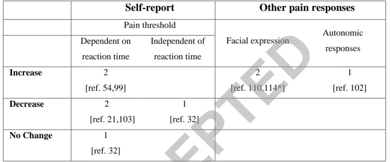
With regard to facial expressions of pain, we found only two studies in which facial expressions were analysed following four innocuous stimuli (heat, cold, pressure and touch) and one presumably noxious stimulus (pin prick) among adults with severe and profound ID [110,114]. Simons and co-workers found similar increases in facial activity following all of these stimuli compared to baseline (Table 2) with no differences between responses to innocuous and noxious stimuli. However, given that pinprick was applied using a Neuropen, and that the stimuli were not tested among controls, there is no assurance that pinprick was perceived by the subjects as painful. To the best of our knowledge, autonomic responses to experimental pain stimuli were not measured among individuals with developmental disability. There is evidence, however, that individuals with ID present elevated heart rate during venepuncture compared to unimpaired individuals [102] suggesting that evaluation of objective responses to experimental pain among these individuals is called for.
Conclusions and recommendations for future studies: On the basis of the majority of
existing findings we may conclude that individuals with developmental disability are more sensitive to pain than control subjects. This finding corresponds with imaging studies showing
damage to structures involved in pain modulation, i.e. the brain stem and frontal cortex as well as structures involved in pain processing (e.g. cingulate, insula and sensory cortex). Still, some inconsistency exists in pain threshold among the different impairment types. This inconsistency could reflect differences in pain processing between developmental disability syndromes despite seemingly similar structural alterations in the brain. Alterations in
peripheral and/or central conduction of sensory signals may also be responsible for variations in the sensitivity to pain, especially if pain threshold is measured with methods that include reaction time. While such alterations have been reported for innocuous stimuli [19,99] studies are needed to test whether such alterations occur in nociceptive pathways. Due to the
possibility of delayed reaction time, measuring pain threshold with methods that bypass this limitation, i.e. reaction-time free methods, is preferable. However, pain threshold
measurement is suitable primarily for individuals with mild and perhaps for some individuals with moderate cognitive impairment. Thus, the use of indirect indices of pain is necessary. Although behavioural indices of pain such as facial expressions were mostly analysed in the clinical setting, additional studies are needed in order to explore which indices best reflect pain in the various types of developmental disability.
5. Cognitive impairment secondary to vascular and traumatic insults
Introduction: CI is a frequent outcome after vascular or traumatic insults to the brain. The
degree of CI, however, can vary largely between only slight impairments (e.g. after mild traumatic brain injuries) to extreme impairments (e.g. after severe brain damage as in patients in vegetative state). Vascular and traumatic insults to the brain can alter pain processing directly by affecting ascending and descending nociceptive pathways, and indirectly, by affecting cognitive and emotional pathways. The two possibilities may also exist
simultaneously.
5.1. Stroke
Stroke and cerebral small vessel disease is a common cause of CI and the most common pathology underlying vascular dementia. Connections between areas of the cortex associated with complex information processing are disrupted thus, leading to impaired cognition and function, motor disability, psychological or emotional impairments and in some cases communication disorders such as aphasia. Fatigue and chronic pain are also common after stroke [86]. The most frequent types of pain after stroke are hemiplegic shoulder pain, central post-stroke pain and post-stroke headache [9,65]. Thus, stroke seems to render the patient more vulnerable to pain. Several studies have investigated the mechanism underlying the development of pain after stroke and found alterations in pain processing, surprisingly
manifested in increased pain thresholds [e.g. 63,125]. However, to the best of our knowledge, in none of these studies were patients with CI included and therefore the specific effects of CI on pain processing among stroke patients are unknown.
5.2. Traumatic brain injury
Traumatic brain injury (TBI) can cause long term CI (attention, memory, and executive functioning) in addition to changes in personality, disturbances of mood and emotion
regulation, the degree of which depends on the extent, location and severity of TBI. Chronic pain is frequent after TBI. The most frequent type of pain after TBI is chronic post traumatic headache, followed by central pain, musculoskeletal pain and peripheral neuropathic pain [90,116]. Thus, like stroke, TBI seems to render patients more vulnerable to pain. Indeed, when using experimental pain, it was found that TBI patients showed heightened pain
sensitivity as indicated by decreased pressure pain threshold, hyper excitability in the painful regions and reduced pain habituation and modulation compared to controls [33,34,94]. However, these studies did not include patients with known CI and therefore the specific effects of CI on pain processing among TBI patients are yet to be discovered.
5.3. Vegetative state and minimal conscious state
Many individuals who acquire severe brain injury experience prolonged disorders of consciousness. Persons in a vegetative state (VS) differ from persons in a minimal conscious state (MCS) for the absence of discernible, even if inconsistent, awareness of self or
environment [43]. The diagnosis of VS and MCS is usually based on clinical judgment. In recent years however, the results of neuroimaging [16] and neurophysiological studies [22] and studies of technology-based learning set-ups have been a supplement to the diagnosis [69]. By showing that arousal reaction toward multimodal and especially painful stimuli may be present even in vegetative state, these studies have fundamentally changed the way one thinks about these conditions. A controversial issue in the management of patients in a vegetative or a minimally conscious state concerns their hypothetical capacity to continue to experience pain despite an apparent absence of self- and environmental awareness. Thus, recently clinical, functional and neuroimaging studies have been conducted in order to address this important concern.
Pain measurements: Recent functional neuroimaging studies have shown a greater
responsiveness to pain in patients in MCS compared with patients in vegetative state. Using PET imaging, electrical noxious stimulation activated similar regions of the thalamus, primary and secondary somatosensory cortices and the frontoparietal and anterior cingulate cortex in patients in MCS and in healthy controls [16]. Activity in these areas - known as the ‘pain matrix’- was markedly greater in patients in MCS than in patients in VS, who show no evidence of self or environmental awareness. Furthermore, VS patients only showed activity in the primary somatosensory but not the associative cortex following electrical stimulation [72]. However, the findings of these two studies are limited by the use of electrical stimuli which are not specific to nociceptive processing and thus may activate a cortical network devoted to salient stimuli rather than noxious stimuli. In another study where O15–H2O PET was employed to explore the responsiveness of VS patients to painful electrical stimuli,
Kassubeck et al [60] observed posterior insula/secondary somatosensory cortex (SII), post-central gyrus/primary somatosensory cortex (SI) and cingulate cortex activation during stimulation which was perceived as noxious in controls. In a recent study, laser evoked responses, which are a specific tool to explore pain pathways, were present among patients in MCS and VS, albeit with prolonged latencies [30]. Interestingly, the cortical responses to electrical stimuli, delivered at an intensity which was perceived as painful in controls, were absent in most of the VS and MCS patients. Importantly, electrical stimuli primarily activate A-beta fibres although activation of nociceptive fibers is possible depending on stimulation intensity. However, given the relatively high activation threshold of nociceptive pathways in VS and MCS patients, the employment of laser stimuli which specifically activate A-delta and C afferents, seems more appropriate to test pain related reactions in such patients.
Conclusions and recommendation for future studies: The use of cerebral functional
studies is changing our opinion about the possible absence of pain sensation and reaction in patients with severe brain damage (MCS and even VS). Several studies have found cortical responses to painful stimuli even in VS patients. This suggests that even after severe brain damage, cortical areas are activated for a potential response against dangerous environmental factors. Functional studies employing stimuli that specifically activate nociceptive pathways , such as laser stimuli, are needed to confirm the cortical arousal of severe brain damaged patients and to identify the response to noxious stimuli as a condition of minimal cortical preservation.
6. Translational studies: what can we learn from animal models of cognitive impairment?
Introduction: The homology of basic biological processes such as 'nociception' among
animals and humans has rendered animals - mostly rodents - a relevant tool in basic and preclinical pain research [52]. In addition, modelling of psychiatric and neurological diseases
in animals by means of classical neuropsychopharmacological approaches, spontaneous mutants or the most recent molecular biology engineered models has provided new opportunities to study pain comorbidity [17]. However, so far only a limited number of experimental studies have examined pain in animal models of CI or dementia [45].
In the absence of the equivalence to self-reports, nociception tests must rely on observable behaviours that range from a simple nocifensive withdrawal reflex to complex operant behaviours. Thus, some tests measure the latency of the avoidance behaviour in response to thermal (e.g. tail-flick test, hot-plate test), mechanical (e.g. von Frey test, paw pressure test), chemical (e.g. formalin test) or electrical stimuli, as an indicator of the
sensitivity of the animal to stimuli. Others record spontaneously emitted behaviours following noxious stimulation (e.g. abnormal gait or hunched posture) or operant behaviours (e.g. learned escape or place aversion). Changes induced by pain in cognitive (e.g. attention), emotional (e.g. freezing), physical (e.g. changes in body weight) and social behaviours (e.g. aggression) are also considered important as pain-related correlates [87]. Interestingly, behavioural responses such as facial expression and vocalizations – which are the focus in observational pain rating scales for patients with dementia - are starting to be considered and have been successfully translated to non-human animals [70,112,51,120].
CI in animal models is usually evaluated with behavioral tests for the assessment of learning and memory and spatial orientation or place navigation. The Morris water maze for example, a circular maze filled with opaque water,in which a rodent searches for an escape platform hidden under the water surface, is probably the most widely used test of spatial learning and memory. This maze is believed to evaluate deficits in hippocampal-related behaviors that can be found in models of Alzheimer's disease [47].Since deficits in gating functions and signal processing are also found in neuropsychiatric disorders, sensorimotor gating tests such as the prepulse inhibition (PPI) of the startle reflex elicited by a strong
sensory stimulus, are also common [119]. Additional behavioral approaches are used to study executive function in rodents, especially working memory and cognitive flexibility, which are sensitive to decline with age across species and for which well validated rodent models currently exist (for review see [15].
Pain measurements: What has been found with regard to pain processing in the different
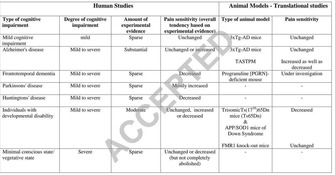
animal-models of cognitive impairment? Are these findings in line with the findings in human subjects? Table 3 addresses these questions by providing broaden overview on the main outcomes for human and animal models of different types of CI. The Table summarizes studies addressing face validity (features), predictive validity (outcome of interventions), and construct validity (neurological basis). Age-related cognitive decline in C57BL/6J mice is characterized by impairment of motor skills, acquisition and memory consolidation, yet in contrast to findings in human subjects, the mice show well-preserved noxious threshold and working memory [37].
Mild cognitive impairment (MCI) is difficult to model in animals except when it is regarded as the transitional state preceding AD. In such a case, MCI is studied as the
prodromal stage that follows the premorbid period and precedes the moderate and advanced stages of AD disease. Mutations of amyloid precursor protein (APP) and presenilins (PS) 1 and 2, associated with amyloid deposition, brain structural change and cognitive decline, are used to emulate AD in transgenic animals but different models may present with different results regarding nocifensive responses and variations may also exists within a model depending on the stimulation modality. For example, the sensitivity to cold stimuli is
unaltered in 3xTg-AD mice that progressively develop both beta-amyloid and tau in cortical and limbic areas [46]. Responses of the old mutants in the hot-plate test were also unaltered [41]. However, the avoidance response latency in the tail flick test was increased in an age-dependent manner equally to its background C57BL/6 strain [7]. Other models for AD such as
the double mutant TASTPM mice expressing human APP(695(K595N, M596L)) x
PS1(M146V ) also show differential responsiveness: lower sensitivity in the hot water tail-flick test but increased sensitivity in the hot-plate test [27]. As inconsistencies in responses to pain exist also among patients with AD, these models show the important role of key
neurobiological hallmarks of the disease and help to depict their contribution in the derangement of the neuronal pathways related to pain.
There are very few well-characterized animal models for FTD. One of these models is the progranuline [PGRN]-deficient mouse based in the loss-of-function mutations of PGRN as the cause of familial ubiquitin-positive FTD. These mice show progressive neuropathology, signs of premature aging and behavioural deficits [76]. They are currently used to study pain defence after nerve injury as well as the development or maintenance of neuropathic pain. Much more common in experimental use is the partial trisomic Ts(1716)65Dn mice (Ts65Dn) based on genetic homology to model Down’s syndrome, the most common genetic cause of developmental disability in humans. The Ts65Dn mice show reduced thermal sensitivity threshold in the tail-flick test but normal sensitivity to morphine assessed in this test as compared to control littermates [82]. Furthermore, Ts65Dn mice display the expected
biphasic (early and late) behavioural response in the formalin test but with reduced sensitivity. It is uncertain if the reduced sensitivity is due to diminished peripheral nociceptor
responsiveness and/or less effective central processing of nociceptive signals [35,92]. It is noteworthy that these results are similar to those of two studies showing increased pain threshold among individuals with Down’s syndrome when measured with a reaction-time inclusive method [32,54]. The double transgenic APP/SOD1 mouse model of Down syndrome shows reduced sensitivity for neuropathic pain associated with neuroma and a decreased autotomy response [64]. The murine model for fragile X mental retardation 1, the FMR1 knock-out mice, shows normal acute responses to noxious stimuli assessed in the hot
plate and tail-flick [61,127]. In the rat valproic acid (VPA) model of autism, adolescent rats prenatally exposed to VPA exhibited hypoalgesia on the hot plate test [61] supporting reports of altered pain sensitivity among individuals with autism.
Conclusions and recommendation for future studies: Although important advances have
been achieved in recent years using animal models of CI, a few limitations should be considered regarding their use. First, although executive functions can be demonstrated in various animals, they are substantially more developed and, as a consequence, more vulnerable to impairment in humans. Second, the relatively short length of the ‘aging’ processes in rodents may not mimic time-dependent neuronal modifications involved in nociceptive processing among elderly patients. However, large animals, mostly those whose pain is naturally developed over time such as cats and dogs [55], are proposed as better models to study pain in aged-related CI. This is also the case for the ‘Cognitive Dysfunction Syndrome’, a naturally developed neurodegenerative disease characterized in cats and dogs that is also considered a model of Alzheimer’s disease [71]. Third, despite the fact that mammals have a high degree of neuroanatomical similarity to humans (as opposed to non-mammalian models), they are generally expensive and time-consuming to use. Fourth, the concerns about the poor predictive validity of experimental pain for chronic pain conditions that is applied to studies of experimental pain in humans can also be applied to animal models.
Certainly, further studies of pain in naturally occurring or induced animal models of
cognitive impairment and dementia are required. Nevertheless, at present, studies of
transgenic rodents harbouring the human familial AD mutations that closely mimic the temporal, neuroanatomical and behavioural patterns of the human disease offer great advantages due to their shorter time requirements, lower maintenance expenses and ethical
considerations. These models are especially promising for the development of new treatments for AD and other age-related cognitive impairments.
7. Overall conclusions
CI is a frequent consequence of many neurological diseases, of neurodegenerative
changes and of vascular and traumatic insults to the brain. CI also may gradually appear in the course of healthy ageing. The literature on the experimental analyses of effects of CI on pain processing is sparse. Nevertheless, existing data suggest that pain processing is frequently altered compared to cognitively intact matched controls. There seems to be no common denominator for all types of CI reviewed herein, with regard to the nature of this alteration. Rather, variability exists across different types of CI, with increased or decreased pain threshold and pain tolerance and decreased or lack of change in pain ratings (see Table 3). Obviously, this variability may stem from structural variability in the brain regions affected by each condition and the extent of the damage as well as of the functional consequences and their counter-regulation. Variability may also stem from between-studies differences in the experimental protocol and the severity as well as exact type of CI within the sample tested.
To date, studies of pain processing in animal models of cognitive impairment and dementia have been sparse. As can be seen in Table 3, some of the findings derived from animal models are in agreement with findings in CI patients, whereas others point in a different direction. Novel developments in animal research that make use of non-verbal behaviours like facial expressions, seem promising approaches that allow assessment of similar behaviours in human and animals and thus, can help to better translate findings from animal to human research.
Importantly, because studies in individuals with CI in general, and in individuals who cannot report their pain in particular, are uniquely challenging, it is critical that researchers adhere to the declaration of Helsinki and conduct the studies in the most empathic,
compassionate and ethically sound manner possible. It is also important that researchers have sufficient experience in pain assessment and that all testing protocols would first be tested among healthy volunteers and adjusted prior to testing individuals with CI. In the search for additional knowledge that can help future patients, one must carefully consider all risks and benefits and keep in mind the interests and vulnerability of the patients at hand.
Despite the diversity between CI types, it appears that those with widespread brain atrophy or neural degeneration (ageing, MCI, Alzheimer’s disease, traumatic brain injuries, multiple sclerosis, autisms, cerebral palsy) all show increased pain responses and/or greater pain sensitivity. We can only speculate that the widespread brain damages, especially those involving white matter, affect descending pain modulation pathways which in turn lead to reduced inhibitory control over the pain system. It is possible that these descending pain modulation pathways are more susceptible to such damage whereas ascending pathways are - in comparison - more robust. In cases of severe brain damage that is associated with severe CI such as in patients in VS and MCS, both the ascending and descending pain pathways might be disrupted to a similar degree resulting perhaps in reduced sensitivity to pain. Neural atrophies restricted to regions associated with processing of ascending nociceptive input, such as in patients with FTD, may also render patients less sensitive to pain. Regardless of the etiology of CI, however, the scientific evidence suggests that the majority of patients do seem to feel noxious stimuli. Thus, despite the communication difficulties characteristic of CI rendering pain assessment a challenge in this population, true changes in pain processing have to be assumed. Therefore, and in light of the high rates of chronic pain, special attention
Acknowledgements: The work was supported by the COST program (European
Cooperation in the field of Scientific and Technical Research; COST TD1005). The authors have no conflict of interests related with this work.
Authors' contribution: Authors RD and MK conceived the paper, supervised and
organized its structure and content. All the authors have contributed to different sections of the paper. All the authors have read and approved the paper.
References
[1] Achterberg WP, Pieper MJ, van Dalen-Kok AH, de Waal MW, Husebo BS, Lautenbacher S, Kunz M, Scherder EJ, Corbett A. Pain management in patients with dementia. Clin Interv Aging 2013;8:1471-82.
[2] Achterberg WP, Pot AM, Scherder EJ, Ribbe MW. Pain in the nursing home: assessment and treatment on different types of care wards. J Pain Symptom Manage 2007;34:480-7.
[3] Amanzio M, Benedetti F, Vase L. A systematic review of adverse events in the placebo arm of donepezil trials: the role of cognitive impairment. Int Psychogeriatr 2012;4:1-10.
[4] Apkarian AV, Bushnell MC, Treede RD, Zubieta JK. Human brain mechanisms of pain perception and regulation in health and disease. Eur J Pain 2005;9:463–484.
[5] Arendt-Nielsen L, Lautenbacher S. Assessment of pain perception. In: Pathophysiology of Pain Perception, S. Lautenbacher and R.B. Fillingim (eds), New York : Plenum Publishing Co., NY 2004:25-42.
[6] Attal N, Masselin-Dubois A, Martinez V, Jayr C, Albi A, Fermanian J, Bouhassira D, Baudic S. Does cognitive functioning predict chronic pain? Results from a prospective surgical cohort. Brain 2014;137:904–17.
[7] Baeta-Corral R, Pick CG, Giménez-Llort L. Progression of BPSD-like symptoms and pain perception in aging and Alzheimer’s disease: studies in 3xTg-AD mice. Proceedings of the 6th International Conference on Pain and Impaired Cognition (PAIC) Aalborg, Denmark. 28-29th March 2014.
[8] Bathgate D, Snowden JS, Varma A, Blackshaw A, Neary D. Behaviour in frontotemporal dementia, Alzheimer’s disease and vascular dementia. Acta Neurol Scand 2001;103:367–78. [9] Bender L & McKenna K. Hemiplegic shoulder pain: defining the problem and its management.
Disability Rehab 2001;23:698-705.
[10]Benedetti F, Arduino C, Costa S, Vighetti S, Tarenzi L, Rainero I, Asteggiano G. Loss of expectation-related mechanisms in Alzheimer’s disease makes analgesic therapies less effective. Pain 2006;121:133–44.
[11]Benedetti F, Arduino C, Vighetti S, Asteggiano G, Tarenzi L, Rainero I. Pain reactivity in Alzheimer patients with different degrees of cognitive impairment and brain electrical activity deterioration. Pain 2004;111:22–9.
[12]Benedetti F, Carlino E, Pollo A. How placebos change the patient’s brain. Neuropsychopharm 2011;36:339-54.
[13]Benedetti F, Vighetti S, Ricco C, Lagna E, Bergamasco B, Pinessi L, Rainero I. Pain threshold and tolerance in Alzheimer’s disease. Pain 1999;80:377–82.
[14]Biersdorff K. Incidence of significantly altered pain experience among individuals with developmental disabilities. Am J Mental Retardation 1994;98:619-31.
[15]Bizon JL, Foster TC, Alexander GE, Glisky EL. Characterizing cognitive aging of working memory and executive function in animal models. Front Aging Neurosci 2012;12;4:19.
[16]Boly M, Faymonville ME, Schnakers C, Peigneux P, Lambermont B, Phillips C, Lancellotti P, Luxen A, Lamy M, Moonen G, et al. Perception of pain in the minimally conscious state with PET activation: an observational study. Lancet Neurol 2008;7:1013–20.
[17]Borsook. Neurological diseases and pain. Brain 2012;135:320-44.
[18]Braak E, Griffing K, Arai K, Bohl J, Bratzke H, Braak H. Neuropathology of Alzheimer's disease: what is new since A. Alzheimer? Eur Arch Psychiatry Clin Neurosci 1999;249 Suppl 3:14-22.
[19]Brandt BR, Rosén I. Impaired peripheral somatosensory function in children with Down syndrome. Neuropediatrics 1995;26:310-2.
[20]Carlino E, Benedetti F, Rainero I, Asteggiano G, Cappa G, Tarenzi L, Vighetti S, Pollo A. Pain perception and tolerance in patients with frontotemporal dementia. Pain 2010;151:783-9. [21]Cascio CJ, McGlone F, Folger S, Tannan V, Baranek G, Pelphrey KA, Essick G. Tactile
perception in adults with autism: a multidimensional psychophysical study. J Autism Det Disord 2008;38:127–37.
[22]Cavinato M, Volpato C, Silvoni S, Sacchetto M, Merico A, Piccione F. Event-related brain potential modulation in patients with severe brain damage. Clin Neurophys 2011;122:719–24. [23]Chakrabarty T, Sepehry AA, Jacova C, Hsiung GY. The prevalence of depressive symptoms
in frontotemporal dementia: a meta-analysis. Dement Geriatr Cogn Disord 2015;93:257-71. [24]Cole LJ, Farrell MJ, Duff EP, Barber JB, Egan GF, Gibson SJ. Pain sensitivity and fMRI
pain-related brain activity in Alzheimer’s disease. Brain 2006;129:2957–3265.
[25]Cole LJ, Farrell MJ, Gibson SJ, Egan GF. Age-related differences in pain sensitivity and regional brain activity evoked by noxious pressure. Neurobiol Aging 2010;31:494–503. [26]Cole LJ, Gavrilescu M, Johnston LA, Gibson SJ, Farrell MJ, Egan GF. The impact of
Alzheimer's disease on the functional connectivity between brain regions underlying pain perception. Eur J Pain 2011;15:568-11.
[27]Corbett A, Husebo B, Malcangio M, Staniland A, Cohen-Mansfield J, Aarsland D, Ballard C. Assessment and treatment of pain in people with dementia. Nature Rev Neurol 2012;8:264-74. [28]Craik FIM, Salthouse TA, editors. The handbook of aging and cognition. 3. New York:
Psychology Press 2011; pp. 491–556.
[29]de Knegt NC, Pieper MJ, Lobbezoo F, Schuengel C, Evenhuis HM, Passchier J, Scherder EJ. Behavioral pain indicators in people with intellectual disabilities: a systematic review. J Pain 2013;14(9):885-96.
[30]de Tommaso M, Navarro J, Ricci K, Lorenzo M, Lanzillotti C, Colonna F, Resta M, Lancioni G, Livrea P. Pain in prolonged disorders of consciousness: laser evoked potentials findings in patients with vegetative and minimally conscious states. Brain Inj 2013;27:962-72.
[31]de Tommaso M, Serpino C, Difruscolo O, Cormio C, Sciruicchio V, Franco G, Farella I, Livrea P. Nociceptive inputs transmission in Huntington’s disease: a study by laser evoked potentials. Acta Neurol Belg 2011;111:33-40.
[32]Defrin R, Pick CG, Peretz C, Carmeli E. A quantitative somatosensory testing of pain threshold in individuals with mental retardation. Pain 2004;108:58–66.
[33]Defrin R, Riabinin M, Feingold Y, Schreiber S, Pick CG. Deficient Pain Modulatory Systems in Patients with Mild Traumatic Brain and Chronic Post-Traumatic Headache: Implications for its Mechanism. J Neurotrauma 2015;32(1):28-37.
[34]Defrin R. Chronic post-traumatic headache: clinical findings and possible mechanisms. J Man Manip Ther 2014;22:36-44.
[35]Dierssen M, Vallina IF, Baamonde C, García-Calatayud S, Lumbreras MA, Flórez J. Alterations of central noradrenergic transmission in Ts65Dn mouse, a model for Down syndrome. Brain Res 1997;749:238-44.
[36]Englander ZA, Pizoli CE, Batrachenko A, Sun J, Worley G, Mikati MA, Kurtzberg J, Song AW. Diffuse reduction of white matter connectivity in cerebral palsy with specific vulnerability of long range fiber tracts. NeuroImage: Clinical 2013;2:440-447.
[37]Fahlström A, Zeberg H, Ulfhake B. Changes in behaviors of male C57BL/6J mice across adult life span and effects of dietary restriction. Age (Dordr) 2012;34:1435-52.
[38]Feldt KS, Ryden MB, Miles S. Treatment of pain in cognitively impaired compared with cognitively intact older patients with hip-fracture. J Am Geriatr Soc 1998;46:1079-85.
[39]Femminella GD, Rengo G, Komici K, Iacotucci P, Petraglia L, Pagano G, Ferrara N. Autonomic Dysfunction in Alzheimer's Disease: Tools for Assessment and Review of the Literature. J Alzheimer's Disease 2014;42:369-377.
[40]Fernández-de-Las-Peñas C, Ortega-Santiago R, Ortíz-Gutiérrez R, Caminero AB, Salom-Moreno J, Arendt-Nielsen L. Widespread Pressure Pain Hypersensitivity in Patients with Multiple Sclerosis with and Without Pain as Sign of Central Sensitization. Clin J Pain 2014. [41]Filiali M, Lalonde R, Theriault P, Julien C, Calon F, Planel E. Cognitive and
noncognitivebehaviors in the triple-transgenic mouse model of Alzheimer's disease expressing mutated APP, PS1 and Papt (3xTg-AD). Behav Brain Res 2012;234:334-42.
[42]Gerdelat-Mas A, Simonetta-Moreau M, Thalamas C, Ory-Magne F, Slaoui T, Rascol O, Brefel-Courbon C. Levodopa raises objective pain threshold in Parkinson’s disease: a RIII reflex study. J Neurol Neurosurg Psychiatry 2007;78:1140–2.
[43]Giacino JT, Ashwal S, Childs N, Cranford R, Jennett B, Katz DI, Kelly JP, Rosenberg JH, Whyte J, Zafonte RD, Zasler ND. The minimally conscious state: definition and diagnostic criteria. Neurol 2002;58:349–53.
[44]Gibson SJ, Voukelatos X, Ames D, Flicker L, Helme RD. An examination of pain perception and cerebral event-related potentials following carbon dioxide laser stimulation in patients with Alzheimer’s disease and age-matched control volunteers. Pain Res Manag 2001;6:126–32. [45]Giménez-Llort L, Pick CG. Animal models for pain in dementia. In: Pain in dementia. IASP
press, 2014, in press.
[46]Giménez-Llort L, Torres-Lista V, De la Fuente, M. Crosstalk between behavior and immune system during the prodromal stage of Alzheimer's disease. Curr Pharm Des 2014;20(29):4723-32.