Alma Mater Studiorum – Università di Bologna
DOTTORATO DI RICERCA IN
Biologia Cellulare e Molecolare
Ciclo XXX
Settore Concorsuale di afferenza: 05/E1 Settore Scientifico disciplinare: BIO/10
TITOLO TESI
Biochemical characterization and validation of a novel cell
model for dominant optic atrophy.
Presentata da: Mario Fogazza
Coordinatore Dottorato Prof. Giovanni Capranico
Relatore Prof.ssa Michela Rugolo
Correlatore Dott.ssa Claudia Zanna
ABSTRACT
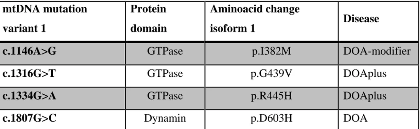
Mutations in the OPA1 gene, encoding the mitochondrial dynamin-like GTPase OPA1, are well known to cause Dominant Optic Atrophy (DOA), the most common inherited optic neuropathy. The missense variants, envisaged to exert a dominant-negative effect, are associated with high risk to develop the severe multisystem disorder (DOA “plus”), characterized by extra-ocular features, including sensorineural deafness, ataxia, myopathy, chronic progressive external ophthalmoplegia, and peripheral neuropathy. Primary skin fibroblasts derived from patients bearing OPA1 mutations represent the cell model for studying DOA pathophysiology, although they often reveal a mild phenotype, as a consequence of the autosomal genetic transmission of DOA. Other genetically modified cellular models characterized by a phenotype strikingly different from wild-type, are therefore desirable.
In this study we describe a novel cell model obtained from Opa1-/- MEFs, where human OPA1 isoform 1 bearing OPA1 mutations was expressed. Under this setting, all OPA1 protein is mutated, ruling out the effect of the wild-type allele. We present here a detailed molecular and biochemical analysis in parallel of fibroblasts and MEFs bearing three known OPA1 pathogenic mutations (I382M, G439V, R445H) and a novel one (D603H), selected on the basis of their clinical phenotypes, ranging from very mild associated with pure optic atrophy to more detrimental causing severe syndromic forms. The results indicate that MEFs bearing OPA1 mutations are a model useful to predict the pathogenicity of new mutations. In fact, according with the severity of the clinical phenotype of patients, the MEFs exhibit an increased number of mitochondrial dysfunctions.
In addition, we propose this cell model as a suitable tool to test drugs with potential therapeutic effect on mitochondrial diseases associated with OPA1 mutations. Indeed, in a preliminary study we were able to to confirm the efficacy of few molecules previously identified in a yeast hight throuput screening as able to revert the pathological phenotype of a mutant Mgm1-OPA1 yeast chimera.
INTRODUCTION _________________________________________________________ 1
Historic overview ____________________________________________________________ 2 From the gene to the function __________________________________________________ 3
OPA1 gene and protein _____________________________________________________________ 3 The role of the eight isoforms ________________________________________________________ 4 Transcriptional regulation ___________________________________________________________ 6 Proteolytic processing ______________________________________________________________ 7 Protein structure ___________________________________________________________________ 8 Oligomerization and interactors ______________________________________________________ 10
The functions in the mitochondrial landscape ____________________________________ 15
Mitochondrial dynamics and long/short balance ________________________________________ 15
Cristae structure and bioenergetics ___________________________________________________ 17
mtDNA maintenance ______________________________________________________________ 20 Apoptosis _______________________________________________________________________ 21 Autophagy and mitophagy __________________________________________________________ 22 Calcium homeostasis and glutamate excitotoxicity _______________________________________ 23 Other functions and implications _____________________________________________________ 25
OPA1 dysfunctions: mutations and pathophysiology ______________________________ 28
DOA pathophysiology ______________________________________________________________ 28 OPA1 mutations: DOA and DOA “plus” ________________________________________________ 30 OPA1 mutations: other neurological disorders __________________________________________ 31
AIMS _________________________________________________________________ 32 MATERIALS AND METHODS _______________________________________________ 34
Cells culture conditions ______________________________________________________ 35 Plasmid construction and retroviral transduction _________________________________ 36 Cellular ATP content _________________________________________________________ 36 Mitochondrial ATP Synthesis __________________________________________________ 36 Citrate synthase activity ______________________________________________________ 37 Mitochondrial network morphology ____________________________________________ 37
Cristae architecture _________________________________________________________ 37
mtDNA sequencing __________________________________________________________ 38 Cellular viability ____________________________________________________________ 39 Western blotting ___________________________________________________________ 39 Respiratory supercomplexes analysis by BN-PAGE ________________________________ 39 OPA1 oligomerization analysis by BN-PAGE ______________________________________ 40 Reagents __________________________________________________________________ 40 Statistical analysis __________________________________________________________ 40
RESULTS ______________________________________________________________ 41
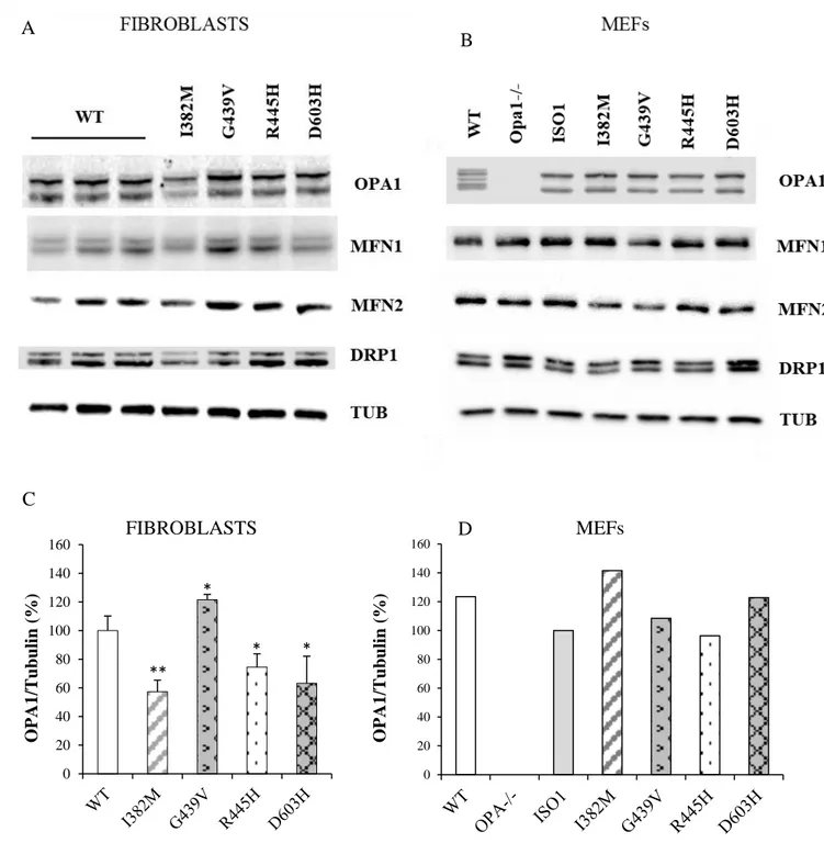
The mitochondrial dynamics machinery _________________________________________ 42
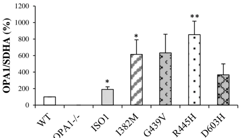
Fusion and fission proteins __________________________________________________________ 42 OPA1 oligomers __________________________________________________________________ 44
The mtDNA ________________________________________________________________ 46
Copy number ____________________________________________________________________ 46 NGS analysis _____________________________________________________________________ 46
Mitochondrial morphology ___________________________________________________ 48
Mitochondrial network morphology __________________________________________________ 48
Cristae architecture _______________________________________________________________ 50 Energetic profile ____________________________________________________________ 51
Cell viability ______________________________________________________________________ 51 Cellular ATP content _______________________________________________________________ 51 Mitochondrial ATP synthesis ________________________________________________________ 52 Organization of RCSs _______________________________________________________________ 53
Other cell models of DOA by using CRISPR/Cas9 gene editing technology _____________ 57 Search for therapeutic molecules: the ORMs _____________________________________ 59
Determination of ORMs dose-responses _______________________________________________ 59 Mitochondrial network morphology __________________________________________________ 61 Cellular viability after metabolic stress ________________________________________________ 63 Cellular ATP content _______________________________________________________________ 64
DISCUSSION AND CONCLUSIONS ___________________________________________ 65 BIBLIOGRAPHY _________________________________________________________ 72
1
2
Historic overview
OPA1 (Optic Atrophy 1) is a nuclear gene, first mapped in the 3q28-qter region (1), that
encodes for a dynamin-related protein localized in the inter-membrane space (IMS) of mitochondria, anchored to the mitochondrial inner membrane (IMM) (2,3). The name derives from Dominant Optic Atrophy (DOA), a disease caused by mutations in this gene (4,5), disease firstly described in 1959 (6) and characterized by degeneration of the retinal ganglion cells (RGCs) and optic nerve atrophy.
OPA1 is a conserved dynamin-related GTPase, its expression is ubiquitous, but quantitatively variable depending to the organ or tissue examined. High mRNA expression levels are present in retina, brain, liver, heart and pancreas (4,7,8).
OPA1 was initially associated with the mechanism of fusion of the IMM (9), but as time went on OPA1 has been recognized to be implicated also in other important mitochondrial functions, such as the maintenance of the complex architecture of cristae (10), which in turn controls the onset of apoptotic process by regulating the release of the cytochrome c from the cristae into the cytoplasm. OPA1 was also shown to be able to directly interacts with some respiratory complexes, thus stabilizing the oxidative phosphorylation system (11). It is also required for the supramolecular organization of respiratory supercomplexes, which is strictly linked to energetic efficiency (12), and, finally, it contributes to maintenance of the mitochondrial DNA stability, probably by anchoring this genome to the IMM (13).
3
From the gene to the function
OPA1 gene and protein
Mapped by linkage studies short tandem repeat polymorphisms (1), in 2000, using different approaches, two European groups simultaneously identified the first mutations in the OPA1 gene (4,5) the human of yeast ortholog Mgm1p/Msp1p.
The gene spans more than 100kb and is composed of 31 exons, and the alternative splicing of the exons 4, 4b and 5b generate 8 different isoforms with a great variability of expression between them in different human tissues, suggesting a fine regulation of OPA1 mRNAs. All OPA1 isoforms are ubiquitously expressed, nevertheless splicing variants containing exon 4 are consistently more represented. In the brain the exons 4 and 4b, alone or combined, are predominant. The exon 4 is evolutionarily conserved, while the exons 4b and 5b are both specific to vertebrates (8,14).
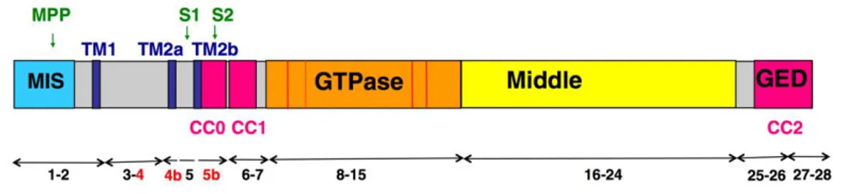
The eight OPA1 mRNA splice forms encode proteins of 924–1014 aminoacids, presenting at the N-terminus an amphiphilic mitochondria targeting sequence (MTS) followed by a transmembrane domain (TM), acting as a stop transfer signal, anchoring the protein at the IMM and leaving most of it in the IMS (2), and the three alternate spliced exons 4, 4b and 5b (8). While apparently the exon 4 does not include any noteworthy domain, exon 4b and 5b encode two additional hydrophobic domains, TM2a and TM2b. The 5b exon also presents a coiled-coil domain (CC0). The following portion of the protein contains the conserved dynamin regions: the GTPase domain, with a coiled-coil domain (CC1), the middle domain, whose function is unknown, and the C-terminus GTPase effector domain (GED) also presenting a coiled-coil domain (CC2) (8,15).
The two main coiled-coil domains, CC1 and CC2, exhibited only the capacity to self-interact to form homo-oligomers, without showing the ability to cross-self-interact with other peptides. In support of this, mutations known to cause DOA located on the CC2 were shown to abolish its capacity of self-interact. Conversely, the analysis of the CC0 domain supported the hypothesis of a hetero-interaction with CC1 domain on the same OPA1 protein, rather than an homo-interaction (16).
After import of the precursors through the mitochondrial membranes and cleavage of the MTS by the mitochondrial processing peptidase (MPP), OPA1 may be further processed at two N terminus cleavage sites, named S1 and S2, located on exons 5 and 5b respectively. The proteolytic cleavage is carried out by the two IMM peptidases YME1L and OMA1 to
4
produce a defined combination of membrane-anchored long forms (l-forms) and short forms (s-forms) soluble in the IMS, which can be peripherally attached to the IMM or diffuse in the IMS and associate to the OM (17). Furthermore, the four isoforms including the exon 4b are completely cleaved into the short forms (18).
Finally YME1L and OMA1 are reciprocally degraded in response to distinct types of cellular stress, thus modulating the proteolytic processing of OPA1 (19).
Figure 1. Schematic representation of OPA1 protein domains. Cleavage sites are highlighted in
green, trans-membrane domains in blue, coiled-coil domains in pink, the three alternatively spliced exons in red (from Belenguer et all., 2013)
. The role of the eight isoforms
As mentioned above, eight isoforms of OPA1 exist, formed by different combinations of the three alternatively spliced exons 4, 4b and 5b. Thus, question has been raised whether each of these exons were associated with a specific OPA1 function, to assess which each of the three alternative exons has been selectively silenced in HeLa cells. Since the variants including the exon 4 represents ~90% of the total protein, its silencing provoked a drastic decrease of OPA1 level, causing mitochondria network fragmentation and depolarization, without signs of cytochrome c release or apoptosis, whereas the silencing of exons 4b and 5b provoked apoptosis, with a slow cytochrome c release, only minor cristae modifications and without mitochondrial fission or depolarization (8).
Later, the same group proved how silencing of the exon 4b, but not the others, caused significant mtDNA depletion and an uneven distribution of the nucleoprotein complexes or nucleoids throughout the mitochondrial network. Furthermore, the N-terminal OPA1 peptide including the and exon 4b, released after the proteolytic cleavage, was shown to physically interact with mitochondrial transcription factor A (TFAM) and DNA polymerase gamma (POLG), the main protein components of nucleoids, and with mtDNA. It was therefore proposed that exon 4b containing OPA1 variants may promote mtDNA
5
stability by anchoring the nucleoids to the IMM, which in turn guarantees mtDNA replication, nucleoids abundance and proper distribution along the network (13).
It seems therefore that specific mitochondrial functions may be associated with the three differentially spliced exons of OPA1, i.e. the exon 4 containing variants could be responsible for fusion of the IMM, the exon 5b variants could preserve the tightness of the cristae junctions, preventing the release of cytochrome c, and the exon 4b variants could promote the mtDNA stability, but these data give us no indication about the need of having eight different isoforms.
Thus, in order to evaluate whether a single OPA1 isoform is specifically associated with a definite mitochondrial function, we carried out a detailed molecular and biochemical analysis in a murine cellular model where OPA1 was deleted, the Opa1-null murine embryonal fibroblasts (MEFs), where every individual OPA1 splice form has been stably expressed alone. Our analysis highlighted that every isoform was able to recover the major phonotypes affected by Opa1 depletion, such as mtDNA content, cristae organization and energetic competence (20). However, the completely fragmented mitochondrial network observed in Opa1-/- MEFs was only in part rescued by mRNA splice forms generating both long and short forms, in accord with a previous report (18). Using two different approaches, co-expressing different couples of isoforms in Opa1-/- MEFs, and co-silencing with different combinations two out of the three alternatively spliced exons in HeLa cells, we concluded that to fully recover even the mitochondrial network morphology, both an adequate amount of OPA1 protein and at least two isoforms with a defined long/short forms ratio are needed (20).
We therefore sustain the presence of a hierarchy in the mitochondrial features the cell first needs to recover, being mtDNA, cristae organization and energetic competence equally important to restore the cellular metabolic efficiency and strictly related one with each other. We proved that every OPA1 isoform has, alone, the capacity to recover these main features, apparently in contrast with transient silencing experiments results, but actually, the two models give us different information. In fact, while the “chronic” model, with the stable expression of any single isoform, reveals that each isoform has the potential to recover those features, the “acute” model, with the transient silencing, suggests that in a physiological context each variant, moreover expressed at different levels, can carry out preferentially a specific function (20).
Thus, each OPA1 isoform is not specifically associated with a definite mitochondrial function, but rather the redundancy of their potentials provides mitochondria of the
6
necessary flexibility to withstand and adapt to different metabolic and stress conditions in highly specialized tissues.
Transcriptional regulation
OPA1 expression is ubiquitous, but its regulation is still not fully understood, although it seems to lay downstream several pathways.
Together with MFN2, OPA1 is reported to be upregulated during bone marrow progenitor differentiation and to promote the migration of immature dendritic cells (21).
Novel in vitro evidence indicates that TNFR2 activation upregulates OPA1 expression, with the acetylation of STAT3 at lysine 370 and/or 383 by p300 playing an essential role, enabling the interaction of STAT3 with RelA to bind to the promoter region of OPA1 and enhance transcription. TNFR2 activation in an in vivo transverse aortic constriction-induced heart failure mouse model exerted beneficial effects on OPA1 expression, improving mitochondrial morphology and respiratory activity, leading to improved cardiac function and survival rate (22).
Treatment of cardiomyocytes in vitro and in vivo with insulin also increased Opa-1 protein levels, ameliorating mitochondrial functions as fusion, membrane potential, ATP levels and oxygen consumption. This has been achieved through the Akt-mTOR-NF-κB signaling pathway, highlighting the existence of a link between mitochondrial morphology and insulin signaling in cardiac and skeletal muscle cells and potentially with the onset of insulin resistance (23). It is known that under stress condition, OPA1 is transcriptionally upregulated via NF-kB-responsive promoter elements for maintenance of mitochondrial integrity and protection from stress-induced cell death.
Some studies suggest that this is due to parkin recruitment to the linear ubiquitin assembly complex and increases linear ubiquitination of NF-kB essential modulator (NEMO), which is essential for canonical NF-κB signaling. Accordingly, linear ubiquitination of NEMO, activation of NF-κB and upregulation of OPA1 are significantly reduced in response to TNF-a stimulation in parkin-deficient cells (24).
In contrast to this, a more recent study proposes that the protective effect of parkin may rather be related to the ubiquitination of Bax impairing its mitochondrial translocation. Indeed this study shows that the absence of IKKα, with or without IKKβ, has an impact on OPA1 expression and mitochondrial network morphology, pointing out a role of the
7
nonclassical NF-κB pathway rather than the canonical one, in the regulation of mitochondrial dynamics and OPA1 expression (25)
Another recent study displays that T-cell intracellular antigens (TIA1b/TIARb) and Hu antigen R (HuR) exert antagonistic roles in regulating expression of mitochondrial shaping proteins. In particular, while HuR functions as a translational activator increasing steady-state levels of the protein, TIARb operates as a translational repressor, both in a 3’-UTR-dependent manner on the OPA1 mRNA. Moreover, TIA1 and TIAR modulate alternative splicing of OPA1 pre-mRNA, promoting exon 4b inclusion and exon 5b skipping, facilitating the production of short OPA1 forms (26).
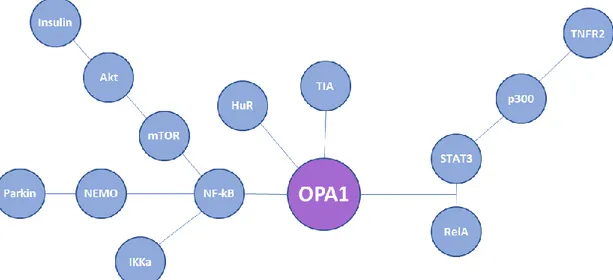
Figure 2. Schematic representation of factors known to be involved in the transcriptional and
post-transcriptional regulation of OPA1 mRNA.
Proteolytic processing
The primary sequence of OPA1 presents two cleavage sites, S1, present in all the isoforms and located at exon 5, and S2, present only in the isoforms containing exon 5b. Thus, each mRNA splice form can generate a long form, produced by cleavage with MPP, and one or more short isoforms (produced by cleavage at S1 or S2). But, as mentioned above, OPA1 isoforms containing exon 4b are totally processed into short forms (18,27).
Several and sometimes discordant studies have identified different proteases recognizing the two cleavage sites of OPA1 in human cells. The presilin-associated rhomboid-like protease (PARL) seems to be involved in the generation of a soluble short form of OPA1 in the IMS (28). The m-AAA proteases, are present in the IMM as AFG3L2
homo-8
oligomers or AFG3L2-paraplegin hetero-oligomeric complexes (29). Overexpression of paraplegin induces the accumulation of short forms of OPA1 by cleavage at S1 (27). Down-regulation of AFG3L2 decreased the stability of long OPA1 forms (30). Nevertheless, neither PARL nor paraplegin are involved exclusively in OPA1 processing. Indeed, knocking them down/out does not alter the long/short forms ratio (27).
Down-regulation of ATP-independent protease OMA1 lightly decreased the levels of OPA1 short forms, generated by cleavage at S1 site and accumulated at low levels in MEFs (30,31). The cleavage at S2 is ascribed to the ATP-dependent AAA+ protease YME1L (18,32). The MEFs OMA1- and YME1L- double knockout contained only long forms of OPA1 (33). OMA1 and YME1L have many independent functions but cooperate to regulate their differential processing of OPA1. Moreover, these two proteases are reciprocally degraded in response to insults that depolarize mitochondria in a process dictated by cellular energetic status. OMA1 is degraded through a YME1L-dependent mechanism following insults that depolarize mitochondria. Alternatively, YME1L is degraded in response to insults that depolarize mitochondria and deplete cellular ATP through a mechanism involving OMA1. This differential degradation alters their proteolytic processing of OPA1 (19).
Moreover, even prohibitins are involved in the processing of OPA1 Indeed, their deletion results in selective loss of long forms of OPA1 and concomitant increase of the short forms (34,35), although the mechanism underlying the effect of prohibitins is not known.
Post-translational regulation of OPA1 comprises, in addition to proteolytic processing, also other modifications that take part in the regulation of other proteins involved in the mitochondrial dynamics. In this respect, the deacetylation of OPA1 lysines 926 and 931 by SIRT3 has been shown to increase OPA1 GTPase activity and to recover mitochondrial functions in OPA1 -/- cells (36). Moreover, OPA1 has been demonstrated to be the substrate of the leucine-rich repeat kinase 2 (LRRK2, PARK8), whose mutations are commonly associated with autosomal dominant familial Parkinson’s disease. In this regard, mutations in the kinase domain of LRRK2 proved to reduce the steady-state levels of short forms of OPA1 in human Parkinson’s brain (37).
Protein structure
OPA1 is a member of a family of highly conserved GTPases related to dynamin, has the same domain architecture as the dynamin-like proteins, compared to the classical dynamins
9
lack of the prolin-rich domain, but has an additional amino-terminal mitochondrial import sequence that is followed by a transmembrane and coiled-coil sequence (38). While the primary structure of the protein, in every of its isoform, is well known, the information we have about the upper level structure are fragmentary because the three-dimensional structure of OPA1 has not be resolved by experimental procedures such as NMR and X-ray crystal analysis and there is little information on the structure of the complex between OPA1 and GTP.
First attempt to obtain a structural homology model of the OPA1 GTPase domain yielded the Dictyostelium dynamin A GTPase domain as the most similar structure (39). After manual refinement, OPA1 and Dictyostelium dynamin A GTPase domains could be superposed, showing that the resolved G1, G3 and G4 signatures, involved in the coordination of GDP/GTP and the Mg2+ ion, essential for GTP hydrolysis, could be
structurally and functionally mirrored in the OPA1 GTPase domain model (40).
Later, bacterial dynamin like protein (BDLP) was identified as the most significant hit in a profile–profile sequence searches with a sequence identity of 13% in the C terminal region of OPA1 (residues 220–960), thus the BDLP coordinates (41) were used as a template for modelling the OPA1 structure. The model obtained was used to map some missense mutations found in DOA patients, most of which reside in the highly conserved GTPase domain. These missense mutations (A357T, G439V, R445H, S545R) affect the GTPase domain just adjacent to its active site potentially interfering with nucleotide binding and altering the affinity and hydrolysis rate of the GTPase domain. The only missense mutation differently located (V910D) resides at the interface of the two effector domains performing the conformational change (42).
Recently, homodimer structural models of wild-type and mutant OPA1 were predicated. Molecular modeling was performed with a region containing the GTPase domain and part of the middle domain of the dimer crystal structure of human Dynamin 1 (43) as a template. The analyses predicted decreased dimer formation of OPA1 and decreased GTP binding as the causes of the disease symptoms associated with these mutations (44). Indeed, dynamin-related proteins are known to homo- and hetero-oligomerize (45).
10
Figure 3. Human OPA1 homology model from residue 220 to 960. GDP depicted in sticks, DOA
mutations depicted in spheres. (from Amati-Bonneau et al., 2008)
Oligomerization and interactors
Peptide analysis of the OPA1 protein demonstrated specific self-interaction of two coiled-coil domains, the CC1 in the GTPase domain and CC2 in the C-terminal GED domain, while the CC0 in the exon 5b could only hetero-interact with the CC1. Being so near to the TM domain, the CC1 domain of the long forms may be sterically hampered from interaction because of the membrane bound, while their CC2 domain could still interact with other OPA1 molecules in the IMS. Instead, the processing to short forms would allow free interaction even of the CC1 domain supporting the formation of larger aggregates via both coiled-coil domains. However, in short forms of isoforms bearing the exon 5b could only a dimer formation via CC2, because of the presence of CC0, which blocks CC1 interaction. (16)
First evidence that of OPA1 can form oligomers of different molecular weight in the IMS came by experiments where mitochondria isolated from HeLa cells were solubilized with 1% Triton X-100 and subjected to gel filtration. Western blots of each fraction revealed
11
that the long and the short forms of OPA1 were eluted at distinctly different peak with apparent molecular weight of 440 and 158 kDa, respectively, with the short form being found in a broad molecular weight range, approximately from 400 to 150 kDa. This analysis could not clarify if these complexes were homo- or hetero-oligomers. Given their differential sub-mitochondrial localization, since OPA1 long forms were found associated to the IMM and the short one both in the IMS and associated with OMM, these different molecular mass complexes should consist of different proteins (3).
Similar results have been obtained in purified mitochondria isolated from different mouse tissues by using the same purification protocol, but with milder detergents to avoid aberrant migration. OPA1 was detected in fractions, corresponding to the apparent molecular weight of 285 and 184 kDa, respectively. In all tissues the peak levels of long forms and the short forms without the 5b were found in fractions corresponding to the 285 kDa complexes. Whether the different isoforms form individual homomeric complexes or interact in one large heteromeric complex remains to be elucidated. In contrast, the highest peak intensity for short form with the exon 5b was observed in the fraction corresponding to the small complex of 184 kDa (16)
Another group identified by chemical crosslinking an ~290 kDa OPA1 immunoreactive band that disappeared when cristae membranes were separated by osmotic swelling. Using tagged versions of long and short forms of OPA1, they demonstrated that this oligomer contained both. The size of the OPA1 oligomer suggested the presence of at least a trimer comprising two long and one short form of OPA1. They also found OPA1 in a ~230 to ~180 kDa fraction after in vitro treatment of mitochondria with cleaved p7/p15 BID (cBID), suggesting that OPA1 can associate with other proteins during apoptosis. (10). During apoptosis, these oligomers are early targets of BID, BIM-S, and BNIP3, the latter being proved to co-immunoprecipitate with OPA1, as well as of intrinsic death stimuli, with their disruption being associated with cristae remodeling (10,46,47).
Further analysis by western blot of blue native gel electrophoresis (BNGE) of mitochondrial proteins revealed four major OPA1-containing complexes, the heavier of which, ~720 kDa molecular weight complex, rapidly disappeared upon treatment with cBID (12). Accordingly, the OPA1 oligomers targeted by cBID to trigger cristae remodeling and cytochrome c redistribution were stabilized in Opa1 isoform1-overexpressing mice mitochondria. All together, these data show that mild OPA1 overexpression hampers apoptotic cristae remodeling in vivo (48).
12
A fraction of OPA1 was found to co-immunoprecipitated with MIC60 (49) and MIC25 (50), core proteins of the mitochondrial contact site and cristae organizing system (MICOS). Moreover, a ~180–190 kDa complex stabilized by crosslinking was found to be immunoreactive for both OPA1 and MIC60, and this complex was also reduced in apoptotic cBID-treated mitochondria. Not only the ~720 kDa OPA1 but also complexes that partially overlap with it containing MIC60 and MIC19, another crucial MICOS component that regulate cristae junctions’ biogenesis, were selectively destabilized during apoptotic
cristae remodeling, data confirmed also by mass spectrometry analysis and quantitative
proteomic analysis. All these evidence together show that OPA1 not only interact, but is also epistatic to MICOS in the regulation of cristae shape (49).
The list of the proteins proved to interact with OPA1 become longer over time, most of them being involved in the energy production or in apoptosis and mitophagy mechanisms. To the first category belong the RCSs. Indeed, it has been shown that, within the cristae membrane, OPA1 directly interacts with subunits of CI, CII, CIII (11) and CIV (51). Furthermore, recently two proteins involved in the GTP fueling of OPA1, NDPK-D (mitochondrial nucleoside diphosphate Kinase, also called nonmetastatic protein 23-H4 or Nm23-H4) and WBSCR16 (Williams-Beuren syndrome critical region 16), were found to physically interact with OPA1, being located in the IMS bound to the IM (52–54).
Both long and short forms of OPA1 co-immunoprecipitated even with SIRT4, a stress-responsive mitochondrial sirtuin that controls cellular energy metabolism in a NAD+ -dependent manner and is implicated in cellular senescence and aging. Only the enzymatically active SIRT4 triggered an unbalance of the long/short OPA1 ratio toward the long forms, and interacted with the long form of OPA1. This OPA1 long form stabilization could involve direct or indirect protein-protein interaction or even a mechanism of protection from stress-induced or protease mediated processing (55). Among the proteins that proved to co-immunoprecipitate with OPA1, the reactive oxygen species modulator 1 (ROMO1), belong to the protein involved in the mitochondria quality control mechanisms, being a redox-regulated protein shown to be important for mitochondrial fusion activity and normal cristae morphology. Not only knockdown of ROMO1 promoted mitochondrial fission and led to an imbalance in OPA1 isoforms abundance that favored the accumulation of the short form of isoform 1, but ROMO1 also proved to be essential for the oligomerization of OPA1 (56).
It seems also that overexpression of Hypoxia-induced gene domain protein-1a (Higd-1a), a IMM protein that plays a role in cell survival under hypoxic conditions, directly inhibits
13
the processing of Opa1 induced by hypoxia and CCCP, finding also corroborated by the co-immunoprecipitation of Higd-1a with Opa1, and in particular only with the long forms of it. Indeed, the soluble short forms of Opa1 did not interact with Higd-1a at all, so the N-terminal domain of Opa1 appears essential for its interaction. Moreover, the deletion of a N-terminal portion of Higd-1a, a region that includes some highly conserved basic amino acids and is located in the IMS proximal to the TM domain, completely eliminated its interaction with Opa1 and its fusogenic activity. Thus, due to its position Higd-1a can approach the N-terminal domain of Opa1. Furthermore, even MFN1 can be co-immunoprecipitated together with OPA1 and Higd-1a, but the interaction with the latter seems to be indirect, because Opa1 knockdown or cleavage eliminated also the interaction of MFN1 with Higd-1a (57).
Another protein, involved in apoptosis and in the mitophagy mechanism, proved to co-immunoprecipitate with OPA1, is the FUN-14 domain containing protein 1 (FUNDC1), that is able to anchor OPA1 through its lysine 70 residue toward the inner face of OMM. Under mitochondrial stresses conditions, OPA1 is cleaved or even degraded, thus promoting mitochondrial fission, required for mitophagy (58).
Furthermore, OPA1 was found to co-immunoprecipitate also with Omi/HtrA2, a serine protease released as a pro-apoptotic factor from the IMS into the cytosol. The loss of this protein is known to cause nerve cell loss in mouse models and has been linked to neurodegeneration in Parkinson's and Huntington's diseases. In cells, loss of Omi/HtrA2 provoked a selective up-regulation of more soluble OPA1 protein. Interestingly, the accumulated long forms of OPA1 were degraded more rapidly upon proteinase K digestion. Also, an increase of the small cytosolic pool of OPA1 in the Omi/HtrA2 KO cells has been found (59). The release of OPA1 were already been described upon disruption of OPA1 engagement in cristae junctions (10).
Finally, two of the already discussed post-translational regulators of OPA1, in particular SIRT3 and LRRK2, have been demonstrated to physically interact with OPA1 (36,37).
14
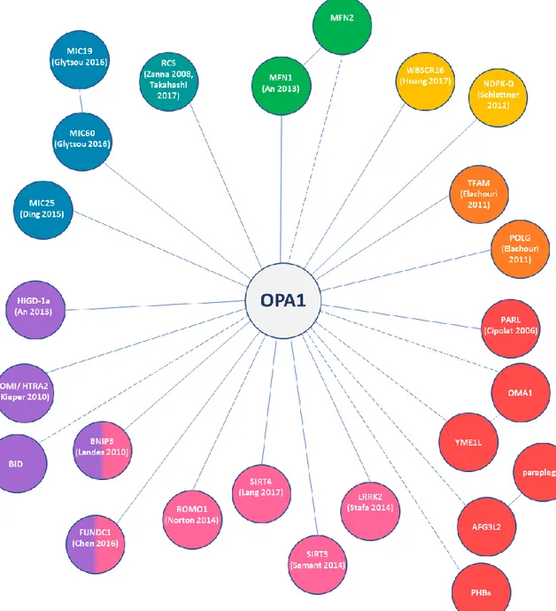
Figure 4. Schematic representation of all the proteins known (solid lines) or supposed (dashed
lines) to physically interact with OPA1. The colors identified proteins involved in: green – mitochondrial fusion, petrol blue – energetics, blue – cristae, purple – apoptosis, pink – mitochondria quality control, red – proteases, orange – nucleoids, yellow – ATP/GTP exchange.
15
The functions in the mitochondrial landscape
Mitochondrial dynamics and long/short balance
When observed by live-cell imaging, mitochondria appear as an interconnected network that spreads through the cell. This structure is not static, being dynamically regulated by constitutively ongoing fusion and fission processes occurring at the two mitochondrial membranes. In mammals, the mitochondrial network dynamics involves four proteins, each playing a specific role: DRP1 is in charge of mitochondrial fission, MFN-1 and -2 and OPA1, are responsible for the fusion of OMM, and IMM, respectively.
The OPA1 orthologs in yeasts, Mgm1p and Msp1p, were initially identified for their involvement in the maintenance of mitochondrial genome, and only later associated with mitochondrial fusion. OPA1 was first identified by linkage studies, and from the beginning associated with mitochondrial dynamics. In agreement with its localization in the IMS, its primary function was demonstrated to be the fusion of the IMM. Indeed, several studies disclosed that OPA1 loss of function, by gene knock-out or knock-down, leads to a fragmented mitochondrial network (9,18,60–62). Noteworthy, the OPA1 overexpression in a physiological context also induces network fragmentation, whereas in cells where mitochondrial network was already fragmented the overexpression of OPA1 promotes its elongation (2,60). A mild OPA1 overexpression also inhibited apoptotic cristae remodeling and corrected the altered cristae shape and defective mitochondrial bioenergetics in mouse models of primary mitochondrial diseases (12,48,49,63).
The fusogenic activity has been initially ascribed to the OPA1 variants bearing the exon 4, as suggested by silencing experiments (8), but recently our and another group independently established that any isoform processed in both long and short forms has the capability to restore mitochondrial network morphology in Opa1-/- MEFs. (20,64). Moreover, both studies demonstrated that the expression of an un-cleavable isoform 1 in Opa1-/- MEFs allows for mitochondrial fusion, beside not being able to restore the interconnected network morphology (20,64). Indeed, is now clear that even if the mitochondrial fusion is indispensable to the network to be interconnected, the ability to fuse does not assure a filamentous and interconnected mitochondrial morphology. Thus, fusion capability and mitochondrial morphology have to be considered as distinct phenotypes.
16
The initial hypothesis was that the OPA1 long forms only were fusion competent, whereas short forms were unable to promote fusion (27). Indeed, in vitro experiments evidenced that a recombinant OPA1 short form was able to tubulate membranes, but could not induce membrane fusion (65). More recently, by using an in vitro fusion assay, the same group clearly demonstrated that OPA1 long forms on one membrane and cardiolipin on the other are the minimal components sufficient and necessary for the two membrane to fuse (66). Moreover, this study confirmed that the short forms are involved in the fusion process, but are not able to promote it without the long forms. Indeed, addition of the short forms to the minimal components accelerated the fusion process and promoted liposome binding, suggesting that the soluble short forms may act like a bridge between the two membranes, linking the long forms on one side and the cardiolipin on the opposite one (66).
Still, a residual fusogenic activity of short forms can be detected when expressed in Opa1-/- MEFs (20,64), whereas mutations that ablate the GTPase activity totally prevent fusion (20). Accordingly, the artificial anchoring of the short forms to the membrane via a lipid tail (66), or by fusion of the N-terminal portion of the IMM protein AIF (20), resulted in a significant increase in membrane fusion. Taken together, these results support the hypothesis that both the functional GTPase domain and the membrane anchoring are necessary to promote fusion, in accord with previous studies on Oma1 and Yme1l double knockout MEFs, where the formation of short forms is blocked, and long forms alone are sufficient to promote mitochondrial fusion (Anand et al., 2014).
Conversely, the role of the short forms is still debated. Anand and colleagues suggest that they are involved in mitochondrial fission, given that the expression of a chimeric AIF-short form did not modify the fusion rate while increasing mitochondria fragmentation in a GTPase activity dependent manner. Moreover the GTPase-inactive AIF-short form co-localizes with sites of mitochondrial division (33). Still, it must be noticed that the AIF domain of this chimaera contains a trans-membrane sequence that may compromise the solubility of this short form, resulting in a shorter membrane anchored form.
Thus, the long/short forms ratio seems to play a major role in the mitochondrial network morphology. In Opa1-/- MEFs, several studies pointed out that only the expression of the long and short forms together was able to elongate the mitochondrial network, whereas it remained completely fragmented after the expression of an uncleavable long version of the isoform 1, despite its proved fusogenic capability (18,20,64). Still, another group is discordant about the uncleavable form fragmentation recover, stating that that OPA1 processing is dispensable for the ability to maintain tubular interconnected mitochondria
17
(33). However, for a full recovery of the mitochondrial network morphology due to ablation of Opa1 in MEFs, only partially rescued by the expression of one isoform generating both long and short forms, the expression of at least two isoforms with a balanced long/short forms ratio is required (20).
Conversely, the overexpression of long form re-equilibrated the accumulation of OPA1 short forms in rat retinal cells exposed to ischemia-reperfusion injury, preventing fragmentation of mitochondrial network and cell death (67). Furthermore, in both Oma1 and Yme1l double knockout MEFs and cardiomyocytes, in which only the long forms are present, an interconnected mitochondrial network was observed (33,68).
These data are not necessarily in disagreement with each other, as they were obtained on different models, where the different multiplicity of OPA1 variants expression may be the keystone.
Cristae structure and bioenergetics
As already mentioned OPA1 is mainly found in the IMS of mitochondria, soluble or anchored to the IMM (2,7) with a small amount of the short soluble forms found to be associated with the OMM (3). Due to the presence of the narrow tubular cristae junctions (CJ), the IMM can be divided in two sub-compartments: the cristae membrane and the inner boundary membrane, that face the OMM (69). It was hypothesized that OPA1 sustain
cristae architecture by acting as a dynamic intra-mitochondrial skeleton (70). Silencing
experiments of OPA1 in HeLa cells showed drastically disorganized IMM structures with irregular cristae shape and fragmentation of the mitochondrial network, well before appearance of apoptosis hallmarks (2,61). Accordingly, the alteration of cristae shape was also described in several OPA1 deficiency mouse models, attesting the relevance of OPA1 in the maintenance of cristae architecture (71–74).
Opa1 genetic depletion also caused dramatic ultrastructural changes, such as mitochondrial swelling and loss of cristae organization (75), as well as energetic impairment, mirrored by severe perturbation of the respiratory chain supercomplexes (RCS) and complex V organization (20,64).
Similar ultrastructural defects were detected even in conditional Opa1 ablation mouse model, exhibiting altered mitochondrial network morphology, increased cristae width and reduced amount of assembled RCS, but without affecting mtDNA content or translation. In these studies the link between OPA1, cristae architecture and energetic features was further
18
corroborated by evidencing that a mild over-expression of Opa1 promoted cristae tightening, RCS assembly and mitochondrial energetic efficiency in the conditional Opa1 ablation mouse (12).
In this regard, it was reported that the protease PARL could generate a soluble OPA1 short form that, binding to the long forms, contribute to preserve the integrity of the CJ. Noticeably, in PARL−/− MEFs the loss of PARL reduced the levels of OPA1 short form, leading to faster apoptotic cristae remodeling and cytochrome c release due to proapoptotic stimuli (Cipolat et al., 2006).
Remarkably, in the absence of respiratory substrates, the level of OPA1 oligomers increased in parallel with significant narrowing of the cristae width, promoting ATP synthase assembly and granting the maintenance of mitochondrial functions in a fusion-independent manner (76). Accordingly, even starvation induced an increase in the density of cristae in mitochondria of both wildtype and MFN2-/- cells, but not in those of OPA1-/- cells (77).
Increased mitochondrial network fragmentation, cristae structure alterations and variable degree of energetic impairments have been often reported in several studies in fibroblast and lymphoblasts derived from DOA patients (8,42,78), highlighting lowered mitochondrial ATP synthesis and uncoupling of OXPHOS (79,80). Interestingly, as far as lymphoblasts from DOA patients with nonsense mutations concerns, OXPHOS dysfunction arise only in those with severe vision loss. Patients with relative preserved vision maintained a normal mitochondrial ATP production, likely compensating through increases in the distal complexes of the respiratory chain (81).
Our group showed that the mitochondria of fibroblasts derived from patients with different mutations causing haploinsufficiency displayed a significant reduction in the number and organization of cristae, which dramatically worsened when cells were forced to rely on OXPHOS only for ATP production. Moreover, in these fibroblasts the ATP synthesis driven by CI substrates was significantly impaired and the mitochondrial network much less interconnected. Furthermore, OPA1 was shown to interact with CI, CII and CIII, providing a potential direct link between OPA1 mutations and the energetic defects (11,78). Remarkably, defective OXPHOS was confirmed in vivo in muscle from DOA patients bearing several different OPA1 mutations (82,83).
The silencing of the alternate spliced OPA1 exons showed that the exon 4 was also involved with the ΔΨm maintenance (8). Accordingly, even the depletion of OPA1 via RNAi in MEFs cells induced loss of membrane potential and drastic reduction of basal respiration,
19
unresponsive to uncoupler (84). In this regard OPA1 plays a role in transient matrix contraction coupling to mitochondrial depolarization (85) and is necessary for spontaneous mitochondrial depolarization induced matrix alkalinization (pH flash), a mechanism propose to electrically couple non fused mitochondria (86). OPA1 is supposed to stabilize RCS in a conformation that enables mitochondria respiration to compensate drops in mitochondrial membrane potential by an explosive pH flash (87)
It remains still unclear the respective role of long and short forms in maintaining the mitochondrial respiratory competence and cristae architecture.
While, when expressed in Opa1-/- MEFs, both long and short forms proved to be alone equally effective in maintaining the mitochondrial energetic competence, keeping the
cristae density and width and CJ density (64), short forms proved to be more effective than
the long ones in recovering bioenergetic features (20). Indeed, even if long forms have a similar mtDNA amount and cristae organization, nevertheless exhibited limited oxygen consumption rate (OCR) and reduced amount of assembled RCS (20). Contrarily, the in
vitro manipulation of the mitochondrial proteases involved in OPA1 processing, result in a
different outcome. In fact, while OMA1-/- cells show normally shaped cristae, YME1L-/- cells, that exhibit a decrease in the long/short forms ratio, display disorganized cristae morphology. Finally, the double KO cell model of OMA1 and YME1L, that present only the long forms of OPA1, present normal cristae morphology (33). Accordingly, even the knockdown of other proteins that perturb the long/short forms ratio causing an accumulation of the short one causes disorganization of cristae structure (30,35,56,57,88). In view of the above, we could speculate that, even if both long and short forms have the capability to organize the cristae structure, only the long ones are involved in the cristae maintenance.
Still, in these last models OPA1 long/short ratio variation may not be the cause of the observed cristae structure impairment, this being directly chargeable to the loss of the different proteins analyzed. In agreement with this, in vivo loss of OMA1 prevents brain atrophy in Phb2-knockout mice (89) and cardiomyopathy in heart-specific Yme1l-knockout mice (68) stabilizing the long forms of OPA1 but without restoring cristae morphology.
Our proposal is a model in which the short forms may act as a passive scaffold for the cristae to wrap on, anchoring the mtDNA and possibly interacting with components of MICOS complex (20), accordingly with other groups studies (50). Another model has been
20
proposed, with the short forms requiring prohibitins to furnish the congenial lipid environment, necessary for membrane binding and cristae maintenance (64)
mtDNA maintenance
The role carried out by Mgm1p in the mtDNA maintenance has been critical for its identification in yeast (90,91). As OPA1 deficiency alters IMM morphology and cristae architecture, it stands to reason it affects mtDNA stability too. Indeed mtDNA is known to be associated with proteins in nucleoids and to be anchored to IMM on the matrix side (92), regulating mtDNA replication and transcription (93). Thus, OPA1 depletion or mutation, impairing cristae morphology, could restrain mtDNA anchoring to the IMM and affect its properties. In alternative, altered mitochondrial fusion could hamper the mixing of intra-mitochondrial content which has the potential to dilute out the damaged components, thus repairing the damaged organelle through functional complementation (94). Thus OPA1 mutations could contribute to mtDNA instability, precluding in particular the repair of damaged mtDNA, which would perturb nucleoids abundance and distribution along the network (95).
The role of OPA1 in mtDNA stability was described for the first time in a multicenter clinical study revealing the accumulation of mtDNA multiple deletions in skeletal muscle biopsies of DOA patients bearing OPA1 missense mutations (42,96). Another study reported, in the COX-negative fibers, a 2- to 4-fold increase in mtDNA copy number (97). Conversely, fibroblasts and lymphocytes from DOA patients failed to exhibit significant changes in the mtDNA copy number (11,98) or any deletion (78).
The differential silencing of the three exons involved in the alternative splicing showed that down-regulation of the isoforms bearing the exon 4b in HeLa cells leads to a reduction in the mtDNA content and replication as well as an altered distribution of nucleoids through the mitochondrial network. The reintroduction of the N-terminal portion of OPA1 containing the exon 4b, upstream the cleavage site, reverted the phenotype, and this peptide was also shown to colocalize with mtDNA. These findings led to propose that this N-terminal peptide containing the exon 4b might anchor the nucleoids to the IMM (13), similarly to what observed in yeast for the Mgm1p (99).
21
Figure 5. Schematic representation of the mechanism through the N-terminal OPA1 peptide,
including the exon 4b, is proposed to anchor the nucleoids to IMM (from Elachouri et al., 2011)
Apoptosis
Another cellular function in which OPA1 is implicated, directly linked to its role in CJ organization, is apoptosis, as demonstrated by the increased sensitivity of cells to spontaneous and induced apoptosis following the down-regulation or expression of pathogenic mutants of OPA1 (9,100,101).
Indeed, the OPA1 role in the apoptotic process has to be searched in the cristae architecture organization and the following compartmentalization of the pro-apoptogenic factor cytochrome c (9,10). As previously stated, has been proposed that soluble short forms of OPA1, produced by PARL, may interact with the long one to maintain the CJ bottleneck, holding cytochrome c within the cristae volume and regulating its release during apoptosis (28). Expression of functional OPA1 is able to protect cells from death induced by intrinsic apoptotic stimuli independently from mitochondrial fusion, evidence given by the observation of apoptotic protection in MFN1−/− and double MFN1−/− and MFN2−/− cells (10).
Even the differential silencing of the three spliced exons confirmed the uncoupling of the fusogenic and antiapoptotic functions of OPA1. Indeed, silencing of isoforms bearing either exon 4b or exon 5b favorited cytochrome c release without mitochondrial network
22
fragmentation or mitochondrial membrane depolarization. Furthermore, the overexpression of OPA1 isoforms bearing exon 5b seems to positively affect cytochrome c compartmentalization (8). Similarly, fibroblasts from a DOA patient with a mutation in the exon 5b of OPA1 displayed an increased susceptibility to apoptosis and minor mitochondrial respiration defects, but no augmented fragmentation of mitochondrial network (102).
During the apoptotic process, the BH3-only pro-apoptotic protein tBid induces a striking remodeling of IMM structure, with the opening of the CJ (103), by inducing the disassembly of OPA1 oligomers, and thus the release of cytochrome c (10,47).
Cristae architecture alterations and an increased susceptibility to external apoptotic stimuli
have also been shown by our group in DOA patients fibroblasts (11). Moreover, the OPA1 oligomers disassembly in cytochrome c release from the cristae was highlighted in BNIP3-induced apoptosis. OPA1 and BNIP3 was proved to co-immunoprecipitate, ant their interaction is necessary to trigger Opa1 oligomers disassembly in a Bax- and/or Bak-dependent manner, inducing mitochondrial network fragmentation and apoptosis (104,105).
Another model to explain the anti-apoptotic role of OPA1 is based on the evidence that OPA1 is more efficient in binding liposomes that containing cardiolipin (65), an anionic phospholipid present predominantly in the IMM, to which cytochrome c is associated (106). Nonetheless, recent studies failed to confirm these hypotheses neither in human cells bearing OPA1 pathogenic variants nor in DOA animal models (107). Moreover deletion of Opa1 in MEFs was shown to delay staurosporine-induced apoptosis (108). Nevertheless, it cannot be ruled out that OPA1 dysfunctions may sensitize cells to alternative cell-death pathways such as autophagic cell death (109).
Autophagy and mitophagy
Autophagy belongs to cellular cytoprotective pathways. It consists in the recycling of cellular material, ensuring its lysosomal degradation. The selective degradation of damaged mitochondria by autophagy is referred to as mitophagy. In addition to cell survival, an autophagic type of cell death or type II cell death has also been described (110,111)
In DOA mouse models, autophagic vesicles were detected in RGCs and in other tissues, supporting the hypothesis of RGCs loss by programmed autophagic cell death (73,112).
23
Mitophagy alteration, together with imbalance of mitochondrial dynamics and respiratory chain function, has been associated with neurodegenerative disorders (113). The strict interplay between mitochondrial dynamics/energetics and the autophagic machinery ensures the maintenance of a cohort of healthy mitochondria, through fragmentation of the mitochondrial network, selective targetting of dysfunctional fragments and their delivery to autophagosomes for removal by lysosomes. Indeed, targetting damaged and depolarized mitochondria to lysosomes needs the mitochondrial reticulum to be fragmented (114). Conversely, its elongation by fission inhibition protected cells from autophagic degradation during starvation (77).
New evidence suggests that in the early stages of DOA pathogenesis, the down-regulation of OPA1 could lead to a reduction of BNIP3 and consequently of autophagy and mitophagy levels, contributing to desensitization of NGCs to acute or chronic stresses. With the disease progression, OPA1 down-regulation leads to a redox imbalance that cannot be compensated thus increasing ROS production, restoring the BNIP3 level, whose long-term expression has pro-apoptotic function (115).
Accordingly, a recent study in DOA patients’ fibroblasts showed different alterations of mitochondrial functions and turnover in relation to the type of OPA1 mutation considered, with an intrisical activation of the autophagic machinery associated with dominant negative mutations but not with the haploinsufficient ones (116). Increased mitophagy levels and mitochondrial network fragmentation have been also found in DOA plus patients’ fibroblasts carrying biallelic OPA1 mutations (117). Thus, alterations in OPA1 protein levels and pro-fusion activity may influence the autophagic and mitophagic response, in accordance with the evidence that mitophagy constitutes a pro-survival pathway through the up-regulation of OPA1 expression (24). Moreover, recently a coupling mechanism between mitochondrial dyamics and mitophagy has been found to involve FUNDC1. This protein interacts with OPA1 in the IMS in normal conditions, while the interaction is reduced under mitochondrial stress conditions, when it recruit the fission protein DRP1 toward mitochondria from its normal cytosolic localization (58).
Calcium homeostasis and glutamate excitotoxicity
OPA1 seems to be also implicated in calcium homeostasis, even if is still not clear if the disfunctions observed are part of the pathogenic process or rather a consequence of ATP level depletion. Indeed, it has been shown that after the knockdown of OPA1 in cells, an
24
increase in the rate and amplitude of mitochondrial [Ca2+] rise evoked with K+ and histamine was registered, despite reduced mitochondrial membrane potential. Moreover, in permeabilized cells the rate of Ca2+ uptake by depolarized mitochondria was also increased in OPA1-silenced cells, suggesting the involvement of Na+/Ca2+ and Ca2+/H+ antiporters, as indicated by pharmacological inhibitors of these carriers (118)
The same group therefore studied mitochondrial Ca2+ homeostasis in fibroblasts obtained
from members of a DOA family. The ophthalmological parameters were inversely correlated to the evoked mitochondrial Ca2+ signals, indicating the importance of enhanced
mitochondrial Ca2+ uptake as a pathogenic factor in the progress of DOA and the
significance of OPA1 in the control of mitochondrial Ca2+ homeostasis (119)
Also, OPA1 loss by RNA interference in cell lines and RGCs results in reduced mitochondrial Ca2+ retention capacity. OPA1-depleted cells exhibit decreased
histamine-evoked mitochondrial Ca2+ uptake and a reduction of NAD+ to NADH. Although in this
study OPA1 loss in RGCs has no apparent impact on mitochondrial morphology, it decreases buffering of cytosolic Ca2+ and sensitizes RGCs to excitotoxic injury (120) Moreover, treatment with CCCP on cells silenced for OPA1 during the recovery phase after high K+ stimulation, shown how in the absence of a normal ΔΨm and of OPA1 activity, Ca2+ recapture is highly defective, leading to drastic impairment in Ca2+ clearance and ultimately to cell death. (121)
Neurons could suffer excitotoxic damage if glutamate, the principal excitatory transmitter within the vertebrate nervous system, is released in excess into the extracellular space, activating ionotropic and metabotropic receptors and resulting in toxic cytoplasmic Ca2+ accumulation. Glutamate excitotoxicity can alter mitochondrial dynamics in a process that could be mediated by oxidative stress. (122)
Besides, during acute glutamate exposure in cell culture models occurs a phenomenon known as delayed Ca2+ deregulation (DCD) (123,124) that leads to neuronal cell death
(125) and is thought to be a consequence of mitochondrial Ca2+ overload-mediated injury
(126). Remarkably, RGCs are known to be resistant to excitotoxicity in contrast to hippocampal neurons (127). However, when OPA1 is depleted, DCD becomes irreversible and leads RGCs to death. (120)
It’s been reported that increased OPA1 expression restores mitochondrial morphology and promotes neuronal survival following excitotoxicity. (128)
Furthermore a study on the retina of a haploinsufficient mouse model showed that the expression of the glutamate NMDA receptors (NR1, 2A, and 2B) was significantly
25
increased, demonstrating that an imbalance in mitochondrial fission/fusion leads to NMDA receptor upregulation and oxidative stress and so proposing a new vicious cycle involved in neurodegeneration. (129)
On the other hand, as already mentioned, OPA1 mutations can cause ATP production impairment, which results in a diminished Na+/K+ exchange and more depolarized cell
membrane potentials. In this situation the probability of the sodium and calcium voltage-dependent channels opening increases, and excitatory stimuli are more likely to fire action potentials. (130)
Also, this cell membrane partial depolarization will relieve the voltage-dependent magnesium block of NMDA receptor, allowing normal synaptic concentrations of glutamate to activate the NMDA receptor, which could initiate the excitotoxic cascade. (Stavrovskaya et al., 2005)
In MEFs reduced mitochondrial Ca2+ uptake was shown to be caused by the loss of the
mitochondrial protein AFG3L2, component of the m-AAA proteases, due to the fragmentation of the mitochondrial network, secondary to respiratory dysfunction and the consequent processing of OPA1. The majority of mitochondria lose the connections to the ER and thus Ca2+ elevations, interfering with the proper Ca2+ diffusion along the mitochondrial network. The overexpression of OPA1 recover the mitochondrial fragmentation in Afg3l2-/- MEFs and rescues the impaired mitochondrial Ca2+ buffering, but fails in restoring respiration (131)
More recently, in presymptomatic 3-month old DOA mice, metabolomics analysis revealed a Opa1+/- related signature characterized by, among other molecules, the increased concentration of glutamate and carnosine in Opa1+/- optic nerves compared to controls. (132) The increased carnosine may be interpreted as a counteracting mechanism against the increased excitatory glutamate concentration since carnosine decreases neuronal cell death by targeting the glutamate system. (Ouyang L, 2016)
Other functions and implications
Being OPA1 implicated in process like apoptosis and autophagy, that regulate cells’ destiny between life and death, it follows that it may also have a correlation with the process of aging. Indeed, some recent studies linked the protein to this process.
If it was known that in a OPA1 haploinsufficiency mouse model there was a dendritic degeneration with age, characterized by a selective loss of glutamatergic, but not
26
GABAergic, synaptic sites (74), it was recently proved that the activity reduction of CIV in mice with aging coincided with binding reduction of OPA1 to CIV (51).
Moreover, in humans OPA1, together with MFN1, has been proved to regulate a metabolic shift from glycolysis to mitochondrial respiration in old human fibroblasts during chronological lifespan (133), and an age-related decline of its levels has been associated with muscle loss sedentary but not active humans (134).
The regulation of the adipose tissue is another field in which OPA1 showed to have an implication, first seemed to be related to its fusogenic role. Indeed, adipocytes’ mitochondrial morphology turn from filamentous to fragmented upon differentiation to adult adipocytes, with subsequent cellular triacylglycerol accumulation regulated, at least in part, by mitochondrial dynamics (135), and adrenergic stimulation was proved to induce brown adipocytes thermogenic activation by complete mitochondrial fragmentation, through DRP1 phosphorilation and OPA1 cleavage (136).
Still, more recently in this regard an extra-mitochondrial role has been proposed for OPA1, suggesting it may work as A-kinase anchoring protein (AKAP) on lipid droplets, so mediating the adrenergic control of lipolysis (137). Consistently with this hypothesis, the loss of the protease OMA1, known to alter OPA1 processing, has been showed to cause obesity and defective thermogenesis in mice (138).
28
OPA1 dysfunctions: mutations and pathophysiology
DOA pathophysiology
Autosomal dominant optic atrophy (DOA), also known as Kjer’s type dominant optic atrophy or Kjer’s disease (6), is the most common hereditary optic neuropathy, characterized by degeneration of the retinal ganglion cells and optic nerve atrophy, with a moderate to severe loss of visual acuity, blue-yellow dyschromatopsia and central scotoma. Usually the onset occur during the first two decades of life, with an estimated disease prevalence of 1:50000 worldwide, and is associated with mutations in nuclear genes encoding mitochondrial proteins, primarily the OPA1 gene (4,5).
DOA has a manifestation extremely tissue-specific, limitated to RGCs and their axons, that form the optic nerve, with a preferential involvement of the small fibers in the papillomacular bundle that subserve central vision (139). The disease is characterized by a slowly progressive bilateral visual loss, associated with centrocaecal scotomata, impairment of color vision (usually acquired blue-yellow loss, or tritanopia, but also generalized nonspecific dyschromatopsia and rarely red-green defects), pallor of the optic discs temporally and relative preservation of the pupillary reflex (140,141). The onset is usual in school age, although it can manifest later and remain subclinical until early adult life. Visual impairment is irreversible, usually moderate, but can be encountered from subclinical manifestations to extreme severe (legal blindness) (141).
The two postmortem histologic studies available to date on DOA patients identified similar histopathologic changes, with the optic nerves, the optic chiasm and optic tracts showing diffuse atrophy of the RGC layer, an increased content of collagen tissue and a decreased number of neurofibrils and demyelination, situation extended to the lateral geniculate body, with massive loss of ganglion cells, fibrillary gliosis and a great quantity of fine granular lipid in the cytoplasm of the ganglion cells (Johnston et al., 1979; Kjer et al., 1983). With the advent of optical coherence tomography (OCT), a non-invasive diagnostic and monitoring technology, has been clarify that DOA patients exhibit a general decreased retinal nerve fiber layer (RNFL) thickness with a averagely smaller optic disc size, with the peripapillary RNFL loss due to axonal degeneration (142–144). OCT measurements also showed that among patients with different OPA1 mutation, the ones with missense mutations displayed the most severe phenotype (143).
29
The mechanisms that lead to the degeneration of RGCs in DOA patients are still poorly understood, but the particular anatomic features of these cells may be the cause of their susceptibility. RGCs soma and dendrites are constantly exposed to high levels of light, which could enhance ROS production and aggravate the effect of mitochondrial dysfunction. Indeed, rat retinal cells’ light exposure experiments showed an increase in the apoptotic response caused by cleavage and activation of caspase-3 by light (Lascaratos et al., 2007). As already discussed, DOA murine models exhibit and increased autophagy in the RGC layer, with early age-related like signs of optic nerve degeneration. Actually, an increased autophagy may contribute to RGCs loss and thus optic atrophy (White et al., 2009).
Furthermore, RGCs exhibit a particular uneven mitochondrial subcellular distribution. Indeed, mitochondria are more abudant in the unmyelinated portion of the axon and less in the myelinated one. This peculiar pattern mirrors the high-energy requirement of the unmyelinated section, making these cells potentially more vulnerable to energetic impairment. Moreover, due to the pronounced length of RGCs axons, more stringent energetic requirements and an adequate mitochondrial transport along these axons may be required. Thus, due to its role in mitochondrial dynamics and energetic functions, it stands to reason that OPA1 mutations may lead, directly or not, to an impairment of the cellular functions in tissue with high energetic demands (139,145).
Downregulation of OPA1 in cultured RGCs led to abnormal mitochondria aggregation in both the soma and neurites (Kamei et al., 2005). Moreover, from in vitro study of cortical neurons OPA1 seems to be essential for the neuronal maturation. In addition to the phenotypes seen in other models, OPA1 loss provoked reduced dendritic growth and synaptogenesis, which may be linked to impaired synaptic plasticity associated with other neurodegenerative diseases, and that could contribute to the pathogenic mechanism (146). Accordingly, the dendropathy of the RGCs has been shown to be the first morphologic evidence of the disease in OPA1-mutant mouse model, with an increase in the severity with the age. Together with impaired mitochondrial morphology and cristae ultrastructure, this model exhibited changes even in synaptic density and structure, suggesting that the dendritic atrophy could be driven by the synaptic atrophy (74,147).