1
Alma Mater Studiorum – Università di Bologna
DOTTORATO DI RICERCA IN
Scienze cardio-Nefro-Toraciche
Ciclo XXXSettore Concorsuale: 06/D
Settore Scientifico Disciplinare: MED/10
TITOLO TESI
Change in pulmonary mechanics and the effect on breathing pattern of high flow oxygen therapy in stable hypercapnic Chronic Obstructive Pulmonary Disease
Presentata da: Dott.ssa Corcione Nadia
Coordinatore Dottorato Supervisore
Ch.mo Prof. Domenico Gaetano Gargiulo Ch.mo Prof. Stefano Nava
Abstract
Chronic hypercapnic respiratory failure (CHF) represents a major issue in stable chronic obstructive pulmonary disease (COPD). Non-invasive Ventilation (NIV) improves pulmonary gas exchange function with decrease in PaCO2 and rise in pH.
Long-term NIV reduces mortality in these patients and time to first exacerbations, but adherence to ventilatory therapy is poor. High Flow Oxygen Therapy (HFOT) could counterbalance the effect of intrinsic Positive End Expiratory Pressure (PEEPi) and optimize Ventilation/Perfusion ratio through the modification of breathing pattern; then, HFOT could be an appealing alternative to home NIV. Therefore, in order to assess HFOT effects on respiratory work of breathing, compared to NIV as gold standard, we studied the consequences of these two form of non-invasive respiratory support on: inspiratory effort, as assessed by measuring transdiaphragmatic pressure; breathing pattern; gas exchange. Fourteen patients with hypercapnic stable COPD underwent five 30-min trials, in a random order: HFOT at two flow rates (20 L/min and 30 L/min), both with open and closed mouth, and NIV. After each trial, standard oxygen therapy was reinstituted for ten min. Compared with baseline, HFOT and NIV significantly improved breathing pattern, although to different extents, and reduced inspiratory effort; however, arterial carbon dioxide oxygen tension decreased but not significantly. These results indicate a possible role for HFOT in the long-term management of patients with hypercapnic stable COPD, because of no rise in PaCO2,
2
Introduction
1.1 Low-Flow Oxygen Therapy in Chronic Obstructive Pulmonary Disease (COPD) – State of the Art
- Respiratory Failure in COPD Patients
Respiratory failure is a condition in which the respiratory system fails in one or both of its gas exchange functions, i.e. oxygenation of and / or elimination of carbon dioxide from mixed venous blood. It is conventionally defined by an arterial oxygen tension (PaO2) of < 8.0 kPa (60 mmHg), an arterial carbon dioxide tension (PaCO2)
of > 6.0 kPa (45 mmHg) or both. Failure of the lung, i.e. the gas-exchanger organ, and of the pump (i.e., chest wall, respiratory muscles and respiratory central nervous controllers) that ventilates the lung, may coexist in Chronic Obstructive Pulmonary Disease (COPD). COPD is marked by an airflow limitation with persistent and progressive courses of breathlessness, frequently associated with chronic productive cough and chest tightness [1, 2]; according to the WHO, COPD is the fourth leading cause of death and is supposed to become the third leading cause by 2020 [3]. Ventilation / Perfusion mismatch and decreased capillary surface area due to emphysema account for COPD - related hypoxemia, while airflow obstruction leading to hypoventilation and respiratory muscles impairment account for COPD – related hypercapnia. More importantly, the dynamic hyperinflation that develops in these patients, due to limitation of expiratory flow, imposes a severe strain on the respiratory muscles because of the additional load that is placed on them (intrinsic
3
positive end-expiratory pressure (PEEPi). Carbon dioxide levels and ventilatory capacities are the best indicators of a poor prognosis in COPD patients [4, 5]
- Acute Respiratory failure and COPD
According British Thoracic Society Guidelines for oxygen use in adults in healthcare and emergency settings [6], oxygen therapy is aimed to achieve, in acutely ill patients, a normal or near-normal saturation, being the recommended target of SpO2
94-98%; in patients at risk for hypercapnic respiratory failure (i.e., Chronic Obstructive Pulmonary Disease – COPD), target SpO2 ranges between 89-92%.
Latters have an increased hazard to get worse carbon dioxide arterial content and pH, because of reduced respiratory drive as oxygen arterial content rises. Furthermore, as reported by ERS/ATS guidelines [7], for patients with Acute Respiratory Failure (ARF) leading to acute or acute-on-chronic respiratory acidosis (pH ≤ 7.35 + PaCO2
> 45 mmHg) due to COPD exacerbation, bilevel Non Invasive Ventilation (NIV) is strongly recommended: indeed, the task force recommends a trial of bilevel NIV in patients considered to require endotracheal intubation and mechanical ventilation, unless the patient is immediately deteriorating. Actually, the common devices for initial oxygen therapy are: nasal cannulae (oxygen delivery up to 6 L/min) or simple face mask (oxygen delivery between 5–10 L/min); Venturi mask (low-flow masks that use the Bernoulli principle to entrain room air when pure oxygen is delivered through a small orifice, resulting in a large total flow at predictable FiO2, up to 60%);
4
provided to the patients is however unpredictable, because of the entrainment of room air depending on individual breathing pattern and peak inspiratory flow rate. Indeed, during acute phase of illness, patient’s inspiratory flow rate frequently exceeds the flow capabilities of the afore mentioned low-flow oxygen delivery devices. Conventional Oxygen Therapy (COT), carrying cool and anhydrous gas, induces nasal and oral discomfort, interfering with patient’s compliance to treatment: if inspired humidity deviates from an optimal level, a progressive dysfunction of upper airway mucosa begins. Furthermore, inspired humidity and temperature influences physiologically lung mechanics, by directly affecting airway patency (airway resistances increase as lower temperature) and lung compliance (damage of surfactant at low, non-isothermal temperature, with reabsorption atelectasis) [8].
- Chronic Respiratory Failure and COPD
Chronic respiratory failure is associated with adverse health outcomes in COPD: impaired exercise tolerance [9], pulmonary hypertension [10], skeletal muscle dysfunction [11], polycythemia, impaired health related quality of life, increased risk of hospitalization [12] and earlier death [13]. As reported recently in a longitudinal analysis of data from the Swedish National register of COPD, performed by Sundh and Elkstrom, the prevalence and incidence of hypoxemic respiratory failure in COPD patients is low (2.4%) and is well predicted by more severe air flow limitation and worse health status, being the risk higher in patients with GOLD stages IV and GOLD groups C or D [14]. The degree of hypoxemia is directly related to mortality
5
in COPD, and correcting hypoxemia with LTOT increases survival [15]. As reported by Ergan and Nava [16], persistent hypoxemia induces pulmonary vasoconstriction and remodeling of pulmonary circle, resulting in right hearth failure; the secondary erythrocytosis causes increased serum viscosity. These consequences increase mortality in COPD patients. Oxygen therapy may reverse pulmonary vasoconstriction and stabilize or reduce pulmonary artery pressure [17]. Improvement of pulmonary hemodynamics is enhanced if oxygen is used continuously, and this effect seems to be sustainable over the years [18]. A trial conducted in 1980 [19], showed that continuous long term treatment with supplemental oxygen reduced mortality among COPD patients and severe resting hypoxemia (PaO2 < 55 mmHg), compared to
nocturnal oxygen therapy alone; this trend was particularly striking for patients with carbon dioxide retention. In the same years, another controlled trial of long term oxygen therapy (LTOT) in COPD patients, showed that in long term survivors (5 years of follow-up) arterial oxygenation did seem to stop worsening of respiratory failure [20]. In 1990s, two trials evaluated long term treatment with supplemental oxygen in COPD patients who had a mild to moderate daytime hypoxemia; neither trial showed a mortality benefit [21, 22]. More recently, a randomized trial of long term oxygen therapy for stable COPD patients with resting or exercise-induced moderate desaturation (Long Term Oxygen Treatment Trial – LOTT) failed to demonstrate that the use of long term supplemental oxygen had effect on the time until the first hospitalization or death [23]. On the other hand, Ekstrom and coworkers, in a systematic review, have reported that LTOT in COPD patient who
6
would not otherwise qualify for home oxygen therapy, decreases breathlessness – a cardinal symptom of the disease – during exercise tests [24]. Instead, Literature data describe more evidences in favor of long term NIV in stable hypercapnic COPD. Patients with chronic respiratory failure and hypercapnia have severe dyspnea, lower quality of life and even increased mortality. NIV could improve gas exchange function by increasing tidal volume and ameliorating Ventilation / Perfusion ratio; by improving respiratory centers’ sensitivity to CO2; by relaxing respiratory muscle
fatigue [25, 26]. In a recent meta-analysis of randomized controlled trials, Liao et al. have reported that long term NIV in stable hypercapnic COPD patients decreases PaCO2, compared to LTOT alone; in particular, patients in the NIV group mainly
aimed at reducing PaCO2 had a lower risk of mortality than those in the control group
[27]. In a randomized clinical trial of COPD patients with persistent hypercapnia [28], Murphy at al. randomized 59 patients to home oxygen alone and 57 patients to home oxygen plus home NIV, 64 patients completing 12 months of study-period. The median time of hospital readmission or death was significantly lower in NIV group compared to the home oxygen alone group. Poor adherence to long term NIV (discomfort, inability to fit interface, poor tolerance) represents actually a major issue in this patients’ population.
7
1.2 High Flow Oxygen Therapy
– Mechanism of Action and Rationale of Application
High Flow Nasal Cannula Oxygen Therapy (HFOT) is a non-invasive respiratory support consisting of an air-oxygen blender with adjustable FiO2 (21-100%), that
delivers a modifiable gas flow (2L/min up to 60 L/min) to a heated chamber where the gas is heated and humidified. The gas mixture is then routed through a highly performance circuit, containing water (44 mg H2O/L) to be delivered at 37°C (range:
37-34-31°C) to the patient via short, wide bore binasal prongs [29]. Potential mechanisms of action and clinical accomplishment are here described [30]:
HFOT use has been widely reported either in pediatric or adult patients for treatment of AHRF, while the effects in hypercapnic patients have been less investigated. First Tobin and Groves [31] observed that studies on pediatric patients using HFOT reported similar efficacy compared to nasal continuous positive airway pressure (CPAP); so in a study on healthy adult volunteers they applied high flow nasal
8
interface and recorded pharyngeal pressure with flow from 0 to 60 L/min. They found that high flow nasal therapy was associated with the generation of significant positive pharyngeal airway pressure in volunteers (~ 4-5cmH2O), which is flow dependent
and also dependent on whether the person is breathing with mouth open or closed. Nevertheless, as reported in Literature [32, 33], pressure and air flow dynamics markedly differ between HFOT and CPAP. During CPAP, pressure is generated within the device and is dependent on the flow in the inspiratory line; the device supplies a constant and steady pressure at mouth and nares and resistance is provided by the expiratory valve. During HFOT, pressure is developed within the nasal cavity and results from the flow through the cannula in combination with patient’s breathing; the resistance is determined by patient’s breathing out against the high velocity of continuous incoming gas flow, the leak between the nares and cannula and by alae nasae muscle activation that stiffens the airway. Without HFOT, the inspiratory pressure in the nasal cavity becomes negative with onset of inspiration, but when HFOT is present, the pressure remains above atmosphere for most of inspiratory phase. This increase would raise the driving pressure for inspiration (∆P N-T = pressure gradient between the Nose and the Trachea) and the low level of positive airway pressure could reduce the Work Of Breathing (WOB) by providing a small amount of inspiratory assistance. In particular, Park et al [34] observed that for each increase of 10 L/min in flow rate, mean airway pressure increases by 0,69 cmH2O (mouth closed) or 0,35 cmH2O (mouth open). So, HFOT increases expiratory
9
contributes up to half of total airway resistance) while CPAP does not alter resistances throughout the respiratory cycle compared to normal breathing. HFOT differs from NIV: in the first case, airway pressure is variable, and depends on flow (higher the flow, higher the pressure), on type of breathing (lower pressure in mouth breathers) and on phase of respiratory cycle (inspiration-expiration); more, no trigger system occurs. During NIV, a constant level of pressure(s) or volume(s) is applied to the airways, and patient and ventilator interact through a trigger system. NIV reduces respiratory WOB in direct proportion to level of inspiratory pressure assist and application of Positive End Expiratory Pressure (PEEPe) counterbalancing intrinsic PEEP (PEEPi); such a number of Literature data have reported a reduction of relative risk in mortality, re-intubation and treatment failure for patients affected by respiratory failure due to COPD exacerbation and acute cardiogenic pulmonary edema. Actually, one of major indication for NIV use is the treatment of hypercapnic respiratory failure due to COPD exacerbation, because NIV improves pulmonary gas exchange function either unloading respiratory muscle or through optimization Ventilation / Perfusion Ratio (Va/Q) with arterial PaCO2 reduction and rise in pH.
HFOT seems to interfere with CO2 production and CO2 elimination and there are
different possible explanations for this. HFNC generates a small amount of positive airway pressure (~ 5 cmH2O) that could counterbalance PEEPi in COPD patients,
reducing the resistive load imposed at the beginning of inspiration and allowing CO2
clearance. Furthermore, HFOT seems to reduce metabolic cost of breathing: the provision of adequately warmed and humidified gas through the nasal pharynx
10
reduces the metabolic work associated with gas conditioning and improves conductance and pulmonary compliance compared to dry, cooler gas [35]. Furthermore, flushing the dead space of the nasopharyngeal cavity results in refresh of overall dead space (upper airway becomes a reservoir of humid, warmed gases readily available at each inspiration) and in alveolar ventilation as a greater fraction of minute ventilation. Thus, there is an improvement in the efficiency of respiratory efforts. The essential clinical criteria to remain on non-invasive respiratory support modes are effective spontaneous respiratory effort and CO2 elimination. Hypercapnia
is the most common reasons for progressing to more invasive forms of ventilatory support. Therefore, if CO2 retention can be reduced or eliminated, many patients can
be spared noninvasive or invasive mechanical ventilation and the associated potential lung injury [36, 37]. During HFOT a reduction of respiratory rate and minute ventilation have been reported, with no changes in paCO2, probably because of the
increase of tidal volume and/or decrease of dead space volume [38].
1.3 High Flow Oxygen Therapy and Hypercapnic Respiratory Failure in COPD Patients
– Data from Literature
In 2016, Fraser and coworkers performed a randomized cross-over study aimed to assess short-term physiological responses to HFNC therapy in 30 COPD males chronically treated with LTOT(> 15 hours/day) [39]. LTOT (2-4 L/min) through nasal cannula was compared with HFOT at 30 L/min plus supplemental oxygen.
11
They found that transcutaneous carbon dioxide, transcutaneous oxygen, Inspiratory / Expiratory Ratio and respiratory rate were significantly lower in HFOT ; tidal volume and end-expiratory lung volume, measured via electrical impedance tomography, were significantly higher in HFOT group. In the same year, Fricke et al. measured, in a tracheostomized COPD patient, carbon dioxide and airway pressure via sealed ports in the tracheostomy cap and monitored transcutaneous CO2 and tidal volumes [40].
HFOT (30 L/min mixed with 3 L/min oxygen) was administered repeatedly at 15-minutes intervals; inspired CO2 decreased instantly with onset of HFOT, followed by
a reduction in transcutaneous/arterial CO2. Minute ventilation on nasal high flow was
also reduced by 700 ml, indicating that nasal high flow led to a reduction of dead space ventilation thereby improving alveolar ventilation. Since the reduction in hypercapnia was similar to that reported with effective NIV treatment, the Authors conclude that HFOT may become an alternative to NIV in hypercapnic respiratory failure. Braunlich and coworkers, in order to describe the possible longtime effectiveness of HFNC in hypercapnic respiratory failure, enrolled eleven COPD patients with stable hypercapnia [41]. Patients were adjusted to HFOT system with a flow of 20 L/min and after six weeks were switched to NIV for another six weeks period. Results were that six weeks of HFNC led to significant decreases in resting paCO2. After decreasing by HFOT therapy, NIV was able to preserve normocapnia;
no differences in PaCO2 were observed between the two methods of noninvasive
ventilatory support. More recently Vogelsinger et al. performed a study in order to compare the efficacy and safety of HFOT with those of COT in normo- and
12
hypercapnic stable COPD patients [42]. Despite the brief period of observation (COT and HFOT for 60 min each, separated by a 30 min washout phase), the Authors observed a significant decrease in PaCO2 and increased PaO2; lower oxygen levels
were effective in correcting hypoxemic respiratory failure and reducing hypercapnia, leading to a reduced amount of oxygen consumption. These findings open the way to further studies, in order to assess the feasibility of home HFOT for chronic hypercapnic respiratory failure in stable COPD patients requiring noninvasive mechanical ventilation.
13
Aim of the Study
Compare the physiological effects of standard oxygen therapy, NIV and HFOT oxygen therapy in patients with stable hypercapnic COPD, as assessed at two flow rates with open and closed mouth. In addition to breathing pattern and arterial blood gases (ABGs), we measured the inspiratory effort by measuring the trans-diaphragmatic pressure (Pdi).
Patients
Fourteen consecutive COPD patients with stable chronic hypercapnic respiratory failure (CHRF), referring to our outpatient clinic for periodical controls, were enrolled in the study. Patients’ characteristics are shown in table R1. The study was approved by the Sant’Orsola Malpighi Hospital. Written informed consent was obtained before enrolment. The study is registered with ClinicalTrials.gov, number NCT02363920. Clinical stability was defined as lack of 1) exacerbations in the previous three months, and 2) changes in the medications, as assessed from electronic records. Enrolment criteria were pH > 7.35 < 7.45 and PaCO2 > 45 mmHg with
PaO2/FiO2 > 200. Patients with cancer, neuromuscular disease or chronic heart failure
14
Protocol
The patients were studied in a semi-recumbent position. They were asked to breathe for a few minutes through a standard nasal cannula (baseline). Data were recorded during five 30-minute trials applied according to a predetermined computer-generated random sequence,. After each trial standard oxygen therapy through a nasal cannula was reinstituted for 10 min (baseline). In all of the trials the oxygen was administered to maintain SpO2 between 91% and 94%, keeping the FiO2 constant for the NIV and
HFOT trials.
Noninvasive ventilation
Noninvasive ventilation (NIV) was administered using ventilators equipped with a dedicated NIV software (Vivo 50 Breas, Molnlycke, Sweden) or V60 (Respironics V60 ventilator, Philips), through a full-face mask (PerforMax Face Mask,- Philips Respironics). Great care was taken to avoid any possible air leaks. Correct positioning of the mask was reassessed at the beginning of each trial. The expiratory pressure (EPAP) was set by default to 4 cmH20 (the minimum allowed by the ventilators), while the peak inspiratory pressure (IPAP) was set according to the patient’s tolerance and to avoid tidal volumes (VTs) >7 ml/Kg/Predicted Body Weight.
15
High Flow Oxygen Therapy
HFOT was delivered at two flow rates, 20 and 30 L/min. At each flow rates we performed two separate trials, asking the patients to breathe with their mouth open or closed. For this purpose, the patient was requested and instructed to breathe through both the nose and mouth in one trial and with the nose only in the other one trial. The presence or absence of flow at the mouth was assessed using a pneumotachograph connected to a mouthpiece. The two flow rates used for the investigation had been previously defined by assessing patient comfort during HFOT, ion a flow range between 10 and 50 L/min, through using a visual analog scale, in eight different COPD patients with CHRF. It is worth mentioning, that six patients scored the highest comfort at 30 L/min, which was then selected as the highest value flow rate for the study. NHFOT was delivered by using an AIRVO through an Optiflow nasal interface (Fisher & Paykel Healthcare, Auckland, New Zealand). With this system, the gas mixture is routed through a circuit to be delivered to the patient via short, large bore nasal prongs at 37°C with 100% relative humidity.
Measurements
Esophageal (Pes) and gastric (Pga) pressures were measured using separate balloon-tipped catheters (Microtek, Medical B.V., Zutphen, Netherlands) positioned in the mid-esophagus and in the stomach respectively and connected to two differential pressure transducers (Micro Switch, Honeywell, USA). Esophageal and gastric balloons were filled with 0.5 and 1 ml of air, respectively [43]. Correct positioning of
16
the catheters was evaluated as previously described [44]. Transdiaphragmatic (Pdi) pressure was obtained by subtracting Pes from Pga. The inspiratory efforts generated by the diaphragm was assessed by the inspiratory swing in Pdi , i.e., baseline to peak. The Pdi-time product of the was calculated as the integral of inspiratory Pdi over one minute, as already described [45]. Flow was measured using a pneumotachograph (Hans Rudolph, Model 3700; Shawnee, KS, USA) connected to a mouthpiece during the baseline and HFOT trials with a closed mouth, while and at the airways opening, between the mask, and the Y-piece during trials with an open mouth. Tidal volume was obtained by the integration of the flow, in the NHF trials with the mouth open. Flow and VT were obviously not determined in the two HFOT sessions with open mouth. The patient’s own inspiratory time (TI,p) and expiratory time (TE,p) time were obtained from the Pdi tracing. TI,p was determined as the time lag between the onset of the positive Pdi swing above baseline (i.e., start of inspiratory effort) and the point where Pdi started to fall toward baseline,. while TE,p was determined as the time distance between this latter point and the onset of the following inspiration [46]. The Ppatient’s respiratory rate (RRp) was also determined from the Pdi tracing [46]. PEEPi (PEEPi,dyn) was obtained from the Pdi signal, as the value of Pes at the point of zero flow, only in the trials where flow was measured [47]. Patient comfort was assessed during each trial using a visual analogue scale from 0 to 10, where the former is the worst comfort and the latter the best. best. At the end of each run, just before ABG assessment, the patient was asked to score her/his comfort on the scale [48].
17
Statistical Analysis
The results are expressed as mean ± standard deviation (SD). Differences between the different trials were determined using the Friedman test. and The difference between the two HFOT trials with the mouth open and closed interfaces was assessed using the Wilcoxon signed-rank test. P values < 0.05 were considered statistically significant. Comparison between groups, were adjusted for multiplicity in post-hoc analysis using the Bonferroni’s test. All of the analyses were performed using statistical package Stata Intercooled for Windows, version 12.0 (STATA, College Station, TX).
18
Table 1. Baseline Characteristics of the Patients
n=14
Age, yrs
73.5± 5.2Sex (M/F) 9/6
Body Mass Index (BMI) 26.65±4.8 FEV1 (% pred) 44.29 ± 13.59 LTOT duration (months) 37.3±12
Previous “acute” NIV (n/total) 5/14 Actual Smokers (n/total) 6/14 Charlson Index 2-11 Secretions 2 ± 1.1 (0-3)
Ex- Smokers (n/total) 8/14
Data are presented as mean ± SD, minimum and maximum value. NIV=Non Invasive Ventilation, FEV1= Forced Expiratory Volue in the 1st second, LTOT=Long Term Oxygen Therapy
19
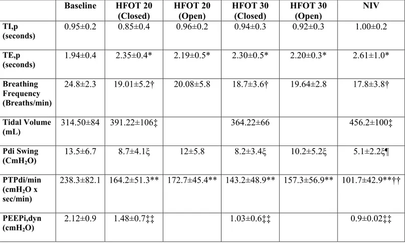
Table 2. Breathing Pattern, Inspiratory Efforts and Lung Mechanics in different setting
Legend
*p= 0.006 HFOT 20 closed vs baseline; p= 0.01 HFOT 20 open vs baseline; p= 0.007 HFOT 30 closed vs baseline; p= 0.02 HFOT 30 open vs baseline; p= 0.002 NIV vs baseline.
†p= 0.022 HFOT 20 closed vs baseline; p= 0.007 HFOT 30 closed vs baseline; p= 0.002 NIV vs baseline.
‡p= 0.015 HFOT 20 closed vs baseline; p= 0.007 NIV vs baseline.
ξp= 0.005 HFOT 20 closed vs baseline; p=0.005 HFOT 30 closed vs baseline; p=0.03 HFOT 30 open vs baseline; p=0.001 NIV vs baseline.
¶p<0.003 NIV vs HFOT 20 closed; p=0.003 NIV vs HFOT 20 open; p=0.007 NIV vs HFOT 30 closed; p=0.005 NIV vs HFOT 30 open.
**p=0.005 HFOT 20 closed vs baseline; p=0.002 HFOT 20 open vs baseline; p=0.004 HFOT 30 closed vs baseline; p=0.015 HFOT 30 open vs baseline; p=0.001 NIV vs baseline. Baseline HFOT 20 (Closed) HFOT 20 (Open) HFOT 30 (Closed) HFOT 30 (Open) NIV TI,p (seconds) 0.95±0.2 0.85±0.4 0.96±0.2 0.94±0.3 0.92±0.3 1.00±0.2 TE,p (seconds) 1.94±0.4 2.35±0.4* 2.19±0.5* 2.30±0.5* 2.20±0.3* 2.61±1.0* Breathing Frequency (Breaths/min) 24.8±2.3 19.01±5.2† 20.08±5.8 18.7±3.6† 19.64±2.8 17.8±3.8† Tidal Volume (mL) 314.50±84 391.22±106‡ 364.22±66 456.2±100‡ Pdi Swing (CmH2O) 13.5±6.7 8.7±4.1ξ 12±5.8 8.2±3.4ξ 10.2±5.2ξ 5.1±2.2ξ¶ PTPdi/min (cmH2O x sec/min) 238.3±82.1 164.2±51.3** 172.7±45.4** 143.2±48.9** 157.3±56.9** 101.7±42.9**†† PEEPi,dyn (cmH2O) 2.12±0.9 1.48±0.7‡‡ 1.03±0.6‡‡ 0.9±0.02‡‡
20
††p<0.004 NIV vs HFOT 20 closed; p=0.006 NIV vs HFOT 20 open; p=0.016 NIV vs HFOT 30 closed; p=0.02 NIV vs HFOT 30 open.
‡‡p=0.01 HFOT 20 closed vs baseline; p=0.003 HFOT 30 closed vs baseline; p=0.001 NIV vs baseline.
Data are presented as mean±SD. HFOT, high flow oxygen therapy; NIV, noninvasive ventilation; Pdi, trans-diaphragmatic pressure; PEEPi,dyn, intrinsic dynamic positive end expiratory pressure; PTPdi, pressure–time product of the trans-diaphragmatic pressure; TE,p, patient’s expiratory time; TI,p, patient’s inspiratory time.
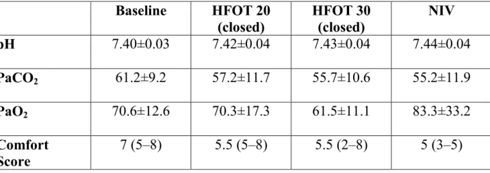
Table 3. Arterial blood gas values and comfort scores at different settings Baseline HFOT 20 (closed) HFOT 30 (closed) NIV pH 7.40±0.03 7.42±0.04 7.43±0.04 7.44±0.04 PaCO2 61.2±9.2 57.2±11.7 55.7±10.6 55.2±11.9 PaO2 70.6±12.6 70.3±17.3 61.5±11.1 83.3±33.2 Comfort Score 7 (5–8) 5.5 (5–8) 5.5 (2–8) 5 (3–5)
Data are presented as mean ± SD unless indicated otherwise. Comfort score was assessed with a scale where 0 is the worst comfort and 10 the best. The data are presented as the median (interquartile 25–75).
21
Results
As shown in table 2, compared with baseline, breathing frequency was significantly reduced in HFOT trials with the mouth closed and with NIV. Patient’s own expiratory time (TE,p) was significantly prolonged and VT higher compared with baseline for all the settings. The patient’s own inspiratory time (TI,p) was no different between trials. Pdi swing and diaphragm pressure time product (PTPdi) were reduced compared with baseline in all trials. However, the reductions observed during NIV were significantly larger, as opposed to all of the HFOT trials. Dynamic intrinsic positive end expiratory pressure (PEEPi, dyn) was significantly reduced compared with baseline in all trials. Breathing frequency, TI.p and TE.p, did not change between the different HFOT trials with the mouth closed or open, while Pdi at HFOT 20 L/min, was statistically higher with the mouth closed compared with open. As shown in table 2, the PaCO2 level decreased but not significantly with HFOT at 30 L/min and NIV compared with standard oxygen. Also shown in table 3, comfort did not vary among the different trials.
22
Discussion
The main findings in our study are threefold. First, compared with low flow oxygen, HFOT and NIV both reduce the respiratory muscle load, as well documented by Pdi swing and Diaphragm Pressure Time Product (PTPdi) reduction. Second, breathing frequency was significantly reduced during HFOT with mouth closed and with NIV, compared with baseline. Third, there was a trend toward PaCO2 reduction during
HFOT 30 L/min trial and NIV, juxtaposed to low flow oxygen, also if, in this last case, statistical significance wasn’t reached. Respiratory failure is still an important complication of chronic obstructive pulmonary disease (COPD) and hospitalization with an acute episode being a poor prognostic marker. Changes in lung mechanics are thought to be the major determinants of the physiological abnormalities that characterize hypercapnic respiratory failure. In practice, a subject would need to increase their ventilation very substantially to overcome the wasted ventilation in high ventilation/perfusion ratio units, but their inability to do so despite the respiratory stimulus that a rising PaCO2 tension provides has been the subject of
much debate. At last, as well described by Moxham [55], respiratory muscle pump plays a central role in the development of hypercapnia, being affected to some extent by the load that it has to overcome, i.e. the expiratory airflow limitation seen in severe COPD, but also by its own capacity to generate pressure, which is significantly reduced by the respiratory muscle shortening that accompanies pulmonary hyperinflation. Furthermore, chronic respiratory failure is associated with
23
adverse health outcomes in COPD: impaired exercise tolerance [9], pulmonary hypertension [10], skeletal muscle dysfunction [11], polycythemia, impaired health related quality of life, increased risk of hospitalization [12] and earlier death [13].Two trials that were conducted in the 1970s showed that long term treatment with supplemental oxygen reduced mortality among patients with COPD and severe resting hypoxemia [19,20]. More recently, the Long Term Oxygen Treatment Trial (LOTT), designed to test whether long term treatment with supplemental oxygen would result in a longer time to death or hospital admission than no use in stable COPD patients with resting or exercise induced desaturation, has failed to prove it. As reported by a recent Cochrane review [24], the efficacy of oxygen therapy for breathlessness and health-related QoL was assessed in COPD patients who did not meet the criteria for LTOT; the authors concluded that oxygen can relieve breathlessness when given during exercise to mildly hypoxemic and non-hypoxemic people with COPD who would not otherwise qualify for home oxygen therapy. However, actually there are more evidences in favor of the use of home noninvasive ventilation + oxygen compared to oxygen alone in stable hypercapnic COPD patients [27, 28]. It seems that home noninvasive ventilation unloads respiratory muscles and improves ventilatory response to hypercapnia, which could be expected to act as a relevant effect of treatment, allowing a more robust and adaptive response to the adverse physiological challenge of an acute PaCO2 increase during an exacerbation.
Furthermore, long term NIV in COPD patients may contribute to airway remodeling and improved ventilation / perfusion matching [56]. However, intolerance and major
24
side effects (incorrect use of interface, gas – humidification, ventilator settings) led to poor adherence to ventilatory therapy in stable COPD patients. HFOT could represent an innovative, efficient third option in this population. Physiologically, pressure loads and ventilation loads imposed to respiratory system are balanced with capabilities – that is, neural drive and muscles’ strength and endurance. HFOT acts on both the arms of this balance. The design of the nasopharynx facilitates humidification and warming of inspired gas by contact with the large surface area. By definition, this large wet surface area and nasopharyngeal gas volume can account for an appreciable resistance to gas flow. In addition, after analyzing nasal and oral flow-volume loops, Shepard and Burger showed that the nasopharynx has a distensibility that contributes to variable resistance [50]. When inspiratory gas is drawn across this large surface area, retraction of the nasopharyngeal boundaries results in a significant increase in inspiratory resistance compared to expiratory resistance. CPAP has been shown to reduce this supraglottic resistance up to 60% by mechanically splinting the airways [51]. HFOT most likely minimizes the inspiratory resistance associated with the nasopharynx by providing nasopharyngeal gas flows that match or exceed a patient’s peak inspiratory flow. This change in resistance translates to a decrease in resistive work of breathing, and so in reduction of pressure loads. Furthermore, HFOT indirectly influences respiratory muscles’ metabolism: we found a reduction of respiratory rate compared to low flow oxygen therapy; these results are analogous to those of other studies [35,40,52]. The reduction of respiratory rate reduces CO2
25
various remarkable changes in breathing efforts in COPD patients: a decrease in respiratory rate and an increase in tidal volume result in a reduced minute ventilation, without rising in PaCO2, suggesting an improvement of alveolar ventilation. The
most likely explanation for this response seems to be related to the increase in the expiratory resistance, with a mechanism different from that of CPAP.During CPAP, pressure is generated within the device and is dependent on the flow in the inspiratory line; the device supplies a constant and steady pressure at mouth and nares and resistance is provided by the expiratory valve. During HFOT, expiratory resistance are provided by patient’s effort against continuous incoming, at high velocity ,of fresh gases in nasal cavity, and by the leak between cannula and nares. The respiratory pattern elicited by HFOT resembles pursed lip breathing which is, however, associated with increased work of breathing and patients cannot maintain this pattern over a longer time period [25]. In contrast, during HFOT we could demonstrate for the first time that inspiratory effort was reduced. Several mechanisms have been advocated to explain the effect of HFOT on work of breathing, such as minimization of inspiratory resistance, attenuation of the activation of cold receptors or osmoreceptors in the nasal mucosa inducing bronchoconstriction, and reducing the anatomical dead space in the upper airways. Data from published clinical studies support the theory that HFT eliminates dead space because of the immediate impact on ventilation rates. A study by Dewan and Bell investigated exercise tolerance in COPD patients receiving respiratory support by transtracheal catheters (TTC), and compared low and high flows through nasal cannulae to low and high flow through
26
the TTCs [54]. TTCs are catheters placed in the patient’s trachea for the direct purpose of increasing respiratory efficiency by tracheal gas insufflation dead space washout. Dewan and Bell showed that exercise tolerance was greater for high flow than low flow regardless of method of administration (p < 0.01), but high flow via nasal cannula was just as effective for dead space washout as with TTC. These data confirm that dead space washout is a primary mechanism of action during HFOT. Furthermore, it’s well known that providing distending pressure to the lungs results in improved ventilatory mechanics by optimizing lung compliance and assists with gas exchange by maintaining patency of alveoli. Whereas HFOT is not necessarily intended to provide CPAP, if gas flow and nasal prong dimensions are set appropriately for patient size, distending pressure can be accomplished, quantifiable in a modest degree of positive pressure, unlikely to be above 5–6 cmH2O; this small
amount of positive pressure may also partially counteract the threshold load imposed by the presence of PEEPi. The reduction in transcutaneous CO2 in Fraser’s study and
in PaCO2, despite not being statistically significant, in our investigation, support the
hypothesis that it is possible to reduce hypercapnia using HFOT. Indeed, carbon dioxide directly controls the activity of inspiratory muscles alone and therefore its reduction may lead to a decrease in diaphragmatic effort. We cannot rule out the effect of a higher PaO2/FiO2 ratio as explanation for the PaCO2 increase during
baseline conditions. Moreover baseline conditions consisted of breathing oxygen through nasal cannula, and under these conditions, FiO2 cannot be controlled
27
patients breathe predominantly through the mouth or the nose. Therefore, the decrease in PaO2 during HFOT can be explained by a higher actual FiO2 under nasal
oxygen therapy compared with HFOT. We have further explored the physiological changes induced by mouth or nasal breathing, since it is totally unrealistic to assume that patients recruited for long-term treatment will always breathe with their mouth perfectly ‘sealed’. It has been shown that breathing with the mouth open negatively influences the generation of a positive pressure. Despite this, we were unable to demonstrate any ‘detrimental’ effect of this behavior on the breathing pattern and inspiratory effort compared with breathing with the mouth closed. Similar results have been recentely obtained by Vogelsinger et al. [42]. The Authors performed a study in order to compare the efficacy and safety of HFOT with those of conventional oxygen therapy (COT) in normo- and hypercapnic stable COPD patients. Despite the brief period of observation (COT and HFOT for 60 min each, separated by a 30 min washout phase), a significant decrease in PaCO2 and increased PaO2 was assessed; lower oxygen levels were effective in correcting hypoxemic respiratory failure and reducing hypercapnia, leading to a reduced amount of oxygen consumption. The results of this study show overall similar acute physiological changes between HFOT and NIV, and support the need for further investigations to assess the effectiveness of domiciliary HFOT versus NIV in patients with stable hypercapnia. Obviously our findings could not be translated to the situation of an acute exacerbation of COPD.In conclusion, HFOT is an appealing technique as a potential alternative to NIV in stable hypercapnic COPD because is less of a burden, and because HFOT provides a
28
more physiological humidification and heating of the airways, a more ‘easy to fit’ interface, a breathing pattern swinging in favor of alveolar ventilation and not of dead space volume, and a lung distending pressure that seems to impact on work of breathing of COPD patients.
29
Bibliography
1. Global strategy for the diagnosis, management and prevention of chronic obstructive pulmonary disease. 2018 Report Global Initiative for Chronic Obstructive Lung Disease (GOLD)
2. Kessler R, Partridge MR, Miravitlles M, Cazzola M, Vogelmeier C, Leynaud D, Ostinelli J. Symptom variability in patients with severe COPD: a pan-European cross-sectional study. Eur Respir J. 2011 Feb;37(2):264-72.
3. Rycroft CE, Heyes A, Lanza L, Becker K. Epidemiology of chronic obstructive pulmonary disease: a literature review. Int J Chron Obstruct Pulmon Dis. 2012;7:457-94.
4. Burrows B, Earle RH. Course and prognosis of chronic obstructive lung disease. A prospective study of 200 patients. N Engl J Med. 1969 Feb 20;280(8):397-404.
5. Roussos C, Koutsoukou A. Respiratory failure. Eur Respir J Suppl. 2003 Nov;47:3s-14s.
6. B R O’Driscoll, L S Howard, J Earis, V Mak, on behalf of the British Thoracic Society Emergency Oxygen Guideline Group BTS guideline for oxygen use in adults in healthcare and emergency settings Thorax 2017;72:i1–i90. 7. B Rochwerg, L Brochard, M. W. Elliott, D Hess, N. S. Hill, S Nava, P
Navalesi, M Antonelli, J Brozek, G Conti, M Ferrer, K Guntupalli, S Jaber, S Keenan, J Mancebo, S Mehta and Su Raoof Official ERS/ATS clinical practice guidelines: noninvasive ventilation for acute respiratory failure Eur Respir J 2017; 50: 1602426
8. Williams R, Rankin N, Smith T, Galler D, Seakins P. Relationship between the humidity and temperature of inspired gas and the function of the airway mucosa. Crit Care Med. 1996 Nov;24(11):1920-9. Review.
9. Antonucci R, Berton E, Huertas A, Laveneziana P, Palange P. Exercise physiology in COPD. Monaldi Arch Chest Dis. 2003 Apr-Jun;59(2):134-9.
30
10. Calcaianu G, Canuet M, Schuller A, Enache I, Chaouat A, Kessler R. Pulmonary Arterial Hypertension-Specific Drug Therapy in COPD Patients with Severe Pulmonary Hypertension and Mild-to-Moderate Airflow Limitation. Respiration. 2016;91(1):9-17.
11. Kim HC, Mofarrahi M, Hussain SN. Skeletal muscle dysfunction in patients with chronic obstructive pulmonary disease. Int J Chron Obstruct Pulmon Dis. 2008;3(4):637-58.
12. Garcia-Aymerich J, Monsó E, Marrades RM, Escarrabill J, Félez MA, Sunyer J, Antó JM; EFRAM Investigators. Risk factors for hospitalization for a chronic obstructive pulmonary disease exacerbation. EFRAM study. Am J Respir Crit Care Med. 2001 Sep 15;164(6):1002-7.
13. Anthonisen NR, Wright EC, Hodgkin JE. Prognosis in chronic obstructive pulmonary disease. Am Rev Respir Dis. 1986 Jan;133(1):14-20.
14. Sundh J, Ekström M. Risk factors for developing hypoxic respiratory failure in COPD. Int J Chron Obstruct Pulmon Dis. 2017 Jul 20;12:2095-2100.
15. Pierson DJ. Pathophysiology and clinical effects of chronic hypoxia. Respir Care. 2000 Jan;45(1):39-51;
16. Ergan B, Nava S. Long-Term Oxygen Therapy in COPD Patients Who Do Not Meet the Actual Recommendations. COPD. 2017 Jun;14(3):351-366. 17. Scharf SM, Iqbal M, Keller C, Criner G, Lee S, Fessler HE, National
Emphysema Treatment Trial (NETT) Group. Hemodynamic characterization of patients with severe emphysema. Am J Respir Crit Care Med 2002; 16683:314–322.
18. Rowan SC, Keane MP, Gaine S, McLoughlin P. Hypoxic pulmonary hypertension in chronic lung diseases: novel vasoconstrictor pathways. Lancet Respir Med 2016; 4(3):225–236.
31
19. Continuous or nocturnal oxygen therapy in hypoxemic chronic obstructive lung disease: a clinical trial. Nocturnal Oxygen Therapy Trial Group. Ann Intern Med.1980 Sep;93(3):391-8.
20. Long term domiciliary oxygen therapy in chronic hypoxic cor pulmonale complicating chronic bronchitis and emphysema. Report of the Medical Research Council Working Party.Lancet. 1981 Mar 28;1(8222):681-6.
21. Chaouat A, Weitzenblum E, Kessler R, Charpentier C, Enrhart M, Schott R, Levi-Valensi P, Zielinski J, Delaunois L, Cornudella R, Moutinho dos Santos J. A randomized trial of nocturnal oxygen therapy in chronic obstructive pulmonary disease patients. Eur Respir J. 1999 Nov;14(5):1002-8.
22. Górecka D, Gorzelak K, Sliwiński P, Tobiasz M, Zieliński J. Effect of long-term oxygen therapy on survival in patients with chronic obstructive pulmonary disease with moderate hypoxaemia. Thorax. 1997 Aug;52(8):674-9.
23. Long-Term Oxygen Treatment Trial Research Group, Albert RK, Au DH, Blackford AL, Casaburi R, Cooper JA Jr, Criner GJ, Diaz P, Fuhlbrigge AL, Gay SE, Kanner RE, MacIntyre N, Martinez FJ, Panos RJ, Piantadosi S, Sciurba F, Shade D, Stibolt T, Stoller JK, Wise R, Yusen RD, Tonascia J, Sternberg AL, Bailey W. A Randomized Trial of Long-Term Oxygen for COPD with Moderate Desaturation. N Engl J Med. 2016 Oct 27;375(17):1617-1627.
24. Ekström M, Ahmadi Z, Bornefalk-Hermansson A, Abernethy A, Currow D. Oxygen for breathlessness in patients with chronic obstructive pulmonary disease who do not qualify for home oxygen therapy. Cochrane Database Syst Rev. 2016 Nov 25;11:CD006429.
25. Windisch W, Kostić S, Dreher M, Virchow JC Jr, Sorichter S. Outcome of patients with stable COPD receiving controlled noninvasive positive pressure ventilation aimed at a maximal reduction of Pa(CO2). Chest. 2005 Aug;128(2):657-62.
26. Nickol AH, Hart N, Hopkinson NS, Hamnegård CH, Moxham J, Simonds A, Polkey MI. Mechanisms of improvement of respiratory failure in patients
32
with COPD treated with NIV. Int J Chron Obstruct Pulmon Dis. 2008;3(3):453-62.
27. Liao H, Pei W, Li H, Luo Y, Wang K, Li R, Xu L, Chen X. Efficacy of long-term noninvasive positive pressure ventilation in stable hypercapnic COPD patients with respiratory failure: a meta-analysis of randomized controlled trials. Int J Chron Obstruct Pulmon Dis. 2017 Oct 10;12:2977-2985.
28. Murphy PB, Rehal S, Arbane G, Bourke S, Calverley PMA, Crook AM, Dowson L, Duffy N, Gibson GJ, Hughes PD, Hurst JR, Lewis KE, Mukherjee R, Nickol A, Oscroft N, Patout M, Pepperell J, Smith I, Stradling JR, Wedzicha JA, Polkey MI, Elliott MW, Hart N. Effect of Home Noninvasive Ventilation With Oxygen Therapy vs Oxygen Therapy Alone on Hospital Readmission or Death After an Acute COPD Exacerbation: A Randomized Clinical Trial. JAMA. 2017 Jun 6;317(21):2177-2186.
29. Papazian L, Corley A, Hess D, Fraser JF, Frat JP, Guitton C, Jaber S, Maggiore SM, Nava S, Rello J, Ricard JD, Stephan F, Trisolini R, Azoulay E. Use of high-flow nasal cannula oxygenation in ICU adults: a narrative review. Intensive Care Med. 2016 Sep;42(9):1336-49.
30. Spoletini G, Alotaibi M, Blasi F, Hill NS. Heated Humidified High-Flow Nasal Oxygen in Adults: Mechanisms of Action and Clinical Implications. Chest. 2015 Jul;148(1):253-261.
31. Groves N, Tobin A. High flow nasal oxygen generates positive airway pressure in adult volunteers. Aust Crit Care. 2007 Nov;20(4):126-31.
32. Parke RL, McGuinness SP. Pressures delivered by nasal high flow oxygen during all phases of the respiratory cycle. Respir Care. 2013 Oct;58(10):1621-4.
33. Mündel T, Feng S, Tatkov S, Schneider H. Mechanisms of nasal high flow on ventilation during wakefulness and sleep. J Appl Physiol (1985). 2013 Apr;114(8):1058-65.
33
34. Parke RL, Eccleston ML, McGuinness SP. The effects of flow on airway pressure during nasal high-flow oxygen therapy. Respir Care. 2011 Aug;56(8):1151-5.
35. Greenspan JS, Wolfson MR, Shaffer TH. Airway responsiveness to low inspired gas temperature in preterm neonates. J Pediatr 1991;118(3):443e5. 36. Dysart K, Miller TL, Wolfson MR, Shaffer TH. Research in high flow
therapy: mechanisms of action. Respir Med. 2009 Oct;103(10):1400-5.
37. Spoletini G, Hill NS. High-flow nasal oxygen versus noninvasive ventilation for hypoxemic respiratory failure: Do we know enough? Ann Thorac Med. 2016 Jul-Sep;11(3):163-6.
38. Corley A, Caruana LR, Barnett AG, Tronstad O, Fraser JF. Oxygen delivery through high-flow nasal cannulae increase end-expiratory lung volume and reduce respiratory rate in post-cardiac surgical patients. Br J Anaesth. 2011 Dec;107(6):998-1004.
39. Fraser JF, Spooner AJ, Dunster KR, Anstey CM, Corley A. Nasal high flow oxygen therapy in patients with COPD reduces respiratory rate and tissue carbon dioxide while increasing tidal and end-expiratory lung volumes: a randomized crossover trial. Thorax. 2016 Aug;71(8):759-61.
40. Fricke K, Tatkov S, Domanski U, Franke KJ, Nilius G, Schneider H. Nasal high flow reduces hypercapnia by clearance of anatomical dead space in a COPD patient. Respir Med Case Rep. 2016 Aug 26;19:115-7.
41. Bräunlich J, Seyfarth HJ, Wirtz H. Nasal High-flow versus non-invasive ventilation in stable hypercapnic COPD: a preliminary report. Multidiscip Respir Med. 2015 Sep 3;10(1):27.
42. Vogelsinger H, Halank M, Braun S, Wilkens H, Geiser T, Ott S, Stucki A, Kaehler CM. Efficacy and safety of nasal high-flow oxygen in COPD patients. BMC Pulm Med. 2017 Nov 17;17(1):143.
43. Navalesi P, Costa R, Ceriana P, Carlucci A, Prinianakis G, Antonelli M, Conti G, Nava S. Non-invasive ventilation in chronic obstructive pulmonary
34
disease patients: helmet versus facial mask. Intensive Care Med. 2007 Jan;33(1):74-81.
44. Tobin MJ. Monitoring respiratory mechanics in spontaneously breathing patients. In: Tobin MJ, ed. Principles and Practice of Intensive Care Monitoring. Columbus, McGraw-Hill, 1998; pp. 621–628.
45. Baydur A, Behrakis PK, Zin WA, Jaeger M, Milic-Emili J. A simple method for assessing the validity of the esophageal balloon technique. Am Rev Respir Dis. 1982 Nov;126(5):788-91.
46. Sassoon CS, Light RW, Lodia R Pressure-time product during continuous positive airway pressure, pressure support ventilation, and T-piece during weaning from mechanical ventilation. Am Rev Respir Dis 1991; 143:469-75. 47. Appendini L, Purro A, Gudjonsdottir M et al. Partitioning of inspiratory muscle workload and pressure assistance in ventilator-dependent COPD patients. Am J Respir Crit Care Med 1996;154:1301-9
48. Navalesi P, Fanfulla F, Frigerio P et al. Physiologic evaluation of noninvasive mechanical ventilation delivered with three types of mask in patients with chronic hypercapnic respiratory failure. Crit Care Med 2000; 28:1785–1790.
49. Spahija J, de Marchie M, Grassino A. Effects of imposed pursed-lips breathing on respiratory mechanics and dyspnea at rest and during exercise in COPD. Chest 2005;128:640–50.
50. Shepard Jr JW, Burger CD. Nasal and oral flow-volume loops in normal subjects and patients with obstructive sleep apnea. Am Rev Respir Dis 1990;142(6 Pt 1):1288e93.
51. Miller MJ, DiFiore JM, Strohl KP, et al. Effects of nasal CPAP on supraglottic and total pulmonary resistance in preterm infants. J Appl Phys 1990;68(1):141e6.
52. Mauri T, Turrini C, Eronia N, Grasselli G, Volta CA, Bellani G, Pesenti A. Physiologic Effects of High-Flow Nasal Cannula in Acute Hypoxemic
35
Respiratory Failure. Am J Respir Crit Care Med. 2017 May 1;195(9):1207-1215.
53. Riera J , Pérez P , Cortés J , Roca O , Masclans JR , Rello J . Effect of high-fl ow nasal cannula and body position on end-expiratory lung volume: a cohort study using electrical impedance tomography. Respir Care . 2013 ; 58 ( 4 ): 589 - 596 .
54. Dewan NA, Bell CW. Effect of low flow and high flow oxygen delivery on exercise tolerance and sensation of dyspnea. A study comparing the transtracheal catheter and nasal prongs. Chest 1994;105(4):1061e5.
55. Moxham J. Respiratory failure: definitions and causes. In: Wetherall DA, Ledingham JM, Warrell D, eds. Oxford Textbook of Medicine. Oxford, Oxford University Press, 1996; pp. 2901–2906.
56. De Backer L, Vos W, Dieriks B, Daems D, Verhulst S, Vinchurkar S, Ides K, De Backer J, Germonpre P, De Backer W. The effects of long-term noninvasive ventilation in hypercapnic COPD patients: a randomized controlled pilot study. Int J Chron Obstruct Pulmon Dis. 2011;6:615-24.