© Copyright by ICR Publishers 2017
Calogera Pisano, Claudia Calia, Alessandro Ricasoli, Oreste Fabio Triolo, Vincenzo Argano
Unit of Cardiac Surgery, Department of Surgery and Oncology, University Hospital ‘P. Giaccone’, Palermo, ItalyBackground and aim of the study: Segmental analysis
of diseased mitral valves is important to predict a successful surgical valve repair. An assessment was made of the comparative accuracy of intraoperative three-dimensional (3D) and two-dimensional (2D) transesophageal echocardiography (TEE) in the evaluation of mitral valve lesions when compared with intraoperative surgical segmental analysis.
Methods: A total of 42 consecutive patients (12
females, 30 males; mean age 70.5 ± 14 years) with severe mitral valve regurgitation due to degenerative disease and who underwent mitral valve repair was enrolled in the study. Complete 2D- and 3D-TEE were performed before surgery. The findings obtained using the different echocardiographic techniques were compared with intraoperative segmental analysis performed by a single operator who was blinded to the 2D- and 3D-TEE findings until the end of the inspection. The sensitivity and specificity of
echocardiographic evaluations of involved scallops were compared with surgical inspection.
Results: 3D-TEE allowed an accurate identification
of all mitral lesions. Thirty-three patients had simple lesions at 3D-TEE and underwent a simple surgical procedure, while nine patients had complex lesions; in these latter cases complex surgical procedures were performed. 3D-TEE showed more sensitivity than 2D-TEE in the analysis of the anterior leaflet (A), in particular for A3 lesion (100% versus 25%, p <0.001) and for complex lesion (100% versus 33.3%, p <0.009).
Conclusion: 3D-TEE allowed a more accurate
identification of mitral valve lesions compared with 2D-TEE. The greatest accuracy was achieved for analysis of the anterior leaflet. 3D-TEE should be regarded as an important adjunct to standard 2D-TEE in decisions regarding mitral valve repair.
The Journal of Heart Valve Disease 2017;26:
Mitral valve repair has become preferential to replacement, and is now used in the overwhelming majority of patients with mitral valve prolapse (1). The procedure is associated with a low mortality, and is highly durable when performed by experienced surgeons. The reported operative mortality is estimated at 1.4% for valve repair compared to 3.8% for valve replacement (2). Recent guidelines have underlined the importance of early surgical intervention to preserve long-term left ventricular function in severe mitral regurgitation (3,4). In this regard, a preoperative assessment of mitral valve anatomy is essential to define the feasibility and complexity of a repair (5). Transesophageal
echocardiography (TEE) affords high-quality, real-time assessments of mitral valve structure and function, and is uniquely suited to intraoperative use (6,7). Consequently, TEE is considered an essential tool during mitral valve surgery. Three-dimensional (3D) echocardiography is a new emerging technique that allows a very precise localization of mitral valve lesions (8-11). The aim of the present study was to assess the comparative accuracies of intraoperative 3D and two-dimensional (2D) TEE for the evaluation of mitral valve lesions.
Clinical material and methods
Patient population
Forty-two consecutive patients (12 females, 30 males; mean age 70.5 ± 14 years) with severe mitral valve regurgitation due to degenerative disease (31
Address for correspondence:
Dr. Calogera Pisano, Unit of Cardiac Surgery, Department of Surgery and Oncology, Liborio Giuffrè Street n. 5, 90100 Palermo, Italy
cases of fibro-elastic deficiency, 11 cases of Barlow disease) were enrolled in the study. All patients were suitable for mitral valve repair (Table I).
Study protocol
Complete 2D- and 3D-TEE were each performed before surgery. The findings obtained when using the different echocardiographic techniques were separately recorded by two cardiologists and compared with intraoperative segmental analysis performed by a single operator who was blinded to the 2D- and 3D-TEE findings until the end of his inspection.
Informed consent to participate was obtained from each patient, and the study protocol conformed to the ethical guidelines of the 1975 Declaration of Helsinki as reflected in a priori approval by the institution’s human research committee.
2D- and 3D-TEE
2D-TEE was performed in all patients intraoperatively after the induction of anesthesia. A 5 MHz multi-plane probe (TEE X7-2t) and a Ie33 Philips Echocardiography Medical System was utilized. Real-time 3D-TEE was performed on completion of the 2D examination. Multiplane 2D and 3D evaluations included a complete standard protocol for evaluation of the mitral valve, allowing a complete description of all segments of the valve. Carpentier’s nomenclature (12,13) was applied to the mitral valve leaflets. Each scallop of the posterior leaflet was defined through the indentations (or cleft when present) which anatomically divided its lateral (P1), central (P2), and medial (P3) sections. The anterior leaflet scallops facing the posterior scallops were classified as A1, A2, and A3. The anterolateral (AL) and posteromedial (PM) commissures were also evaluated.
All segments were classified as normal, prolapsing (>2 mm beyond the annulus plane in 2D-TEE and a bright convexity of bulge in 3D), and flail. The presence of ruptured chordae was also annotated. The 3D acquisition and reconstruction times were measured in each patient. Color-3D analysis was used. Prolapse lesions were defined as either simple or complex. Simple anatomic lesions included isolated P2 prolapse or P2 associated with P1 or P3. All other lesions, including those involving the entire posterior leaflet, lesion or anterior leaflet, bi-leaflet prolapsed, and commissural lesions were defined as complex. This definition was based on published data, which suggested that simple lesions are generally treated surgically with simple techniques, while lesions defined as complex may require a more complex procedural approach (14-16).
Imaging analysis and intra- and inter-observer variability
All 3D-TEE images were analyzed by two experts in 3D echocardiography, who were blinded to the surgical findings. 3D-TEE images were analyzed in a random sequence. The quality of the 3D images, judged on the basis of the resolution of mitral valve
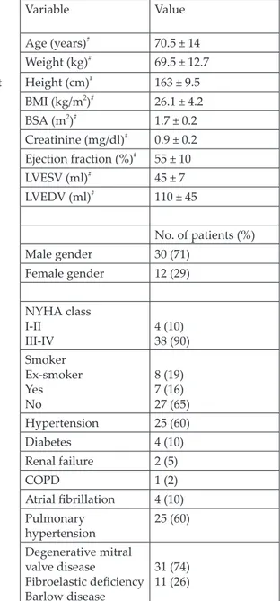
Table 1. Demographic and clinical characteristics of 42 patients
BMI:
Body Variable Value Age (years)# 70.5 ± 14 Weight (kg)# 69.5 ± 12.7 end-left Height (cm)# 163 ± 9.5 BMI (kg/m2)# 26.1 ± 4.2 BSA (m2)# 1.7 ± 0.2 Creatinine (mg/dl)# 0.9 ± 0.2 Ejection fraction (%)# 55 ± 10 LVESV (ml)# 45 ± 7 LVEDV (ml)# 110 ± 45 No. of patients (%) Male gender 30 (71) Female gender 12 (29) NYHA class I-II III-IV 4 (10)38 (90) Smoker Ex-smoker Yes No 8 (19) 7 (16) 27 (65) Hypertension 25 (60) Diabetes 4 (10) Renal failure 2 (5) COPD 1 (2) Atrial fibrillation 4 (10) Pulmonary hypertension 25 (60) Degenerative mitral valve disease Fibroelastic deficiency Barlow disease 31 (74) 11 (26)
#Values are mean ± SD.
BMI: Body mass index; BSA: Body surface area; COPD: Chronic obstructive pulmonary disease;
LVEDV: Left ventricular end-diastolic volume; LVESV: Left ventricular end-systolic volume.
anatomy and on the presence or absence of artifacts throughout the cardiac cycle, was rated as optimal, good, sufficient, or insufficient.
Surgical inspection and validation
The surgeon described the anatomy of the valve using the same Carpentier’s classification, and was aware of the 2D-transthoracic echocardiography finding, but not of the 2D-and 3D-TEE analyses.
Surgical techniques
The surgical techniques utilized to repair the mitral valve in the present series included the following: quadrangular resection of the posterior leaflet (50%); triangular resection of the anterior leaflet (7%); replacement of chordae tendineae with Gore-Tex neo-chordae (21%); transfer of neo-chordae tendineae (14%); and annuloplasty (14%) (Table II).
Statistical analysis
The sensitivity and specificity of echocardiographic evaluation of the involved scallops (or chordal rupture) was calculated, with surgical findings as a reference. The sensitivity and specificity of the two diagnostic tests were compared using an exact McNemar’s test because paired data were available. Predictive positive values were compared using a weighted generalized score statistic (17). A p-value <0.05 was considered to be statistically significant. R software (version 3.2.4) was used for the data analysis.
Results
Three-dimensional TEE was performed in all patients. The mean (± SD) time to obtain 3D-TEE images was 5 ± 3 min, and the quality of 3D-TEE was good in 67% of cases, optimal in 21%, and suboptimal in 12%. The major reasons for insufficient Figure 1: Posterior leaflet analysis. Example of a patient with a P2 prolapse in whom 2D-TEE (A) allowed a good
quality were suboptimal echocardiograph windows, minimal patient movements, respiration, and cardiac arrhythmia. However, anatomic visualization of the mitral valve during the end-systolic frame allowed diagnostic information also in cases with suboptimal quality, and therefore all patients were included in the study. The sensitivity of 3D-TEE was higher in the
case of optimal images than in suboptimal images and arrhythmic patients.
On surgical inspection, 12 patients had isolated P2 lesions (28%), six had isolated A2 lesions (14%), three had isolated A3 lesions (7%), while the others had more complex pathologies. Chordal rupture was present in 24 cases (57%).
Figure 2: Anterior leaflet analysis. Example of a patient in whom 2D-TEE (A) showed A2 prolapse and chordal rupture, whereas 3D-TEE (B) showed A1 prolapse and chordal rupture in A1 and A2, confirmed at surgical inspection (C).
Table II: Surgical technique for mitral valve repair.
Surgical technique Percentage
Quadrangular resection of posterior leaflet 50% Triangular resection of the anterior leaflet 7% Replacement of chordae tendineae with Gore-Tex neo-chordae 21%
Transfer of chordae tendineae 14%
Figure 3: Comparison between 2D-TEE, 3D-TEE and surgical inspection in analysis of the posterior and anterior leaflets. Table III: Detection of the pathology with transesophageal (TEE) two-dimensional (2D)
and three-dimensional (3D) echocardiography.
3D-TEE
sensitivity 2D-TEE sensitivity p-value 3D-TEE specificity 2D-TEE specificity p-value 3D-TEE PPV 2D-TEE PPV p-value Posterior leaflet P1 100 100 1 92,3 92,3 1 50 50 1 P2 100 100 1 71.4 71.4 1 77.8 77.8 1 P3 100 66.7 0.206 90.9 90.9 1 75 66.7 0.146 Posterior scallops >1 100 50 0.182 83.3 83.3 1 50 33.3 1 Anterior leaflet A1 100 50 0.182 91.7 83.3 0.347 66.7 33.3 1 A2 100 85.7 0.232 71.4 71.4 1 77.8 75 0.006 A3 100 25 <0.001 90 90 1 80 50 0.035 Anterior scallops >1 100 33.3 0.009 81.8 81.8 1 60 33.3 0.607 Chordal rupture 100 87.5 0.234 100 100 1 100 100 1
Sensitivity, specificity and positive predictive values of the 3D- and 2D-TEE methods for each of the segmental lesions and for chordal rupture are listed in Table III. For analysis of the posterior leaflet, 2D- and 3D-TEE had similar sensitivities and specificities (Fig. 1). However, 3D-TEE showed a slightly higher sensitivity than 2D-TEE in P3 analysis (100% versus 66.7%, p = 0.2). For analysis of the anterior leaflet, 3D-TEE showed a significantly higher sensitivity compared with 2D-TEE (Fig. 2), in particular for the lesions localized in A3 (sensitivity 100% versus 25%, p = 0.001) and in complex lesions (sensitivity 100% versus 33.3%, p = 0.009). In addition, the positive predictive value of 3D-TEE was significantly higher than for 2D-TEE in the analysis of A2 (77.8% versus 25%, p = 0.06) and A3 lesions (80% versus 50%, p = 0.035). Finally, according to the present results, 3D-TEE had a slightly higher sensitivity than 2D-TEE in the chordal rupture analysis (100% versus 87.5%, p = 0.23).
Quadrangular resection, either as an isolated procedure or in combination with other techniques, was the most common intervention. Twenty-one patients (50%) had simple lesions. In these cases, simple techniques were utilized in 98% of patients, while only in two cases a complex procedure was performed. No patients in this group underwent mitral valve replacement. The other 21 patients (50%) had 3D-TEE lesions classified as complex; in these latter cases surgeons performed a complex procedure in 14 patients (66%), a simple procedure in five patients (22%), and in two patients (12%) mitral valve replacement was necessary after the first repair attempt.
Early and late outcomes and echocardiographic evaluation of the study population are listed in Tables IV and Table V. The mean duration of follow up was 18 ± 6 months.
Discussion
Degenerative mitral valve disease encompasses a range of pathology, including annular dilatation, chordal stretching or rupture, leaflet thickening and redundancy, and calcification of the leaflets and chordae (18). Two forms of degenerative disease are recognized, namely fibroelastic deficiency (FED) and Barlow disease (19).
Although FED and Barlow disease are separate clinical entities, they represent two ends of a disease spectrum. The successful repair of degenerative mitral valve disease is possible in the majority of patients, particularly when disease is limited to the posterior leaflet. Anterior and bileaflet repair are more challenging and are associated with a lower rate of success and a higher need for reoperation (20). However, success for all types of repair is increasing with, in recent series, success rates approaching 100%
being reported for isolated posterior leaflet repairs (21,22). For complex anterior and bi-leaflet repair, success rates of more than 90% have been reported at high-volume centers (23,24).
In this regard, a non-invasive preoperative assessment of mitral valve anatomy is essential to define the feasibility and complexity of repair. TEE affords high-quality, real-time assessment of mitral valve structure
Table IV: Early postoperative outcomes after mitral valve repair.
Early postoperative outcome Value Days in intensive care unit# 2.7 ± 1.8
Days in hospital# 7.5 ± 1.8 Creatinine (mg/dl)# 0.8 ± 0.2 Arrhythmias (n) 8 (19) Pulmonary failure (n) 0 (0.000) Renal failure (n) 0 (0.000) Cerebrovascular complications (n) 1 (2.3) Cardiac failure (n) 0 (0.000) Thromboembolism (n) 0 (0.000) Infections (n) 0 (0.000) Echocardiographic variable at discharge
Ejection fraction (%)# 55.5 ± 7.4
Left atrial diameter (mm)# 29.5 ± 7
LVEDD (mm)# 48.7 ± 6.1 LVEDV (ml)# 88.7 ± 24.7 LVESD (mm)# 35.5 ± 6.6 LVESV (ml)# 39 ± 4.9 SPAP (mmHg)# 25 ± 7.6 Mitral regurgitation (n) None Faint Moderate Severe 36 (86) 4 (10) 2 (4) 0 (0) Tricuspid regurgitation (n) None Faint Moderate Severe 25 (60) 15 (36) 2 (4) 0 (0) Pulmonary hypertension (mmHg)# 8 ± 19 #Values are mean ± SD.
Values in parentheses are percentages.
LVEDD: Left ventricular end-diastolic diameter; LVEDV: Left ventricular end-diastolic volume; LVESD: Left ventricular end-systolic diameter; LVESV: Left ventricular end-systolic volume; SPAP: Systolic pulmonary artery pressure.
and function, and is uniquely suited to intraoperative use. Consequently, TEE is an essential tool during mitral valve repair surgery (25). In fact, in order to obtain appropriate information to guide surgical decision making, perioperative echocardiographers must understand the etiology and mechanisms of mitral regurgitation, have an appreciation of surgical techniques and, most importantly, to be able to perform a comprehensive assessment of mitral valve structure and function in the operating room environment (26).
Interesting data showed that 3D echocardiography is superior to the corresponding 2D techniques when describing mitral valve pathology (8-11). Indeed, the possibility of visualizing mitral valve leaflets, commissures, annulus dilatation and subvalvular structures from different and unique planes, both from the atrium or ventricle with access to ‘en-face’ views, facilitates the understanding of this complex apparatus. The involvement of commissures and subvalvular apparatus (tips of leaflets, chordae, papillary muscles) can be accurately assessed (27). In the qualitative analysis of mitral valve disease - in particular for mitral valve prolapse - 3D echocardiography seems to have a greater accuracy than 2D echocardiography to investigate the details of mitral valve leaflet anatomy and motion, while full-motion acquisition is required when the entire valvular apparatus is to be assessed.
In terms of accuracy, 3D echocardiography has recently been compared to surgical inspection. In a consecutive series of 200 cases (10), the overall accuracy of the method was high (sensitivity 92.5%, specificity 96%, accuracy 95%). The sensitivity of 3D echocardiography was slightly lower for the anterolateral commissure, P1 and A1, while specificity was very high in all segments. The present study also investigated differences in the accuracy of 3D echocardiography not only in simple but also in complex cases, which involved the majority of the study population. The accuracy was very high in both simple and complex lesions (98% and 93%, respectively), even if slightly lower in complex lesions.
In the present analysis, 3D-TEE showed a significantly greater sensitivity than 2D-TEE in the analysis of lesions localized in A3 (sensitivity 100% versus 25%, p <0.001) and in complex lesions (sensitivity 100% versus 33.3%, p <0.009) (Fig. 3). In addition, the positive predictive value of 3D-TEE was significantly higher than for 2D-TEE in the analysis of A2 (77.8% versus 25%, p = 0.06) and A3 lesions (80% versus 50%, p = 0.035). Finally, the study results showed that the complexity of mitral valve lesions as assessed with 3D-TEE correlates to surgical assessment and to the surgical procedure performed. Twenty-one patients (50%) had simple lesions; in these cases simple techniques were utilized in 98% of patients, while only in two cases a complex procedure was Table V: Late postoperative outcomes and
echocardiographic variables.
Variable Value
Late postoperative outcomes
Pulmonary failure (n) 3 (7) Renal failure (n) 0 (0) Cerebrovascular complications (n) 2 (5) Cardiac failure (n) 1 (2) Thromboembolism (n) 0 (0) Infections (n) 0 (0) Mortality (n) 0 (0) Echocardiographic variable Cardiac rate (bpm)# 74.8 ± 12.8 Systolic pressure (mmHg)# 130 ± 16.9 Diastolic pressure (mmHg)# 69 ± 9.5 Mean pressure (mmHg)# 82 ± 9.5 Ejection fraction (%)# 56.1 ± 9.1
Left atrial diameter (mm)# 28.0 ± 11.1
LVEDD (mm)# 51.3 ± 6.3
LVEDV (ml)# 92.3 ± 21.9
LVESD (mm)# 40.6 ± 7.1
LVESV (ml)# 42.8 ± 16.0
TAPSE (m)# 18.2 ± 2.3
Tricuspid annulus diameter (mm)# 29.5 ± 7.4
SPAP (mmHg)# 25 ± 6.1
Interventricular septum thickness (mm)# 9.6 ± 1.7
Posterior ventricular wall thickness (mm)# 10 ± 1.8
Diastolic dysfunction (n) None I grade II grade III grade 30 (72) 12 (28) 0 (0) 0 (0) Mitral regurgitation (n) None I grade II grade III grade 32 (76) 8 (19) 2 (5) 0 (0) Tricuspid regurgitation (n) None Faint Moderate Severe 25 (60) 15 (36) 2 (4) 0 Pulmonary hypertension (mmHg)# 8 ± 19 #Values are mean ± SD.
Values in parentheses are percentages.
TAPSE: Tricuspid annular plane systolic excursion. Other abbreviations as Table IV.
performed. The other 21 patients (50%) had 3D-TEE lesions classified as complex. In these cases, surgeons performed complex procedures in 14 patients (66%), simple procedures in five patients (22%), and in two patients (12%) mitral valve replacement was necessary after the first repair attempt.
Additional data (28) have shown that 3D echocardiography appears to have an important role in the identification of posterior leaflet deep cleft-like indentations (CLIs) in degenerative mitral valve disease. One typical aspect of myxomatous mitral valve disease are the deep ‘clefts’ of the posterior mitral leaflet (29), which do not really cleave the leaflet but rather occupy the position of normal indentations between its three scallops. Normal indentations are usually not deep, but abnormal indentations - although different from true clefts - form when there are deep CLIs that can be associated with mitral regurgitation and failure of mitral repair. 3D-TEE provides mechanistic insights that CLIs are associated mostly with single scallop prolapse and insufficient tissue to cover the scallop separation, probably combining conformational and acquired valvular patterns.
These data further emphasize the complex combination of various morphological elements of myxomatous mitral valve disease, and warrants detailed physiological future studies of annular and valvular dynamics with quantitative mitral regurgitation dynamic assessment to characterize the complete phenotypical manifestation of myxomatous mitral valve disease.
Finally, real-time 3D echocardiography has been used to assess the annular saddle shape in the pathogenesis of mitral regurgitation. 3D reconstructions of the mitral valve annulus, in fact, have permitted a characterization of the link between mitral morphology and mitral regurgitation severity in patients with degenerative mitral valve disease. Using real-time 3D echocardiography, Lee et al. (30) showed, for the first time in humans, that annular flattening (as represented by a reduced ratio of annular height to commissural width) is strongly associated with progressive leaflet billowing, higher frequencies of chordal rupture, and greater regurgitant orifices. The lower limit of the ratio of annular height to commissural width in a healthy population appears to be 15%, and a ratio <15% is strongly associated with moderate or severe mitral regurgitation among patients with mitral valve prolapse.
Based on the present results, 3D echocardiography is very important in both the qualitative and quantitative analysis of degenerative mitral valve disease, and the achievement of good surgical exposure is paramount for an excellent mitral valve repair. A perfect 3D-TEE image of the valve will not compensate for poor
exposure. In addition, it is important to remember that the entire subvalvular apparatus can be examined by the surgeon, including the subtle pathology of the chords and leaflet that may not be apparent on 3D-TEE. With this incomplete information, a less-experienced surgeon may settle for a repair that addresses the major part of the pathology but may leave behind an untreated pathology. This may lead to an unacceptably high rate of residual or recurrent mitral regurgitation, or the unexpected pathology may lead to valve replacement. For example, the use of artificial chords (neochords) without resection will also change the way in which the valve closes, perhaps making other valve lesions more prominent, which may in turn lead to residual mitral regurgitation.
Study limitations
The main limitation of quantitative mitral valve imaging is that the procedure involved in segmentation of the valve is currently too time-consuming to be incorporated into routine clinical use. Moreover, manual input to define the anatomic landmarks of the mitral valve tends to introduce bias, measurement errors, and variability. Automated morphological quantification using intelligent algorithms for anatomic recognition will likely improve the efficiency and reproducibility of the mitral valve modeling process to a degree that is optimal for day-to-day diagnostic use. Advances in hardware and software leading to improvements in the 3D-TEE image quality will also enhance the robustness of the segmentation process.
A second limitation was that the sample size was too small. In fact, a sample size of 42 patients achieved 77% power to detect an odds ratio of 2,75 using a two-sided McNemar test with a significance level of 0.05. The odds ratio is equivalent to a difference between two paired proportions of 0.35, and the proportion of discordant pairs is 0.750. To achieve an 80% power, and to identify a difference in sensibility of 0.35, it would have been necessary to include 51 patients in the study. Hence, a process has been undertaken to enroll more patients and it is hoped that these data can be updated in about one year, with a larger sample size.
In conclusion, 3D-TEE has allowed a more accurate identification of mitral valve lesions compared to 2D-TEE, with the greatest accuracy being achieved for analysis of the anterior leaflet. 3D-TEE should be regarded as an important adjunct to standard 2D-TEE when making decisions regarding mitral valve repair.
Acknowledgements
The authors gratefully acknowledge Dr. Veronica Capuccio, who contributed to the statistical analysis of the data.
References
1. Nishimura RA, Otto CM, Bonow RO, et al. 2014 AHA/ACC guideline for the management of patients with valvular heart disease: A report of the American College of Cardiology/American Heart Association task force on practice guidelines. J Am Coll Cardiol 2014;63:2438-2488
2. Gammie JS, Sheng S, Griffith BP, et al. Trends in mitral valve surgery in the United States: Results from the Society of Thoracic Surgeons Adult Cardiac Surgery Database. Ann Thorac Surg 2009;87:1431-1437
3. Gaasch WH, Meyer TE. Left ventricular response to mitral regurgitation: Implications for management. Circulation 2008;118:2298-2303
4. Zoghbi WA, Adams D, Bonow RO, et al. Recommendations for noninvasive evaluation of native valvular regurgitation: A report from the American Society of Echocardiography developed in collaboration with the Society for Cardiovascular Magnetic Resonance. J Am Soc Echocardiogr 2017;30:303-371
5. Silbiger JJ. Anatomy, mechanics and pathophysiology of mitral annulus. Am Heart J 2012;164:163-176
6. Sugeng L, Shernan SK, Weinert L, et al. Real-time three-dimensional transesophageal echocardiography in valve disease: Comparison with surgical findings and evaluation of prosthetic valves. J Am Soc Echocardiogr 2008,21:1347-1354 7. Grewal J, Mankad S, Freeman WK, et al.
Real-time three-dimensional transesophageal echocardiography in the intraoperative assessment of mitral valve disease. J Am Soc Echocardiogr 2009;22:34-41
8. Chikwe J, Adams DH, Su KN, et al. Can three-dimensional echocardiography accurately predict complexity of mitral valve repair? Eur J Cardiothorac Surg 2012;41:518-524
9. de Groot-de Laat LE, Ren B, McGhie J, et al. The role of experience in echocardiographic identification of location and extent of mitral valve prolapse with 2D and 3D echocardiography. Int J Cardiovasc Imaging 2016;32:1171-1177
10. Tamborini G, Muratori M, Maltagliati A, et al. Pre-operative transthoracic real-time three-dimensional echocardiography in patients undergoing mitral valve repair: Accuracy in cases with simple vs. complex prolapse lesions. Eur J Echocardiogr 2010;11:778-785
11. Hien MD, Rauch H, Lichtenberg A, et al. Real-time three-dimensional transesophageal echocardiography: Improvements in intraoperative mitral valve imaging. Anesth Analg 2013; 116:287-295
12. Carpentier A, Adams DH, Filsoufi F. Carpentier’s Reconstructive Valve Surgery. Elsevier Health Sciences, 2011
13. McCarthy KP, Ring L, Rana BS. Anatomy of the mitral valve: Understanding the mitral valve complex in mitral regurgitation. Eur J Echocardiogr 2010;11:i3-i9
14. Schäfers H-J, Langer F, Kunihara T. Chordal replacement in mitral repair. Multimed Man Cardiothorac Surg/Eur Assoc Cardio-Thoracic Surg 2010 (402):mmcts.2009.003962. doi: 10.1510/ mmcts.2009.003962
15. David TE, Ivanov J, Armstrong S, et al. A comparison of outcomes of mitral valve repair for degenerative disease with posterior, anterior, and bileaflet prolapse. J Thorac Cardiovasc Surg 2005;130:1242-1249
16. Perier P, Hohenberger W, Lakew F, et al. Toward a new paradigm for the reconstruction of posterior leaflet prolapse: Midterm results of the ‘respect rather than resect’ approach. Ann Thorac Surg 2008;86:718-725
17. Kosinski AS. A weighted generalized score statistic for comparison of predictive values of diagnostic tests. Stat Med 2013;32:964-977
18. Han RI, Black A, Culshaw G, et al. Structural and cellular changes in canine myxomatous mitral valve disease: An image analysis study. J Heart Valve Dis 2010;19:60-70
19. Adams DH, Anyanwu AC. Seeking a higher standard for degenerative mitral valve repair: Begin with etiology. J Thorac Cardiovasc Surg 2008;136:551-556
20. Seeburger J, Borger MA, Doll N, et al. Comparison of outcomes of minimally invasive mitral valve surgery for posterior, anterior and bileaflet prolapse. Eur J Cardiothorac Surg 2009;36:532-538
21. Johnston DR, Gillinov AM, Blackstone EH, et al. Surgical repair of posterior mitral valve prolapse: Implications for guidelines and percutaneous repair. Ann Thorac Surg 2010;89:1385-1394
22. Mihaljevic T, Jarrett CM, Gillinov AM, et al. Robotic repair of posterior mitral valve prolapse versus conventional approaches: Potential realized. J Thorac Cardiovasc Surg 2011;141:72-80
23. Seeburger J, Borger MA, Doll N, et al. Comparison of outcomes of minimally invasive mitral valve surgery for posterior, anterior and bileaflet prolapse. Eur J Cardiothorac Surg 2009; 36:532-538
24. Castillo JG, Anyanwu AC, Fuster V, et al. A near 100% repair rate for mitral valve prolapse is achievable in a reference center: Implications for future guidelines. J Thorac Cardiovasc Surg 2012;144:308-312
25. American Society of Anesthesiologists and Society of Cardiovascular Anesthesiologists Task Force on Transesophageal Echocardiography. Practice guidelines for perioperative transesophageal echocardiography. Anesthesiology 2010;112: 1084-1096
26. Wei J, Hsiung MC, Tsai SK, et al. The routine use of live three-dimensional transesophageal echocardiography in mitral valve surgery: Clinical experience. Eur J Echocardiogr 2010;11:14-18 27. Laug RM, Tsang W, Weinerr L, et al. Valvular
heart disease: The value of 3-dimensional echocardiography. J Am Coll Cardiol 2011;58: 1933-1934
28. Mantovani F, Clavel MA, Vatury O, et al. Cleft-like indentations in myxomatous mitral valves by three-dimensional echocardiographic imaging. Heart 2015;101:1111-1117
29. Ring L, Rana BS, Ho SY, Wells FC. The prevalence and impact of deep clefts in the mitral leaflets in mitral valve prolapse. Eur Heart J Cardiovasc Imaging 2013;14:595-602
30. Lee AP, Hsiung MC, Salgo IS, et al. Quantitative analysis of mitral valve morphology in mitral valve prolapse with real-time 3-dimensional echocardiography: Importance of annular saddle shape in the pathogenesis of mitral regurgitation. Circulation 2013;127:832-841