Alma Mater Studiorum – University of Bologna
Submitted in accordance with the requirement for the degree of
DOCTOR OF PHILOSOPHY IN BIOENGINEERING
XXVI CYCLE
IN VIVO EVALUATION OF THE TRANSLATIONS OF THE
GLENO-HUMERAL JOINT USING MAGNETIC RESONANCE
IMAGING
Manuela Calderone
Supervisor
Prof. Ugo Della Croce
Co-supervisor
Dr. Andrea Cereatti
Examiners
Prof. Luca Cristofolini Dr. Ara Nazaria
TABLE OF CONTENTS
SUMMARY……….……….v
SOMMARIO….………...….…...vii
STRUCTURE OF THE THESIS……….…….ix
GLOSSARY OF TERMS………...……….x
CHAPTER 1... Aims of the thesis..………...1
CHAPTER 2... The shoulder joint complex and the gleno-humeral joint 2.1 Anatomy of the shoulder joint complex………..3
2.2 Gleno-humeral joint……….5
2.2.1 Normal gleno-humeral relationship.………..…...9
2.2.2 Gleno-humeral joint stability………..10
2.2.3 Gleno-humeral joint laxity and instability………..13
CHAPTER 3... Quantitative measurement of the gleno-humeral joint translations 3.1 Background………....18
3.1.1 Instrumental techniques for the assessment of the gleno-humeral joint translations………. ………....18
3.2 Magnetic Resonance Imaging………23
3.2.1 Basic principles...………...23
3.2.2 Image formation, contrast and weighting………...28
3.2.3 Magnetic Resonance Imaging acquisition parameters………...………32
CHAPTER 4... Scapular and humeral anatomical coordinate systems 4.1 Literature review………35
4.2 Proposal for a novel humerus and scapula anatomical coordinate system definition……… 48
4.2.1 Introduction………48
4.2.2 Materials and methods....….………..49
4.2.3 Results………54
4.3 An alternative method for the anatomical coordinate system definition on incomplete 3D bone
model: an application for the humerus………..58
4.3.1 Introduction………58
4.3.2 Materials and methods..………..59
4.3.3 Results………61
4.3.4 Conclusion………..61
CHAPTER 5... Magnetic Resonance Imaging based methodology for the estimation of gleno-humeral joint translations 5.1 Introduction……… 63
5.2 Materials and methods………..64
5.2.1 Subjects selection………...64 5.2.2 Experimental set-up………....64 5.2.3 Experimental protocol………65 5.2.4 Repeatability assessment………67 5.2.5 Statistical analysis………..68 5.3 Results………...68 5.4 Discussion………..70 CONCLUSIONS………....73 ACKNOWLEDGMENTS……….75 REFERENCES………..76
v
SUMMARY
Gleno-humeral joint (GHJ) is the most mobile joint of the human body. This is related to the incongruence between the large humeral head articulating with the much smaller glenoid (ratio 3:1). The GHJ laxity is the ability of the humeral head to be passively translated on the glenoid fossa and, when physiological, it guarantees the normal range of motion of the joint. Three-dimensional GHJ linear displacements have been measured, both in vivo and in vitro by means of different instrumental
techniques. In vivo gleno-humeral displacements have been assessed by means of
stereophotogrammetry, electromagnetic tracking sensors, and bio-imaging techniques. Both stereophotogrammetric systems and electromagnetic tracking devices, due to the deformation of the soft tissues surrounding the bones, are not capable to accurately assess small displacements, such as gleno-humeral joint translations. The bio-imaging techniques can ensure for an accurate joint kinematic (linear and angular displacement) description, but, due to the radiation exposure, most of these techniques, such as computer tomography or fluoroscopy, are invasive for patients. Among the bio-imaging techniques, an alternative which could provide an acceptable level of accuracy and that is innocuous for patients is represented by magnetic resonance imaging (MRI). Unfortunately, only few studies have been conducted for three-dimensional analysis and very limited data is available in situations where preset loads are being applied.
The general aim of this doctoral thesis is to develop a non-invasive methodology based on open-MRI for in-vivo evaluation of the gleno-humeral translation components in healthy subjects under the application of external loads. To achieve this goal it was necessary to take action on two critical points related to the use of MR scanner: (1) the definition of scapula and humerus anatomical coordinate systems which are suitable to be used with 3D incomplete bone models obtained from MRI images; (2) the development of a device for applying an external force during a MRI exam and which was compatible, in terms of material, with the MR scanner.
For the research study thirteen asymptomatic shoulders were acquired using a horizontal open magnetic resonance scanner. Recordings were made with the subjects in the supine position both at 15 deg and 90 deg of arm abduction with and without an anterior force of 20 N applied to the humerus. The results showed that when no load was applied, from 15 deg to 90 deg of arm abduction, the translation of the humeral head center with respect the glenoid fossa were greater in the anterior and superior direction
vi
than in the medio-lateral direction. Under the application of the anterior force no statistically significant differences were found in the GHJ laxity between 15 deg and 90 deg of arm abduction. The translations observed in vivo in this study were significantly smaller than those observed in previous cadaver studies under the application of an anterior load of 20 N. This discrepancy can be ascribed to the total or partial lack of the shoulder muscles and differences in muscular tone. The results also showed a level of precision associated to the GHJ translation estimates of one order of magnitude smaller than the relevant translations.
vii
SOMMARIO
L’articolazione gleno-omerale rappresenta l’articolazione più mobile del corpo umano. Le ragioni di ciò sono da ricondursi alla parziale congruenza tra la testa omerale che si articola con la più piccola cavità glenoidea (rapporto 3:1). La lassità dell’articolazione gleno-omerale rappresenta l’attitudine della testa omerale a essere traslata passivamente rispetto alla cavità glenoidea; essa garantisce, quando fisiologica, il normale range di movimento dell’articolazione. Gli spostamenti lineari tridimensionali (lassità) sono stati misurati, sia in vivo sia in vitro per mezzo di diverse tecniche strumentali. In vivo gli spostamenti dell’articolazione gleno-omerale sono stati valutati con sistemi stereofotogrammetrici, sensori di tracciamento elettromagnetici, e tecniche di bio-imaging.
Sia i sistemi stereofotogrammetrici sia i dispositivi di tracciamento elettromagnetici, a causa della deformazione dei tessuti molli che circondano le ossa, non sono adatti a stimare accuratamente piccoli spostamenti, come possono essere le traslazioni dell’articolazione gleno-omerale. Le tecniche di
bio-imaging possono garantire un’accurata descrizione della cinematica articolare (spostamenti lineari e
angolari), ma a causa dell’esposizione alle radiazioni molte di queste tecniche, come la tomografia assiale computerizzata e la fluoroscopia, sono invasive per i pazienti. Tra le tecniche di bio-imaging, un’alternativa che può garantire un accettabile livello di accuratezza e che risulta innocua per i pazienti è rappresentata dall’imaging di risonanza magnetica (RM). Sfortunatamente, solo pochi studi sono stati condotti sull’analisi tridimensionale e pochi dati sono disponibili in situazioni in cui l’articolazione è soggetta all’azione di carichi esterni noti.
L’obiettivo generale di questa tesi di dottorato è di sviluppare una metodologia non invasiva basata sulla RM aperta per la valutazione in vivo delle componenti traslazionali dell’articolazione gleno-omerale in soggetti sani e con l’applicazione di carichi esterni. Per raggiungere quest’obiettivo è stato necessario intervenire su due punti critici legati all’uso della RM: (1) la definizione dei sistemi di riferimento anatomici di scapola e omero, compatibili con l’uso di modelli ossei 3D incompleti ottenuti da immagini di RM; (2) lo sviluppo di un dispositivo per l’applicazione di carichi esterni durante gli esami di RM e che fosse compatibile, in termini di materiali, con lo scanner di RM.
Per lo studio sono state acquisite tredici spalle asintomatiche per mezzo uno scanner di RM aperta orizzontale. Le acquisizioni sono state fatte con il soggetto in posizione supina con il braccio abdotto a 15 e a 90 gradi, in presenza e in assenza di un carico esterno di intensità pari a 20 N applicato all’omero
viii
e diretto anteriormente. I risultati hanno mostrato che in assenza di carco, da 15 a 90 gradi di abduzione dell’omero, le traslazioni del centro della testa dell’omero rispetto alla cavità glenoidea erano maggiori in direzione anteriore e superiore rispetto alla direzione medio laterale. Non sono state trovate differenze significative nella lassità dell’articolazione gleno-omerale nelle due posizioni del braccio analizzate (15 e 90 gradi di abduzione) a seguito dell’applicazione del carico. Le traslazioni osservate in vivo in questo studio sono significativamente più piccole rispetto a quelle osservate in studi precedenti svolti su cadavere e con l’applicazione di un carico esterno d’intensità pari a 20 N diretto anteriormente. Questa discrepanza può essere attribuita alla totale o parziale mancanza dei muscoli della spalla e alle differenze nel tono muscolare. I risultati hanno mostrato inoltre un livello di precisione associata alla stima delle traslazioni dell’articolazione gleno-omerale di un ordine di grandezza più piccolo rispetto alle effettive traslazioni.
ix
STRUCTURE OF THE THESIS
The present doctoral thesis fits in the broader context of the gleno-humeral joint laxity evaluation which represents a topic of high relevance both in biomechanics and orthopedic medicine. Specifically, the general objective of the present thesis concerns the evaluation of the translational components of the joint under the action of an anterior directed force.
Primary and secondary aims of the doctoral thesis and the main issues addressed during the research work were discussed in CHAPTER 1.
In CHAPTER 2, a brief description of the shoulder joint complex and a detailed description of the anatomy and biomechanics of the gleno-humeral joint are reported.
The CHAPTER 3 reports a literature review of the in vivo and in vitro techniques proposed for the evaluation and quantification of the translation of the gleno-humeral joint. This section also focuses on the magnetic resonance imaging technique by covering the following topics physical principles, image formation, and acquisition parameters.
The CHAPTER 4 contains a review of the literature on scapula and humerus anatomical reference systems. In addition a novel proposal for the definition of scapula and humerus anatomical reference systems is presented. In this chapter it is also investigated an alternative method for the definition of the humerus anatomical coordinate system.
In CHAPTER 5 is presented a MRI based methodology for the evaluation of the translations of the gleno-humeral joint. The thesis ends with a section reporting the general conclusions.
x
GLOSSARY OF TERMS
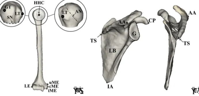
3D: Three-dimensional LB: Lateral Border
AA:Angulus Acromialis LE: Lateral Epicondyle
ACS: Anatomical Coordinate System LT:Lesser Tubercle
AL: Anatomical landmark
z y x
MAD, , / MADx,y,z:Mean Absolute Angular Deviation Value
AN: anatomical Neck MEc: Medial epicondyle (central)
A-P: Anterior-Posterior M-L: Medio-Lateral
AR: Anatomical Region MRI: Magnetic Resonance Imaging
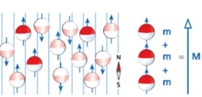
B0: Magnetic Field Strength NMV: Net Magnetization Vector
CP:Tip of Coracoid Process RSA: Radio Stereometric Analisis
CT: Computed Tomography RF: Radio Frequency
DoF: Degree of Freedom SBP:Portion of the Subject Specific Bone Model
DRRs: Digitally Reconstructed Radiographs SD: Standard Deviation
EBCT: Electron Beam Computed Tomography S-I: Superior-Inferior
FFT: Fast Fourier Transform SS: Root of Scapula Spine
FoV: Field of View TBC: Template of a Complete Bone Model
GHJ: Gleno Humeral Joint TE: Echo Time
GHJC: Gleno Humeral Joint Center TR: Repetition Time
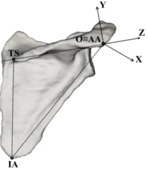
G:Glenoid TS: Trigonium Spinae Scapula
GT: Greater tubercle HHC: Humera Head Center IA: Inferior Angle
CHAPTER 1
1
Traumatic joint dislocations represent a frequent and important problem in orthopaedic surgery and sports medicine. The most frequently dislocated joint by far is the shoulder, or the gleno-humeral joint (GHJ), which is characterized by the widest range of motion among the human joints and by very little coverage of the humeral head by the joint socket. The incidence of gleno-humeral dislocations has been reported to be 11.2 per 100`000 per year and a high number of recurrences after the first dislocation have been reported, especially in young patients. The investigation of gleno-humeral kinematics of the shoulder, in terms of rotations and translations and joint stability, has been a primary focus of orthopaedic research and it is instrumental in understanding and thus preventing primary and repeated shoulder dislocations. In vivo kinematics of the shoulder joint has been studied by means of different instrumental techniques (Hill et al., 2007). Both using stereo-photogrammetric systems and electromagnetic tracking devices, the orientation and position of the relevant body segments are estimated using sensors placed on the skin of the subject. Due to the deformation of the soft tissues surrounding the bones, there is an ongoing debate about the capability to accurately track the movement of the underlying bony segments from sensors attached to the skin (van Andel et al., 2009). It is especially true, when small displacements, such as gleno-humeral joint translations, need to be assessed. An alternative approach is offered by the use of technologies based on ionizing radiation such as computer tomography (Baeyens et al., 2001), biplanar X-rays (Lagacé et al., 2012), fluoroscopy (San Juan and Karduna, 2010) or a combination of dual-plane fluoroscopy and 3D bone models derived from CT. Major limitations of such techniques include the image geometric distortion and invasiveness due to the radiation exposure. A further alternative, which could provide an acceptable level of accuracy and that is innocuous for patients, is the use of magnetic resonance imaging (MRI) (von Eisenhart-Rothe et al., 2010). GHJ translations have been investigated both in healthy subjects (Rohad et al, 1998; Graichen et al., 2000; Sahara et al., 2007) and patients (von Eisenhart-Rothe et al., 2002; Chhadia et al., 2010) using MRI. However, none of the latter in vivo studies analyzed the GHJ translations under the action of selected external forces. This doctoral research aims at developing and testing a MRI based methodology for in vivo estimation of the GHJ translations with and without an external load. The images from an open-MRI system were used, along with three-dimensional post-processing methods, to analyze gleno-humeral displacements (1) in different shoulder positions and (2) under the application of an anterior load in healthy volunteers. In contrast to earlier conducted cadaveric studies and anatomical studies using bone pin markers, the assessment with open-MRI allowed us to observe and document joint motion at high resolution and with all passive restraints
2
(ligaments, joints capsule) and active stabilizers (musculature) in place and fully, physiologically functioning.
This research represents the first step of a wider research aimed at developing and validating both experimental and analytical methods for evaluating and comparing the outcome of different surgical techniques and rehabilitation protocols for the treatment of gleno-humeral joint dislocation using bio-imaging techniques as an ultimate goal.
While pursuing, the main aim of the present doctoral thesis, the following secondary aims were also considered:
Anatomical coordinate systems definition
The description of joint kinematics requires the definition of anatomical reference systems of the bone segments forming the joint. Most of the definitions in the literature are based either on the identification of anatomical landmarks, or anatomical regions located in the distal portions of the bones under analysis. Often, the relevant anatomical landmarks or anatomical regions are not included in the images acquired using bio-imaging techniques for the clinical examination of the joint. In fact, these techniques have the limitation of being characterized by a restricted field of view (FoV) which may prevent the acquisition of the entire bones.
Development of a device for applying an external force during a MRI exam
To study the gleno-humeral joint in different configurations while an external force is applied, a device for fixing the arm at different degrees of abduction and applying a selected force to the proximal humerus was developed. Since the recordings have to be carried out under high intensity magnetic fields and in small measurement volumes, particular attention was paid in the selection of non-ferromagnetic materials for the construction of the device for the shoulder loading.
CHAPTER 2
3 2.1 Anatomy of the shoulder joint complex
The shoulder joint complex is formed by the shoulder girdle and the humerus. The shoulder or pectoral girdle is the set of bones which connects the upper limb to the axial skeleton. It consists of the clavicle, scapula and sternum. The pectoral girdle is a complex of five joints that can be divided into two groups. Three of these joints are true anatomical joints, while two are physiological joints. Within each group, the joints are mechanically linked so that both groups simultaneously contribute to the different movements of the shoulder to variable degrees.
The scapulo-thoracic joint is not a true joint in anatomical sense, it has no capsule or ligamentous
attachments, but is a physiological joint formed by articulation of the anterior scapula with the posterior thoracic rib cage. The scapula attachment to the axial skeleton in a healthy shoulder is purely musculo-tendinous, formed by the trapezius and serratus muscles. Its gliding movement patterns consist of elevation/depression, retraction/protraction, and superior/inferior rotation. Scapulo-thoracic joint function enhances arm-trunk motion and gleno-humeral stability as the scapula orients the glenoid to the humeral head.
Sterno-clavicular joint represents the single bony articulation between the axial skeleton and upper
ex-tremity (Dempster, 1965). It is formed by the articulation of the manubrium of the sternum and the first costal cartilage with the medial end of the clavicle. The sterno-clavicular joint serves as the pivot point for scapular elevation-depression and abduction-adduction (Doody et al., 1970).
Acromio-clavicular joint is a plane synovial joint formed by the articulation of the distal clavicle with
the acromion of the scapula. The acromion of the scapula rotates on the acromial end of the clavicle. Three degrees of freedom are available at the acromio-clavicular joint. Movement can occur between the acromion and lateral end of the clavicle, about a vertical axis, around a frontal axis, and about a sagittal axis (Peat, 1986).
Supra-humeral joint (subacromial joint) is a physiological joint formed by an articulation of the
coraco-acromial ligament and the head of the humerus. It is formed by the gap between the humerus and the acromion process of the scapula. This joint plays a role during complex movements while the arm is fully flexed at the gleno-humeral joint.
4
Gleno-humeral joint is the articulation between the head of the humerus and the glenoid cavity of the
scapula. It is a ball-and-socket type of synovial joint. It represents the most important joint of the shoulder. In this chapter we will focus on the gleno-humeral joint, its components and the mechanism which confer mobility to the joint.
Plane of motion of the shoulder complex
Motion of the shoulder complex is described in relation to the cardinal planes, sagittal, coronal, and horizontal (Fig. 2.1). Shoulder flexion and extension occur in the sagittal plane, abduction and adduction in the coronal plane, and horizontal abduction and adduction in the horizontal plane. Internal and external rotation occurs through the long axis of the humerus, affording a high degree of mobility in an in finite number of planes. This is typically assessed at 90 deg of coronal plane abduction or with the arm at the side (Kelley et al., 1995).
Figure 2.1: Motion of the shoulder in the coronal (abduction-adduction), sagittal (flexion-extension) and horizontal (internal-external rotation) planes.
Scapulo-humeral rhythm
During arm elevation the humerus rotates around the scapula, at the gleno-humeral joint, and the scapula moves around the thorax, at the scapulo-thoracic joint. The result is a synchronized movement of the shoulder girdle and humerus, described as the scapulo-humeral rhythm (Inman et al., 1944; Groot et al., 1999; McQuade et al., 1998; Meskers et al., 1998).
The scapulo-humeral rhythm describes the relationship of motion between the scapula and humerus, and is influenced by the movement of the sterno-clavicular and acromio-clavicular joints.
With active humeral elevation up to 30 degrees (deg) in the coronal or scapular abduction planes and up to 60 deg of sagittal plane flexion, the scapula seeks a position of stability (setting phase). The setting phase is variable and individualized (Inman et al., 1944). Following the setting phase, the humerus and scapula maintain a particular relationship during arm elevation (a ratio of movement). The relationship between gleno-humeral and scapulo-thoracic motion is critical and is generally considered to be 2:1, culminating in 120 and 60 deg, respectively.
5
Taking into account only the GHJ, the active abduction up to 120 deg occurs only if the humerus extra rotates of 90 deg. Then, the movement is due to the tilting of the scapula. The shoulder blade glides over the thoracic wall thanks to the scapulo-thoracic and acromion-clavicular joints, and the muscular activity. The full abduction of the arm in the frontal plane is therefore the consequence of a harmonic sequence of actions, for every 15 deg of abduction 10 deg achieved on the gleno-humeral joint and 5 deg at scapulo-thoracic level, with an integrated scheme and rhythm. For the deltoid, the main muscle of abduction, it is important the scapular rotation to maintain the tension necessary for its contraction. The stability of the shoulder, especially after the first 90 deg of abduction, is guaranteed by the tilting of the scapula that changes the relationship between the humeral head and the glenoid so that at 180 deg the deltoid almost does not work because the glenoid socket is located below the humeral head. In this rhythm also the sterno-clavicular joint steps in. In the excursion between 0 deg and 90 deg the scapula rotates 30 deg and the clavicle rises equally; beyond 90 deg of abduction (at sterno-clavear level), however, it is no longer possible for the scapula to move. For this reason the clavicle rotates 45 deg around the axis of the diaphysal, in order to raise its lateral end of the remaining 30 deg required to complete the movement of abduction.
2.2 Gleno-humeral joint
The gleno-humeral joint is an enarthrosis (ball-and-socket joint). The bones entering into its formation are the humeral head and the glenoid cavity of the scapula.Only 25% to 30% of the humeral head is covered by the glenoid surface in any given anatomic position. Although the bony surfaces of the humeral head and glenoid fossa have slightly different curvatures, their cartilaginous articular surfaces have approximately the same radius of curvature. This joint has three rotational axes of motion along the cardinal planes of the body: sagittal, frontal, and horizontal.
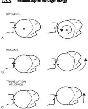
The humeral head spins, rotates, and glides or translates, on the face of the glenoid during arm elevation and rotation (Fig. 2.2) (Hart and Carmichael, 1985).
Figure 2.2: Three types of articular movement occur at
the gleno-humeral joint. A, Rotation. B, Rolling. C, Gliding. (From Matzen FA III, Zuckerman J:
Biome-chanics of the shoulder. In Frankel VH, Mordin M [eds]: Basic Biomechanics of the Musculoskeletal System, 2nd ed. Philadelphia, Lea & Febiger, 1989, p 231).
6
The direction of rolling and gliding components is dependent on whether the concave or convex surface is moving. The more congruent the surfaces, the more gliding occur and the more incongruent, the more rolling takes place (Kaltenborn, 1980). If a convex surface moves on a concave surface, then gliding occurs in the opposite direction to the rolling; if a concave surface moves on a convex surface, then rolling and gliding occur in the same direction. Therefore, due to the disproportion between the gleno-humeral articular surfaces rolling would be dominant.
Humerus
The humerus (Fig. 2.3) is the longest and largest bone of the upper extremity. Three parts can be distinguished: the body and two extremities. The upper extremity consists of a large rounded head joined to the body by a constricted portion called the neck, and two eminences, the greater and lesser tubercles.
The head (nearly hemispherical in form) is the humeral portion that articulates with the glenoid cavity of the scapula. The circumference of its articular surface is slightly constricted and it is referred to as the anatomical neck, in contradistinction to a constriction below the tubercles called the surgical neck. The anatomical neck is obliquely directed, forming an obtuse angle with the body. It is best marked in the lower half of its circumference; in the upper half, it is represented by a narrow groove separating the head from the tubercles. It affords
attachment to the articular capsule of the shoulder-joint.
The greater tubercle (greater tuberosity) is situated laterally to the head and lesser tubercle. Its upper surface is rounded and marked by three flat impressions which give insertion to the muscle tendons (supra-spinatus; infra-spinatus and teres minor).
The lesser tubercle (lesser tuberosity) although smaller, is more prominent than the greater and it is located anteriorly.
The body or shaft is almost cylindrical in the upper half of its extent, prismatic and flattened below, and has three borders and three surfaces.
The lower extremity includes, projected on either side, the lateral and medial epicondyles. The lateral epicondyle is a small, tuberculated eminence, curved a little forward. The medial epicondyle, larger and
7
more prominent than the lateral, is directed slightly backwards. The epicondyles serve as insertion points for tendons and ligaments of the elbow-joint (Standring, 2008).
Scapula
The scapula forms the posterior part of the shoulder girdle. It is a flat, triangular bone, with two surfaces, three borders, and three angles. The costal or ventral surface (Fig. 2.4) presents a broad concavity, the sub-scapular fossa. The medial two-thirds of this fossa are marked by several oblique ridges, which run laterally and upward. The ridges provide the attachment to the tendinous insertions.
The dorsal surface (Fig.2.4) is arched from above downward, and it is divided into two unequal parts by the spine; the portion above the spine is called the supra-spinatous fossa, and that below it is the infra-spinatous fossa.
The spine is a prominent plate of bone, which crosses obliquely the medial four-fifths of the dorsal surface of the scapula at its upper part, and separates the supra- from the infra-spinatous fossa. It begins at the vertical border by a smooth triangular area and ends in the acromion. The spine is triangular, and flattened from above downward. It presents two surfaces (superior and inferior) and three borders (anterior, posterior and lateral).
The acromion forms the summit of the shoulder. It is a large, somewhat triangular or oblong process, curving forward and upward, so as to overhang the glenoid cavity.
The scapula has three borders and three angles. The superior is the shortest and thinnest of the three borders of the scapula, it is concave, and it extends from the medial angle to the base of the coracoid process. The axillary border is the thickest of the three. It begins above at the lower margin of the glenoid cavity, and inclines obliquely downward and backward to the inferior angle. The vertebral border is the longest of the three, and extends from the medial to the inferior angle.
The inferior angle is formed by the union of the vertebral and axillary borders. The lateral angle is the thickest part of the bone, and it is sometimes called the head of the scapula. The articular surface, the glenoid cavity, is directed laterally and forward and articulates with the head of the humerus; it is broader at the bottom than at the top and its vertical diameter is the longest. The surface is covered with
8
cartilage in the fresh state; and its margins, slightly raised, give attachment to a fibro-cartilaginous structure, the glenoidal labrum, which deepens the cavity. At its apex is a slight elevation, the supra-glenoid tuberosity. The neck of the scapula is the slightly constricted portion which surrounds the head. The coracoid process is a thick curved process attached by a broad base to the upper part of the neck of the scapula; it runs at first upward and medially, then, becoming smaller, it changes its direction and it projects forward and laterally (Standring, 2008).
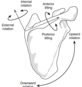
The scapula is involved in various movements of the shoulder. In particular, Kibler et al. (1998) have described five roles attributed to the scapula: (1) it represents a stable part of the gleno-humeral joint; (2) it allows for retraction and protraction along the thoracic wall; (3) it elevates the acromion to decrease impingement and coraco-acromial arch compression in the throwing and serving motion; (4) it serves as a base for muscle attachment; and (5) it functions as a link in the proximal to distal sequencing of the kinetic chain (Fig. 2.5).
Posterior and anterior tilt: posterior and anterior
tilt is the scapular rotation about an oblique medial-lateral axis (Karduna et al., 2000); posterior tilt occurs as the acromion moves backward and anterior tilt occurs as the acromion moves forward.
Internal and external rotation: internal and
external rotation are described as scapular rotation about an oblique superior-inferior axis (Karduna et al., 2000); external rotation can be visualized as the acromion moving posteriorly with the medial border of the scapula moving in an anterior direction.
Downward (medial) and upward (lateral) rotation: scapular downward and upward rotation occurs
about an axis in the scapular body (Karduna et al., 2000); downward rotation is defined by the rotation of the glenoid downward and the inferior angle of the scapula toward the spine; upward rotation is the rotation of the glenoid superiorly and movement of the inferior angle away from the spine.
Abduction and adduction: abduction is the movement of the medial border of the scapula away from the vertebral column; adduction is defined as movement of the medial border of the scapula toward the vertebral column (Oatis, 2004).
Figure 2.5: Scapular variables: posterior tilt, upward
rotation, and external rotation. (From Dayanidhi S, Orlin
M, Kozin S, et al: Scapular kinematics during humeral elevation in adults and children. Clin Biomech (Bristol, Avon) 20:600-606, 2005).
9
Elevation and depression:scapular elevation is the movement of the scapula superiorly on the thorax; depression is the movement of the scapula inferiorly on the thorax (Oatis, 2004). When a patient assumes the prone position, the shoulder girdle falls into a relatively elevated and protracted state.
2.2.1 Normal gleno-humeral relationship
The gleno-humeral joint is a multi-axial ball-and-socket synovial joint. The articular surfaces, the head of the humerus and the glenoid fossa of the scapula, although reciprocally curved, are oval and are not sections of true spheres. It was estimated that the articular surface of the glenoid fossa is one third to one fourth that of the humeral head (Iannotti et al., 1992).
Because the head of the humerus is larger than the glenoid fossa, only part of the humeral head can be in articulation with the glenoid fossa in any position of the joint.
The gleno-humeral congruence (conformity) is the relationship between the radius of curvature of the humeral head and the glenoid (Iannotti et al., 1998). If the radii of curvature of the humeral head and glenoid were the same, i.e. congruency ratio of 1, and then there would be maximum contact between the two surfaces. The most common configuration (90%) is a smaller radius of curvature for the humeral head relative to the glenoid, such that the congruency ratio is less than one. This implies an increased range of movement but a decreased stability. The design characteristics of the joint are typical of an "incongruous" joint. The surfaces are asymmetrical, the joint has a movable axis of rotation, and muscles related to the joint are essential in maintaining stability of the articulation (O’Brien et al., 1990).
The mean humeral head radius and the mean humeral head thickness are correlated with the humeral head offset, which is the distance between the center of the humeral head and the longitudinal axis of the humeral shaft. The ratio of the humeral head thickness to humeral head radius is reliably consistent at 0.7–0.9 (Howell et al., 1989; O’Connell et al., 1990). This ratio is directly proportional to the amount of humeral head which articulates with the glenoid, irrespective of other variables such as length of the humeral shaft or the size of the patient (O’Connell et al., 1990).
The normal glenoid has a pear shaped appearance with a shorter anterior-posterior dimension in the superior half (mean 23 mm) than in the inferior half (mean 29 mm) (Howell et al., 1989). The glenoid offset is the distance between the base of the coracoid and the deepest portion of the glenoid articular surface (Howell et al., 1989). This measurement determines the location of the gleno-humeral joint line and again is not related to the size of the patient. The lateral gleno-humeral offset is the distance
10
between the base of the coracoid and the most lateral aspect of the greater tuberosity. This measurement is important as it determines the resting tension of the rotator cuff and the moment arm of the deltoid.
Gleno-humeral index
This is defined as the maximum transverse diameter of the glenoid divided by the maximum transverse diameter of the humeral head. This ratio is approximately 0.75 in the sagittal plane and 0.6 in the transverse plane (Saha et al., 1971). A low gleno-humeral index is associated with recurrent anterior instability (Randelli et al., 1986).
Gleno-humeral articular constraint
The constraint is the amount of humeral head which is in direct articulation with the glenoid cavity (Iannotti et al., 1998). It is related to the depth of the glenoid but it is independent of articular congruence. The normal glenoid has a depth of 9 mm in the superior-inferior direction and 5 mm in the anterior-posterior direction. As a result, the glenoid is more constrained in the superior-inferior direction than posterior direction, accounting for the more frequently observed anterior-posterior dislocation (Lam et al., 2006).
2.2.2 Gleno-humeral joint stability
The term “gleno-humeral joint stability” refers to the process by which the humeral head remains centered on the glenoid during motion (Halder et al., 2001).
The stability of the joint is maintained by an interconnecting network of static and dynamic restraints (Lam et al., 2006) and it is provided by the articulating surfaces, capsular and ligamentous structures (static stabilizers), and synchronous activity of the rotator cuff, biceps, deltoid, and scapular muscles (dynamic stabilizer). The role of any specific component of the stabilizing system varies with gleno-humeral joint position and direction of the opposing force. A functional interplay or interdependence exists between anterior and posterior and between superior and inferior components of the capsule-ligamentous system. For the stability two aspects are important: conformity and constraint. Conformity is the relative match between the radii of the humeral head and glenoid; i.e. a completely conforming joint has 0 mm mismatch between the respective radii. Constraint is the threshold to dislocation which is related to the depth and size of the socket. Experimental evidence shows greater gleno-humeral translation in non-conforming articular surface (Iannotti et al., 1999). This means that the relationship
11
between gleno-humeral translation and joint conformity only applies under active, not passive joint loading.
Capsulo-ligamentous complex
The gleno-humeral joint capsule originates from the labrum and the margin of the glenoid fossa. It is attached laterally to the anatomical neck of the humerus. It maintains the negative intra-articular pressure, contributing to the stability of the gleno-humeral joint (Lam et al., 2006).
The gleno-humeral joint capsule provides passive stability at the extremes of gleno-humeral motion (Werner et al., 2004). The capsule does not only limit rotation and prevent excessive translations, but also causes a coaptation and obligate translation of the humeral head on the glenoid at the end of passive movements. The superior part of the capsule, together with the coraco-humeral ligament, is important in strengthening the superior aspect of the joint and resisting the effect of gravity on the dependent limb.
Anteriorly and posteriorly the capsule is strengthen by ligaments and tendons. Inferiorly, the capsule is thin and weak and contributes little to the stability of the joint. The inferior part of the capsule is subject to considerable strain because it is tightly stretched across the head of the humerus when the arm is elevated; moreover it is lax and lies in folds when the arm is adducted. The frequency of anterior dislocation seen clinically demonstrates the weakness of the inferior part of the capsule (Lam et al., 2006). The integrity of the capsule and the maintenance of the normal gleno-humeral relationship depend on the reinforcement of the capsule by ligaments and the attachment of the muscle tendons of the rotator cuff mechanism.
Glenoid labrum
The glenoid labrum is a rim of fibro-cartilaginous tissue attached around the margin of the glenoid fossa, deepening the glenoid cavity and serving to bridge bone to the gleno-humeral ligaments and biceps tendon. The inner surface of the labrum is covered with synovium; the outer surface attaches to the capsule and is continuous with the periosteum of the scapular neck (Lam et al., 2006). The glenoid labrum accounts for about 10–20% of the static stability by deepening the glenoid socket 50%, and detachment of the labrum anteriorly may reduce the depth of the socket in the anterior-posterior direction. The shape of the labrum adapts to accommodate rotation of the humeral head, adding flexibility to the edges of the glenoid fossa. Resection of the glenoid labrum has been reported to reduce the effectiveness of compression-stabilization by approximately 10% to 20% (Halder et al., 2001).
12
Compression by muscle activity and capsule-ligamentous tightening increases the stability of the labrum. Superior labral defects have been found to decrease torsional rigidity and increase inferior gleno-humeral ligament strain, which contributes to anterior instability (Abboud et al., 2002).
Gleno-humeral ligaments
The gleno-humeral ligaments are thickenings of the joint capsule and consist of superior, middle and inferior portions. The superior gleno-humeral ligament functions as the primary restraint to inferior translation of the humeral head. The middle gleno-humeral ligament is the most variable and it limits external rotation and anterior subluxation of the humeral head when the arm is in mid-abduction. The inferior gleno-humeral ligament is the most important ligament in maintaining joint stability (Lam et al., 2006).
Coraco-humeral ligament
The coraco-humeral ligament is one of the most important ligamentous structures in the shoulder complex as it is involved in maintaining the gleno-humeral relationship. The downward pull of gravity on the arm is counteracted largely by the superior capsule and the coraco-humeral ligament. Because the coraco-humeral ligament is located anterior to the vertical axis about which the humerus rotates axially, the ligament checks lateral rotation and extension. Shortening of the ligament would maintain the gleno-humeral relationship in medial rotation and would restrict lateral rotation severely.
Together with the acromion and the coracoid processes, the ligament forms an important protective arch over the gleno-humeral joint. The arch forms a secondary restraining socket for the humeral head, protecting the joint from trauma and preventing dislocation of the humeral head superiorly.
Rotator cuff
The rotator cuff is the musculo-tendinous complex formed by the attachment to the capsule of the supraspinatus muscle superiorly, the subscapularis muscle anteriorly, and the teres minor and infraspinatus muscles posteriorly. All of their tendons blend intricately with the fibrous capsule. They provide active support for the joint and can be considered true dynamic ligaments. The rotator cuff, acting as a dynamic compound musculo-tendinous unit, plays an essential role in movements of the gleno-humeral joint. The combined effect of the cuff muscles works as a force-couple to keep the humeral head centered on the glenoid. The dynamic restraints stabilize the joint by several mechanisms (Lam et al., 2006):
13
2. by dynamic contraction thereby causing compression of the humeral head into the glenoid (Halder et al., 2001);
3. by determining a secondary tightening effect on the static constraints; 4. by exerting a direct barrier effect.
2.2.3 Gleno-humeral joint laxity and instability
The term shoulder laxity refers to the physiological motion of the gleno-humeral joint that allows a normal range of motion. It was defined as the ability of the humeral head to be passively translated on the glenoid fossa (Matsen et al., 1991). This obligate translation of the humeral head on the glenoid is physiologic and in fact necessary in order to achieve the large degrees of freedom afforded the highly mobile shoulder (Saurer et al., 2001).Translation can occur in any direction as the humeral head moves on the glenoid face during humeral elevation and rotation.
The position at which the maximal mobility occurs is defined resting position (Lin et al., 2007). The resting position is the position of a joint in which the joint tissues are under the least amount of stress and in which the joint capsule has its greatest laxity (Lin et al., 2007). It is also called the “loosely packed position” as opposed to the “closely packed position” (Lin et al., 2007). This position is also the position of minimal congruence between joint surfaces allowing the greatest passive separation between articular surfaces (Lin et al., 2007).
For the in vivo gleno-humeral joint, the resting position was found to be located at a position in neutral rotation between 30 and 60 deg of shoulder abduction with respect to the trunk in the plane of the scapula (commonly defined as the plane 30 deg anterior to the frontal plane) (Lin et al., 2007) .
Quantitative assessment of gleno-humeral laxity has been performed both in vivo and in vitro. Poppen and Walker (1976) observed from X-rays that the humeral head moved upward relative to the glenoid between 0 and 30 deg of abduction and translated inferiorly by 2-3 mm throughout the rest of abduction. Harryman et al. (1990) in their cadaveric study demonstrate the obligate nature of translation. In order to determine whether the observed translations were forced by asymmetrical tightness of the capsule, rather than being associated with laxity of the capsule, they attempted to prevent the anterior translation of the humeral head during flexion of shoulders that had an intact capsule by applying an oppositely directed (posterior) force to the humeral head. Herryman et al. (1990) concluded that given the obligate nature of these translations they may also occur with active motions. To confirm this, Howell et al. (1989) reported evidence that translation occurs with combined
14
abduction, extension, and external rotation and that normal translation is reduced or eliminated when there is gleno-humeral instability.
Rhoad et al. (1998) used MRI to examine normal relationships about the gleno-humeral joint in internal and external rotation. They found that during active motion the humeral head translation averaged 2.1 mm in the anterior-posterior plane and that during passive positioning average translations increased to 8.2 mm in the anterior-posterior plane.
Graichen et al. (2000) used 3D MRI to demonstrate the centering of the humeral head during abduction and rotation. They found that during passive elevation the humeral head translated slightly inferiorly at low angles of elevation (from +1.58 mm at 30 deg to +0.36 mm at 150 deg of abduction ) and slightly posteriorly at higher angles of elevation (from +1.55 mm at 30 deg to -0.07 mm at 150 deg of abduction). With muscle activity, the respective translations were smaller, particularly at low angles of elevation (1.0±1.3 mm at 60 deg of abduction, 0.04±1.3 mm at 90 deg and of -0.02 ±1.4 mm at 120 deg of abduction). In their study, Graichen et al. (2000) reported an inferior translation of 1.2 mm (from 30 to 150 deg). While at 60 deg of abduction the humerus is still 1 mm superior to the glenoid, it is more centered at 90 and 120 deg. The superior position at 60 deg may be caused by the dominance of the deltoid with its cranial force direction, while at 90 deg and 120 deg the rotator cuff muscles with their centralizing effect are more active (Graichen 2000).
Saurer et al. (2001) in their study on the gleno-humeral joint laxity using an instrumented arthrometer found a greater posterior translation than anterior. This is probably due to the fact that the posterior capsule is thinner than the anterior capsulo-ligamentous structures and therefore may provide less resistance to translation than the thicker anterior capsule and supporting gleno-humeral ligaments (O’Brien et al., 1995). Additionally, the subscapularis tendon is also reported to resist anterior translation when the humerus is below 90 deg of abduction (McFarland et al., 1996).
Schiffern et al. (2002) conducted a study to analyze the humeral head centering in the absence of voluntary positioning. They found that the humeral head remained centered within the glenoid, especially in the midrange positions of passive rotation in which the gleno-humeral ligaments and capsule are known to be lax (from 0 deg to 45 deg of external rotation, 35 deg of gleno-humeral abduction, cadaveric studies) (Matsen et al., 1994; Matsen et al., 1998). Within this midrange of rotation, none of the humeral heads tested translated more than 2.2 mm either anteriorly or posteriorly in relation to the glenoid center.
The findings of the Schiffern et al. (2002) study demonstrate that active muscle effort is not required to stabilize the shoulder in midrange positions. They demonstrated that the humeral head remains centered
15
in the glenoid in the absence of voluntary muscle contraction when the shoulder is passively positioned in midrange positions in which the capsule and ligaments are lax. This observation leads to the consideration of what mechanisms other than ligament stabilization or dynamic neuromuscular control might maintain the desired position of the humeral head in the glenoid (Schiffern et al, 2002).
They justified their findings with the conformity of the joint. In a high conforming joint, the stability ratio (the ratio of force necessary to translate the humeral head to the load compressing the humeral head into the glenoid) is maximal when the humeral head is centered in the glenoid. In a system of this sort, the stabilizing effect of a given compressive load is greatest when the head is centered in the glenoid concavity, and very low compressive loads, such as those from resting muscle tone, may be sufficient to center the humeral head. Thus, a high degree of conformation of the glenoid concavity to the humeral head provides an anatomic situation that optimizes the centering effect of concavity compression (Schiffern et al, 2002).
This tendency for the gleno-humeral joint to become uncentred in positions near the end of the range of motion is thought to be related to an unopposed translatory force applied to the humeral head, forcing it from the centered position (Matsen et al., 1994; Matsen et al., 1998).
Alberta and colleagues (Alberta et al., 2006) tested six cadaver shoulders with an intact capsule and all muscles removed, under an external load of 15 and 20 N and found an anterior translation of 12.8 ± 1.9 mm and of 13.4 ± 2.0 mm at 90 deg of shoulder abduction, respectively. Sahara et al. (2007) in their in
vivo study investigated the gleno-humeral joint motion during isometric arm abduction by means of an
open and vertical MR scanner. As reported by the authors, in the superior-inferior direction, the humeral head translated inferiorly from 1.9 mm at 0 deg to 0.8 mm at the maximum abduction. In anterior-posterior direction, the humeral head translated anteriorly from 0 to 90 deg (mean 2.4 mm) and posteriorly from 90 to 150 deg of abduction (mean -1.4 mm). Marquardt et al. (2006) by means of robot-assisted shoulder simulator tested twelve cadaveric shoulders with all soft tissues removed except for the tendons of the rotator cuff, the pectoralis major and the deltoid muscle. They found a translation of 6.8 ± 2.4 mm at 0 deg and 5.1 ± 3.1 mm at 90 deg of gleno-humeral abduction. Su et al., (2009) investigate the translations in five cadaveric intact shoulders in both the superior and anterior-superior directions. The study design also provided the rotator cuff tendons were individually loaded, to simulate the muscle forces acting in vivo. They found an average translation of 2.0 ± 0.5 mm at 45 deg of gleno-humeral abduction. The studies on cadaver demonstrate the importance of muscle forces and joint conformity in the limitation of humeral head translations, which can lead to the instability.
16
Shoulder instability is the inability to maintain the humeral head within the center of the glenoid during active motion of the arm (Lippitt and Matsen, 1993).
As above mentioned, gleno-humeral stability is influenced by a variety of factors, such as adhesion-cohesion, concavity-compression, negative intra-articular pressure, and limited joint volume. All these factors are combined with the static ligamentous restraints and dynamic muscular control to provide stability within the large range of motion.
Gleno-humeral instability leads to shoulder dislocations in 2-8% of cases (Grumet et al., 2010). Although the natural history of the dislocated shoulder depends on a variety of factors, it is the age of the patient at the time of the initial dislocation that is most important (VandenBerghe et al., 2005). Shoulder dislocations most commonly occur in young males of less than 25 years of age, with a prevalence of anterior dislocations (about 90–95% of all shoulder dislocations). The etiology of shoulder instability is most commonly traumatic, and results in a Bankart lesion or disruption of the labrum and anterior-inferior gleno-humeral ligament complex from the glenoid as well as an impaction fracture on the postero-lateral aspect of the humeral head. Among the different dislocations the posterior are less common (only 3-5% of dislocations), instead the sub-acromial dislocation (humeral head posterior to the glenoid and inferior to the acromion) is the most common.
The incidence of shoulder dislocations has been reported to be 11.2 per 100`000 per year (Simonet et al., 1984). Major deficits in shoulder function and a high number of recurrences after the first dislocation have been reported especially in young patients (Robinson et al., 2006). According to Robinson et al. (2006), recurrences are found in 56% of young patients aged between 15 and 35 within the first year (Robinson et al., 2006).
Many classification systems have been developed to describe shoulder instability. Schemes related to direction (anterior, posterior, multidirectional), etiology (traumatic, atraumatic, overuse), degree of instability (subluxation versus dislocation), and duration of symptoms (acute, recurrent, fixed) are described in the literature.
A new classification, which distinguishes among static, dynamic, and voluntary dislocation, have been proposed (Gerber et al., 2002) and provides for a division into classes and subclasses.
Class A-static instabilities: they are defined by the absence of classic symptoms of instability
and are associated with rotator cuff tears and degenerative joint disease (radiological diagnosis) (VandenBerghe et al., 2005).
Classe B-dynamic instabilities: they are symptomatic and have traumatic etiology (Hawkins et
17
Class C: they are voluntary dislocation and are of two types: muscular and positional. This type
of dislocation is most frequently among children and the preadolescent population. The first group of patients is able to sublux or dislocate the shoulder by selective muscle activation, whereas the others need to place the arm in appropriate position (such as forward flexion, adduction, and internal rotation for posterior instability) to induce the subluxation or dislocation.
CHAPTER 3
QUANTITATIVE MEASUREMENT OF THE GLENO-HUMERAL JOINT
TRANSLATIONS
18 3.1 Background
Three-dimensional (3D) gleno-humeral joint linear displacements have been measured, both in vivo and in vitro by means of different instrumental techniques. In vitro studies allow for a very accurate and repeatable joint kinematics reconstruction permitted by the use of different invasive approaches (Bourne et al., 2010; Bull et al., 1997; Ludewig et al., 2009; Massimini et al., 2010; Milne et al., 1996; Nishinaka et al., 2008). Studies on anatomical specimens also enable researchers to study patterns of injury and changes in joint kinematics that would be impossible in vivo studies (Su et al., 2009). Cadaveric experiments (Halder et al., 2001; Payne et al., 1997; Sharkey et al., 1995; Wuelker et al., 1994; Wuelker et al., 1995) can provide highly accurate measures of joint position or motion, because of the use of bone-embedded sensors (pins drilled into the bone) (Karduna et al., 1997), but are unable to accurately duplicate the complex motions, muscle forces, or joint forces associated with dynamic in
vivo conditions (Bey et al., 2006). Extensive analyses of the biomechanical role of soft tissues and
articular surfaces as joint constraints under the application of selected external forces has been conducted on cadavers mainly by means of shoulder testing device that allow six degrees of freedom for gleno-humeral positioning (Grossman et al., 2005) coupled with microscribe, used to digitize three-dimensional anatomic landmarks on the bone and record the humeral shift and translation data (Grossman et al., 2005); linear transducers for linear displacements measurement, or magnetic tracking device to determine position and orientation of a user in the working space (Su et al., 2009, Lin et al., 2007).The main limitations associated with published in vitro models are the inactivity of the muscles involved and that the gleno-humeral joint is often analyzed in isolation, without consideration of scapulo-thoracic motion or, the position of the clavicle. In vivo kinematics of the shoulder joint has been studied by means of stereophotogrammetry, electromagnetic tracking sensors, single and dual-plane fluoroscopy and open-MRI. Due to various factors such as the total or partial lack of the shoulder muscles and differences in muscular tone in cadaveric experiments, there are discrepancies between the phenomena observed in vivo and in vitro conditions.
3.1.1 Instrumental techniques for the assessment of the gleno-humeral joint translations
After a brief overview on in vivo and in vitro studies and the main characteristics, in this chapter we will focus on the principal techniques employed, both in vivo and in vitro, for the assessment of the GHJ translations.
19
Stereophotogrammetry
Video-based optoelectronic systems represent conventional motion measurement methods used to track the instantaneous position of markers attached to the skin. These systems are used to track the 3D position of a set of fiducial points, constituted from either retro-reflective (passive) or light-emitting (active) markers. In order to reconstruct 3D positions of markers captured by two or more cameras, the extrinsic parameters of each camera have to be known. These extrinsic parameters describe the geometrical relation between the camera and the captured calibration body (Lujan et al., 2006; Everaert et al., 1999).
These systems are non-invasive, easy to use and represent a convenient solution for many clinical and research applications (Tashman et al., 2003). However, several studies have shown that markers affixed to the skin shift relative to underlying bone by as much as 30 mm, particularly during rapid movements (Holden et al., 1997; Reinschmidt et al., 1997; Lafortune et al., 1992). This marker tracking error varies with the body segment analyzed, the marker position, the type of motor task under analysis and the angular joint excursion. Due to the deformation of the soft tissues surrounding the bones, there is an ongoing debate about the capability to accurately track the movement of the underlying bony segments from sensors attached to the skin (van Andel et al., 2009). It is especially true, when small displacements, such as gleno-humeral joint translations, require to be assessed.
Electromagnetic tracking devices
Six-degree-of-freedom (DoF) electromagnetic trackers provide both the position and orientation of the sensor with respect to a laboratory coordinate system by exploiting orthogonal electromagnetic fields (Raab, Blood, Steioner, & Jones, 1979; Foxlin et al., 1998; Kindratenko et al., 1999, Kindratenko et al., 2000; Borstad et al., 2002; Karduna et al., 1996; Karduna et al., 1997; Karduna et al., 2000; Bull et al., 1998; Harryman et al., 1990; Herryman et al., 1992). The system is compound by a source (transmitter) and a detector (sensor attached to the skin). Similarly to the video-based optoelectronic systems, when using electromagnetic tracking devices for in vivo evaluation, the sensors are placed on the skin of the subject and the measurements are affected by the presence of soft tissue artifacts which can conceal the measurement of the small displacements involved (Bey et al., 2006). Moreover due to the dependence of the measurements on the local electromagnetic field, the tracking systems are sensitive to the ambient electromagnetic environment and the transmitter signals could be distorted and the resulting measurements contain errors (Kindratenko et al., 2001).
20
To overcome the error imparted by soft tissue deformation some in vivo studies have used bone-embedded sensors that is trans-cortical pins drilled into the bone to hold marker cluster(Bourne et al., 2010, Stereophotogrammetry) or sensors (Ludewing et al., 2009, Electromagnetic tracking devices). Since these systems use pins anchored directly to the bone segments, they allow obtaining a highly accurate estimation of joint kinematics. Unfortunately, bone-pin cluster markers are associated with the risk of infection, change in pattern of motion due to pain, and translational/rotational instability of the actual pin (Bourne et al., 2010). The pain intensity of 4 out of 10 (Visual Analog Scale, VAS) have been reported when bone pins were used (Ludewing et al., 2009).
Bio-imaging techniques
The term bio-imaging refers indiscriminately to a wide range of techniques either based on the use of ionizing (e.g. X-ray based techniques) and non-ionizing radiations (e.g. MRI) (Hill et al., 2007). Among the techniques based on ionizing radiation the main used for the shoulder analysis are: computer tomography (CT) (Baeyens et al., 2001), biplanar X-rays (Lagacé et al., 2012), fluoroscopy (Karduna et al., 1997; San Juan et al., 2010) or a combination of dual-plane fluoroscopy (Fleisig et al., 1995) and 3D bone models derived from CT. An alternative which avoid the radiation exposure and then is noninvasive for the patient is represented by the magnetic resonance imaging (MRI) (Bey et al., 2006; Nishinaka et al., 2008; Massimini et al., 2012).
-
Techniques based on ionizing radiationX-ray based techniques have been employed to measure the position of bones relative to one another, or the position of implanted markers (RSA), either from static or dynamic images. Below the main techniques employed for the assessment of the gleno-humeral joint displacement have been reported.
Planar rontegenography (planar X-ray) is more commonly used in the clinical practice for evaluating
pathological abnormalities than as a tool for the kinematic study (Hill et al., 2007). Several studies used the X-ray for the imaging of the shoulder in the plane of the scapula (de Luca et al., 1973; Freedman et al., 1966; Poppen and Walker, 1976; Johnston et al., 1937; Saha et al., 1950) and the evaluation of the parameters of motion in normal and abnormal shoulders, such as the excursion of the humeral ball on the face of the glenoid (Poppen and Walker 1976).
However, this technique is affected by projective artifacts, which can be responsible of the many differences reported in the scapular-humeral rhythm in the different studies (de Groot et al., 1998). Moreover, for some applications, the 2D assessments of gleno-humeral joint motion, cannot be
21
sufficient to characterize the relative movement between the humerus and the glenoid which is characterized by rotations and translation along three axes (Bey et al., 2006)
Fluoroscopy is an imaging technique that uses X-rays to obtain real-time moving images of the internal
body structures. Fluoroscopy can be mono-planar, which allows investigating a larger volume with a reduced dose of X-rays, or bi-planar which is more accurate but also expose the patient to a higher radiation dose. Mono- and bi-planar X-ray fluoroscopy besides allowing the acquisition of a wide range of motion (Anderst et al., 2009; Dennis et al., 2005; Torry et al., 2010) enable to measure the joint motion during dynamic activities (Burkhart et al., 1992; Mandalidis et al., 1999; Pfirrmann et al., 2002; Werner et al., 2004).
Unfortunately, the low quality of the fluoroscopic images and especially the motion artifact (blur) may introduce errors which could affect the estimation of the relevant variables. In fact, the errors associated to the geometry distortion are comparable to those found in static planar radiography, if not greater due to the dynamic nature of the modality (Hill et al., 2007). In addition to the image geometric distortion, another important limitation is related to the radiation exposure which makes this approach invasive for the patients (Massimini et al., 2010; Nishinaka et al., 2008).
Radiostereometric analysis consists in the implantation of tantalum spheres into the bony segments to
be analyzed and to reconstruct their 3D positions from repeated multi-planar radiographic examinations. This methodology is very accurate (Selvik et al., 1983) and it has been applied to study prosthetic fixation (Ryd 1992) and joint stability (Uvehammer et al., 2000). RSA can be also used to dynamically study joint kinematics. Dynamic RSA has been used to study joint movements (Kärrholm et al. 1988; Brandsson et al., 2002; Saari et al., 2005), and in particular gleno-humeral joint kinematics (Bey et al., 2006; Hallström and Kärrholm, 2009). Use of biplane radiographic film methods (RSA) for 3D studies of static bone position has been well established (Kärrholm et al., 1988; Selvik et al., 1990), with precision reported in the ±10–250 µm range (Kärrholm et al., 1989). However, given the high invasiveness, the RSA is adopted only if there is the opportunity to insert marker of tantalum during the operating phase (such as in prosthetic implantation),
X-ray computed tomography(x-ray CT) is a technology that uses computer-processed x-ray to form a series of electronically collected projections, which are later reconstructed to a topographic imageof specific areas of the scanned body. Digital geometry processing is used to generate a three-dimensional image of the inside of the body from a large series of two-dimensional radiographic images taken around a single axis of rotation (Herman et al., 2009).