Introduction
In the adult mouse testis, undifferentiated spermatogonial stem cells (As spermatogonia) have been described as single cells that are able both to renew themselves and to produce more differentiated Apr (paired) spermatogonia. The Apr cells then divide into Aal (aligned) spermatogonia that further differentiate into A1spermatogonia (for a review, see de Roij, 2001). Appearance of A1 spermatogonia coincides with the expression of c-kit, encoding the receptor for Kit ligand (Kitl) (Manova et al., 1990; Yoshinaga et al., 1991; Sorrentino et al., 1991; Schranss-Stassen et al., 1999). Kit is a tyrosine kinase receptor that mediates proliferation/survival signals in type A spermatogonia (Rossi et al., 1993; Feng et al., 2000; Dolci et al., 2001). Upon Kit expression, spermatogonia become sensitive to Kitl produced by Sertoli cells (Rossi et al., 1992; Rossi et al., 1993) and undergo a definite number of proliferative cycles (forming the A2-A4, intermediate, and B spermatogonia), before entering meiosis. The temporal appearance of Kit expression and of Kitl sensitivity in spermatogonia, at around 6-7 days postnatum (dpn) (Manova et al., 1990; Yoshinaga et al., 1991; Dolci et al., 2001), marks the switch from the Aal spermatogonia to the A1-B differentiating cell types, at puberty.
The factors controlling spermatogonia differentiation to the Kit expressing stage are not known; however, some clues in addressing this topic can be gathered by analogies between spermatogonia and their fetal precursors, the primordial germ cells (PGCs). In fact, proliferation and differentiation of PGCs, as for spermatogonia, depend on a functional Kitl/Kit system
(Dolci et al., 1991), as demonstrated by their absence or severe reduction in W and Sl mutations, in which the c-kit or Kitl genes are inactivated. A further analogy between spermatogonia and PGCs is observed in the responsiveness to BMP8b, a growth factor belonging to the TGFβ-BMP superfamily. BMP8b–/– mice show impairment of PGC commitment (Ying et al., 2000), defects of spermatogonia proliferation, and spermatocyte apoptosis (Zhao et al., 1996). The incomplete abolishment of proliferation both in PGCs and spermatogonia from BMP8b null mice might be explained by the redundant action of other members of the BMP family within the embryo and the prepuberal testis. Indeed, a member of this family, BMP4, has been shown to induce PGC formation, to act as a PGC survival and localization factor within the allantois (Lawson et al., 1999) and as a mitogen in in vitro cultured PGCs (Pesce et al., 2002). Furthermore, two genes, encoding for proteins Smad1 and Smad5, known to be the substrates of the signaling cascade induced by BMPs (Hoodless et al., 1996), have been shown to control PGC differentiation and survival (Tremblay et al., 2001; Chang and Matzuk, 2001). An eventual effect of BMP4, Smad1 or Smad5 deficiencies in postnatal spermatogenesis cannot be ruled out, due to the lethal phenotype of their knockouts during the embryonic stages of development.
To address these points we undertook an in vitro study to investigate the possibility that BMP4 and its transduction pathway might be involved in the initiation of spermatogenesis. We show that BMP4 is expressed and developmentally regulated in Sertoli cells, and that its receptors Alk3 and It is well established that the c-kit gene plays an essential
role in the proliferation of differentiating spermatogonia in prepuberal mice. However, the mechanisms that regulate the onset of spermatogenesis, i.e. differentiation of spermatogonial stem cells and c-kit expression, are poorly understood. Here we identify a novel signal transduction system in mouse prepuberal testis regulating this developmental event, involving bone morphogenetic protein 4 (BMP4) and its transduction machinery. BMP4 is produced by Sertoli cells very early in the postnatal life and is successively down regulated in peri-puberal Sertoli cells. Its receptor Alk3 and the R-Smad Smad5 are
specifically expressed both in proliferating primordial germ cells and in postnatal spermatogonia. BMP4 stimulation of cultured spermatogonia induces Smad4/5 nuclear translocation and the formation of a DNA-binding complex with the transcriptional coactivator p300/CBP. In vitro exposure of undifferentiated spermatogonia to BMP4 exerts both mitogenic and differentiative effects, inducing [3H]thymidine incorporation and Kit expression. As a result of the latter event, Kit-negative spermatogonia acquire sensitivity to Stem Cell Factor.
Key words: BMP4, Alk3, Smad5, Kit, Spermatogonia Summary
Developmental expression of BMP4/ALK3/SMAD5
signaling pathway in the mouse testis: a potential role
of BMP4 in spermatogonia differentiation
Manuela Pellegrini, Paola Grimaldi, Pellegrino Rossi, Raffaele Geremia and Susanna Dolci* Dipartimento di Sanita’ Pubblica e Biologia Cellulare, Sezione di Anatomia, Universita’ di Roma Tor Vergata, Rome, Italy
*Author for correspondence (e-mail: [email protected]) Accepted 6 May 2003
Journal of Cell Science 116, 3363-3372 © 2003 The Company of Biologists Ltd doi:10.1242/jcs.00650
BMPIIR are specifically expressed in mitotic spermatogonia during the first week postnatum. Furthermore, we demonstrate that BMP4 is able to induce DNA synthesis in spermatogonia both from 4 and 7 days postnatum. BMP4 action is mediated by a rapid nuclear translocation of Smad4, which associates with Smad5. Upon nuclear translocation, the Smad4/Smad5 complexes are able to recruit the transactivating factor CBP and to bind Smad-responsive DNA sequences. We also found that Alk3 and Smad5 are exclusively expressed in the germline compartment of the postnatal testis and in proliferating PGCs. Finally, we show that BMP4 is able to induce c-kit expression in Kit-negative spermatogonia, thus conferring Kitl sensitivity in these cells. The present data indicate that the BMP4/ Alk3/Smad5 pathway is expressed and operates in the postnatal testis and is involved in the regulation of spermatogonia differentiation.
Materials and Methods
Cell cultures
Spermatogonia were obtained from 4 and 7 day old Swiss CD-1 mice, as previously reported by Rossi et al. (Rossi et al., 1993). Briefly, germ cell suspensions were obtained by sequential collagenase-hyaluronidase-trypsin digestions of freshly withdrawn testes. A 3 hour period of culture in E-MEM with 10% FCS was performed to facilitate adhesion of contaminating somatic cells to the plastic dishes. At the end of this pre-plating treatment, which was considered T0, enriched germ cell suspensions were rinsed from FCS and spermatogonia were then cultured in E-MEM supplemented with 2 mM Na-pyruvate and 1 mM Na-lactate in the presence or absence of BMP4 and/or Kitl (100 ng/ml; Genzyme, MA). Purity of 7 day postnatal (dpn) spermatogonia was about 80% after the pre-plating treatment, and about 50% for 4 dpn spermatogonia. During the 24 hours of culture, most of the contaminating somatic cells attached to the dish, and floating spermatogonia were recovered in the suspension at a purity higher than 90%.
At different times of culture an equal number of surviving cells, judged as Trypan Blue negative, were fixed or frozen for immunofluorescence, western and northern blotting, and electrophoretic mobility shift assay (EMSA). Immunoprecipitation DNA synthesis was studied by [3H]thymidine incorporation followed by autoradiography as previously described (Rossi et al., 1993). In these experiments, incubation with [3H]thymidine was performed during the last 4 hours of the 24 or 48 hour culture periods. The Student t-test was used to assess the significance of [3H]thymidine incorporation. Sertoli cells from different developmental ages, spermatocytes and spermatids were prepared as previously reported (Grimaldi et al., 1993; Sorrentino et al., 1991). Embryos were staged considering 0.5 the day of the vaginal plug.
Western-blot analysis, immunoprecipitation and antibodies Cells were harvested, washed in cold PBS and homogenized at 4°C in lysis buffer containing 10 mM Hepes pH 7.9, 10 mM KCl, 1.5 mM MgCl2, 0.1 mM EGTA, 0.5 mM DTT, 10 mM β-glycerophosphate, 0.1 mM sodium vanadate and a protease inhibitor cocktail (P8340; Sigma). Total cellular proteins were transferred to polyvinylidene difluoride membranes after SDS/PAGE. Membranes were blocked with PBS buffer containing 5% BSA and 0.1% Tween 20 for 1 hour at room temperature and then hybridized overnight at 4°C with primary antibodies. After hybridization with secondary antibodies conjugated to horseradish peroxidase, the immunocomplexes were detected with Supersignal West Pico detection reagent (Pierce, NJ).
Primary antibodies Smad4 goat polyclonal (sc-1909), anti-Smad5 goat polyclonal 7443), anti-Smad8 goat polyclonal
(sc-7442), anti-BMPR-IA goat polyclonal (sc-5676), anti-CBP rabbit polyclonal (sc-1211 X), anti-c-Kit rabbit polyclonal (sc-6283) and anti-actin rabbit polyclonal (sc-7210) were from Santa Cruz Biotechnology (CA), and anti-histone H3 (06-755) was from Upstate Biotechnology (Milton Keynes, UK). All primary antibodies were used at a 1:1000 dilution.
For immunoprecipitation, 70 µg of total protein from control or treated samples was incubated in the presence of 2 µg anti-Smad4 antibody for 2 hours. The immunocomplexes were recovered with protein G-Sepharose presaturated with PBS containing 0.05% BSA (Sigma, Milan, Italy). After three washes in PBS at 4°C, under constant shaking, immunocomplexes were eluted from beads with SDS-samples buffer. Proteins were separated by SDS/PAGE, blotted and probed with anti-Smad5 antibody or with anti-CBP antibody.
Northern-blot analysis
BMP4, Alk3, BMPIIR, c-kit and βactin probes were obtained by RT-PCR amplification of total testis RNA using the following primer pairs: BMP4 forward 5′TTTGGCCATGATGGCCGGGGCCATAC-CTT and reverse 5′TCAGCGGCATCCACACCCCTCTACCACCAT; Alk3 forward 5′ACTTTAGCACCAGAGGATACC and reverse 5′TT-TTCACCACGCCATTTACCC; BMP-IIR forward 5′TGCGGCTA-TAAGTGAGGTTGG and reverse 5′AGCAGTTGACATTGGG-TTGAC; β-actin forward 5′GGTTCCGATGCCCTGAGGCTC and reverse 5′ACTTGCGGTGCACGATGGAGG; c-kit forward 5′TA-TGGACATGAAGCCTGGCGT and reverse 5′CATTCCTGATGT-CTCTGGCTAGC. RNA was prepared from, 7 dpn testis, Sertoli cell cultures, spermatogonia, spermatocytes, spermatids and STO (embryonic fibroblast cell line) using the TRIZOL system (Gibco BRL). Total RNA (15 µg) was separated in denaturing agarose gel electrophoresis, blotted onto nylon membrane (Hybond-N, Amersham, UK) using 10× saline sodium citrate (SSC) buffer. Hybridization was carried out following Quick Hybrid System’s instruction (Stratagene, CA).
Autophosphorylation assay
Viable cells (106) obtained from 4 dpn testes were harvested and homogenized in 40 µl of a modified lysis buffer [50 mM Hepes pH 7.5, 150 mM NaCl, 1 mM EDTA, 1 mM EGTA, 10% glycerol, 1 mM DTT, 0.1% Tween 20, 10 mM β-glycerophosphate, 1 mM NaF, 0.1 mM sodium vanadate, 0.1 mM PMSF and protease inhibitor cocktail (P8340; Sigma)]. Clarified supernatants were prepared from whole cell lysates and 100 µg of protein from the supernatants was incubated in 50 µl of the lysis buffer with 20 µg/ml anti-c-kit polyclonal antibody for 2 hours at 4°C on a rotating shaker. The immunocomplexes were recovered with protein A sepharose (Sigma) for 1 hour at 4°C, while the supernatants were saved for normalization with an anti-β-actin antibody in western blotting. The immunocomplexes were then washed three times at 4°C with PBS containing 0.05% BSA and once with the specific kinase reaction buffer (50 mM Hepes pH 7.5, 10 mM MgCl2and 1 mM DTT). Kinase assays were performed at 30°C for 30 minutes in a 20 µl volume of kinase reaction buffer containing 10 mM β-glycerophosphate, 0.1 mM sodium vanadate, protease inhibitor cocktail (Sigma), 2.5 mM EGTA, 50 µM ATP, 0.1 mM PKAi, and 3 µCi of [α-32P]ATP/ reaction. Beads were washed three times with PBS and boiled in 4× Laemmli buffer. The immunocomplexes were separated by SDS/PAGE. Phosphorylated Kit protein was visualized by autoradiography of the dried gel and quantified by densitometric analysis.
Immunofluorescence analysis
Cryostat sections were obtained from unfixed frozen embryo testes at different ages of development. Control and treated spermatogonia were adhered onto poly-L-lysine glass slides. Both tissues and cells
were fixed for 10 minutes at room temperature in 2% paraformaldhyde, washed in PBS, permeabilized for 10 minutes with PBS containing 0.1% Triton X-100, and incubated for 30 minutes at room temperature with PBS containing 0.5% BSA. Samples were then incubated overnight at 4°C in a humidified chamber with the respective antibodies at a final concentration of 2 µg/ml and then for 1 hour at room temperature with cyanin 3 (AP 180 C; CHEMICON, CA) or FITC-conjugated secondary antibodies (401319 anti-rabbit IgG and 401514 anti-goat IgG; Calbiochem, Darmstadt, Germany). Slides were washed and mounted in 50% glycerol in PBS and immediately examined by fluorescence microscopy. Nuclei were counterstained with 1 µg/ml Hoechst (33342; Sigma). Control experiments were performed using non-immune immunoglobulins instead of the specific antibody. For histological analysis, representative frozen sections of 4 and 7 dpn testes were stained with 0.05% Toluidine Blue.
Electrophoretic mobility shift assay (EMSA)
Gel mobility shift assay was performed as previously reported (Grimaldi et al., 1993). Briefly, nuclear extracts were prepared from spermatogonia at 7 dpn treated or untreated with BMP4 after 1 hour of culture. After cell lysis, nuclei were isolated by centrifugation and extracted for 30 minutes at 4°C with extraction buffer containing 490 mM NaCl, 10 mM Hepes pH 7.9, 10 mM KCl, 1.5 mM MgCl2, 0.1 mM EGTA, 0.5 mM DTT, 10 mM β-glycerophosphate, 0.1 mM sodium vanadate, 1/100 (v/v) of protease inhibitors mixture and 5% glycerol. Nuclear extracts were collected after centrifugation at 100,000 g for 30 minutes and concentrated on Centricon-10 membranes after NaCl adjustment to the final concentration of 60 mM NaCl. EMSAs were performed as previously described using 9 µg of nuclear proteins added to a premix solution containing 2 µg double-stranded poly(dI-dC) (Pharmacia, Piscataway, NJ), and a 0.5-1 ng [α-32P]dATP-labeled double-stranded GCCGnCGC (GCCG motif)-containing oligonucleotide (5′GATATCTGCCGCCGCTTTGCCG-CCGCTTTGCCGCCGCG3′ and 3′ATCCGCGGCGGCAAAGCG-GCGGCAAAGCGGCGGCA5′) (see Kusanagi et al., 2000). For binding specificity, extracts were incubated with 100 fold molar excess of unlabeled oligonucleotide or with a mutated form of the oligonucleotide (5′GATCTGCCGTCGCTTTGCCGTCGCTTTGCC-GTCGCG3′ and 3′GATCCGCGACGGCAAAGCGACGGCAAAG-CGACGGCA5′; mut1) (Kusanagi et al., 2000). Anti-Smad4 and anti-Smad5 antibodies were added to nuclear extracts for 1 hour at 4°C before incubating with probe to prevent or to supershift the formation of the specific retarded band. Anti-Smad8 was used as negative
control. After 30 minutes of incubation at room temperature, the reaction mixture was loaded on a 6% polyacrylamide gel in 1× Tris-borate-EDTA buffer. Gels were dried and analyzed by autoradiography.
Results
BMP4 and its receptors are differentially expressed in testicular cells
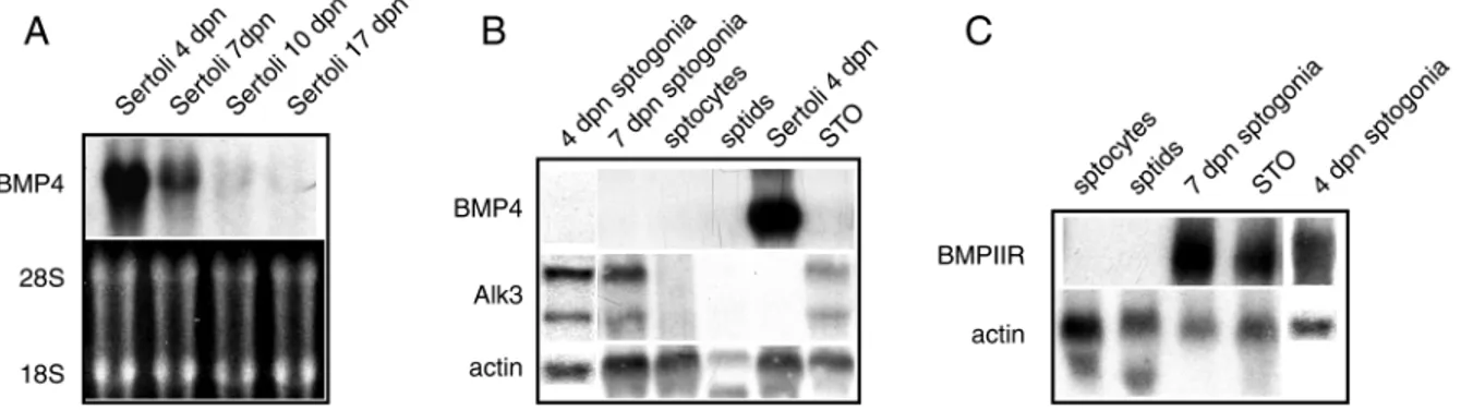
Fig. 1A and B show northern blot analyses of BMP4 expression in purified testicular cells obtained at various ages of development. A strong band corresponding to BMP4 mRNA was observed only in Sertoli cells from 4 dpn animals with a decreasing intensity in cells from 7, 10 and 17 dpn animals (Fig. 1A). BMP4 RNA levels did not change upon stimulation with dibutyril-cAMP at any developmental age (data not shown), differently from what was previously observed for Kitl (Rossi et al., 1991; Rossi et al., 1993). Spermatogonia, pachytene spermatocytes and spermatids were negative for BMP4 expression (Fig. 1B). It is known that BMPs ligands activate signaling in the target cells via heteromeric complexes composed by type II (BMPRII, ActRIIA/IIB) and type I (Alk3, Alk6, Alk2) serine/threonine kinase receptors (Hoodless et al., 1996; Nishimura et al., 1998). To investigate whether these classes of receptors are expressed in testicular cells, parallel northern analyses were performed. Fig. 1B and C show that Alk3 and BMPIIR transcripts are detectable only in spermatogonia, both at 4 and 7 dpn, whereas Alk6 gave negative results in all the samples tested (not shown).
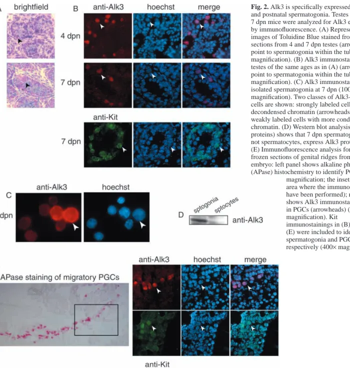
Immunofluorescence analysis on frozen cryostat sections of 4 and 7 dpn testes using an anti-Alk3 antibody showed that the only cells expressing Alk3 are spermatogonia (Fig. 2B). The same analysis carried out on isolated spermatogonia showed that at 7 dpn two classes of cells could be identified: a strongly labeled spermatogonia cell type with a large nucleus and decondensed chromatin, and a weaker labeled class of spermatogonia with a smaller nucleus and condensed chromatin (Fig. 2C). Western blot analysis showed a strong Alk3 immunoreactive band in spermatogonia and the absence of a specific signal in spermatocytes (Fig. 2D).
To investigate whether Alk3 was expressed also in PGCs,
Fig. 1. Expression pattern of BMP4, Alk3 and BMPIIR in the testicular cell types. (A) Northern blot analyses of BMP4 expression in Sertoli
cells obtained at different ages of postnatal development (upper panel), compared to ethidium bromide staining of the same gel (bottom panel). (B) Northern blot analyses of BMP4 (upper panel) and Alk3 (middle panel) expression in different cell types from testis at different ages of development (sptogonia=spermatogonia at 4 and 7 dpn; sptocytes=spermatocytes at 60 dpn; sptids=spermatids at 60 dpn), compared to actin expression (lower panel). (C) Northern blot analyses of BMPIIR expression (upper panel) within the different cell types of the testis, compared to actin expression (lower panel).
which are the cell types primarily affected by BMP mutations, we performed an immunolocalization experiment during their migratory period. PGCs at 10.5 days post coitum (dpc), identified by intense alkaline phosphatase activity (Fig. 2E, left panel), showed specific staining for Alk3 as well as for Kit (Fig. 2E, right panels, respectively), the same as observed for mitotic postnatal spermatogonia (Fig. 2B).
BMP4 induces [3H]thymidine incorporation in Kit-negative and Kit-expressing spermatogonia cultures
Because spermatogonia express high levels of both Alk3 and BMPIIR, and Sertoli cells of the corresponding age express high levels of BMP4 transcripts, we evaluated the effect of BMP4 on spermatogonia proliferation by a [3H]thymidine
incorporation assay (Fig. 3). BMP4 treatment induced a 2.5 to 3 fold increase in the percentage of [3H]thymidine incorporating cells in cultures of spermatogonia isolated from 4 dpn animals, and a 2 to 2.5 fold increase in 7 dpn cells. Parallel experiments were conducted including Kitl as a mitogen and an increase of 3.9 fold of [3H]thymidine incorporating cells was observed in 7 dpn spermatogonia, as previously reported (Rossi et al., 1993; Dolci et al., 2001). On the contrary, 4 dpn spermatogonia did not respond to Kitl, confirming that at this age spermatogonia expressing Kit are rare (Dolci et al., 2001). Addition of both BMP4 and Kitl in 7 dpn spermatogonia was not significantly additive in terms of fold increase of [3H]thymidine incorporating cells, suggesting that, at this developmental age, BMP4 is acting in spermatogonia co-expressing Kit and Alk3. BMP4 did not
Fig. 2. Alk3 is specifically expressed in PGCs
and postnatal spermatogonia. Testes from 4 and 7 dpn mice were analyzed for Alk3 expression by immunofluorescence. (A) Representative images of Toluidine Blue stained frozen sections from 4 and 7 dpn testes (arrowheads point to spermatogonia within the tubules; 400× magnification). (B) Alk3 immunostainings in testes of the same ages as in (A) (arrowheads point to spermatogonia within the tubules; 400× magnification). (C) Alk3 immunostainings in isolated spermatogonia at 7 dpn (1000× magnification). Two classes of Alk3-positive cells are shown: strongly labeled cells with decondensed chromatin (arrowheads) and weakly labeled cells with more condensed chromatin. (D) Western blot analysis (20 µg of proteins) shows that 7 dpn spermatogonia, but not spermatocytes, express Alk3 protein. (E) Immunofluorescence analysis for Alk3 in frozen sections of genital ridges from a 10.5 dpc embryo: left panel shows alkaline phosphatase (APase) histochemistry to identify PGCs (200×
magnification; the inset shows the area where the immunostainings have been performed); right panel shows Alk3 immunostaining only in PGCs (arrowheads) (400× magnification). Kit
immunostainings in (B) and (E) were included to identify spermatogonia and PGCs, respectively (400×magnification).
influence cell viability, since in TUNEL assays the percentage of apoptosis was similar in BMP4 treated samples compared to the control (not shown).
BMP4 induces a rapid nuclear translocation of Smad4 and Smad5
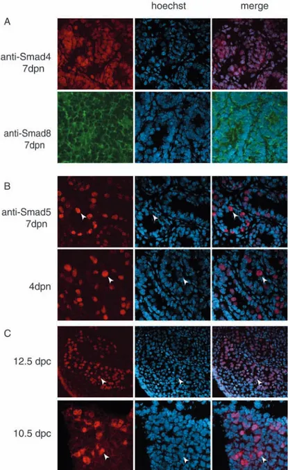
Activation of type II-type I receptors of the TGFβ family members leads to the phosphorylation of receptor activated Smads (R-Smads: Smad 1, Smad2, Smad3, Smad5, and Smad8) (Graff et al., 1996). Among the R-Smads, Smad1, Smad5 and Smad8 have been shown to be preferential substrates for type I BMP receptors (Hoodless et al., 1996; Nishimura et al., 1998). Upon phosphorylation, R-Smads oligomerize with common mediator Smad (Smad4) (Zhang et al., 1997) and translocate to the nucleus. Since we observed that BMP4 induced an increase of the proliferation rate in spermatogonia, we analyzed which of the transduction molecules among the BMP-activated Smads were activated by BMP4. We first performed immunofluorescence experiments on testicular sections at 4 and 7 dpn of development using antibodies against Smad1, Smad4, Smad5 and Smad8 (Fig. 4). The anti-Smad4 and anti-Smad8 antibodies stained all the cellular types within the testis, and Smad8 was localized predominantly in the cytoplasm (Fig. 4A). In agreement with previous observations (Zhao and Hogan, 1997), Smad1 was negative at both ages (not shown). Interestingly, anti-Smad5 antibodies specifically decorated the nuclear compartment of spermatogonia at both 4 dpn and 7 dpn (Fig. 4B). Smad5 was also expressed in PGCs at 10.5 dpc, during their migratory period, and at 12.5 dpc, when they have already colonized the gonads (Fig. 4C).
To test which of the Smads was actually transducing BMP4 signaling in spermatogonia, cells, isolated from 4 and 7 dpn animals, were stimulated by the factor and after 1 hour they were subjected to immunofluorescence. Fig. 5A shows that
BMP4 induces nuclear translocation of Smad4 in 4 dpn spermatogonia, while in the controls a cytoplasmatic ring was evident. Similar studies were performed using anti-Smad5 and anti-Smad8 antibodies. Smad5 showed nuclear localization in control spermatogonia; however, after 1 hour of BMP4 stimulation, the intensity of the immunostaining in the nuclear compartment increased (Fig. 5B). On the contrary, Smad8 was predominantly cytoplasmic both in the control and BMP-treated cells (Fig. 5C). Nuclear translocation of Smad4 and the increase of Smad5 nuclear levels were also confirmed by western blot analysis of nuclear and cytoplasmic extracts obtained from control and BMP4-treated 7 dpn spermatogonia (Fig. 5D).
Smad4/Smad5 form a functional DNA-binding complex in BMP4-treated spermatogonia
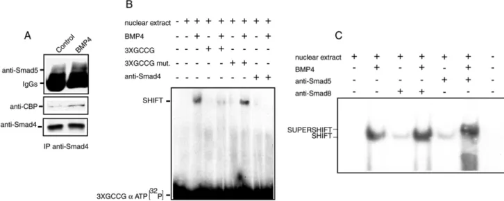
Because both Smad4 and Smad5 levels increased in the nuclear compartment of spermatogonia treated with BMP4 for 1 hour, we tested whether they were interacting in the BMP4-treated cells. After immunoprecipitation with anti-Smad4 antibodies, an increase of Smad5 association was evident in BMP4-treated cell extracts (Fig. 6A). It has been shown that the transcriptional activity of the Smad4/R-Smad complex can be mediated by the recruitment of the transcriptional coactivator CBP/p300 in the complex (Pouponnot et al., 1998). To verify whether this event was also occurring in spermatogonia, the Smad4 immunoprecipitates were probed with an anti-CBP/p300 antibody. Fig. 6A shows that, only in the BMP-treated cells, CBP is actually associated to the complex.
We then investigated whether a GC-rich synthetic double-stranded oligomer, which has been previously shown to be recognized by the Smad4/Smad5 complex (Kusanagi et al., 2000), was able to form DNA-protein complexes in the presence of nuclear extracts from BMP4-treated or untreated spermatogonia. Fig. 5B shows an electrophoretic mobility shift assay (EMSA), in which after 1 hour of BMP4 treatment a discrete DNA-protein complex was evident in nuclear extracts from BMP4-treated cells. The presence of a molar excess of unlabeled double-stranded oligomer was able to prevent the formation of the complex, whereas the presence of a molar excess of a mutated oligomer did not compete for the complex formation. Pre-incubation of the extracts with anti-Smad4 (Fig. 6B) was able to inhibit the formation of the specific retarded band but this effect was not observed with anti-Smad8 antibody (Fig. 6C). The presence of anti-Smad5 resulted in supershift of the DNA complex, indicating that both Smad4 and Smad5 are present in the BMP4-induced DNA-protein complex (Fig. 6C).
BMP4 regulates c-kit expression in spermatogonia
It has been suggested that BMP4 may play a potential role in PGC differentiation by inducing receptors for survival factors (Fujiwara et al., 2001). Since Kit is a receptor that mediates proliferation and survival of differentiated spermatogonia through its ligand Kitl, we hypothesized that BMP4 treatment could induce c-kit expression in undifferentiated 4 dpn spermatogonia, which are not sensitive to Kitl [(Dolci et al., 2001); see also Fig. 2]. Cells were cultured for 48 hours in the presence or absence of BMP4, and Kitl was added for the last 24 hours in a set of cultures for both conditions. We found that
CONTROL BMP4 Kitl BMP4+Kitl * 0 0.5 1 1.5 2 2.5 3 3.5 4 4.5 5 4dpn 7dpn F OL D O F I NCR EASE I N 3H T HY M IDI N E I NCOR P OR A T ION * * * *
Fig. 3. BMP4 induces [3H]thymidine incorporation in postnatal spermatogonia. Proliferative response to BMP4 and Kitl in 4 and 7 dpn isolated spermatogonia. Mean of the fold increase of
[3H]thymidine incorporating cells for at least four independent experiments. Bars represent the standard deviation. Asterisks indicate the presence of statistical significance (P<0.001).
BMP4 pretreatment actually induces Kitl responsiveness in Kit-negative spermatogonia, since a significant additive effect of the two growth factors in the last 24 hours of culture was observed (Fig. 7A). To acquire Kitl sensitivity, 4 dpn spermatogonia had to be incubated in the presence of BMP4 at least for 24 hours, since when BMP4 and Kitl were added simultaneously for 24 hours, no additive effect of Kitl was observed (see Fig. 3).
A probable explanation for the induction of Kitl responsiveness in these cells would be the induction of c-kit expression. To test this hypothesis, we treated 4 dpn spermatogonia for 24 hours with or without 100 ng/ml of BMP4 and immunoprecipitated the cell extracts with an anti-Kit antibody. Because of the low levels of anti-Kit expression at this age, the immunoprecipitates were subjected to constitutive autophosphorylation in a kinase assay by the addition of [γ-32P]ATP prior to the electrophoretic separation. Fig. 7B shows that BMP4 induces a dramatic increase of Kit expression
in 4 dpn spermatogonia. Kit induction by BMP4 was confirmed also in spermatogonia isolated from 7 dpn mice by northern blot analysis (Fig. 7C).
Discussion
Proliferation and differentiation of mitotic spermatogonia in the prepuberal testis depend on the production of secreted growth factors by the nursing Sertoli cells within the seminiferous tubules. The Kitl/Kit system plays an essential role in the initiation and maintenance of spermatogenesis, since artificially induced Kit point mutations (Kissel et al., 2000; Blume-Jensen et al., 2000) or naturally occurring Kitl mutations (Brannan et al., 1992) block spermatogonia proliferation and/or differentiation. BMP8b exerts proliferative and antiapoptotic effects on spermatogonia and spermatocytes (Zhao et al., 1996); however, the penetrance of its disruption in knockout animals suggests that other factors might compensate its loss within the seminiferous tubules. In the present study we identify BMP4 as a proliferation/differentiation factor for prepuberal Kit-negative spermatogonia just before they enter into the Kitl-dependent cycle of spermatogenesis.
BMP4/Alk3/Smad5 are differentially and
developmentally regulated in the prepuberal testis
The role of BMP4 in the generation of PGCs has been clearly shown in knockout mice (Lawson et al., 1999). In the early mouse embryo, at around 5.5 dpc, BMP4 is produced by the extraembryonic ectoderm, and in combination with BMP8b (Ying et al., 2001) and with BMP2 (from endodermal origin) (Ying and Zhao, 2001), it plays a role in the induction of PGC precursors. At later stages (7 dpc) it is produced by extraembryonic mesoderm and it has been shown to be required for the proper allocation within the allantois, survival and differentiation of PGCs (Fujiwara et al., 2001). Here we report that, in the postnatal development, BMP4 regulates proliferation and differentiation of the descendants of male PGCs, i.e. spermatogonia. BMP8b has been shown to be produced by germ cells (Zhao et al., 1996), whereas BMP4 is expressed at high levels in the early postnatal testis by Sertoli cells and its expression is developmentally regulated. The maximal BMP4 expression in the testis coincides with the presence of Kit-negative spermatogonia within the seminiferous tubules, suggesting that these cells might be the natural target of BMP4 action in the prepuberal testis. Indeed,
Fig. 4. Smads expression in postnatal testis and in PGCs.
(A) Frozen sections from testes at 7 dpn were analyzed by immunofluorescence for the expression of Smad4 and Smad8, which shows ubiquitous immunostaining. (B) Frozen sections from testes at 4 and 7 dpn mice, (C) from 12.5 dpc fetal testis (200×magnification) and from 10.5 dpc genital ridges were analyzed for Smad5 expression, which shows a nuclear immunostaining pattern only in the germ cell compartment (arrowheads point to germ cells, within the seminiferous tubules or within the genital ridge; 400× magnifications).
spermatogonia express the BMP receptor Alk3 at 4 dpn and are sensitive to BMP4, as demonstrated by the in vitro
proliferation assay. Activation of the BMP4 signal transduction pathway in spermatogonia appears to be mediated by the Smad4/5 complex. Smad5 is specifically expressed in mitotic spermatogonia, but not in somatic cells, and mediates a rapid Smad4 nuclear translocation upon BMP4 treatment of spermatogonia. Since Smad1 is not expressed in mitotic spermatogonia and Smad8 does not change its cytoplasmic distribution after BMP4 stimulation, Smad5 is the molecular target of Alk3 activation in these cell types.
BMP-activated Smads are known to play an essential role in PGC generation and localization in the early mouse embryo (Tremblay et al., 2001; Chang and Matzuk, 2001); however, it has not been formally shown whether they are expressed in PGCs. Here we show that both the receptor Alk3 and Smad5 are expressed in migratory PGCs and in the germ cell compartment of the male fetal gonad, and that their expression is maintained in the postnatal life in mitotic spermatogonia, thus suggesting that Alk3 and Smad5 mediate BMP signaling throughout the development of mitotic germ cells.
Smad4/5 and CBP form an active complex upon BMP4 stimulation
We demonstrate that Smad5 activation in response to BMP4 leads to Smad4 association and nuclear translocation after 1 hour of stimulation. The GCCG motif has been shown to be a Smad1/5/4-binding element (Kusanagi et al., 2000; Ishida et al., 2000) and appears to be conserved in Drosophila Dpp activated promoters (Kim et al., 1997; Xu et al., 1998). We found that the BMP4-induced Smad4/5 complex formation has DNA-binding ability. When an oligomer containing the GCCG motif was used as a probe in EMSA experiments, it was specifically retarded in BMP4-treated extracts. It is known that Smad4 is able to form homo- and hetero-oligomers with R-Smads upon activation (for a review, see Heldin et al., 1997). The DNA-binding activity of the heteromeric complex induced by BMP4 was inhibited by the presence of an anti-Smad4 antibody against the MH2 domain (oligomerization domain). However, since the anti-Smad4 antibody is able to co-immunoprecipitate Smad5, it is possible that the antibody inhibits Smad4-Smad4 interactions in the formation of the heteromeric complex, rather than interfering with the physical association of Smad4 to Smad5.
Just as with many transcription factors, the R-Smad proteins interact with the transcriptional coactivators CBP and p300 through their MH2 domains (Feng et al., 1998; Janknecht et al., 1998; Pouponnot et al., 1998; Topper et al., 1998). CBP/p300 are essential coactivators for a wide variety of transcriptional promoters (Mannervik and Akusjarvi, 1997) and possess intrinsic histone acetyltransferase activity. CBP and p300 contain three highly conserved cysteine-histidine-rich domains, two of which have been shown to interact with the Smad activation
Fig. 5. Nuclear translocation of Smad4 upon BMP4 stimulation in postnatal
spermatogonia. Immunofluorescence analysis for Smad4 in control and BMP4-treated spermatogonia isolated at 4 dpn with (A) Smad4, (B) Smad5, and (C) Smad8 (400×magnifications). The merged images show the nuclear localization of Smad4 and Smad5, but not of Smad8. (D) Western blot analysis for Smad4, Smad5 and Smad8 in nuclear and cytoplasmic extracts from control and BMP4-treated 7 dpn spermatogonia. Twenty µg of proteins were loaded in each lane.
domain of Smad4, (de Caestecker et al., 2000), and with R-Smads (Feng et al., 1998; Janknecht et al., 1998; Nishihara et al., 1998), respectively. According to this model, we found that
only in extracts from BMP4-treated spermatogonia is CBP associated to the Smad4/5 heteromeric complex, suggesting that cooperation between Smads and CBP enhances BMP4-mediated activation of transcription in spermatogonia.
Kit is upregulated by BMP4 in mitotic spermatogonia
It has been shown that TGF-β can downregulate c-kit expression in hemopoietic cells (Sansilvestri et al., 1995; Heinrich et al., 1995), while it upregulates c-kit expression in T-leukemia cells and in melanoblasts (Tomeczkowski et al., 1998; Kawakami et al., 2002). To date no reports have been obtained on the factors that regulate c-kit expression in germ cells. We found that exposure of spermatogonia to exogenous BMP4 is able to upregulate Kit expression both at the RNA and protein level, conferring Kitl responsiveness to spermatogonia at 4 dpn, when the majority of germ cells are Kit negative. It is possible that BMP4 activation of Smad4/5 directly mediates c-kit expression in germ cells. Indeed, there are several GCCG elements within the 4225 bp of the c-kit promoter region (accession no. X86451), suggesting potential binding sites for Smad4/5. An alternative possibility is that the BMP4-activated signaling might regulate the expression of germ cell specific transcription factor(s), which in turn activates c-kit gene expression. It has been shown, in fact, that the c-kit gene is specifically regulated by tal1/SCL transcription factor in hematopoietic cells (Krosl et al.,1998) and by MITF in mast cells (Tsujimura et al., 1996).
Our data show that BMP4 acts both as proliferation and differentiation factor in undifferentiated spermatogonia in vitro. Proliferation of undifferentiated spermatogonia has also been shown to be stimulated by GDNF produced by Sertoli cells within the prepuberal testis (Meng et al., 2000; Viglietto et al., 2000); however, GDNF seems rather to block their
Fig. 6. BMP4 stimulation induces Smad4/Smad5/CBP association and DNA-binding activity in spermatogonia. (A) Eighty µg of total cell protein extracts from control or BMP4-treated spermatogonia were immunoprecipitated for Smad4 (lower panel) and immunoblotted for Smad5 and CBP (upper and middle panel, respectively). (B) DNA-binding activity in nuclear extracts from control and BMP4-treated spermatogonia probed with a 3×GCCG-[α-32P]ATP double-stranded oligonucleotide, in the presence of 100×molar excess of competitors (wild-type or mutated 3×GCCG double-stranded oligonucleotides) or in the presence of an anti-Smad4 antibody. (C) DNA-binding activity in nuclear extracts from control and BMP4-treated spermatogonia probed with a 3×GCCG-[α-32P]ATP double-stranded oligonucleotide, in the presence or absence of anti-Smad5 and anti-Smad8 antibodies.
Fig. 7. BMP4 treatment induces Kitl sensitivity and Kit expression in
Kit-negative and Kit-positive spermatogonia. (A) Mean of the fold increase of [3H]thymidine incorporating cells from three independent experiments. Asterisks indicate the presence of statistical
significance (P<0.001). (B) Kit immunoprecipitation and in vitro autophosphorylation from 4 dpn spermatogonia total extracts, treated or untreated with BMP4 for 24 hours. (C) Northern blot analysis for c-kit expression in 7 dpn spermatogonia RNAs, treated or untreated with BMP4 for 24 hours.
differentiation, as shown by overexpression experiments in transgenic mice (Meng et al., 2000). Since spermatogonia stem cells are able to renew themselves and at the same time to progress through differentiation (i.e. to the Kit-dependent stages of proliferation), BMP4 could be one of the factors that regulates such process. Alternatively, BMP4 could act on a subset of spermatogonia that has lost stem cell features but still has to become a Kit-positive class of germ cells.
This work was supported by grants from the Agenzia Spaziale Italiana (ASI) and MIUR National project on “Development and Differentiation of Germ Cells”.
References
Blume-Jensen, P., Jiang, G., Hyman, R., Lee, K. F., O’Gorman, S. and Hunter, T. (2000). Kit/stem cell factor receptor-induced activation of phosphatidylinositol 3′-kinase is essential for male fertility. Nat. Genet. 24, 157-162.
Brannan, C. I., Bedell, M. A., Resnick, J. L., Eppig, J. J., Handel, M. A., Williams, D. E., Lyman, S. D., Donovan, P. J., Jenkins, N. A. and Copeland, N. G. (1992). Developmental abnormalities in Steel17H mice result from a splicing defect in the steel factor cytoplasmic tail. Genes Dev. 6, 1832-1842.
Chang, H. and Matzuk, M. (2001). Smad5 is required for mouse primordial germ cell development. Mech. Dev. 104, 61-67.
De Caestecker, M. P., Yahata, T., Wang, D., Parks, W. T., Huang, S., Hill, C. S., Shioda, T., Roberts, A. B. and Lechleider, R. J. (2000). The Smad4 activation domain (SAD) is a proline-rich, p300-dependent transcriptional activation domain. J. Biol. Chem. 275, 2115-2122.
De Rooij, D. G. (2001). Proliferation and differentiation of spermatogonial stem cells. Reproduction 121, 347-354.
Dolci, S., Williams, D. E., Ernst, M. K., Resnick, J. L., Brannan, C. I., Lock, L. F., Lyman, S. D., Boswell, H. S. and Donovan, P. J. (1991). Requirement for mast cell growth factor for primordial germ cell survival in culture. Nature 352, 809-811.
Dolci, S., Pellegrini, M., di Agostino, S., Geremia, R. and Rossi, P. (2001). Signaling through extracellular signal-regulated kinase is required for spermatogonial proliferative response to stem cell factor. J. Biol. Chem. 276, 40225-40233.
Feng, X. H., Zhang, Y., Wu, R. Y. and Derynck, R. (1998). The tumor suppressor Smad4/DPC4 and transcriptional adaptor CBP/p300 are coactivators for smad3 in TGF-beta-induced transcriptional activation. Genes Dev. 12, 2153-2163.
Feng, L. X., Ravindranath, N. and Dym, M. (2000). Stem cell factor/c-kit up-regulates cyclin D3 and promotes cell cycle progression via the phosphoinositide 3-kinase/p70 S6 kinase pathway in spermatogonia. J. Biol. Chem. 275, 25572-25576.
Fujiwara, T., Dunn, N. R. and Hogan, B. L. (2001). Bone morphogenetic protein 4 in the extraembryonic mesoderm is required for allantois development and the localization and survival of primordial germ cells in the mouse. Proc. Natl. Acad. Sci. USA 98, 13739-13744.
Graff, J. M., Bansal, A. and Melton, D. A. (1996). Xenopus Mad proteins transduce distinct subsets of signals for the TGF beta superfamily. Cell 85, 479-487.
Grimaldi, P., Piscitelli, D., Albanesi, C., Blasi, F., Geremia, R. and Rossi, P. (1993). Identification of 3′,5′-cyclic adenosine monophosphate-inducible nuclear factors binding to the human urokinase promoter in mouse Sertoli cells. Mol. Endocrinol. 7, 1217-1225.
Heinrich, M. C., Dooley, D. C. and Keeble, W. W. (1995). Transforming growth factor beta 1 inhibits expression of the gene products for steel factor and its receptor (c-kit). Blood 85, 1769-1780.
Heldin, C. H., Miyazono, K. and ten Dijke, P. (1997). TGF-beta signalling from cell membrane to nucleus through SMAD proteins. Nature 390, 465-471.
Hoodless, P. A., Haerry, T., Abdollah, S., Stapleton, M., O’Connor, M. B., Attisano, L. and Wrana J. L. (1996). MADR1, a MAD-related protein that functions in BMP2 signaling pathways. Cell 85, 489-500.
Ishida, W., Hamamoto, T., Kusanagi, K., Yagi, K., Kawabata, M., Takehara, K., Sampath, T. K., Kato, M. and Miyazono, K. (2000). Smad6 is a Smad1/5-induced smad inhibitor. Characterization of bone
morphogenetic protein-responsive element in the mouse Smad6 promoter. J. Biol. Chem. 275, 6075-6079.
Janknecht, R., Wells, N. J. and Hunter, T. (1998). TGF-beta-stimulated cooperation of smad proteins with the coactivators CBP/p300. Genes Dev. 12, 2114-2119.
Kawakami, T., Soma, Y., Kawa, Y., Ito, M., Yamasaki, E., Watabe, H., Hosaka, E., Yajima, K., Ohsumi, K. and Mizoguchi, M. (2002). Transforming growth factor beta1 regulates melanocyte proliferation and differentiation in mouse neural crest cells via stem cell factor/KIT signaling. J. Invest. Dermatol. 118, 471-478.
Kim, J., Johnson, K., Chen, H. J., Carroll, S. and Laughon, A. (1997). Drosophila Mad binds to DNA and directly mediates activation of vestigial by Decapentaplegic. Nature 388, 304-308.
Kissel, H., Timokhina, I., Hardy, M. P., Rotschild, G., Tajima, Y., Soares, V., Angeles, M., Whitlow, S. R., Manova, K. and Bessmer P. (2000). Point mutation in Kit receptor tyrosine kinase reveals essential roles for kit signaling in oogenesis and spermatogenesis without affecting other kit responses. EMBO J. 19, 1312-1326.
Kusanagi, K., Inoue, H., Ishidou, Y., Mishima, H. K., Kawabata, M. and Miyazono, K. (2000). Characterization of a bone morphogenetic protein-responsive Smad-binding element. Mol. Biol. Cell 11, 555-565.
Krosl, G., He, G., Lefrancois, M., Charron, F., Romeo, P. H., Jolicoeur, P., Kirsch, I. R., Nemer, M. and Hoang, T. (1998). Transcription factor SCL is required for c-kit expression and c-Kit function in hemopoietic cells. J. Exp. Med. 188, 439-450.
Lawson, K. A., Dunn, N. R., Roelen, B. A., Zeinstra, L. M., Davis, A. M., Wright, C. V., Korving, J. P. and Hogan, B. L. (1999). Bmp4 is required for the generation of primordial germ cells in the mouse embryo. Genes Dev. 13, 424-436.
Mannervik, M. and Akusjarvi, G. (1997). The transcriptional co-activator proteins p300 and CBP stimulate adenovirus E1A conserved region 1 transactivation independent of a direct interaction. FEBS Lett. 414, 111-116. Manova, K., Nocka, K., Besmer, P. and Bachvarova, R. F. (1990). Gonadal expression of c-kit encoded at the W locus of the mouse. Development 110, 1057-1069.
Meng, X., Lindahl, M., Hyvonen, M. E., Parvinen, M., de Rooij, D. G., Hess, M. W., Raatikainen-Ahokas, A., Sainio, K., Rauvala, H., Lakso, M. et al. (2000). Regulation of cell fate decision of undifferentiated spermatogonia by GDNF. Science 287, 1489-1493.
Nishihara, A., Hanai, J. I., Okamoto, N., Yanagisawa, J., Kato, S., Miyazono, K. and Kawabata, M. (1998). Role of p300, a transcriptional coactivator, in signalling of TGF-beta. Genes Cells 3, 613-623.
Nishimura, R., Kato, Y., Chen, D., Harris, S. E., Mundy, G. R. and Yoneda, T. (1998). Smad5 and DPC4 are key molecules in mediating BMP-2-induced osteoblastic differentiation of the pluripotent mesenchymal precursor cell line C2C12. J. Biol. Chem. 273, 1872-1879.
Pesce, M., Klinger, G. F. and de Felici, M. (2002). Derivation in culture of primordial germ cells from cells of the mouse epiblast: phenotypic induction and growth control by Bmp4 signalling. Mech. Dev. 112, 15-24.
Pouponnot, C., Jayaraman, L. and Massague, J. (1998). Physical and functional interaction of SMADs and p300/CBP. J. Biol. Chem. 273, 22865-22868.
Rossi, P., Albanesi, C., Grimaldi, P. and Geremia, R. (1991). Expression of the mRNA for the ligand of c-kit in mouse sertoli cells. Biochem. Biophys. Res. Commun. 176, 910-914.
Rossi, P., Marziali, G., Albanesi, C., Charlesworth, A., Geremia, R. and Sorrentino, V. (1992). A novel c-kit transcript, potentially encoding a truncated receptor, originates within a kit gene intron in mouse spermatids. Dev. Biol. 152, 203-207.
Rossi, P., Dolci, S., Albanesi, C., Grimaldi, P., Ricca, R. and Geremia, R. (1993). Follicle-stimulating hormone induction of steel factor (SLF) mRNA in mouse sertoli cells and stimulation of DNA synthesis in spermatogonia by soluble SLF. Dev. Biol. 155, 68-74.
Sansilvestri, P., Cardoso, A. A., Batard, P., Panterne, B., Hatzfeld, A., Lim, B., Levesque, P., Monier, M. N. and Hatzfeld, J. (1995). Early CD34high cells can be separated into KIT high cells in which transforming growth factor-beta (TGF-beta) downmodulates c-kit and KIT low cells in which anti-TGF-beta upmodulates c-kit. Blood 86, 1729-1735.
Schrans-Stassen, B. H., van de Kant, H. J., de Rooij, D. G. and van Pelt, A. M. (1999). Differential expression of c-kit in mouse undifferentiated and differentiating type A spermatogonia. Endocrinology 140, 5894-5900. Sorrentino, V., Giorgi, M., Geremia, R., Besmer, P. and Rossi, P. (1991).
Expression of the c-kit proto-oncogene in the murine male germ cells. Oncogene 6, 149-151.
Tomeczkowski, J., Frick, D., Schwinzer, B., Wittner, N., Ludwig, W. D., Reiter, A., Welte, K. and Sykora, K. W. (1998). Expression and regulation of c-kit receptor and response to stem cell factor in childhood malignant T-lymphoblastic cells. Leukemia 12, 1221-1229.
Topper, J. N., DiChiara, M. R., Brown, J. D., Williams, A. J., Falb, D., Collins, T. and Gimbrone, M. A. Jr (1998). CREB binding protein is a required coactivator for Smad-dependent, transforming growth factor beta transcriptional responses in endothelial cells. Proc. Natl. Acad. Sci. USA 95, 9506-9511.
Tremblay, K. D., Dunn, N. R. and Robertson, E. J. (2001). Mouse embryos lacking Smad1 signals display defects in extra-embryonic tissues and germ cell formation. Development 128, 3609-3621.
Tsujimura, T., Morii, E., Nozaki, M., Hashimoto, K., Moriyama, Y., Takebayashi, K., Kondo, T., Kanakura, Y. and Kitamura, Y. (1996). Involvement of transcription factor encoded by the mi locus in the expression of c-kit receptor tyrosine kinase in cultured mast cells of mice. Blood 88, 1225-1233.
Viglietto, G., Dolci, S., Bruni, P., Baldassarre, G., Chiariotti, L., Melillo, R. M., Salvatore G., Chiappetta, G., Sferratore, F., Fusco, A. and Santoro, M. (2000). Glial cell line-derived neutrotrophic factor and neurturin can act as paracrine growth factors stimulating DNA synthesis of Ret-expressing spermatogonia. Int. J. Oncol. 16, 689-694.
Xu, X., Yin, Z., Hudson, J. B., Ferguson, E. L. and Frasch, M. (1998). Smad proteins act in combination with synergistic and antagonistic regulators to
target Dpp responses to the Drosophila mesoderm. Genes Dev. 12, 2354-2370.
Ying, Y., Liu, X. M., Marble, A., Lawson, K. A. and Zhao, G. Q. (2000). Requirement of Bmp8b for the generation of primordial germ cells in the mouse. Mol. Endocrinol. 14, 1053-1063.
Ying, Y. and Zhao, G. Q. (2001) Cooperation of endoderm-derived BMP2 and extraembryonic ectoderm-derived BMP4 in primordial germ cell generation in the mouse. Dev. Biol. 232, 484-492.
Ying, Y., Qi, X. and Zhao, G. Q. (2001). Induction of primordial germ cells from murine epiblasts by synergistic action of BMP4 and BMP8B signaling pathways. Proc. Natl. Acad. Sci. USA 98, 7858-7862.
Yoshinaga, K., Nishikawa, S., Ogawa, M., Hayashi, S., Kunisada, T., Fujimoto, T. and Nishikawa, S. (1991). Role of c-kit in mouse spermatogenesis: identification of spermatogonia as a specific site of c-kit expression and function. Development 113, 689-699.
Zhang, Y., Musci, T. and Derynck, R. (1997). The tumor suppressor Smad4/DPC 4 as a central mediator of Smad function. Curr. Biol. 7, 270-276. Zhao, G. Q., Deng, K., Labosky, P. A., Liaw, L. and Hogan, B. L. (1996). The gene encoding bone morphogenetic protein 8B is required for the initiation and maintenance of spermatogenesis in the mouse. Genes Dev. 10, 1657-1669.
Zhao, G. Q. and Hogan, B. L. (1997). Evidence that Mothers-against-dpp-related 1 (Madr1) plays a role in the initiation and maintenance of spermatogenesis in the mouse. Mech. Dev. 61, 63-73.
![Fig. 3. BMP4 induces [ 3 H]thymidine incorporation in postnatal spermatogonia. Proliferative response to BMP4 and Kitl in 4 and 7 dpn isolated spermatogonia](https://thumb-eu.123doks.com/thumbv2/123dokorg/8043601.122930/5.918.99.425.108.378/thymidine-incorporation-postnatal-spermatogonia-proliferative-response-isolated-spermatogonia.webp)