Review Article
Meiotic double strand breaks repair in sexually reproducing
eukaryotes: We are not all equal
Adriana La Volpe
a,⁎
, Marco Barchi
b,⁎⁎
aInstitute of Genetics and Biophysics‘Adriano Buzzati-Traverso’ CNR, Via Pietro Castellino 111, Napoli 80131, Italy
bDepartment of Public Health and Cell Biology, section of Anatomy, University of Rome“Tor Vergata”, Via Montpellier n.1, Rome 00133, Italy
A R T I C L E I N F O R M A T I O N A B S T R A C T Article Chronology:
Received 17 January 2012 Revised version received 14 March 2012 Accepted 14 March 2012 Available online 27 March 2012 Keywords:
Meiosis Mus musculus C. elegans
Meiotic recombination © 2012 Elsevier Inc. All rights reserved.
Contents
Introduction . . . 1333
Recombination initiation . . . 1334
Processing of meiotic DSBs and strand invasion . . . 1335
Interhomolog vs. sister bias . . . 1336
CO vs. NCO and crossover interference . . . 1336
Crossover resolution . . . 1337
Acknowledgments . . . 1337
References . . . 1337
Introduction
The defining event of meiosis is prophase-I, during which the maternal and paternal chromosome find each other in the nucleus,
pair, and align in a process called“synapses of the homologues.” Their faithful segregation during the first meiotic division (MI) re-quires meiotic recombination and in particular at least one crossing over (CO) per pair of homologous chromosomes. COs are needed, ⁎ Correspondence to: A. La Volpe, Institute of Genetics and Biophysics ‘Adriano Buzzati-Traverso’ CNR, Italy. Fax: +39 081 6132706.
⁎⁎ Correspondence to: M. Barchi, University of Rome Tor Vergata, Italy. Fax: +39 06 72596268. E-mail addresses:[email protected](A. La Volpe),[email protected](M. Barchi). 0014-4827/$– see front matter © 2012 Elsevier Inc. All rights reserved.
doi:10.1016/j.yexcr.2012.03.014
A v a i l a b l e o n l i n e a t w w w . s c i e n c e d i r e c t . c o m
not only to generate diversity within a population, but (along with sister chromatids cohesion) to form the chiasmata that are the physical connection between the homologues that ensures their faithful segregation at metaphase-I. Our under-standing of recombination in higher eukaryotes has comes from studies in model organisms such as yeasts, flies and worms. Although there are clear differences among organisms, most of the genes and proteins that are required in these pro-cesses are conserved and have orthologues in mammals. How-ever, their null mutations in mice (Mus musculus) do not always display the same phenotype as in lower eukaryotes, indicating that along with the increased complexity of the ge-nome, same genes have acquired new or partially overlapping functions. In this review we will focus on the main genes and protein products which are required for meiotic recombi-nation, comparing the simple metazoan C. elegans and the mouse, underlying divergences and similarities between these organisms.
Recombination initiation
Meiotic homologous recombination is initiated by the formation of double strand breaks (DSBs) in one sister chromatid of one ho-mologue (Fig. 1a). The protein responsible for such function is SPO11, a type-II topoisomerase-like DNA trans-esterase highly conserved in all eukaryotic species. While in most organisms ho-mologous chromosome pairing and synapsis is dependent on the appearances of such DSBs, it is not so in C. elegans in which homo-logue recognition and pairing is accomplished by means of spe-cialized genomic sequences and proteins[1]. In this organism, in absence of spo-11, although meiotic recombination does not occur and homologous chromosomes appear as univalents by the end of meiotic prophase and fail to properly segregate, meiotic synapsis takes place and meiosis proceeds[2]. Interestingly, in the meiotic mutant cra-1, unlike in the wild type, DSB formation is re-quired to promote the polymerization of the central region
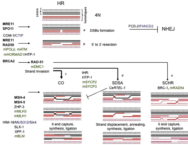
Fig. 1– Homologous Recombination (HR) during meioiosis. Recombination is initiated by SPO11-generated double strand breaks (DSBs) formation (a). Following DSBs, DNA is resected (b) to expose a 3′ single-strand DNA tail which prime the search of the homologue sequence either using one chromatid of the homologous chromosome (c and d) [Inter-homolog recombination, IHR] or the sister chromatid (e) [sister-chromatid homologous recombination, SCHR] leading to DSB repair. When the homolog
chromosome is used for repair, HR might leads to the formation of a crossing over (CO) [c]. Alternatively, the invading single-strand DNA end is displaced and the DSB is repaired by synthesis-dependent strand annealing (SDSA) which results in a non-CO product (d). In black are indicated C. elegans gene products. In black bold are indicated protein names which nomenclature is common in both C. elegans and mice. The blue color identifies the C. elegans hortolog gene product in mammals. In green are indicated mouse gene products. Note that note all the listed protein have a defined function in the specific step of recombination in both organisms (for more details see the text).
components along meiotic chromosome axes. In fact, in the ab-sence of both CRA-1 and SPO11, synaptonemal complex (SC) for-mation is perturbed[3]. This observation and the fact that DSBs are formed in the syp-2 mutants[4], lacking a fundamental synap-tonemal complex (SC) central element, also exclude the possibili-ty that in C. elegans the SC formation might be required for DSBs induction or that it might precede their appearance unlike what has sometimes been suggested[5]. In mice, as in most eukaryotes, chromosome pairing/synapses and DSB formation are instead strictly linked: failure to induce DSBs results in a failure of chro-mosome pairing/synapses in leptonema and zygonema (although some non-homologous synapses does occur) coupled with the absence of meiotic recombination markers. In Spo11−/− female mice, oocytes reach the diplotene/dictyate stage of prophase-I in nearly normal numbers, but cells are soon lost after birth[6–8]
and the few surviving oocytes, that progress further, are defective in chromosome segregation[9].
Processing of meiotic DSBs and strand invasion
In mitotic cells, DSBs can be repaired either by the non-homologous end joining pathway (NHEJ) or via non-homologous re-combination (HR). NHEJ involves direct religation of the DSB and unlike HR is prone to errors. In meiotic cells the NHEJ is repressed, promoting repair by HR[10]. It has been demonstrated that in C. elegans NHEJ during meiosis is repressed by the action of fcd-2 (orthologue of FANCD2) preventing illegitimate repair[11].
In meiosis, the repair of DSBs by HR leads to the formation of COs and non crossovers (NCOs); in C. elegans oogenesis, as in mice, only a fraction of DSBs become COs, but each chromosome pair has at least one (obligatory) CO which is required to lock the homologues together during the first meiotic division.
During HR, following the generation of the meiosis-specific DSBs, Spo11 is removed from DNA, and 5′-ending strands are degraded to expose 3′-ending single-stranded DNA (ssDNA) [Fig. 1b]. These tails, invading an intact homologous DNA duplex, are used as primers for DNA synthesis. Strand invasion of homolo-gous chromosome by the 3′ single strand is mediated by members of the RecA family: namely RAD51 and the meiotic-specific DMC1. Removal of Spo11 from meiotic DSB ends involves in yeast the Sae2 and Mre11-Rad50-Xrs2-dependent endonucleolytic step that re-leases Spo11 bound to a short nucleotide sequence[12,13].
The C. elegans orthologue of Sae2, COM-1, is a conserved eu-karyotic protein known in mammals as CtIP. In com-1 mutants meiotic DSBs are formed, but fail to load the RAD-51 protein and homologous recombination does not occur, although exposure to ionizing radiations induces RAD-51 foci, which suggests that the failure of RAD-51 loading is specific to SPO-11-generated DSBs
[14]. As shown in budding yeast the MRE-11 protein is necessary in C. elegans for DSB induction and end-processing. mre-11 mu-tants are, in fact, unable to repair meiotic chromosomes after ion-izing radiation[15], but intact chromosomes are observed in the absence of both RAD-51 and MRE-11[16]. RAD-51 loading is also RAD-50 dependent in the very early stages of meiotic DSB repair, however at later stages of pachytene (as well as in the premeiotic mitotic zone) RAD-51 loading is independent of RAD-50. Require-ment of RAD-50 for DSBs formation is alleviated in absence of the meiotic cohesin REC-8 suggesting that this cohesin may limit the activity of SPO-11 in DSB formation[17].
In mice a role for CtIP in meiosis has been not established yet. In addition, a role for the MRX complex (consisting of MRE11, RAD50 and NBS1) in the endonucleolytic cleavage that releases the SPO11–oligonucleotide complex from DNA has not been found, in part perhaps, because mutants are unviable. However, the analysis of meiotic events in mice harbouring hypomorphic MRE11 and NBS1 mutations revealed that these proteins are re-quired for processing Spo11-induced DSBs (as evidenced by the persistence Rad51 foci at pachynema) and proper placement of MLH1 foci, a marker of CO formation[18]. Interestingly, even if the molecular players involved in DSB resection in mammals have been not identified, it has recently found that DNA polymer-aseβ is critical for efficient and/or timely removal of the SPO11 complex from DSB ends[19]; and that ATM influences an early step in nucleolytic processing of meiotic DSBs as proposed in yeast, and constrains DSBs formation[20]. Whether this control is also functional in C. elegans is unknown.
A missing actor in the strand invasion step in C. elegans (as well as in Drosophila and Neurospora Crassa) is the RecA-like protein Dmc1. In C. elegans RAD-51 protein is more similar to members of the Rad51 family than to Dmc1, however some conserved Dmc1-specific aminoacids are retained suggesting that the C. ele-gans RAD-51 may represent a hybrid performing the activities of both these strand-invading proteins[16,21].
In most organisms (including mice) in which Dmc1 is present, Dmc1 mutants display meiotic recombination failure and chromo-some synapses defects, indicating that this protein has a specific function in meiotic HR-repair. What functionally distinguishes Rad51 from Dmc1 is still unclear. However in vitro experiments have recently demonstrated that the recombination intermediate formed by Dmc1 is more stable than that formed by Rad51[22], suggesting that it might be required to the formation of stable interhomolog chromosome interactions that promotes pairing and recombination. In both mitotic and meiotic cells the function of RecA family proteins is influenced by accessory proteins. During HR in mitotic cells the processed 3’ ssDNA ends are first loaded by the ssDNA-binding protein RPA, whose loading onto DNA is re-quired to protect the 3’-protruding end, RPA is then replaced by Rad51, with the aid of mediators such as Rad52 and the breast cancer related protein BRCA2.
Another missing actor in C. elegans is the Rad52 protein. Rad52 is also not required for Rad51/Dmc1 loading in mice[23]. Howev-er in both organisms the brc-2 gene (orthologous to the breast cancer susceptibility gene BRCA2 and absent in fungi) is essential for RecA-homologues loading at DSBs sites [24,16]. In nematodes, brc-2 and rad-51 mutants exhibit a similar phenotype resulting in extensive chromatin decondensation/fusion at diakinesis[16,24]. In absence of BRC-2, DNA ends appear over-resected[24]. Further-more, BRC-2 stimulates RAD-51-mediated D-loop formation and reduces the rate of ATP hydrolysis catalyzed by RAD-51 suggesting that BRC-2 can also prevent RAD-51 nucleoprotein filament depo-lymerization[25,26].
In male mice BRCA2 deficiency leads to a marked reduced as-sembly of Rad51/Dmc1 foci and lack of extensive synapses, and both males and females are infertile, indicating that its function is conserved among species[27]. BRCA1 performs a similar func-tion in RAD51 (but not DMC1) loading in male mice, and pachy-nema spermatocytes lack MLH1 foci, a marker of CO formation. Such behaviour however is sexually dimorphic, indeed BRCA1 is not required for oocyte meiosis [28]. In C. elegans oogenesis,
in line with the observation made in mice, brc-1 is dispensable for CO formation since mutations in brc-1 only mildly affect X chromosome non-disjunction and have no effects on autosomes segregation[29].
Interhomolog vs. sister bias
In mitotic cells, the preferred template for repair of DSBs by HR is the sister chromatid (sister chromatid homologous recombination [SCHR]). In meiosis on the contrary, consistent with the require-ment of homologous synapses and COs, the use of the sister tem-plate occurs in yeast at low frequency, and the preferred temtem-plate of choice is one of the chromatids of the homologous chromosome (inter homolog recombination [IHR])(Fig. 1c–d). Observation in yeast indicates that the SC axial element proteins Red1 and Hop1 to-gether with the meiotic specific kinase Mek1 form a“barrier” to SCHR (Fig. 1e) suppressing Rad51-Rad54 complex formation. This would promote the inter-homolog repair function of Dmc1[30,31]. In addition, homologous bias is also implemented by several other factors. For example, Pch2 is required for full Mek1 activity, to estab-lish proper meiotic chromosome axis structure and to regulate mei-otic inter-homolog DSB repair outcomes[32].
SC axial element proteins that as Hop1 contain chromatin binding HORMA domains have been also found in Metazoan.
In mice, the ablation of Hormad1 cause sterility in both sexes; and its protein product is required for the proper formation of the SC and support a wild type level of DSBs and/or single-stranded DSBs ends, thereby facilitating homologous search[33]. HORMAD1, as budding-yeast Hop1, is required for efficient DSB formation, however, whether HORMAD1 may play a specific role in inter-sister vs. sister bias in mice, has not been established yet. Recently a Pch2 orthologue (TRIP13) has also been identified in mice. The analyses of these mutants revealed that TRIP13 is re-quired for the removal of HORMAD1 from synapsed axes [34], completed homologous synapses, and DSB repair, indicating that at least some of the functions of Pch2 are conserved in mammals. However a specific role in inter-homolog vs. sister bias has not been ascertained [35]. Interestingly, Li and co-authors have re-cently provided genetic evidences that in mice females SYCP2 and SYCP3 axial components promotes IHR, and that as proposed in yeast, RAD54 function is required for SCHR recombination. Whether this occurs also in male mice is difficult to test due to their developmental stage arrest, and thus remains an open ques-tion[36].
In C. elegans the HTP-1 (one of the four member of the HORMA domain family involved in coordinating early meiotic events) plays a fundamental role in inhibition of SC assembly between non homologous chromosomes and in conversion of DSBs in inter-homolog crossovers possibly by inhibiting SC formation be-fore homologues are aligned and CO initiated. A further role, as suggested for Hop1 in yeast, is that of preventing the use of the sister chromatid as recombination template[37,38]therefore the specialized mode of interhomolog meiotic repair is established at the onset of prophase and then released later between middle and late pachytene stages by profound changes in the axis com-position of the HORMA family proteins and HTP-1/2 in particular [17,39]. Once the constraint is released, homologous repair via inter-sister is allowed in order to guarantee complete repair of chro-mosomes before segregation. In C. elegans homolog-independent
recombination is in fact crucial for germ cell genomic stability. SMC-5 and 6 proteins, dispensable for chiasmata formation and accurate chromosome segregation in meiosis, are however required for the successful completion of meiotic homologous recombination repair. Mutations in the smc-5 and smc-6 genes induce chromosome fragmentation and dismorphology. Genetic analysis suggests that the C. elegans SMC-5/6 proteins are required in meiosis for the processing of homolog-independent, presumably SCH-mediated, recombination repair[40]. Analogously, the brc-1 gene appears nec-essary for inter-sister homologous recombination in meiosis[29].
CO vs. NCO and crossover interference
In all the organisms the number of DSBs is higher than the number of the COs and only a fraction of DSBs is repaired as a CO, while the other repair events results in a non-COs (NCOs). In the CO path-way, following strand invasion the second processed end of a DSB is captured to form a double Holliday junction. Such specific enzymes can resolve recombination products into either CO, or NCO products. DSBs not involved in CO are thought to be repaired by synthesis-dependent strand annealing (SDSA). In SDSA the invading strand is released and the ssDNA re-anneal to the other ssDNA end of the DSB, resulting in a gene conversion with NCO (Fig. 1d). In yeast the so called ZMM proteins (ZIP1-4, MER3 MSH4 and MSH5) have been shown to be specifically required for the CO pathway; such that, when these genes are deleted, NCOs remain unaffected while COs are reduced [41]. In mice MSH4 and MSH5 have an early role in DSB repair. Indeed meio-cytes arrest at zygonema/early pachynema[42–45]precluding a cytological analysis of CO formation. However, the co-localization (on meiotic chromosome spreads) of MSH4 with a marker of pro-CO and pro-CO events (MLH3 and MLH1 respectively)[46,47]and the ability of hMSH4/hMSH5 heterodimer to bind Holliday Junctions in vitro[48], indicates that these proteins have also a role in late recombination. In addition the observation that the number of MSH4 (and presumably MSH5) foci that associate with meiotic chromosomes exceed 10-fold the number of COs, and that foci number gets pared down in the transition form zygonema to pachynema[43]has suggested that MSH4 foci might take part in a protein-directed selection of crossover sites, perhaps before the NCO/CO bias is established.
The expected phenotypes for the above-described roles in CO formation have been described for mutants in the corresponding C. elegans genes him-14/msh4 and msh-5[49,50]. Although it is often stated that deletion either in him-14/msh-4 or msh-5 abol-ishes all CO events in C. elegans, also in this nematode, as in other organisms, a small number of meiotic recombination events can occur in absence of the him-14/msh4 gene. Simple comparison of embryonic lethality (due to non-disjunction and consequent aneuploidy) and of the average number of chromatin bodies/ nuclei at diakinesis among spo-11, and him-14/msh4, and him-14/ msh4, lig-4 mutants suggest the presence of him-14/msh4 inde-pendent CO events. In fact, a spo-11 deletion strain, where no mei-otic CO occur, exhibits an embryonic lethality close to 99,7% while him-14/msh4 mutant has an average lethality of 95%. Survival can-not be simply explained by random chromosome segregation that should be equal to that of the spo-11 mutant. Furthermore about 10% of the him-14/msh4 diakinesis nuclei show 11 DAPI staining bodies or less instead of the expected 12 bodies representing the
complete set of univalent chromosomes. Finally, the appearance of linked chromosomes at diakinesis is likely to represent the forma-tion of true bivalents resulting from CO recombinaforma-tion since they do not disappear in the him-14/msh4, lig-4 background where non-homologous end joining is abolished (Adamo and Gareri, unpublished).
The C. elegans ZHP-3 is distantly related to the yeast Zip3 protein although it is not a structural component of the SC. Homozygous zhp-3 knockout worms show normal homologue pairing, SC struc-ture, and RAD-51 foci formation. However, ZHP-3 protein is essential for reciprocal recombination between homologous chromosomes and chiasma formation. In the absence of ZHP-3, reciprocal recombi-nation is abolished and double-strand breaks seem to be repaired via alternative pathways[51]. The ZHP-3 is proposed to couple synapto-nemal complex morphogenesis and crossover formation and to mark precursor sites for inter-homolog crossover in late pachytene
[52]. Interestingly, in humans, based on data from a sequencing project of the Icelandic population, it has been observed that old mothers, that have given birth to normal children, seem to share a polymorphism in the RNF212 gene (orthologue of zhp-3) associated with a higher number of COs[53]. The protein is present in mouse but its function has not been established yet.
Beside MSH4/5, two key players in CO formation in mice are MLH1 and MLH3. Mlh1−/− males and females are sterile as re-sult of about 10-fold fewer chiasmata, which cause chromosome segregation defects, indicating that MLH1 is required to form proper COs [54]. Cytologically MLH3 foci formation precedes that of MLH1 (MLH3 appears in early pachynema while MLH1 is first detected at mid-pachynema), and while MLH3 foci form in Mlh1−/− mice, no MLH1 foci are detectable in Mlh3−/−, in-dicating that MLH3 might localize to sites of exchange that are then recognized and stabilized by MLH1. Indeed all MLH1 foci, that appear at mid pachynema, co-localize with MLH3 [47], further supporting this interpretation.
In organisms where multiple COs occur on a given chromo-some, COs are spaced farther apart than predicted in a random distribution, a phenomenon known as CO interference. In mice, how interference is imposed is unknown. However it has been demonstrated that it is already detectable at late zygonema among prospective CO sites containing MSH4 or RPA foci, and that it occurs at a much higher level in pachytene among MLH1 foci. The colocalization of MSH4 and MLH1 in some early pachy-tene foci[46]is consistent with positioning of MLH1 foci in two steps, with MLH1 foci being recruited from weakly interfering MSH4 foci[55]. However, because MLH1 and MSH4 foci are dis-tributed differently along the SCs, the factors determining MLH1 focus positions along wild-type SCs might differ from those deter-mining MSH4 focus positions; thus the two levels of interference observed might be temporally and mechanistically distinct.
In wild-type C. elegans one and one only CO per pair of homol-ogous chromosomes is observed. However, as in other eukaryotes, each homologs pair is subject to more than one DSB. It has been reported that meiotic DSBs are about twice the number of final CO in C. elegans[56]. Interestingly, using chromosome fusions, it has been demonstrated that two or three chromosomes fused to-gether, when in homozygosis, limit the number of COs to one (rarely two) per couple of homologs irrespectively of their length
[57]. On the other hand, increase in axis length caused by deple-tion of condensin subunits correlates with a dominant change in DSB distribution and CO position[56].
Furthermore, it has been postulated that C. elegans RTEL-1 (orthologue of the human anti-recombinase RTEL1) regulates meiotic recombination and CO homeostasis in C. elegans by phys-ically dissociating strand invasion events[58]and promoting NCO repair by meiotic synthesis dependent strand annealing[59].
Crossover resolution
In C. elegans the orthologue of the Blm gene, him-6, coding for a RecQ elicase, was identified several years ago for its meiotic de-fects. Mutations in him-6, beside genome instability leading to a mutator phenotype and radiation hypersensitivity, lead to gener-alized chromosome non-disjunction during meiosis. Unlike in yeast, where lack of Sgs1 leads to increased recombination fre-quency, him-6 mutants exhibit reduced number of COs. HIM-18, an orthologue of MUS312/Slx4 is also required for processing CO intermediates. A reduction in CO recombination frequencies-accompanied by an increase in RAD-51 foci and subsequent chro-mosome non-disjunction support a role for HIM-18 in converting CO intermediates. Such a role is supported by physical interaction of HIM-18 with the DNA repair nucleases SLX-1 and XPF-1[60].
In mice BLM localizes to discrete foci along meiotic chromo-somes, increasing in number as homologous chromosomes pair and begin to synapse, and is gradually lost in early to mid-pachynema stage[61]. The impact of BLM in mammalian meiosis has been recently investigated in spermatocytes by using a mouse line carrying a conditional knockout (CKO) allele of Blm. The phenotypic analysis revealed that spermatocytes display non-homologous synapse defects regardless from the fact that MSH4/5 and MLH3/MLH1 pathways are intact, thus indicating that BLM prevent non-homologous synapses via a mechanism that does not affect the major DSB repair pathway. In addition, contrary to what is observed in C. elegans, CKO spermatocytes, beside a normal number of MLH1 foci, present an increase in chias-mata or chiaschias-mata-like structures, indicating that BLM is required to prevent or resolve aberrant CO events[62]. Interestingly, Mlh3−/− spermatocytes display an increased number of BLM foci. This has suggested that in normal meiosis the access of BLM to CO interme-diates is prevented (perhaps by the ZMM protein group) and as a consequence its anti-recombination activity would be directed against recombination intermediates to which MLH1-MLH3 do not bind[62].
Acknowledgments
We are grateful to Prof. John F. Pulitzer for the critical reading of the manuscript. The lab. of MB is funded by Associazione Italiana Ricerca sul Cancro (AIRC) MFAG 4765. The lab. of ALV is funded by Progetto Merit RBNE08YFN3 and Associazione Italiana Ricerca sul Cancro (AIRC) IG11422.
R E F E R E N C E S
[1] C.M. Phillips, C. Wong, N. Bhalla, P.M. Carlton, P. Weiser, P.M. Meneely, A.F. Dernburg, HIM-8 binds to the X chromosome pairing center and mediates chromosome-specific meiotic synapsis, Cell 123 (2005) 1051–1063.
[2] A.F. Dernburg, K. McDonald, G. Moulder, R. Barstead, M. Dresser, A.M. Villeneuve, Meiotic recombination in C. elegans initiates by a conserved mechanism and is dispensable for homologous chromosome synapsis, Cell 94 (1998) 387–398.
[3] S. Smolikov, K. Schild-Prufert, M.P. Colaiacovo, CRA-1 uncovers a double-strand break-dependent pathway promoting the assembly of central region proteins on chromosome axes during C. elegans meiosis, PLoS Genet. 4 (2008) e1000088.
[4] M.P. Colaiacovo, A.J. MacQueen, E. Martinez-Perez, K. McDonald, A. Adamo, A. La Volpe, A.M. Villeneuve, Synaptonemal complex assembly in C. elegans is dispensable for loading strand-exchange proteins but critical for proper completion of recombination, Dev. Cell 5 (2003) 463–474.
[5] T. Garcia-Muse, S.J. Boulton, Meiotic recombination in Caenorhabditis elegans, Chromosome Res. 15 (2007) 607–621. [6] P.J. Romanienko, R.D. Camerini-Otero, The mouse Spo11 gene is
required for meiotic chromosome synapsis, Mol. Cell 6 (2000) 975–987.
[7] F. Baudat, K. Manova, J.P. Yuen, M. Jasin, S. Keeney, Chromosome synapsis defects and sexually dimorphic meiotic progression in mice lacking Spo11, Mol. Cell 6 (2000) 989–998.
[8] M. Di Giacomo, M. Barchi, F. Baudat, W. Edelmann, S. Keeney, M. Jasin, Distinct DNA-damage-dependent and -independent responses drive the loss of oocytes in recombination-defective mouse mutants, Proc. Natl. Acad. Sci. U.S.A. 102 (2005) 737–742. [9] F. Cole, S. Keeney, M. Jasin, Evolutionary conservation of meiotic
DSB proteins: more than just Spo11, Genes Dev. 24 (2010) 1201–1207.
[10] W. Goedecke, M. Eijpe, H.H. Offenberg, M. van Aalderen, C. Heyting, Mre11 and Ku70 interact in somatic cells, but are differentially expressed in early meiosis, Nat. Genet. 23 (1999) 194–198.
[11] A. Adamo, S.J. Collis, C.A. Adelman, N. Silva, Z. Horejsi, J.D. Ward, E. Martinez-Perez, S.J. Boulton, A. La Volpe, Preventing
nonhomologous end joining suppresses DNA repair defects of Fanconi anemia, Mol. Cell 39 (2010) 25–35.
[12] M.J. Neale, J. Pan, S. Keeney, Endonucleolytic processing of covalent protein-linked DNA double-strand breaks, Nature 436 (2005) 1053–1057.
[13] M.J. Neale, S. Keeney, Clarifying the mechanics of DNA strand exchange in meiotic recombination, Nature 442 (2006) 153–158. [14] A. Penkner, Z. Portik-Dobos, L. Tang, R. Schnabel, M. Novatchkova,
V. Jantsch, J. Loidl, A conserved function for a Caenorhabditis elegans Com1/Sae2/CtIP protein homolog in meiotic recombination, EMBO J. 26 (2007) 5071–5082.
[15] G.M. Chin, A.M. Villeneuve, C. elegans mre-11 is required for meiotic recombination and DNA repair but is dispensable for the meiotic G(2) DNA damage checkpoint, Genes Dev. 15 (2001) 522–534.
[16] C. Rinaldo, P. Bazzicalupo, S. Ederle, M. Hilliard, A. La Volpe, Roles for Caenorhabditis elegans rad-51 in meiosis and in resistance to ionizing radiation during development, Genetics 160 (2002) 471–479.
[17] M. Hayashi, G.M. Chin, A.M. Villeneuve, C. elegans Germ Cells Switch between Distinct Modes of Double-Strand Break Repair During Meiotic Prophase Progression, PLoS Genet. 3 (2007) e191. [18] S.M. Cherry, C.A. Adelman, J.W. Theunissen, T.J. Hassold, P.A.
Hunt, J.H. Petrini, The Mre11 complex influences DNA repair, synapsis, and crossing over in murine meiosis, Curr. Biol. 17 (2007) 373–378.
[19] D. Kidane, A.S. Jonason, T.S. Gorton, I. Mihaylov, J. Pan, S. Keeney, D.G. de Rooij, T. Ashley, A. Keh, Y. Liu, U. Banerjee, D. Zelterman, J.B. Sweasy, DNA polymerase beta is critical for mouse meiotic synapsis, EMBO J. 29 (2010) 410–423.
[20] J. Lange, J. Pan, F. Cole, M.P. Thelen, M. Jasin, S. Keeney, ATM controls meiotic double-strand-break formation, Nature 479 (2011) 237–240.
[21] C. Rinaldo, S. Ederle, V. Rocco, A. La Volpe, The Caenorhabditis elegans RAD51 homolog is transcribed into two alternative
mRNAs potentially encoding proteins of different sizes, Mol. Gen. Genet. 260 (1998) 289–294.
[22] D.V. Bugreev, R.J. Pezza, O.M. Mazina, O.N. Voloshin, R.D. Camerini-Otero, A.V. Mazin, The resistance of DMC1 D-loops to dissociation may account for the DMC1 requirement in meiosis, Nat. Struct. Mol. Biol. 18 (2011) 56–60.
[23] T. Rijkers, J. Van Den Ouweland, B. Morolli, A.G. Rolink, W.M. Baarends, P.P. Van Sloun, P.H. Lohman, A. Pastink, Targeted inactivation of mouse RAD52 reduces homologous
recombination but not resistance to ionizing radiation, Mol. Cell. Biol. 18 (1998) 6423–6429.
[24] J.S. Martin, N. Winkelmann, M.I. Petalcorin, M.J. McIlwraith, S.J. Boulton, RAD-51-dependent and -independent roles of a Caenorhabditis elegans BRCA2-related protein during DNA double-strand break repair, Mol. Cell. Biol. 25 (2005) 3127–3139.
[25] M.I. Petalcorin, V.E. Galkin, X. Yu, E.H. Egelman, S.J. Boulton, Stabilization of RAD-51-DNA filaments via an interaction domain in Caenorhabditis elegans BRCA2, Proc. Natl. Acad. Sci. U.S.A. 104 (2007) 8299–8304.
[26] M.I. Petalcorin, J. Sandall, D.B. Wigley, S.J. Boulton, CeBRC-2 stimulates D-loop formation by RAD-51 and promotes DNA single-strand annealing, J. Mol. Biol. 361 (2006) 231–242. [27] S.K. Sharan, A. Pyle, V. Coppola, J. Babus, S. Swaminathan, J.
Benedict, D. Swing, B.K. Martin, L. Tessarollo, J.P. Evans, J.A. Flaws, M.A. Handel, BRCA2 deficiency in mice leads to meiotic impairment and infertility, Development 131 (2004) 131–142.
[28] X. Xu, O. Aprelikova, P. Moens, C.X. Deng, P.A. Furth, Impaired meiotic DNA-damage repair and lack of crossing-over during spermatogenesis in BRCA1 full-length isoform deficient mice, Development 130 (2003) 2001–2012.
[29] A. Adamo, P. Montemauri, N. Silva, J.D. Ward, S.J. Boulton, A. La Volpe, BRC-1 acts in the inter-sister pathway of meiotic double-strand break repair, EMBO Rep. 9 (2008) 287–292. [30] T.L. Callender, N.M. Hollingsworth, Mek1 suppression of meiotic
double-strand break repair is specific to sister chromatids, chromosome autonomous and independent of Rec8 cohesin complexes, Genetics 185 (2010) 771–782.
[31] H. Niu, L. Wan, V. Busygina, Y. Kwon, J.A. Allen, X. Li, R.C. Kunz, K. Kubota, B. Wang, P. Sung, K.M. Shokat, S.P. Gygi, N.M.
Hollingsworth, Regulation of meiotic recombination via Mek1-mediated Rad54 phosphorylation, Mol. Cell 36 (2009) 393–404.
[32] S. Zanders, M.S. Brown, C. Chen, E. Alani, Pch2 modulates chromatid partner choice during meiotic double-strand break repair in Saccharomyces cerevisiae, Genetics 188 (2011) 511–521.
[33] K. Daniel, J. Lange, K. Hached, J. Fu, K. Anastassiadis, I. Roig, H.J. Cooke, A.F. Stewart, K. Wassmann, M. Jasin, S. Keeney, A. Toth, Meiotic homologue alignment and its quality surveillance are controlled by mouse HORMAD1, Nat. Cell Biol. 13 (2011) 599–610.
[34] L. Wojtasz, K. Daniel, I. Roig, E. Bolcun-Filas, H. Xu, V. Boonsanay, C.R. Eckmann, H.J. Cooke, M. Jasin, S. Keeney, M.J. McKay, A. Toth, Mouse HORMAD1 and HORMAD2, two conserved meiotic chromosomal proteins, are depleted from synapsed chromosome axes with the help of TRIP13 AAA-ATPase, PLoS Genet. 5 (2009) e1000702.
[35] I. Roig, J.A. Dowdle, A. Toth, D.G. de Rooij, M. Jasin, S. Keeney, Mouse TRIP13/PCH2 is required for recombination and normal higher-order chromosome structure during meiosis, PLoS Genet. 6 (2010).
[36] X.C. Li, E. Bolcun-Filas, J.C. Schimenti, Genetic evidence that synaptonemal complex axial elements govern recombination pathway choice in mice, Genetics 189 (2011) 71–82.
[37] F. Couteau, M. Zetka, HTP-1 coordinates synaptonemal complex assembly with homolog alignment during meiosis in C. elegans, Genes Dev. 19 (2005) 2744–2756.
[38] E. Martinez-Perez, A.M. Villeneuve, HTP-1-dependent constraints coordinate homolog pairing and synapsis and promote chiasma formation during C. elegans meiosis, Genes Dev. 19 (2005) 2727–2743.
[39] E. Martinez-Perez, M. Schvarzstein, C. Barroso, J. Lightfoot, A.F. Dernburg, A.M. Villeneuve, Crossovers trigger a remodeling of meiotic chromosome axis composition that is linked to two-step loss of sister chromatid cohesion, Genes Dev. 22 (2008) 2886–2901.
[40] J.S. Bickel, L. Chen, J. Hayward, S.L. Yeap, A.E. Alkers, R.C. Chan, Structural maintenance of chromosomes (SMC) proteins promote homolog-independent recombination repair in meiosis crucial for germ cell genomic stability, PLoS Genet. 6 (2010) e1001028.
[41] G.V. Borner, N. Kleckner, N. Hunter, Crossover/noncrossover differentiation, synaptonemal complex formation, and regulatory surveillance at the leptotene/zygotene transition of meiosis, Cell 117 (2004) 29–45.
[42] W. Edelmann, P.E. Cohen, B. Kneitz, N. Winand, M. Lia, J. Heyer, R. Kolodner, J.W. Pollard, R. Kucherlapati, Mammalian MutS homologue 5 is required for chromosome pairing in meiosis, Nat. Genet. 21 (1999) 123–127.
[43] B. Kneitz, P.E. Cohen, E. Avdievich, L. Zhu, M.F. Kane, H. Hou Jr., R.D. Kolodner, R. Kucherlapati, J.W. Pollard, W. Edelmann, MutS homolog 4 localization to meiotic chromosomes is required for chromosome pairing during meiosis in male and female mice, Genes Dev. 14 (2000) 1085–1097.
[44] M. Barchi, S. Mahadevaiah, M. Di Giacomo, F. Baudat, D.G. de Rooij, P.S. Burgoyne, M. Jasin, S. Keeney, Surveillance of different recombination defects in mouse spermatocytes yields distinct responses despite elimination at an identical developmental stage, Mol. Cell. Biol. 25 (2005) 7203–7215.
[45] S.S. de Vries, E.B. Baart, M. Dekker, A. Siezen, D.G. de Rooij, P. de Boer, H. te Riele, Mouse MutS-like protein Msh5 is required for proper chromosome synapsis in male and female meiosis, Genes Dev. 13 (1999) 523–531.
[46] S. Santucci-Darmanin, D. Walpita, F. Lespinasse, C. Desnuelle, T. Ashley, V. Paquis-Flucklinger, MSH4 acts in conjunction with MLH1 during mammalian meiosis, FASEB J. 14 (2000) 1539–1547.
[47] S.M. Lipkin, P.B. Moens, V. Wang, M. Lenzi, D. Shanmugarajah, A. Gilgeous, J. Thomas, J. Cheng, J.W. Touchman, E.D. Green, P. Schwartzberg, F.S. Collins, P.E. Cohen, Meiotic arrest and aneuploidy in MLH3-deficient mice, Nat. Genet. 31 (2002) 385–390.
[48] T. Snowden, S. Acharya, C. Butz, M. Berardini, R. Fishel, hMSH4-hMSH5 recognizes Holliday Junctions and forms a meiosis-specific sliding clamp that embraces homologous chromosomes, Mol. Cell 15 (2004) 437–451.
[49] J. Zalevsky, A.J. MacQueen, J.B. Duffy, K.J. Kemphues, A.M. Villeneuve, Crossing over during Caenorhabditis elegans meiosis requires a conserved MutS-based pathway that is partially dispensable in budding yeast, Genetics 153 (1999) 1271–1283.
[50] K.O. Kelly, A.F. Dernburg, G.M. Stanfield, A.M. Villeneuve, Caenorhabditis elegans msh-5 is required for both normal and radiation-induced meiotic crossing over but not for completion of meiosis, Genetics 156 (2000) 617–630.
[51] V. Jantsch, P. Pasierbek, M.M. Mueller, D. Schweizer, M. Jantsch, J. Loidl, Targeted gene knockout reveals a role in meiotic
recombination for ZHP-3, a Zip3-related protein in Caenorhabditis elegans, Mol. Cell. Biol. 24 (2004) 7998–8006.
[52] N. Bhalla, D.J. Wynne, V. Jantsch, A.F. Dernburg, ZHP-3 acts at crossovers to couple meiotic recombination with synaptonemal complex disassembly and bivalent formation in C. elegans, PLoS Genet. 4 (2008) e1000235.
[53] A. Kong, G. Thorleifsson, H. Stefansson, G. Masson, A. Helgason, D.F. Gudbjartsson, G.M. Jonsdottir, S.A. Gudjonsson, S. Sverrisson, T. Thorlacius, A. Jonasdottir, G.A. Hardarson, S.T. Palsson, M.L. Frigge, J.R. Gulcher, U. Thorsteinsdottir, K. Stefansson, Sequence variants in the RNF212 gene associate with genome-wide recombination rate, Science 319 (2008) 1398–1401.
[54] W. Edelmann, P.E. Cohen, M. Kane, K. Lau, B. Morrow, S. Bennett, A. Umar, T. Kunkel, G. Cattoretti, R. Chaganti, J.W. Pollard, R.D. Kolodner, R. Kucherlapati, Meiotic pachytene arrest in MLH1-deficient mice, Cell 85 (1996) 1125–1134.
[55] E. de Boer, C. Heyting, The diverse roles of transverse filaments of synaptonemal complexes in meiosis, Chromosoma 115 (2006) 220–234.
[56] D.G. Mets, B.J. Meyer, Condensins regulate meiotic DNA break distribution, thus crossover frequency, by controlling chromosome structure, Cell 139 (2009) 73–86.
[57] K.J. Hillers, A.M. Villeneuve, Chromosome-wide control of meiotic crossing over in C. elegans, Curr. Biol. 13 (2003) 1641–1647.
[58] L.J. Barber, J.L. Youds, J.D. Ward, M.J. McIlwraith, N.J. O'Neil, M.I. Petalcorin, J.S. Martin, S.J. Collis, S.B. Cantor, M. Auclair, H. Tissenbaum, S.C. West, A.M. Rose, S.J. Boulton, RTEL1 maintains genomic stability by suppressing homologous recombination, Cell 135 (2008) 261–271.
[59] J.L. Youds, D.G. Mets, M.J. McIlwraith, J.S. Martin, J.D. Ward, O.N. NJ, A.M. Rose, S.C. West, B.J. Meyer, S.J. Boulton, RTEL-1 enforces meiotic crossover interference and homeostasis, Science 327 (2010) 1254–1258.
[60] T.T. Saito, J.L. Youds, S.J. Boulton, M.P. Colaiacovo,
Caenorhabditis elegans HIM-18/SLX-4 interacts with SLX-1 and XPF-1 and maintains genomic integrity in the germline by processing recombination intermediates, PLoS Genet. 5 (2009) e1000735.
[61] D. Walpita, A.W. Plug, N.F. Neff, J. German, T. Ashley, Bloom's syndrome protein, BLM, colocalizes with replication protein A in meiotic prophase nuclei of mammalian spermatocytes, Proc. Natl. Acad. Sci. U.S.A. 96 (1999) 5622–5627.
[62] J.K. Holloway, M.A. Morelli, P.L. Borst, P.E. Cohen,
Mammalian BLM helicase is critical for integrating multiple pathways of meiotic recombination, J. Cell Biol. 188 (2010) 779–789.