haematologica | 2008; 93(12) | 1899|
Blocking the APRIL circuit enhances acute myeloid
leukemia cell chemosensitivity
Désirée Bonci,
1Maria Musumeci,
1Valeria Coppola,
1Antonio Addario,
1Concetta Conticello,
2Michael Hahne,
3Massimo Gulisano,
4Francesco Grignani,
5and Ruggero De Maria
1,21Dept. of Hematology, Oncology and Molecular Medicine, Istituto Superiore Sanità, Rome, Italy;2Mediterranean
Institute of Oncology, Catania, Italy;3Institut de Génétique Moléculaire de Montpellier, France;4IOM Ricerca, Catania,
Italy;5Patologia Generale, Dipartimento di Medicina Clinica e Sperimentale, Perugia University, Policlinico
Monteluce, Perugia, Italy
Brief Report
ABSTRACT
Resistance to chemotherapy-induced cell death represents a major obstacle in the treatment of acute myeloid leukemia. APRIL (A Proliferation Inducing Ligand) is a member of the tumor necrosis factor superfamily that plays a key role in normal B-cell development, while promoting survival and prolifer-ation of malignant B cells. We investigated APRIL expression and activity in acute myeloid leukemia. We found that APRIL mRNA and protein, including the secreted form, are expressed in leukemic cells of patients with M0, M2 and M4 acute myeloid leukemia subtypes but not in normal hematopoiet-ic progenitors. Retrovirus-mediated APRIL expression in normal hematopoiethematopoiet-ic progenitors confers resistance to chemotherapeuthematopoiet-ic drugs-induced apop-tosis. Conversely, blocking APRIL function by recombinant soluble APRIL receptors increased chemotherapeutic drugs-induced cell adeath in acute myeloid leukemia cells. These results indicate that APRIL acts in an autocrine fashion to protect acute myeloid leukemia cells from drug-induced death and foresee a therapeutic potential of APRIL antagonists in the treatment of acute myeloid leukemia.
Key words: acute myeloid leukemia, APRIL, chemosensitivity.
Citation: Bonci D, Musumeci M, Coppola V, Addario A, Conticello C, Hahne M, Gulisano M, Grignani F, and De Maria R. Blocking the APRIL circuit enhances acute myeloid leukemia cell chemosensitivity. Haematologica 2008; 93:1899-1902. doi: 10.3324/haematol.13035
©2008 Ferrata Storti Foundation. This is an open-access paper.
Acknowledgments: we thank G. Loreto for technical assistance.
Funding: this research was supported by grants from AIRC to RDM and FG.
Manuscript received March 10, 2008. Revised version arrived June 13, 2008. Manuscript accepted July 16, 2008.
Correspondence: Ruggero De Maria, MD, Chairman, Department of Hematology, Oncology and Molecular Medicine, Istituto Superiore di Sanità, Viale Regina Elena 299, 0161, Rome, Italy. E-mal: [email protected]
The online version of this article contains a supplementary appendix.
Introduction
A key issue in the treatment of acute leukemia is the devel-opment of resistance to chemotherapy. The identification of the molecular mechanisms promoting the survival of leukemic cells is a mandatory step to improve the efficacy of therapeutic approaches. APRIL (a proliferation-inducing lig-and), is a member of the TNF superfamily expressed in B-cell progenitors, monocytes, dendritic cells, and megakaryoblasts, which may contribute to the development of B-cell malignan-cies, including non-Hodgkin’s lymphoma (NHL), B-CLL and multiple myeloma (MM), through the enhancement of cell survival and proliferation.1-6APRIL is a homotrimeric type 2
transmembrane protein that also exists in soluble form deriv-ing from the intracellular cleavage of the full-length protein. It can bind with high affinity to two members of the TNF-receptor superfamily, the B-cell maturation antigen (BCMA) and the transmembrane activator and calcium modulator and cyclophilin ligand-interactor (TACI).1,7,8 However, APRIL
binding was also shown on cells lacking known receptors, suggesting the existence of another specific interaction.1, 8,9In
B-cell malignancies, the autocrine production of APRIL con-tributes to cancer cell survival and resistance to therapeutic drugs, as indicated by the increased sensitivity to dexametha-sone and flavopiridol observed after neutralization of APRIL expression in B-lymphoma and B-CLL cells, respectively. The chemoresistance acquired by B-CLL cells through the
expres-sion of APRIL prompted us to study its involvement in the increased survival of acute myeloid leukemia (AML) cells. We show that AML blasts express considerable levels of APRIL protein and that its expression results in increased resistance to drug-induced apoptosis. APRIL neutralization, combined with chemotherapeutic drugs, leads to cell death induction in AML cells.
Design and Methods
Cell purification, culture and transduction
CD34+hematopoietic progenitors were purified by peripheral
blood as described.9To induce unilineage granulocytic
differenti-ation, CD34+cells were cultured in serum-free medium
supple-mented with growth factors as previously described.10Leukemia
cells were obtained from the peripheral blood of adult patients with AML diagnosed at IOM (Catania, Italy) after obtaining their informed consent. Mononuclear cells (MNCs) were prepared by Ficoll-Paque density centrifugation. Cells were cultured in RPMI supplemented with 10% fetal bovine serum (GIBCO). Purity of blasts was assessed by staining with fluorescein isothiocyanate (FITC)-conjugated anti-CD34. All samples considered in this study were > 95% CD34+. APRIL cDNA was cloned into the
pcDNA3.1 (by Invitrogen) or PINCO retroviral vector carrying the green fluorescent protein (GFP) as a reporter gene.9Cell
infec-tion and sorting were performed as previously described.9
©Ferrata
Storti
Real time PCR and immunoblot analysis
Total RNA was extracted from cells with RNeasy Mini kit (Qiagen) and 1 µg RNA was reverse-transcribed by using High Capacity cDNA Reverse Transcription Kit and oligo dT (Applied biosystems). Real Time quantitative PCR was performed as described.9Gene expression
val-ues were reported as relative percentages using CD34+
cells as reference control. Commercial ready-to-use primers/probe mixes were used (Assay-on-Demand Gene Expression products, Hs00601664_g1; Applied Bio-systems). For immunoblotting, protein extracts were pre-pared by resuspending cell pellets in 1% nonidet P-40 lysis buffer and analyzed in equal amounts. Immunoblots were probed with rabbit anti-APRIL polyclonal antibody9
and with anti-actin monoclonal antibody (Sigma). For flow cytometry analysis of APRIL expression, cells were incubated for one hour on ice in PBS/1% BSA and with monoclonal anti-APRIL antibody (Aprily-1 Alexis), washed and incubated with a fluorescein isothiocyanate (FITC)-conjugated antimouse secondary antibody (Invi-trogen-Molecular Probes, Eugene, OR, USA).
Drug treatment
Cells were seeded at a density of 1×10⁵ cells/mL and
incubated in the presence of chemotherapeutic agents. A series of dose-ranging studies were performed: 50-200
ng/mL for etoposide and camptothecin, 20-80 µM for Ara C and 5-20 µM for daunorubicine. Choice of the final drug doses was based on the sensitivity of the different subtypes of AML. The dosages were used in combination with 5 µg/mL APRIL blocking receptors, BCMA-Fc and TACI-Fc or Fn14-Fc (Alexis). Cells were incubated overnight and viability was determined by both trypan blue exclusion and acridin orange/ethidium bromide staining, followed by fluorescence microscopy analysis.
Results and Discussion
We evaluated the expression of APRIL mRNA in 15 AML samples (>95% CD34+ blast cells), in normal
hematopoietic CD34+ progenitors (HPCs), and in early
granulocytic precursors generated from normal HPCs cultured in serum-free medium containing interleukin-3,
D. Bonci et al.
| 1900| haematologica | 2008; 93(12)
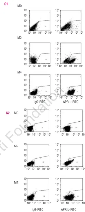
Figure 1. APRIL expression at mRNA and protein level in acute
myeloid leukemia blasts. (A) Real-time PCR analyses of acute myeloid leukemia blasts as compared to normal CD34+cells and
early granulocytic precursors (G) obtained after five days of unilin-eage culture. Normal CD34+
cells were used as reference. Data are mean ± standard deviation of 15 patients in triplicate. (B) Immuno-blot analysis of APRIL expression in CD34+ and acute myeloid
leukemia cells. 293T cells overexpressing APRIL cDNA were used as positive control (293T APRIL). APRIL full-length (APRIL) and soluble forms (sAPRIL) are indicated. Two representative samples for each acute myeloid leukemia subtype (M0, M2 and M4) are shown. (C) Flow cytometry analysis of acute myeloid leukemia blasts labeled with anti-APRIL antibody (right panels) as compared to isotype con-trol staining (left panels). Two samples for each acute myeloid leukemia subtype are shown.
A B C1 C2 CD34+ G M0 M2 M4 30Kd 21Kd APRIL sAPRIL Relative expression (over CD34 +cells) APRIL sAPRIL M4 M2 M0 293T APRIL CD34 + 293T APRIL CD34 + 30Kd 21Kd 104 103 102 10 1 M0 M2 M4 M0 M2 M4 IgG-FITC APRIL-FITC IgG-FITC APRIL-FITC 100 101 102 103104 100101 102 103 104 100 101 102 103104 100101 102 103 104 100 101 102 103104 100 101 102103104 100 101102 103104 100 101 102103 104 100 101102 103104 100 101 102103 104 100 101 102 103104 100 101 102103 104 104 103 102 101 100 104 103 102 101 100 104 103 102 101 100 104 103 102 101 100 104 103 102 101 100 104 103 102 101 100 104 103 102 101 100 104 103 102 101 100 104 103 102 101 100 104 103 102 101 100 104 103 102 101 100 104 103 102 101 100
©Ferrata
Storti
Foundation
GM-CSF and G-CSF.10Real-time PCR analysis showed
APRIL mRNA expression in all AML samples, including 5 cases of M0, 5 cases of M2 and 5 cases of M4 subtype by FAB classification, whereas normal CD34+HPCs and
early granulocytic precursors did not express APRIL (Figure 1A). APRIL expression was confirmed by Western blot analysis, which showed the presence of
both full length and soluble forms of the APRIL protein in AML blasts (Figure 1B). We next performed immuno-fluorescence experiments to determine the expression pattern of APRIL in myeloid leukemia blasts. In 21 cases analyzed, a significant percentage of the cells were stained by anti-APRIL antibodies (Figure 1C). The per-centage of positive cells varied, being higher in the M4 and M2 subtypes than in M0 blasts (Figure 1C). In addi-tion, we evaluated APRIL expression by FACS analysis in other FAB subtypes, including 2 M3 and 2 M5 (Online
Supplementary Figure S1A). Overall, these data indicate
that APRIL expression is specifically found in myeloid leukemia cells, with the exception of M3, but not in nor-mal CD34+ cells or early granulocytic precursors. The
anti-apoptotic activity showed by APRIL in B-CLL, sug-gests that the acquisition of APRIL expression in AML may generate an autocrine loop that could contribute to the leukemia transformed phenotype. To directly evalu-ate the pro-survival effect of APRIL expression on AML cells, we competitively inhibited APRIL activity with an excess of recombinant soluble receptors. Neutralization of the APRIL circuit resulted in significant induction of cell death in all the AML samples, particularly in the M2 subtype (Figure 2A), suggesting that APRIL acts as a sur-vival factor in myeloid leukemia cells. To investigate whether APRIL neutralization results in AML cell apop-tosis, we next evaluated annexin-V/7AAD staining on 3 different M2 samples treated for 24 hours with the blocking reagents. This treatment induced a considerable increase in annexin-V binding (Online Supplementary
Figures S1B and S1C), indicating that constitutive
expres-sion of APRIL protects AML cells from apoptosis. Such an anti-apoptotic activity of APRIL in AML cells might contribute to the increase in resistance to chemotherapy. To investigate this hypothesis, we analyzed the cell death response of 15 fresh AML samples treated with etoposide or camptothecin, two widely used
DNA-dam-APRIL and AML chemosensitivity
haematologica | 2008; 93(12) | 1901|
Figure 2. APRIL promotes the survival of acute myeloid leukemia
cells. (A) acute myeloid leukemia blasts of different subtypes were treated with 5 µg TACI-Fc and 5 µg BCMA-Fc (BCMA/TACI-Fc) or 10 µg Fn14-Fc recombinant soluble receptors. After 24 hours, cell viability was evaluated with acridin orange/ethidium bromide staining and fluorescence microscopy analysis. (B) Acute myeloid leukemia samples were treated for 24h with 50 ng/mL of etopo-side (Eto), 200ng/mL of camptothecin (Cam), 40 µM of cytosine arabinoside (AraC) or 10 µM of daunorubicin (DAU). Data are mean ± SD of 15 patients in triplicate, 5 for each subtype. *0.01<p<0.05; **p<0.01 two-tailed t-test. (C) Immunoblot analy-sis of CD34+cells transduced with the PINCO empty vector (P1) or
the PINCO vector containing APRIL cDNA (P1APRIL). (D) CD34+
cells transduced with P1 and P1APRIL were treated with camp-tothecin, etoposide, daunorubicin and AraC. Cell viability was eval-uated with trypan blue counting after 12 hrs. of treatment. Data are mean ± s.d of three independent experiments in triplicate.
Figure 3. APRIL downstream signaling. (A) Western blotting of CD34+cells transduced with P1 and P1APRIL. (B) Acute myeloid leukemia blasts of M2, M4 and M0 subtypes were treated with 5 µg/L TACI-Fc and 5 µg/L BCMA-Fc (BCMA/TACI-Fc) recombinant soluble receptors and lysated after 24 hrs. One representative of three independent experiments is shown.
A B C D A B M0 M2 M4 BCMA/TACI-Fc Fn14-Fc Untreated P1 P1 APRIL P1 P1 APRIL CD34+ Blasts Bcl-2 relative expression Bcl-2 relative expression M2 M4 M0 M2 M4 M0 BCl-2 α-actin Fc BCl-2 α-actin Cell death (%) Cell viability (%) Cell viability (%) Cell viability (%) Cell viability (%) M0 Fc M2 Fc M4 Fc
Eto Cam AraC DAU
Eto Cam AraC DAU
Eto Cam AraC DAU
Eto Cam AraC DAU
P1 P1 APRIL P1 P1 APRIL − + − + − + − + − + − + − + − + − + − + − + − + 35 30 25 20 15 10 5 0 80 60 40 20 0 80 60 40 20 0 80 60 40 20 0 30 kd 21 kd 60 50 40 30 20 10 0 2.5 2 1.5 1 0.5 0 1.2 1 0.8 0.6 0.4 0.2 0 − + − + − + − + − + − +
©Ferrata
Storti
Foundation
aging drugs. We treated the cells in vitro with either drug in the presence or absence of competing soluble APRIL receptors. The results indicate that inhibition of APRIL signaling significantly increased the apoptotic cell death induced by etoposide or camptothecin, independently of the subtype of the leukemia samples (Figure 2B). These data prompted us to study whether APRIL blocking agents have a synergistic effect with chemotherapeutic drugs currently used in AML. We, therefore, treated leukemia samples representative of the M0, M2 and M4 subtype with cytosine arabinoside (AraC) or daunoru-bicin, in the presence or absence of APRIL soluble recep-tors. As for others chemotherapeutic drugs, the block of APRIL significantly enhanced the induction of cell death by AraC or daunorubicin, independently of the AML subtype (Figure 2B). These data suggest that APRIL activ-ity is part of AML blast response to cytotoxic agents and may play a relevant role in protecting the cells from cell death induced by exogenous stimulation, possibly con-tributing to chemotherapy resistance. To confirm that APRIL upregulation increases the resistance to chemo-therapeutic drugs in myeloid progenitors, we transduced CD34+HPCs with the retroviral PINCO vector carrying
the APRIL gene together with a green fluorescent protein (GFP) reporter.9After flow cytometry sorting, the
virtual-ly pure (>98%) transduced population was anavirtual-lyzed for APRIL expression, which was comparable with that observed in AML samples (Figure 2C and data not shown). In line with the results obtained in AML cells, exogenous APRIL expression in primary HPCs conferred a signifi-cant protection from etoposide, camptothecin, daunoru-bicin, and AraC treatment (Figure 2D). The anti-apoptot-ic protein Bcl-2 is induced by APRIL in B-cell lym-phoma.2,3Since high Bcl-2 expression is associated with
poor chemotherapy response,11,12 we investigated the
relationship between APRIL and Bcl-2 in AML. Western blotting analysis showed that exogenous expression of APRIL up-regulated Bcl-2 in CD34+cells, whereas APRIL
neutralization resulted in Bcl-2 downregulation in pri-mary AML cells (Figure 3A, B), suggesting that APRIL protects AML from chemothereutic drugs through the upregulation of Bcl-2. Thus, APRIL expression promotes
the resistance to chemotherapy of normal myeloid pro-genitors, while APRIL neutralization considerably increases the cytotoxic activity of chemotherapeutic drugs against AML blasts. Transgenic expression of APRIL under the control of the lck distal promoter induces the generation of lymphoid tumors in aged mice,13indicating that the aberrant expression of APRIL
in hematopoietic cells may promote the tumorigenic transformation. The antiapoptotic activity of APRIL has been previously demonstrated in B-lymphoma, multiple myeloma and B-CLL cells, but may be relevant in other cancers. In this study, we demonstrate that APRIL increases the resistance of AML cells to etoposide, camp-tothecin, daunorubicin, and AraC treatment, possibly by the upregulation of Bcl-2. Such an ability to promote the resistance to chemotherapeutic drugs in AML suggests a general oncogenic role for APRIL in hematologic malig-nancies. Biotechnology companies are devoting intense efforts to develop therapeutic molecules that target autoreactive or malignant B cells through the neutraliza-tion of APRIL or its partner Blys in autoimmune diseases, non-Hodgkin’s lymphoma, multiple myeloma and B-CLL.8,14 In xenograft models of lung and colon cancer,
intratumoral delivery of soluble BCMA was able to sig-nificantly reduce tumor growth.8 The increased cell
death induced by chemotherapeutic drugs in AML cells upon inhibition of the APRIL circuit suggests that phar-macological strategies aimed at blocking of APRIL activ-ity may considerably improve the efficacy of chemother-apeutic drugs in myeloid leukemia.
Authorship and Disclosures
All authors contributed significantly to the manu-script. MM, VC and AA conducted the in vitro experi-ments; MH provided anti-APRIL antibody for western blotting; CC collected human samples; DB planned experiments; MG provided intellectual input; DB, RDM, FG wrote the manuscript; RDM was responsible for research co-ordination and strategy. The authors reported no potential conflicts of interest.
D. Bonci et al.
| 1902| haematologica | 2008; 93(12)
References
1. Hahne M, Kataoka T, Schröter M, Hofmann K, Irmler M, Bodmer JL, et al. APRIL, a new ligand of the tumor necro-sis factor family, stimulates tumor cell growth. J Exp Med 1998;188:1185-90. 2. He B, Chadburn A, Jou E, Schattner EJ,
Knowles DM, Cerutti A. Lymphoma B cells evade apoptosis through the TNF family members BAFF/BLyS and APRIL. J Immunol 2004;172:3268-79.
3. Chiu A, Xu W, He B, Dillon SR, Gross JA, Sievers E, et al. Hodgkin lymphoma cells express TACI and BCMA receptors and generate survival and proliferation signals in response to BAFF and APRIL. Blood 2007;109:729-39.
4. Planelles L, Castillo-Gutiérrez S, Medema JP, Morales-Luque A, Merle-Béral H, Hahne M. APRIL but not BLyS serum levels are increased in chronic lymphocytic leukemia: prognostic rele-vance of APRIL for survival. Haematologica 2007;92:1284-5.
5. Endo T, Nishio M, Enzler T, Cottam HB, Fukuda T, James DF, et al. BAFF and APRIL support chronic lymphocytic leukemia B-cell survival through activa-tion of the canonical NF-κB pathway. Blood 2007;109:703-10.
6. Moreaux J, Legouffe E, Jourdan E, Quittet P, Rème T, Lugagne C, et al. BAFF and APRIL protect myeloma cells from apoptosis induced by interleukin 6 deprivation and dexamethasone. Blood 2004;103:3148-57.
7. Ingold K, Zumsteg A, Tardivel A, Huard B, Steiner QG, Cachero TG, et al. Identification of proteoglycans as the APRIL-specific binding partners. J Exp Med 2005;201:1375-83.
8. Dillon SR, Gross JA, Ansell SM, Novak AJ. An APRIL to remember: novel TNF ligands as therapeutic targets. Nat Rev Drug Discov 2006;5:235-46.
9. Bonci D, Hahne M, Felli N, Peschle C, De Maria R. Potential role of APRIL as autocrine growth factor for megakary-ocytopoiesis. Blood 2004;104:3169-72. 10. Ziegler B, Testa U, Condorelli G, Vitelli
L, Valtieri M, Peschle C. Unilineage hematopoietic differentiation in bulk and single cell culture. Stem Cells, 1998; 16(Suppl 1):51-73.
11. Tóthová E, Fricova M, Stecová N, Kafková A, Elbertová A. High expres-sion of Bcl-2 protein in acute myeloid leukemia cells is associated with poor response to chemotherapy. Neoplasma 2002;49:141-4.
12. Lauria F, Raspadori D, Rondelli D, Ventura MA, Fiacchini M, Visani G, et al. High bcl-2 expression in acute myeloid leukemia cells correlates with CD34 positivity and complete remis-sion rate. Leukemia 1997;11:2075-8. 13. Planelles L, Carvalho-Pinto CE,
Hardenberg G, Smaniotto S, Savino W, Gómez-Caro R, et al. APRIL promotes B-1 cell-associated neoplasm. Cancer Cell 2004;6:399-408.
14. Ryan MC, Hering M, Peckham D, McDonagh CF, Brown L, Kim KM, et al. Antibody targeting of B-cell maturation antigen on malignant plasma cells. Mol Cancer Ther 2007;6:3009-18.