UNIVERSITA’ DEGLI STUDI DI CATANIA
DIPARTIMENTO DI SCIENZE BIOMEDICHE E BIOTECNOLOGICHEDOTTORATO DI RICERCA INTERNAZIONALE
BASIC AND APPLIED BIOMEDICAL SCIENCES
TESI DI DOTTORATO
Molecular Mechanisms Involved in Escherichia coli K1 and
Haemophilus influenzae Type a Blood-Brain Barrier impairment
Dott.ssa Nunzia Caporarello
COORDINATORE
TUTOR
Prof. Massimo Libra
Prof.ssa Gabriella Lupo
To my niece Aurora
“Se il seme non si lascia aprire da sole, terra, acqua, accogliendo il suo destino,
rimane sterile. Se invece trova la ragione per rompere il guscio, si lascia ferire ed
entra nel mondo con la sua fioritura e si sperimenta come dono di colori e sapori per
gli altri.
Il prezzo da pagare è un dolore, una morte “apparente”, ma in realtà è “più vita””.
(Alessandro D’Avenia)
ABBREVIATIONS... 5
BACKGROUND: first study ... 6
The Blood-Brain Barrier ... 6
History of the BBB ... 6
Physiology of the BBB ... 7
The Neurovascular Unit ... 8
Endothelial cells ... 9 Pericytes ... 10 Microglia cells ... 11 Astrocytes ... 12 Bacterial Meningitis ... 12 Escherichia coli K1 ... 13
Do the PC have a role in Escherichia coli crossing the BBB? ... 13
Role of phospholipases A2on E. coli K1 invasion of the brain ... 14
The VEGF family ... 14
AIM OF THE STUDY ... 16
MATERIALS AND METHODS ... 17
Chemicals, antibodies, bacteria ... 17
Cell cultures ... 17
Construction of in vitro BBB model ... 18
Electron microscopy ... 18
Evaluation of the barrier integrity... 18
Bacterial invasion and adhesion assays ... 19
Cell viability ... 20
Immunoblotting ... 20
Phospholipase A2 assay ... 20
Determination of PGE2 and VEGF production ... 20
VEGFR-1 blockade experiments ... 21
Statistical analysis ... 21
RESULTS ... 22
E. coli K1 infection determines changes of TEER and permeability to sodium fluorescein in BBEC/BRPC co-cultures ... 22
E. coli K1 stimulates phospholipase A2 activities, PGE2 production and VEGF release ... 23
E. coli adhere to BBEC and BRPC but only in BBEC the invasion occurs ... 26
TEM/SEM ... 27
VEGFR-1 is involved in E. coli adhesion and invasion of BBEC ... 28
VEGFR-1 negatively regulates BRPC survival and its blockade protects the barrier integrity ... 29
Reference list, first study ... 36
BACKGROUND: second study ... 43
Role and metabolism of adenosine ... 43
Adenosine receptors ... 44
HAEMOPHILUS INFLUENZAE MENINGITIS ... 45
Haemophilus influenzae ... 45
Haemophilus influenzae, serotype b ... 45
Haemophilus influenzae, serotype a ... 45
AIM OF THE STUDY ... 47
Cell cultures ... 48
Construction of in vitro BBB model ... 48
H. influenzae preparation and infection ... 49
Electron microscopy ... 49
Immunoblotting ... 49
Fluorescence microscopy ... 50
Evaluation of the barrier integrity... 50
Bacterial invasion assay ... 51
Cell viability ... 51
ENTPDase enzymatic activity assay ... 51
cAMP Detection Assay ... 52
Statistical analysis ... 52
RESULTS ... 53
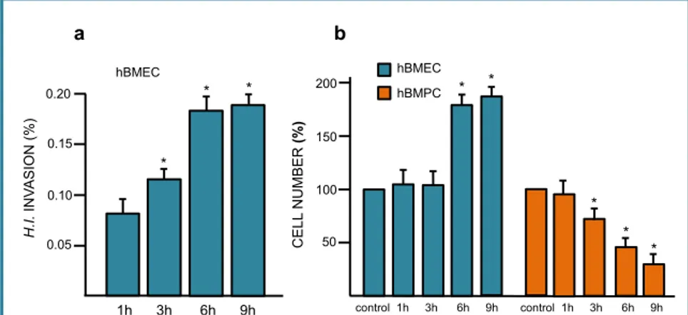
Bacterial invasion modified cell number ... 53
H. influenzae is able to enter hBMECs ... 53
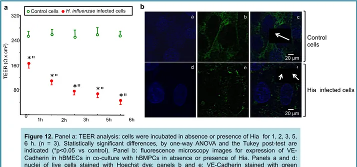
H. influenzae reduced TEER and VE-cadherin expression in hBMEC ... 54
Adenosine receptors were involved in H. influenzae infection ... 55
Ectoenzyme activity ... 57
Bacterial infection increased cAMP production in co-cultures ... 59
H. influenzae stimulated VEGF release ... 60
CREB and Rho activation in co-cultures ... 62
Reference list, second study ... 69
ACKNOWLEDGEMENTS ... 73
Papers published during doctorate ... 74
ABBREVIATIONS
BBB, Blood-Brain Barrier; CNS, Central Nervous System; CSF, Cerebrospinal Fluid; NVU, Neurovascular Unit; AJs, Adherens Junctions; TJs, Tight Junctions;
JAMs, Junctional Adhesion Molecules; ZO, Zonula Occludens;
TGF- β, Transforming Growth Factor β; E.coli, Escherichia coli K1;
EC, Endothelial cells; PC, Pericytes;
PLA2, Phospholipase A2;
VEGF, Vascular Endothelial Growth Factor;
VEGFR, Vascular Endothelial Growth Factor Receptor; PG, Prostaglandin
BBEC, Bovine Brain Endothelial Cells BRPC, Bovine, Retina Pericyte Cells TEER, Transendothelial Electric Resistance Hia, Haemophilus influenzae Type a
BACKGROUND: first study
The Blood-Brain Barrier
The Central Nervous System (CNS), brain and spinal cord, controls the body’s response to different stimuli, both internal and external. The physiological functions of CNS are maintained by a fine regulation if its homeostasis thanks to the blood brain barrier (BBB), located at the level of the cerebral microvasculature. In the adult human brain, the total lenght of capillaries is about 400 miles with a surface area for exchange of 12-18 m2 (Abbott et al, 2010). The BBB acts as a protective barrier against possible neurotoxic molecules, such as proteins or endogenous metabolites, xenobiotics derived from the environment or ingested with the food. Atthe same time, BBB selectively allows the cross of ions and nutrients from blood into the CNS by active transport.
History of the BBB
At the end of the 19th century, during an experiment performed administering intravenously water soluble dyes, the German immunologist Paul Ehrilch observed that all tissue outside CNS were stained, whereas brain and cerebrospinal fluid (CSF) had no color (Ehrlich, 1885). At first, this result was related to a lack of chemical affinity for the dyes specific of the brain tissues. Meanwhile, Bield and Kraus noted that, when injected intravenously, both sodium ferro-cyanide and cholic acids had no pharmacological effects on the CNS but, when they were injected intraventricurarly, the neurological complications occurred (Ribatti et al, 2006) thus providing evidences of the existence of a barrier between CNS and peripheral tissues. In 1890, Max Lewandowsky introduced the term “Blood-Brain Barrier” for the first time. After a few decades, Paul Ehrlich’s student Edwin Goldman performed experiments in rabbits and dogs by using trypan blue dye that later clarify Ehrlich’s result. Goldman observed that, when administered intravenously, trypan blue was able to stain all tissues throughout the body, except the brain and the spinal cord. Surprisingly, injecting
trypan blue directly into the CSF, through subarachnoid space, the nervous tissue was stained while the peripheral tissues was not (Goldman, 1909). Several decades later, Friedemann performed experiments by using lipid soluble dye and observed that it was able to cross the cerebral microvasculature, staining the brain. The anatomical features of BBB was established only with the advent of electron microscopy, in 1967. By using electron-sense tracer horseradish peroxidase (HRP), Reese and Karnovsky demonstrated that endothelial cells in mouse cerebral capillaries was less permeable to HRP in comparison with endothelial cells arising from heart or skeletal muscle (Reese and Karnovsky, 1967), confirming a structural difference in the junctions between cerebral endothelial cells. Now, we know that the BBB is located in the cerebral microvasculature and that it is characterized by controlled transport across, presence of intercellular tight junctions and a lack of fenestration.
Physiology of the BBB
The BBB acts as a filter against all molecules except for small lipophilic molecules with a molecular weight of 400-600 Da (Pardridge, 2001). Nevertheless, it is possible for certain macromolecules to cross the BBB thanks to some receptor-mediated systems, for example GLUT-1 glucose transporter, transferrin receptor, L1 amino acid carrier (Abbott et al, 2006).
TJs of the BBB restrict paracellular diffusion of many substances from the blood while lipophilic molecules are able to enter the brain by passively diffusing across the endothelial cell membranes and could be neurotoxic. In order to avoid the crossing of lipophilic xenobiotics, the BBB expresses high levels of ATP-binding cassette (ABC) transporters to pump indesiderate molecules against their concentration gradients utilizing ATP as energy. The most important ABC transporter is P-glycoprotein (Pgp). Although Pgp is expressed both luminally and abluminally, higher levels are expressed on the luminal side (Ronaldson et al, 2007). Organic cations, weak organic bases with hydrophobic regions and many commonly prescribed drugs represent substrates for Pgp (Miller,
Besides the ABC transporters, there are also different enzymes highly expressed at the BBB plasma membrane that act as a metabolic barrier to detoxify endogenous and exogenous molecules. Exemples are cytochrome P450, aminopeptidases, endopeptidases and cholinesterase (Dauchy et al, 2008).
The Neurovascular Unit
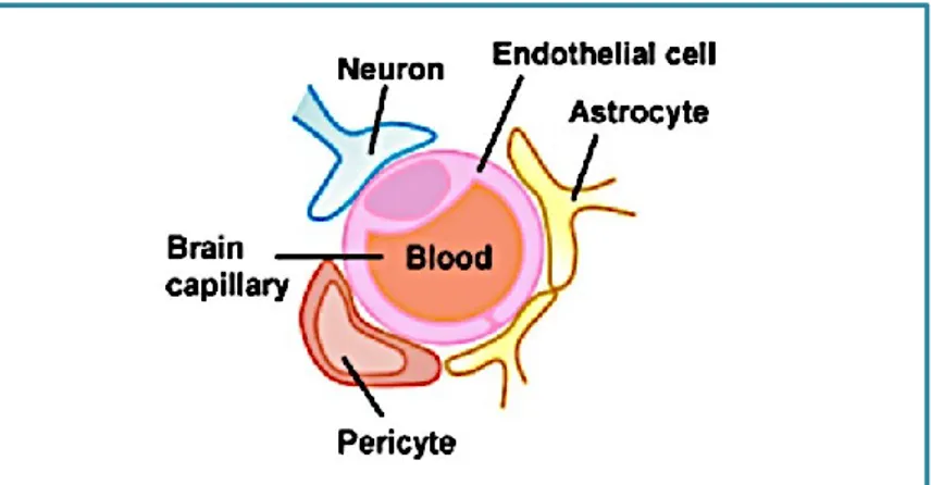
In the brain, the microvessels are phenotypically different from those distribuited in the other districts. But how brain microvessels acquire their barrier properties? In 1980s, Stewart and Wiley performed two sets of experiments: they transplanted embryonic brain fragments into the coelomic cavity, in order to expose them to non neural vessels and fragments of the embryonic mesoderm into the brain, in order to expose them to neural vessels (Stewart and Wiley, 1981). They founded that, when vascularizing into the neural milieu, the abdominal vessels acquired the characteristic features of the BBB, while the brain vessels insered into the non neural milieu were lacking of these features, confirming that the factors present in the neural environment regulate BBB differentiation. BBB is the main component of the Neurovascular Unit (NVU), made of brain microvessels, pericytes, microglia, astrocytes endfeet and neuronal process (Figure 1, Salmeri et al., 2013). All these type of cells are closely connected in order to induce and maintain the physiological function of the BBB (Abbott et al, 2010).
Endothelial cells
Brain endothelium is the first cell type between the blood and the brain and is responsible to bring nutrients to the brain and to avoid the cross of neurotoxic molecules. Each neuron in the human brain has its own capillary (Zlokovic, 2008). Brain endothelial cells are different from peripheric endothelial cells: in fact, they lack fenestrations, present high mitochondrial content to provide energy to support active transport, minimal pinocytotic vesicles and are characterized by the presence of tigh interendothelial junctions (Hawkins and Davis, 2005), which confer them a permeability significantly lower than that of peripheric endothelial cells (Tuma and Hubbard, 2003).
Moreover, brain vasculature is able to increase its diameter in loco in order to supply nutrients when neuronal metabolic demand increase, and this contractile ability is related, at least in part, to presence of pericytes, contractile cells wich also secrete various vasoactive mediators (Hamilton et
al, 2010).
Tight interendothelial junctions are made of adherens junctions (AJ) and tight junctions (TJ) proteins. AJs proteins (such as VE-cadherin and PECAM) give the structural support to the tissue and are critical for tight junctions formation (Abbott et al, 2010). TJs include occludin (Hirase et
al, 1997), claudins (Ohtsuki et al, 2007) and junctional adhesion molecules (JAMs) (Jia et al,
2013). Occludin was the first TJ protein discovered: it is a 522 amminoacids polypeptide with a molecular mass of 59.1 kDa. Occludin has N-terminal and C-terminal cytoplasmatic tails and 2 extracellular loops. N-terminal tail deletion is involved in loss of barrier property of TJs while C-terminal is important for barrier functions as well as for oligomerization of occludin (Chen et al, 1997) and for the interaction with regulatory proteins, such as zonula occludens 1, 2 and 3 (ZO-1, ZO-2 and ZO-3), responsible of anchoring occluding to the actin cytoskeleton (Feldman et al, 2005). Occludin has serin, threonine and tyrosine residues as regulatory site. An overall increase in occludin thyrosine phosphorylation lead to an increase in BBB permeability, while serine and threonine phosphorylation are associated with occludin localization within the membrane
(Takenaga et al, 2009). The human claudin family is made of 23 proteins with a molecular weight between 20-27 kDa and 4 transmembrane regions, 2 extracellular loops and N- and C-terminal cytoplasmic tails. Claudin-5, showing the highest expression levels of any claudin at the BBB (Lippoldt et al, 2000), is found in endothelial and epithelial cells of the choroid plexus.
Depending on the cell type, ZO proteins are located at TJs, adherens junctions (AJ), and/or gap junctions (Bauer et al, 2010). The first ZO protein discovered was ZO-1, a 220 kDa protein. (Stevenson et al. 1986). ZO-2, a 160 kDa protein which has a high sequence homology to ZO-1 (Gumbiner et al, 1991), has been less studied in BBB in comparison to ZO-1 but it is known that alterations in its expression and localization are associated with disruption of BBB (Hom et al, 2007). ZO-3 (130 kDa protein) is found at TJs level, but not in endothelial cells (Inoko et al, 2003). Junctional adhesion molecule 1 (JAM-1) is a 40 kDa protein which contain two extracellular immunoglobulin-like (Ig-like) loops, a short cytoplasmic tail, a single transmembrane segment, and homodimerizes (Severson et al., 2009).
All these proteins play a pivotal role in the restrictive BBB features.
Fig. 2 - Junctional protein of the BBB (Abbott et al., 2010)!!
Pericytes
Pericytes were for the first time described by Rouget in 1879, and for this reason they were named “Rouget cells”. Due to their location on the outer surface of blood capillaries and their stritct
interaction with the underlying endothelial cells, with which they share the basement membrane, in 1923 Zimmermann renamed them “Pericytes” (peri: around; cyte: cell) (Zimmermann, 1923). Pericytes cover between 22 and 37% of the cerebral capillary surface and present processes from their cell body that are in contact with more than one endothelial cell. The ratio endothelial cells/pericytes in the CNS is between 1:1 and 1:3 (Haddad-Tòvolli et al, 2017). Direct physical connection between pericytes and endothelial cells is provided by gap junctions, which enable the exchange of small molecules and ions. Pericytes are anchored to endothelial cells through adhesion plaques, while peg and socket contacts allow the cells to penetrate inside discontinuities in the basement membrane of vessels in contact with each other (Bergers et al, 2005). These junction complexes allow the transmission of mechanical contractile forces from pericytes to endothelial cells, and include cell-adhesion molecules, N-cadherin/β-catenin based adherent junctions and extracellular matrix protein, e.g. fibronectin (Gerhardt et al, 2003).
The role of pericytes in maintaining a proper BBB function was discovered for the first time considering the increased ratio of pericytes to endothelial cells present in the CNS in comparison to other body regions (Dìaz Floreset al, 2009).
Using transgenic mice PDGFRβ knockdown (in order to downregulate pericyte generation), it has been shown that pericytes are necessary for the formation of the BBB (Daneman et al., 2010) and that the vascular permeability is due to pericyte coverage of a capillary (Armulik et al, 2010). In addition to the role on BBB development, pericytes are also involved in maintainance of the BBB during adult and age life: in fact, the number of pericytes decreases with aging while, at the same time, vascular permeability increases (Bellet al, 2010).
Microglia cells
Microglia are cells deputies to immune response within the CNS. In physiological conditions, microglia have small cellular bodies with long processes in direct contact with endothelial cells
processes and enlarge their cellular bodies. Once activated, microglia may secrete inflammatory cytokines and may become reactive, exhibiting phagocytic activity and acting as antigen presenting cells (Zlokovic, 2008).
Astrocytes
The most important function of perivascular astrocyte endfeet, wrapping endothelial cells, is to regulate water transport through aquaporin-4 (Tait et al, 2008). Astrocytes are able to induce barrier properties in endothelial cells both in vivo (Janzer et al, 1987) and in vitro (Rubin et al, 1991). Moreover, astrocytes secrete chemical mediators able to promote the BBB phenotype, such as basic fibroblast growth factor (bFGF), transforming growth factor β (TGF-β) and glial derived neurotrophic factor (GDNF). On the other hand, endothelial cells secrete mediators that promote growth and differentiation of astrocytes, thus underlying the close relation between these two type of cell (Mi et al, 2001).
Bacterial Meningitis
Bacterial Meningitis is a common and severe CNS infection, which is often fatal and over 50% of the survivors develop neurological complications, such as epilepsy, increased intra-cranial pressure, stroke and educational deficits in later life (Hoffman O et al, 2009). There are different pathogens associated with bacterial meningitis: Streptococcus pneumoniae, Streptococcus agalatiae, Neisseria
meningitidis, Listeria monocytogenes, Escherichia coli K1 and Haemophilus influenzae type B. The
bacteria responsible of meningitis are often member of the human microbiome of gut, skin and mucous membranes of healthy humans (Cogen et al., 2008) which can gain virulence factors lead to penetration of epithelial and endothelial barriers and invasive disease. Before to infect CNS, bacteria must survive and persiste within the blood circulation because only the bacteria that survive in the bloodstream possess high penetration rate across the brain endothelium (Dando et al.,
2014). Through the years, bacterial meningitis has remained an infection with a high mortality rate, particularly in very young and elderly patients, despite advances in antimicrobial therapy (Kim,
2008). The reason for the poor outcome has been attributed to limited knowledge of pathogenesis
and pathophysiology of the disease.
Escherichia coli K1
Regarding Escherichia coli, more than 80% of the neuropathogenic strains express the K1 capsule. This capsule is made of homopolymer of -2,8-linked N-acetylneuraminic acid. Although most cases of Escherichia coli meningitis occur via haematogenous spread, microbial and host factors responsible for the ability of neurotropic strains of Escherichia coli to cross the BBB are still missing. After infection, release of pro-inflammatory mediators such as cytokines and prostaglandins (PGs) by leucocytes, endothelial cells, astrocytes, microglial cells and other cells in the central nervous system was stimuled, leading to an increase in the permeability of the BBB
(Engblom et al., 2002; Zhu et al., 2010). E. coli K1 invasion of brain EC is promoted by IQGAP1
(Ras GTPase-activating-like protein) (Krishnan et al., 2012) and is responsible for TJs disruption.
Do the PC have a role in Escherichia coli crossing the BBB?
PCs are structural cells that helps to promote vascular integrity and their loss or dysfunction could play a critical role in the pathogenesis of meningitis.
PC are the cells closest brain EC, with which they share a common basement membrane, but they have not been investigated in a co-culture BBB model understanding the molecular mechanism of bacterial invasion. PC are able to secrete soluble factors, leading to the upregulation of BBB
functions (Hori et al., 2004; Dohgu et al., 2005; 2011; Takata et al., 2007; Nakagawa et al., 2009).
Recently, it has been reported that lipopolysaccharide-induced sepsis in mice is responsible for the
Other studies have revealed that PC deficiency in the CNS leads to BBB breakdown and brain
hypoperfusion resulting in secondary neurodegenerative changes (Winkler et al., 2011) ans that loss
of pericytal function can result in development of CNS disease (Bonkowski et al., 2011).
Role of phospholipases A2on E. coli K1 invasion of the brain
PGs and leukotrienes (LTs) are arachidonate metabolites and contribute to E. coli K1 invasion of
microvascular EC and crossing of BBB (Zhu et al, 2010). Arachidonic acid (AA) derives from
phospholipids by the action of different isoforms of PLA2s and is converted to PGs or LTs by the
action of cyclooxygenase (COX) and 5-lipoxygenase, respectively. There are 3 different PLA2s
isoforms: cytosolic PLA2 (cPLA2), Ca2+-independent intracellular PLA2 (iPLA2) and Ca2+
-dependent secretory PLA2 (sPLA2) that differ from each other in terms of substrate specificity, Ca2+
requirement, lipid modification, translocation to cellular membranes and AA release (Alberghina, 2010).
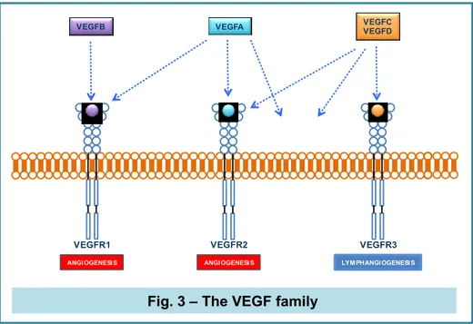
The VEGF family
The vascular endothelial growth factor (VEGF) is a critical regulator of vasculogenesis,
angiogenesis, lymphangiogenesis and vascular permeability in vertebrates. The family is made of
VEGFA, VEGFB, VEGFC, VEGFD, and placental growth factor (PlGF). There are 2 VEGFA isoforms: VEGF121 and VEGF165, and both are principal mediators of tumour angiogenesis. The main signaling tyrosine kinase receptor (TKR) is VEGF receptor 2 (VEGFR2; FLK, KDR in humans), followed by VEGFR1 (FLT1) and VEGFR3 (FLT3). VEGFR1 is a “decoy” receptor, being its affinity for the growth factor high, whose kinase activity is weak, preventing VEGF from binding to VEGFR-2. VEGFR3 is mostly involved in lymphangiogenesis (Ferrara et al., 2005). The PlGF and VEGFB (VEGF-related molecules) bind selectively to VEGFR-1, while VEGFA binds VEGFR-1 and VEGFR-2. VEGFC and -D bind to VEGF3, and, following a proteolytic processing, activate VEGFR-2 after binding (Ferrara et al., 2016).
VEGFR1 VEGFR2
VEGFB VEGFA
VEGFR3
VEGFC VEGFD
LYM PHANGI OGENESIS
ANGIOGENESIS ANGIOGENESI S
Fig. 3 – The VEGF family
VEGFR-1 is a positive regulator of angiogenesis and inflammatory responses in several human diseases such as rheumatoid arthritis (Kong et al., 2011), cancer (Subramanian et al., 2010) and bacterial meningitis. It was demonstrated that E. coli K1 has a critical role in the promotion of the physical association between phosphorylated VEGFR-1 and p85 subunit of PI3K, suggesting the involvement of VEGFR-1 in E. coli K1 invasion of microvascular ECs (Zhao et al., 2010).
High VEGF levels have been measured in the cerebrospinal fluid (CSF) of patients affected by bacterial meningitis with severe BBB disruption, and VEGF immunoreactivity was found in endothelium and smooth-muscle cells arising from brain specimens of patients who died of bacterial meningitis (van der Flier et al., 2001).
VEGF is able to stimulate PGI2 synthesis via cPLA2-mediated AA release via activation of p42/p44 MAP kinases (Wheeler-Jones et al., 1997). So, PKC may be a key mediator of VEGF-induced activation of the ERK pathway via increased association with Raf-1 (Gliki et al., 2001).
AIM OF THE STUDY
The aim of the present study was to understand how E. coli K1 strain is able to cross BBB. To achieve this goal, an in vitro model of BBB obtained by co-culturing BBEC and BRPC in transwell insert was used.
Additionally, in order to identify the contribute of PC in this process, the investigation was also extent to evaluation of the role of the pericytal VEGFR-1.
MATERIALS AND METHODS
Chemicals, antibodies, bacteriaPhospholipase A2 inhibitors, arachidonoyl trifluoromethyl ketone (AACOCF3), bromoenol lactone (BEL) and VEGF A were purchased from Calbiochem (La Jolla, CA). NS-398 (N-[2-(cyclohexyloxy)-4-nitrophenyl]-methanesulfonamide), a selective cyclooxygenase-2 inhibitor, and rabbit polyclonal against iPLA2 antibody were from Cayman Chemical (Ann Arbor, Michigan). Rabbit polyclonal against von Willebrandt factor antibody, mouse monoclonal against cPLA2, VEGFR-2, α-actin and GAPDH antibodies, rabbit polyclonal against VEGFR-1 antibody, were from Santa Cruz Biotechnology (CA). Rabbit polyclonal anti-phospho-VEGFR-2 Tyr1175 was from Cell Signaling Technologies and rabbit polyclonal anti-phospho-VEGFR-1 Tyr1333 was from Sigma Aldrich (St. Louis, MO, USA).
A rifampicin-resistant mutant of E. coli K1 strain (DSMZ 10723) was used for invasion of BBEC and BRPC.
Cell cultures
Primary microvascular endothelial cells from bovine brain (BBEC) were purchased from European
Collection of Cell Cultures (ECACC) fed with Ham's F-10 while pure microvessel pericytes
cultures were prepared from bovine retinas. After isolation, cells were cultured in DMEM with 10% fetal bovine serum, 100 U/ml penicillin and 100 μg/ml streptomycin. Morphological changes and cell viability was determined by MTT test. Pericytes were characterized by their large size and branched morphology, positive immunostaining for a-smooth muscle actin, NG2 chondrotitin sulfate proteoglycan and absence of von Willebrand factor and glial fibrillary acidic protein (GFAP) staining.
Construction of in vitro BBB model
Transwell inserts (Transwells, Corning, Corning, NY) were coated on the upper and bottom side with 2 mg/ml solution of rat tail collagen before being placed in complete medium (half and half
DMEM/HAM’s). To construct an in vitro model of BBB, BRPC (2 × 10⁴ cells cm2) were first plated
on the outside of the Transwell inserts membrane and placed upside down in the well culture plate
to allow adhesion. After adhesion, the Transwells were inverted and BBEC (2 × 10⁴ cells cm2) were
seeded on the top surface of the insert (Fig. 5). Under these conditions, the two type of cells on direct contact established an in vitro BBB model within 3 days.
Electron microscopy
For Scanning Electron Microscopy (SEM) preparations, cells grown in co-colture on filters were fixed with 1.5% glutaraldehyde overnight, following by an additional fix in 1% OsO4 (1 h). Then, cells on the membrane were dehydrated in graded ethanol, critical point dried, sputtered with 5 nm gold layer using an Emscope SM 300 (Emscope Laboratories, Ashford, UK) and observed using a Hitachi S-4000 (Hitachi High-Technologies America, Schaumburg, IL) field emission scanning electron microscope.
For Transmission Electron Microscopy (TEM), after dehydrating in a graded series of acetone, cells were embedded in Durcupan ACM (Fluka Chemika-Biochemika, Buchs, Switzerland). Ultrathin sections were cut using a Reichert Ultracut E microtome and double stained with uranyl acetate and lead citrate. Observations were carried out using a Hitachi H-7000 transmission electron microscope (Hitachi High-Technologies Europe GmbH, Krefeld, Germany).
Evaluation of the barrier integrity
collagen-treated Transwell inserts without cells. Values were expressed as ω x cm2 and were calculated using the formula: (the average resistance of experimental wells – the average resistance of blank wells)*0.33 (the area of the transwell membrane).
For the determination of the flux of sodium fluorescein (Na-F) across endothelial monolayer, inserts containing cells were transferred to 12- well plates containing 1.5 ml of Ringer-Hepes buffer (136
mM NaCl, 0.9 mM CaCl2, 0.5 mM MgCl2, 2.7 mM KCl, 1.5 mM KH2PO4, 10 mM NaH2PO4, 25
mM glucose and 10 mM Hepes, pH 7.4) in the lower compartments (abluminal). In the inserts
(luminal compartment), culture medium was replaced by 0.5 ml of buffer containing 10 μg ml-1
Na-F. The inserts were transferred at 5, 15 and 30 min to a new well containing Ringer-Hepes buffer.
The concentrations of the marker molecule in samples were determined by fluorescence multiwell
plate reader (PerkinElmer; excitation wavelenght: 485 nm, emission wavelenght: 535 nm). Transendothelial permeability coefficient (Pe) was calculated measuring flux across cell-free inserts.
Bacterial invasion and adhesion assays
Bacteria (107 cfu per well) were added to confluent cells in mono-culture or in co-culture on
Transwell inserts (37°C, 60 min). The number of intracellular bacteria was determined after incubation with gentamicin (100 μg/ml) for 1 h at 37°C to kill extracellular bacteria. Cells were washed and lysed with 0.5% Triton X-100. The released intracellular bacteria were evaluated by seeding on LB agar plates and counting the number of colonies the day after. In duplicate experiments, the total cell-associated bacteria were determined as described for invasion, except that the gentamicin step was omitted (in order to include extracellular, adherent to membrane bacteria). Results were expressed as per cent invasion [100 × (number of intracellular bacteria recovered)/(number of bacteria inoculated)].
Cell viability
After incubation of BBEC/BRPC in co-culture with E. coli K1 (60 min), cells from inserts were trypsinized separately, cell suspensions were mixed with a 0.4% (w/v) trypan blue solution, and the number of live cells was determined using a haemocytometer. Cells failing to exclude the dye were considered non-viable. Each infection was performed in triplicate and counted four times each.
Immunoblotting
The lysates of BBEC incubated with E. coli strains for 60 min were prepared for Western blotting
(Giurdanella et al., 2011). Membranes were incubated overnight at 4°C with primary antibodies
against cPLA2, iPLA2, VEGFR-1, VEGFR-2, phospho-VEGFR-2 Tyr1175 or phospho-VEGFR-1 Tyr1333 (dilution, 1:1000) and then incubated with secondary antibodies (dilution, 1200) for 1 h at room temperature.
Phospholipase A2 assay
BBEC and BRPC in mono- or in co-culture were pre-incubated for 60 min in culture medium in the
absence or presence of either 50 μM AACOCF3 or 2.5 μM BEL. The cells were then refed with
fresh culture medium containing the inhibitors in presence or in absence of E. coli K1 for 60 min. Controls were performed by incubation of co-cultures with inhibitors for 120 min in absence of bacteria. After incubations, cells grown on both sides of the inserts were scraped separately, lysed
(Anfuso et al., 2007) and equal amounts of cell lysates were incubated in a 96-well plate with the
substrate arachidonoyl-thio-phosphatidylcholine (ATPC), using cPLA2 assay kit (Cayman
Chemicals, Ann Arbor, MI, USA) according to manufacturer's instructions.
Determination of PGE2 and VEGF production
presence of 50 μM AACOCF3 or 2.5 μM BEL or 5 μM NS-398. The cells were then refed with fresh culture medium containing the inhibitors in presence or in absence of E. coli K1 for 60 min.
Supernatants were collected and used for PGE2 determination (Cayman Chemicals, Ann Arbor, MI,
USA). Conditioned medium was removed from mono- and co-cultures and analysed for VEGF by ELISA (R&D Systems, Minneapolis, MN, USA), as specified by the manufacturer's instructions.
VEGFR-1 blockade experiments
For BBEC/BRPC simultaneous blockade experiments, co-culture were treated with VEGFR-1 Ab
(2 μg ml−1) for 60 min before treatment with E. coli K1 for 60 min. Parallel co-cultures without
VEGFR-1 blockade were performed.
For BBEC (but not BRPC) VEGFR-1 blockade experiments, BBEC were grown on the top surface of the Transwell insert and incubated with VEGR-1 Ab. Then, inserts were inverted and BRPC were seeded on the outside of the insert. After BRPC adhered, the inserts were reinserted into six-well plates and the in vitro BBB model, in which BBEC were blocked by VEGFR-1 Ab, was established within 3 days.
For BRPC (but not BBEC) VEGFR-1 blockade experiments, BRPC were first plated on the outside of the Transwell inserts. After adhesion of BRPC, the inserts were reinserted into six-well and cells
were incubated with VEGFR-1 Ab. Then, BBEC were seeded on the top surface of the insert and
the in vitro BBB model, in which BRPC were blocked by VEGFR-1 Ab, was established within 3 days.
Statistical analysis
Statistical significance between two groups was analysed by Student's test. One-way analysis of variance (anova), followed by Tukey's post hoc test, was used to compare the means for the multiple groups. The P-value < 0.05 was considered statistically significant.
RESULTS
E. coli K1 infection determines changes of TEER and permeability to sodium fluorescein in BBEC/BRPC co-cultures
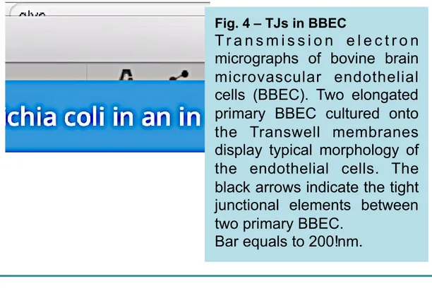
TEM of BBEC growing on Transwell filter in the in vitro BBB model is shown in Fig. 4. TJ sealing
adjoining BBEC are evident.
Fig. 4 – TJs in BBEC
T r a n s m i s s i o n e l e c t r o n
micrographs of bovine brain
microvascular endothelial
cells (BBEC). Two elongated
primary BBEC cultured onto
the Transwell membranes
display typical morphology of
the endothelial cells. The
black arrows indicate the tight
junctional elements between
two primary BBEC.
Bar equals to 200!nm.
The BBB model was validated by measuring TEER and sodium fluorescein flux across BBEC in
monolayer and in co-culture with BRPC (Table 1). Co-culture showed high values of TEER and
very low permeability to sodium fluorescein compared with BBEC mono-culture. Incubation for
60 min with E. coli K1 (107 cfu per well) caused a significant TEER reduction (about 3.2-fold) and
TEER (ω x cm2)
Pe (10-6 cm/s)
mono-culture BBEC 85 ± 21.4 8.1 ± 0.6
mono-culture BBEC + E. coli 60 ± 16.6 6.6 ± 0.8
co-culture
BBEC/BRPC 250 ± 51.1†
3.2 ± 0.15†
co-culture
BBEC/BRPC + E.coli 72 ± 20.3** 7.3 ± 0.5**
Table 1 – Evaluation of the barrier integrity
Evaluation of the barrier integrity. TEER and permeability to sodium fluorescein (Pe) determination in microvascular endothelial cells in mono- and in co-culture, in the absence or
presence of E. coli K1 strain. BBEC (40,000 cells/cm2) were cultured in monolayers in Ham’s
F-10 medium containing 10% FBS or were grown on the top surface of the Transwell insert
(6-well type, 3.0-mm pore size) in which BRPC (40,000 cells/cm2) were first plated on the outside of
the membrane, in 50% DMEM plus 50% F-10 HAM’s containing 10% FBS. After 3 days, the cells
were incubated in the absence or presence of E. coli (107 CFU/well) for 60 min and
measurements of TEER and cell permeability on BBEC were performed. Values were expressed
as w x cm2 and were calculated by the formula: [the average resistance of experimental wells -
the average resistance of blank cells] × 0.33 (the area of the transwell membrane).
For sodium fluorescein determination, flux across cell-free inserts was measured and transendothelial permeability coefficient (Pe) was calculated. Values (means ± SEM) are from three independent experiments (n=3). Statistically significant differences of TEER and Pe,
measured in BBEC in co-culture as compared to mono-culture are indicated by † (†p < 0.01) and
values from E. coli-stimulated BBEC as compared to control non-stimulated BBEC are indicated by asterisks (**p < 0.01).
E. coli K1 stimulates phospholipase A2 activities, PGE2 production and VEGF release
BBEC in mono- and co-culture stimulated with E. coli K1, showed a strong PLA2 activity (about 2.2-fold in mono- and 2.6-fold in co-cultures) compared with the unstimulated BBEC, whereas in
BRPC it was weakly stimulated. The presence of 50 μM AACOCF3 (PLA2 activity dual blocker) or
2.5 μM BEL (iPLA2 inhibitor) for 120 min (pre-incubation of 60 min followed by incubation for 60 min in the presence of bacteria) in control BBEC mono-cultures, reduced enzyme activity of 28% and 19%, respectively; in E. coli K1 treated BBEC mono-cultures, AACOCF3 and BEL
reduced PLA2 activity by 62% and 55%, respectively. In control BRPC mono-cultures, AACOCF3
and BEL caused a decrease by 32% and 24% respectively, and in E. coli stimulated BRPC
In control BBEC/BRPC co-culture, AACOCF3 and BEL reduced PLA2 enzymatic activity by 28%
and 20%, respectively. After E. coli K1 treatment in presence of the inhibitors, the reduction was
stronger (68% and 56%, respectively). Moreover, the two inhibitors decreased PLA2 activity in
control and in E. coli treated BRPC by about 36%. The incubation with BEL allowed us to discriminate between the cPLA2 and iPLA2 activity contribution.
PGE2 in supernatants of BBEC or BRPC in mono- and in co-culture were measured. Table 3 shows
an increase in BBEC monocoltures infected with E. coli K1 (2.5 fold) compared with the respective
control whereas bacterial incubation in presence of AACOCF3 or BEL decreased PGE2 production
by 63% and 60%, respectively. The contribution in PGE2 production from E. coli-treated BRPC monocultures was negligible.
E. coli K1 treatment of BBEC/CRPC co-cultures increased PGE2 production by 3.0 fold in
comparison with the respective untreated co-cultures. Furthermore, in the supernatants of E. coli-stimulated co-cultures, incubated in presence of PLA2 inhibitors AACOCF3 or BEL, PGE2 levels
decreased by about 70% and 68%, respectively.
BBEC monocultures produced low amounts of VEGF in the conditioned medium. Incubation of BBEC monocoltures with E. coli K1 led to a 3.1 fold increase in the release of VEGF (Table 4). Conversely, untreated BRPC monocultures expressed VEGF at basal levels 1.6-fold higher than untreated BBEC monocultures and the presence of E. coli did not changed the secretion. E. coli treatment of cultures induced an 3.2 fold increase in comparison with control untreated co-cultures. Incubation of E. coli-treated BBEC monocultures with AACOCF3, BEL or with COX-2-specific inhibitor NS-398 reduced E. coli-induced VEGF release by 70%, 67% and 71% respectively. Incubation of untreated co-cultures with the three inhibitors reduced VEGF release by
about 33%, while in E. coli-treated co-cultures AACOCF3, BEL and NS-398 reduced VEGF release by 77%, 75% and 79%, respectively. The effect of the inhibitors on VEGF release in untreated and E. coli-treated BRPC monocultures was about 22% and 21% respectively.
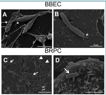
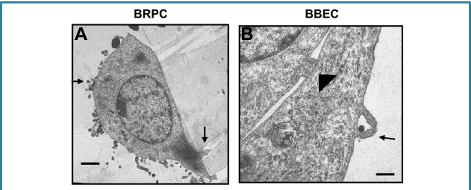
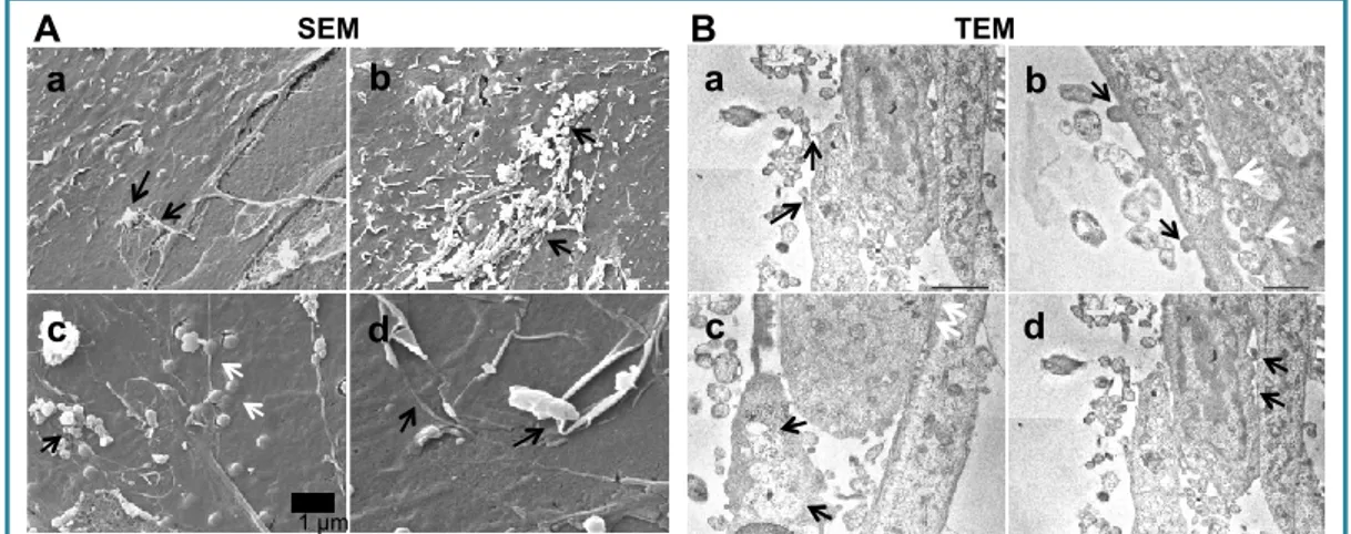
E. coli adhere to BBEC and BRPC but only in BBEC the invasion occurs
SEM images of BBEC, (Fig. 5, A and B) or BRPC (Fig. 5, C and D), after incubation with E. coli
K1, show the cells with an elongated and spindle phenotype, with numerous microvilli distributed
on most of the cell surface (Fig. 5 A). The images at higher magnification show the bacteria
8 mm 2 mm 30 mm
B
A
B
2 mm 7 mmA
Fig. 5 – SEM of BBEC and BRPC at 60 min post infection with E. coli
K1. A. BBECC with elongate and spindle phenotype with microvilli in contact with bacteria (white arrows) magnification, X 1100. B. Higher magnification (X 15 000) showing the microvilli attaching to the bacteria in close and intimate contact with the bacterial surface (white arrow).C. BRPC exhibit a flattened shape with several branches, numerous pseudopodia (thin arrows) and filopodia (arrowheads). The bacteria were found on the cell surface, in proximity of numerous microvilli (magnification, X1000). D. magnification, X 10 000
C
D
30 mm 3 mmBBEC
BRPC
BRPC are branched and flat with short microvilli on the surface area (Fig. 5C). Few bacteria were
found attached to the smooth part of the BRPC surface without any sign of specific membrane interaction (Fig. 5 D).
TEM/SEM
Figure 6 A shows BRPC with numerous bacteria on the cell surface where they remain and no endocytosed bacteria were found. In E. coli–BBEC interaction, electron-dense bacteria both intra- and extracellular were seen and also BBEC formed microvilli-like protrusions that surrounded and
Fig. 6. Transmission electron micrographs of BRPC (A) and BBEC (B–D) infected with E. coli K1for 60 min
A. BRPC on Transwell showing cellular protrusions infiltrating the filter pores (black arrow). Bacteria are present on the surface (dark black) but not inside the cells (bar
= 1 mm). B. Electron-dense (dark black) bacteria were seen extracellularly and
some bacteria were intracellularly engulfed inside membranes-bound vacuoles (thick black arrows) (bar = 1.25 mm).
BRPC BBEC
A B
VEGFR-1 is involved in E. coli adhesion and invasion of BBEC
Western blot analysis showed that VEGFR-1 expression in BRPC mono- and co-cultures was 2.0 fold and 2.2 fold higher than that of BBEC in mono- and in co-cultures, respectively (Fig. 7 A). VEGFR-2 expression did not change in BBEC grown in contact with BRPC in respect to BBEC
monoculture. Interestingly, VEGFR-2 protein was not detected in BRPC (Fig. 7 B).
In order to understand the involvement of VEGF receptors in E. coli K1 adhesion and invasion,
assays were performed in presence of VEGFR-1 or VEGFR-2 antibodies (Ab). Fig. 7 C show that
VEGFR-1 Ab and VEGFR-2 Ab have no effect on E. coli adhesion to BBEC both in mono- and in co-cultures, as well as on E. coli adhesion after addition to BRPC in mono- and in co-cultures
(Fig. 7 D). VEGFR-1 Ab significantly reduced E. coli invasion in BBEC by 30% and by 48% in
mono- and in co-cultures, respectively (Fig. 7 E), while VEGFR-2 did not cause any change in the
E. coli invasion of BBEC. These results indicate a positive correlation between the block of
VEGFR-1 and the block of E. coli K1. Treatment of BBEC with 10 ng/ml VEGF for 15 and 30 min in absence or in presence of VEGFR-2 Ab showed an increase of phosphorylation of VEGFR-2 at Tyr1175, as assessed by Western blot, while treatment with 10 ng/ml of VEGF in presence of
2 μg/ml VEGFR-2 antibody inhibited VEGFR-2 phosphorylation (Fig. 7 F). Experiments were also
VEGFR-1 Ab. Phosphorylation of VEGFR-1 at Tyr1333 was observed in BBEC treated for 15 and 30 min with VEGF.
VEGFR-1 negatively regulates BRPC survival and its blockade protects the barrier integrity
To clarify if the prevention of the pericyte death and monolayer permeability can be attributed to VEGFR-1, experiments blocking the receptor by its specific antibody were performed on co-cultures or single cell type monoco-cultures. After incubation of BBEC/BRPC co-co-cultures with E. coli K1, TEER decreased by 64% and permeability to sodium fluorescein increased by 3.2 fold in
In co-culture, simultaneous block of VEGFR-1 in presence of E. coli K1 was able to preserve BRPC viability by almost 3 fold in comparison with infection in the absence of VEGFR-1 Ab
(Fig. 8 B). Moreover, in presence of E. coli K1 and receptor blockade, TEER values increased by
2.3 fold and permeability decreased by 2.2 fold in comparison with infected co-cultures in absence of receptor blockade. Furthermore, only 10% of TEER reduction and a 20% of increase in
permeability were found in comparison with non-infected control cells in co-culture (Fig. 8 C and
D).
When only the BBEC were treated with VEGFR-1 Ab and E. coli K1, 45% of BRPC adhering to
the co-culture insert was found (Fig. 8 B), while BBEC number was unchanged (Fig. 8 A).
Moreover, a significant decrease in TEER values (about 56%) and a significant increase in permeability (approximately 2.8 fold) in E. coli infected in comparison with non-infected co-cultures were found. Furthermore, a 38% of TEER decrease and a 2 fold permeability increase were
found in comparison with E. coli K1 infected co-cultures in which both BBEC and BRPC were
blocked by VEGFR-1 Ab (Fig. 8C and D). These results demonstrated that VEGFR-1 Ab was able
to partially reduce E. coli K1 internalization (about 50%, as shown in Fig. 7) but not to completely
avoid the activation of signalling pathway leading to VEGF release, detachment of BRPC and loss of barrier properties.
Blockade of only BRPC in presence of E. coli K1 did not change BRPC number in co-culture
(Fig. 8 B), whose value was very similar to that of BRPC in absence of infection. Moreover, the
values of TEER and permeability in this experimental condition were similar to non-infected control cells in co-culture. Furthermore, TEER increased by 1.7 fold and by 2.4 fold and permeability decreased by 60% and 64% in comparison with E. coli K1 infected co-cultures in which only BBEC were blocked by VEGFR-1 Ab and in comparison with E. coli infected
co-cultures in which no blocking was induced, respectively (Fig. 8 C and D). Probably, in absence of
VEGFR-1 Ab on BBEC, E. coli K1 is able to invade BBEC without limits and to activate the signalling pathway leading to VEGF production in large amounts. The VEGF released by BBEC cannot bind VEGFR-1 on BRPC, because blocked by the specific antibody, it therefore does not result in the detachment of pericytes. In this experimental condition, about 90% of BRPC adhered to co-culture and the values of TEER and permeability were very similar to those of the non-infected co-cultures.
These results demonstrate that the prevention of pericyte loss and monolayer permeability can be attributed to VEGF blockade.
To further confirm our results, experiments on BBEC/BRPC co-cultures incubated with E. coli K1 for 30, 60, 90 and 120 min, in absence or in presence of VEGFR-1 or VEGFR-2 Abs were performed.
As shown in Fig. 9, TEER values gradually decreased after 30 min E. coli K1 treatment (37%
reduction) to reach 60% following incubation for 60 and 120 min (panels A, B and C). The presence
incubation time (panel A). No significant change were observed in the TEER values after incubation of the co-cultures with VEGFR-1 Ab and VEGFR-2 Ab in absence of E. coli K1 (panels A and B). When co-cultures were infected with E. coli in the presence of AACOCF3 or BEL, TEER was restored by about 35% at 60 and 120 min (panel C). Regarding permeability studies, fluorescent intensity in the receiver chamber had already increased of about 2 fold after E. coli K1
incubation for 30 min (Fig. 9 D–F) and the fluorescence was more intense at each designated time
point than untreated co-cultures. Incubation in the presence of VEGFR-1 Ab, but not VEGFR-2 Ab, attenuated of about 2.5-fold the diffusion of fluorescein sodium at each studied time (panels D and E). Moreover, when the cells were infected with E. coli K1 for 30, 60, 90 and 120 min in the presence of AACOCF3 or BEL, permeability was restored by about 44% in comparison with cells
E. coli K1 in absence of PLA2 inhibitors (panel F). The incubation of non-infected cells with
DISCUSSION
Neonatal E. coli meningitis is associated with significant morbidity and mortality. In order to reach the brain, circulating bacteria need to cross the BBB, consisting of brain microvascular EC that interact dynamically with other cells nearby, PC, astroglia, perivascular microglia and neurones (Ballabh et al., 2004).
PC communicate with EC releasing soluble factors responsible of the upregulation of BBB
functions (Hori et al., 2004; Dohgu et al., 2005; 2011; Takata et al., 2007; Nakagawa et al., 2009).
Despite the fact that they share a common basement membrane with EC, they were not studied in a co-culture BBB model to test the molecular mechanism of bacterial invasion. Lipopolysaccaride-induced sepsis in mice leads to detachment of brain PC from the basal lamina (Nishioku et al.,
2009), suggesting that brain PC plays a crucial role in BBB integrity.In addition, the genetic animal
models of progressive PC loss with age have shown that BBB integrity is determined by the amount
of PC coverage of cerebral microvessels (Bell et al., 2010). Therefore, loss of PC in brain
microvasculature is often linked to BBB dysfunction.
E. coli K1 exibits brain tropism (Presadarao et al., 1999) and it has been demonstrated that VEGFR-1 is involved in E. coli KVEGFR-1 invasion of human brain EC via recruitment of the PI3K/Akt pathway (Zhao et al., 2010).
By using TEM and SEM, we showed that E. coli K1 is able to adhere and enter BBEC, but not
BRPC, by a vesicle-mediated mechanism and is able to induce PLA2 activation and PGE2
synthesis, these latter contributing to BBB disruption. In fact, anti-inflammatory drugs are
administered for palliative care and treatment during Gram-negative bacterial infection (Kim et al.,
2009; Hulscher et al., 2010) and the elimination of PGE2 across the BBB is inhibited by either intracerebral or intravenous administration of antibiotics and by intracerebral administration of non-steroidal anti-inflammatory drugs as well (Akanuma et al., 2011).
stabilizing role for BBEC. Treatment with AACOCF3 and BEL, as well as COX-2 inhibitor
NS-392, reduces E. coli K1 induced VEGFA secretion of BBEC/BRPC co-cultures, indicating the
involvement of PLA2, AA production and its metabolization in eicosanoids in the production of
VEGFA, in agreement with other studies (Pai et al., 2001). Other studies further confirmed the role of PLA2 activation in secretion of VEGF (Bamba et al., 2000; Ottino et al., 2004; Barnett et al.,
2010). Increase of BBB permeability could be related to PC coverage ablation due to PGs and VEGF biological effects on BBB.
In our model system, permeability increased within 30 min, suggesting that other factors may be involved in the increased permeability induced by VEGF. It is known that VEGF plays a key role in impairment of function of BBB (Murata et al., 1995; Hofman et al., 2000; 2001; Witmer et al., 2003), and activates pathways that were also activated during E. coli K1 invasion of microvascular endothelial cells (Zhao et al., 2010): VEGF induces tyrosine kinase receptor phosphorylation, internalization and cleavage of VE-cadherin, which can cause disruption of adherens junctions coupled with increase of vascular permeability (Dejana et al., 2008).
After E. coli K1 tratment of cultures with VEGFR-1 blocked in both cellular type or in co-cultures in which only BRPC, and not BBEC, were treated with VEGFR-1 Ab, the values of TEER and permeability to sodium fluorescein were very similar to those of non-infected co-cultures. Moreover, the important role played by PLA2s in maintaining barrier properties was demonstrated
performing experiments in which we observed a rescue of almost 40% in TEER and permeability after E. coli K1 infection of cultures in presence of AACOCF3 or BEL in comparison with co-cultures infected with E. coli K1 in absence of PLA2 inhibitors.
Zhao et al. (2010) already showed the involvement of VEGFR-1 during E. coli K1 invasion of brain endothelial, while Salmeri et al. (2019) demonstrated that c- and iPLA2 activities and cPLA2
phosphorylation were stimulated in microvascular endothelial cells after E. coli incubation and that PI3K and ERK 1/2 inhibitors reversed cPLA2 phosphorylation (Salmeri et al., 2012). Arachidonic
production of PGs, whose could exert a proangiogenic influence by inducing VEGF secretion by endothelial cells. VEGF released by endothelial cells could target VEGFR-1 on the membrane of adjacent pericytes and determine their leak, acting as a negative regulator. The VEGF negative role on pericyte function was already shown in the C310T1/2 pericyte line, (Greenberg et al., 2008). It has also been showed that systemic administration of VEGF ablates pericytes from the mature retinal vasculature through the VEGFR-1 mediated signalling pathway, leading to increased vascular (Cao et al., 2010).
Results of this study show the defensive role played by the pericytes during a bacterial attack. The association of an antibiotic therapy with a drug able to block the VEGFR-1 on PC could represent a novel strategy against neonatal bacterial meningitis because would mean slowing down PC loss, thus protecting the anatomical integrity of the microvessels ans BBB overall.
Reference list, first study
Abbott N.J., Rönnbäck L., Hansson E. (2006) Astrocyte-endothelial interactions at the blood-brain barrier. Nature Reviews Neuroscience 7(1): 41-53.
Abbott N.J., Patabendige A.A., Dolman D.E., Yusof S.R., Begley D.J. (2010) Structure and function of the blood-brain barrier. Neurobiol Dis 37(1):13-25.
Akanuma, S., Hosoya, K., Ito, S., Tachikawa, M., Terasaki, T., and Ohtsuki, S. (2010) Involvement of multidrug resistance-associated protein 4 in efflux transport of prostaglandin E2 across mouse blood–brain barrier and its inhibition by intravenous administration of cephalosporins. J Pharmacol
Exp Ther 333: 912–919.
Akanuma, S., Uchida, Y., Ohtsuki, S., Tachikawa, M., Terasaki, T., and Hosoya, K. (2011) Attenuation of prostaglan- din E2 elimination across the mouse blood–brain barrier in lipopolysaccharide-induced inflammation and additive inhibitory effect of cefmetazole. Fluids
Barriers CNS 8: 24.
Alberghina, M. (2010) Phospholipase A(2): new lessons from endothelial cells. Microvasc Res 80: 280–285.
Anfuso, C.D., Lupo, G., Romeo, L., Giurdanella, G., Motta, C., Pascale, A., et al. (2007) Endothelial cell-pericyte cocultures induce PLA2 protein expression through activation of PKCalpha and the MAPK/ERK cascade. J Lipid Res 48: 782–793.
Armulik, A., Genové, G., Mäe, M., Nisancioglu, M.H., Wallgard, E., Niaudet, C., et al. (2010) Pericytes regulate the blood–brain barrier. Nature 468: 557–561.
Ballabh, P., Braun, A., and Nedergaard, M. (2004) The blood– brain barrier: an overview. Structure, regulation and clinical implications. Neurobiol Dis 16: 1– 13
Bamba, H., Ota, S., Kato, A., Kawamoto, C., and Fujiwara, K. (2000) Prostaglandins up-regulate vascular endothelial growth factor production through distinct pathways in differentiated U937 cells. Biochem Biophys Res Commun 273: 485–491.
Barnett, J.M., McCollum, G.W., and Penn, J.S. (2010) Role of cytosolic phospholipase A(2) in retinal neovascularization. Invest Ophthalmol Vis Sci 51: 1136–1142.
Bauer, H., Zweimueller-Mayer J., et al. (2010) The dual role of zonula occludens (ZO) proteins. J
Biomed Biotechnol 2010: 402593.
Bell R.D. and Zlokovic B.V. (2009). Neurovascular mechanisms and blood-brain barrier disorder in Alzheimer's disease. Acta Neuropathol 118(1): 103-13.
Bell, R.D., Winkler, E.A., Sagare, A.P., Singh, I., LaRue, B., Deane, R., and Zlokovic, B.V. (2010) Pericytes control key neurovascular functions and neuronal phenotype in the adult brain and during brain aging. Neuron 68: 409–427.
Bergers G., Song S. (2005) The role of pericytes in blood-vessel formation and maintenance Neuro
Bonkowski, D., Katyshev, V., Balabanov, R.D., Borisov, A., and Dore-Duffy, P. (2011) The CNS microvascular pericyte: pericyte-astrocyte crosstalk in the regulation of tissue survival. Fluids
Barriers CNS 8: 8.
Cao, R., Xue, Y., Hedlund, E.M., Zhong, Z., Tritsaris, K., Tondelli, B., et al. (2010) VEGFR1-mediated pericyte ablation links VEGF and PlGF to cancer-associated retinopathy. Proc Natl Acad
Sci USA 107: 856–861.
Chen, Y., Merzdorf C., et al. (1997). COOH terminus of occludin is required for tight junction barrier function in early Xenopus embryos. J Cell Biol 138(4): 891-9.
Cogen A.L., Nizet V., Gallo R.L. (2008) Skin microbiota: a source of disease or defense? Br J
Dermatol 158(3):442-455.
Dando S.J., Mackay-Sim A., Norton R. et al. (2014) Pathogens penetrating the Central Nervous System: infection pathways and the cellular and molecular mechanisms of invasion. Clin Microbiol
Rev 27(4):691-726.
Daneman R., Zhou L., Kebede A.A. et al. (2010) Pericytes are required for blood-brain barrier integrity during embryogenesis. Nature 468(7323):562-6.
Dauchy, S., Dutheil F. (2008). ABC transporters, cytochromes P450 and their main transcription factors: expression at the human blood-brain barrier. J Neurochem 107(6): 1518-28.
Dejana, E., Orsenigo, F., and Lampugnani, M.G. (2008) The role of adherens junctions and VE-cadherin in the control of vascular permeability. J Cell Sci 121: 2115–2122.
Dìaz-Flores L., Gutiérrez R., Madrid J.F. et al. (2009) Pericytes. Morphofunction, interactions and pathology in a quiescent and activated mesenchymal cell niche. Histol Histopathol 24(7):909-69. Dohgu, S., Takata, F., Yamauchi, A., Nakagawa, S., Egawa, T., Naito, M., et al. (2005) Brain pericytes contribute to the induction and up-regulation of blood–brain barrier functions through transforming growth factor-beta production. Brain Res 1038: 208–215.
Dohgu, S., Takata, F., Matsumoto, J., Oda, M., Harada, E., Watanabe, T., etal. (2011) Autocrine and paracrine up-regulation of blood–brain barrier function by plasminogen activator inhibitor-1.
Microvasc Res 81: 103–107.
Dore-Duffy, P. (2008) Pericytes: pluripotent cells of the blood brain barrier. Curr Pharm Des 14: 1581–1593.
Ehrlich P. (1885) Sauerstoff-Bedürfniss des Organismus Eine farbenanalytische Studie.
Engblom, D., Ek, M., Saha, S., Ericsson-Dahlstrand, A., Jakobsson, P.J., and Blomqvist, A. (2002) Prostaglandins as inflammatory messengers across the blood–brain barrier. J Mol Med 80: 5–15. Esser, S., Lampugnani, M.G., Corada, M., Dejana, E., and Risau, W. (1998) Vascular endothelial growth factor induces VE-cadherin tyrosine phosphorylation in endothelial cells. J Cell Sci 111: 1853–1865.
Evan der Flier, M., Stockhammer, G., Vonk, G.J., Nikkels, P.G., van Diemen-Steenvoorde, R.A., van der Vlist, G.J., et al. (2001) Vascular endothelial growth factor in bacterial men- ingitis: detection in cerebrospinal fluid and localization in postmortem brain. J Infect Dis 183: 149–153.
Feldman, G. J., Mullin J.M., et al. (2005). Occludin: structure, function and regulation. Adv Drug
Deliv Rev 57(6): 883-917.
Ferrara N. and Kerbel R. (2005) Angiogenesis as a therapeutic target. Nature 438: 967-974.
Ferrara N. and Adamis A. (2016) Ten years of anti-vascular endothelial growth factor therapy.
Nature Reviews Drug Discovery 15: 385-403.
Gerhardt H., Betsholtz C. (2003) Endothelial-pericyte interactions in angiogenesis Cell Tissue Res.
314:15-23.
Giurdanella, G., Motta, C., Muriana, S., Arena, V., Anfuso, C.D., Lupo, G., and Alberghina, M. (2011) Cytosolic and calcium-independent phospholipase A2 mediate glioma-enhanced proangiogenic activity of brain endothelial cells. Microvasc Res 81: 1–17.
Gliki, G., Abu-Ghazaleh, R., Jezequel, S., Wheeler-Jones, C., and Zachary, I. (2001) Vascular endothelial growth factor-induced prostacyclin production is mediated by a protein kinase C (PKC)-dependent activation of extracellu- lar signal-regulated protein kinases 1 and 2 involving PKC- delta and by mobilization of intracellular Ca2+. Biochem J 353: 503–512.
Goldmann E.E. (1909) Die äussere und Sekretion des gesunden und kranken Organismus im Lichte der "vitalen Färbung". Beitr Klin Chirurz 64:192–265.
Gumbiner, B., Lowenkopf T., et al. (1991). Identification of a 160-kDa polypeptide that binds to the tight junction protein ZO-1. Proc Natl Acad Sci 88(8): 3460-4.
Haddad-Tòvolli R., Dragano N.R., Ramalho A.F.S. et al. (2017) Development and function of the Blood-Brain Barrier in the context of metabolic control. Front Neurosci 11:224.
Hamilton, N. B., Attwell D. (2010) Pericyte-mediated regulation of capillary diameter: a component of neurovascular coupling in health and disease. Front Neuroenergetics 21;2.
Han H.S. and Suk K. (2005) The function and integrity of the neurovascular unit rests upon the integration of the vascular and inflammatory cell systems. Curr Neurovasc Res 2:409-423.
Hawkins B.T, Davis T.P. (2005) The blood brain barrier/neurovascular unit in health and disease.
Pharmacol Rev 57(2):173-85.
Hirase T., Staddon J.M., Saitou M. et al. (1997) Occludin as a possible determinant of tight junction permeability in endothelial cells. J Cell Sci 110:1603-13.
Hoffman O. and Weber R.J. (2009). Pathophysiology and treatment of bacterial meningitis. Ther
Adv Neurol Disord 2(6):1-7.
Hofman, P., Blaauwgeers, H.G., Tolentino, M.J., Adamis, A.P., Nunes Cardozo, B.J., Vrensen, G.F., and Schlinge- mann, R.O. (2000) VEGF-A induced hyperpermeability of blood-retinal barrier endothelium in vivo is predominantly associated with pinocytotic vesicular transport and not with formation of fenestrations. Vascular endothelial growth factor-A. Curr Eye Res 21: 637–645. Hofman, P., van Blijswijk, B.C., Gaillard, P.J., Vrensen, G.F., and Schlingemann, R.O. (2001) Endothelial cell hypertrophy induced by vascular endothelial growth factor in the retina: new insights into the pathogenesis of capillary non-perfusion. Arch Ophthalmol 119: 861–866.
Hom, S., Fleegal M.A., et al. (2007). Comparative changes in the blood-brain barrier and cerebral infarction of SHR and WKY rats. Am J Physiol Regul Integr Comp Physiol 292(5): R1881-92. Hori, S., Ohtsuki, S., Hosoya, K., Nakashima, E., and Tera- saki, T. (2004) A pericyte-derived angiopoietin-1 multimeric complex induces occludin gene expression in brain capil- lary endothelial cells through Tie-2 activation in vitro. J Neurochem 89: 503–513.
Hulscher, M.E., Grol, R.P., and van der Meer, J.W. (2010) Antibiotic prescribing in hospitals: a social and behavioural scientific approach. Lancet Infect Dis 10: 167–175.
Inoko, A., Itoh M., et al. (2003). Expression and distribution of ZO-3, a tight junction MAGUK protein, in mouse tissues. Genes Cells 8(11): 837-45.
Janzer, R.C. and Raff M.C. (1987). Astrocytes induce blood-brain barrier properties in endothelial cells. Nature 325(6101): 253-7.
Jia W., Martin T.A., Zhang G. et al. (2013) Junctional adhesion molecules in cerebral endothelial tight junction and brain metastasis. Anticancer Res 33(6):2353-9.
Kajdaniuk, D., Marek, B., Foltyn, W., and Kos-Kudła, B. (2011) Vascular endothelial growth factor (VEGF) – part 1: in physiology and pathophysiology. Endokrynol Pol 62: 444–455.
Kim, K.S. (2003) Pathogenesis of bacterial meningitis: from bacteraemia to neuronal injury. Nat
Rev Neuroscience 4: 376–385.
Kim, K.S. (2008) Mechanisms of microbial traversal of the blood–brain barrier. Nat Rev Microbiol
6: 625–634.
Kim, S.Y., Chang, Y.J., Cho, H.M., Hwang, Y.W., and Moon, Y.S. (2009) Non-steroidal antiinflammatory drugs for the common cold. Cochrane Database Syst Rev (3): CD006362.
Kong, J.S., Yoo, S.A., Kang, J.H., Ko, W., Jeon, S., Chae, C.B., et al. (2011) Suppression of neovascularization and experimental arthritis by d-form of anti-flt-1 peptide conjugated with mini-PEG(TM). Angiogenesis 14: 431– 442.
Krishnan, S., Fernandez, G.E., Sacks, D.B., and Prasadarao, N.V. (2012) IQGAP1 mediates the disruption of adherens junctions to promote Escherichia coli K1 invasion of brain endothelial cells.
Cell Microbiol 14: 1415–1433.
Lippoldt, A., Liebner S., et al. (2000). Organization of choroid plexus epithelial and endothelial cell tight junctions and regulation of claudin-1, -2 and -5 expression by protein kinase C. Neuroreport
11(7): 1427-31.
Lupo, G., Anfuso, C.D., Ragusa, N., Strosznajder, R.P., Walski, M., and Alberghina, M. (2001) t-Butyl hydroperoxide and oxidized low density lipoprotein enhance phos- pholipid hydrolysis in lipopolysaccharide-stimulated retinal pericytes. Biochim Biophys Acta 1531: 143–155.
Lupo, G., Assero, G., Anfuso, C.D., Nicotra, A., Palumbo, M., Cannavò, G., et al. (2002) Cytosolic phospholipase A2 mediates arachidonoyl phospholipid hydrolysis in immortalized rat brain endothelial cells stimulated by oxidized LDL. Biochim Biophys Acta 1585: 19–29.
Mi, H., Haeberle H., et al. (2001). Induction of astrocyte differentiation by endothelial cells. J