1
Heme Oxygenase-2 as a novel target to treat
inflammation and chronic neuropathic pain
associated with corneal injury and surgery
Doctorate Thesis
Giuseppina Marrazzo
UNIVERSITY OF CATANIA
NEW YORK MEDICAL COLLEGE
International Ph.D. program in Neuropharmacology
2
Heme Oxygenase-2 as a novel target to treat
inflammation and chronic neuropathic pain
associated with corneal injury and surgery
Doctorate Thesis
Giuseppina Marrazzo
International Ph.D. program in Neuropharmacology
XXIV cycle
Coordinator: Prof. Filippo Drago Tutor: Giovanni Li Volti
3
Table of contents
Acknowledgements 3 List of abbreviations 4 Preface 6 Introduction 1. Cornea 8 2. Corneal innervations 93. Surgical procedures on cornea with epithelial removal 11
4. PRK and LASIK complications 13
4a- Chronic neurophatic corneal pain, inflammation
and impaired wound healing 14
4b- Chronic neurophatic corneal pain treatment and side- effects on epithelial regrow 16 4c- Biochemical pathways involved in corneal
inflammatory response after injury 17
5. Heme oxygenase and its anti-inflammatory, neuroprotective
properties 19
Aim of the study 22
Chapter 1 23
Knockdown of Heme Oxygenase-2 Impairs Corneal Epithelial Cell Wound Healing
4
Chapter 2 59
The Role of Neuthophils in Corneal Wound Healing in HO-2 null mice
6. General Discussion 88
5 Acknowledgements
I wish to thank Professor Filippo Drago, who gave me the opportunity to take part to this prestigious PhD program.
I wish to express my sincere gratitude to Professors Claudio Bucolo, Claudia Di Giacomo for their help and precious suggestions and Professor Giovanni Li Volti as my tutor that allowed me to reach this important goal.
I would like to thank Dr Michal Laniado-Schwartzman that was my mentor. She taught me how to evaluate critically my work and how to love research.
Finally, I would like to thank Adna Halilovic, Lars Bellner and Jason E. Lee that helped me during my laboratory and life experience at the New York Medical College.
6 List of abbreviations
AA arachidonic acid
ATP adenosine-5'-triphosphate
BrdU bromodeoxyuridine (5-bromo-2'-deoxyuridine)
c-TEN Customised, TransEpithelial, No-touch surgery
CO carbon monoxide
COX-2 cyclooxygenase-2
DHA docosahexaenoic acid
DLK diffuse lamellar keratitis
EGF epidermal growth factor
HGF hepatocyte growth factor
HO heme oxygenase
LASEK Laser Epithelial Keratomileusis
LASIK Laser-assisted in situ Keratomileusis
MMP-1 matrix metalloproteinase-1
MMP-9 matrix metalloproteinase-9
MT1-MMP membrane type 1 metalloprotease
NGF nerve growth factor
7
O/E-1 Olf-1/early B-cell factor
ORNs olfactory receptor neurons
PAF platelet-activating factor
PCNA Proliferating Cell Nuclear Antigen
PGE2 Prostaglandin E2
PMNs polymorphonuclear neutrophils
PRK photorefractive keratectomy
ROS reactive oxygen species
ShRNA short hairpin RNA
siRNA small interfering RNA
TUNEL Terminal deoxynucleotidyl transferase dUTP nick
end labeling
VEGF Vascular endothelial growth factor
12(S)-HETE 12-(S)-Hydroxyeicosatetraenoic acid
8 Preface
Corneal refractive surgery aims at correcting alteration of the shape of the cornea correlated with myopia, hyperopia and astigmatism. More than 12 million patients have undergone refractive surgery since it was approved (see http://www. laser-eye-surgery statistics.com/). Laser-assisted in situ keratomileusis (LASIK) and photorefractive keratectomy (PRK) are the most used techniques to perform experimental corneal surgery.
Several studies demonstrated that after epithelial removal (first step of refractive surgery) an inflammatory response arises and corneal subbasal nerve density does not recover for up to five years. Furthermore, the number of stromal nerves decreases by nearly 90% after LASIK leading to possible complications. One of the most notable adverse effects of refractive surgery correlated with the trans-section of basal nerve, is pain, which typically occurs within the first 72 h after surgery.
Topical ocular nonsteroidal anti-inflammatory drugs (NSAIDs) has demonstrated efficacy in controlling pain after surgery and they are commonly used during the postoperative period. However, some studies reported delayed epithelial wound healing as most notable side effect following topic administration of this class of drugs.
During the inflammatory response the epithelial cells activate a series of endogenous protective mechanisms in the attempt to reduce and/or limitate the propagation of the inflammatory response. Among these mechanisms, the heme oxygenases systems seem to play a major role.
9 In the recent years the heme oxygenase system (HO-1 and HO-2) has emerged as a fundamental endogenous cytoprotective and anti-inflammatory system in many tissues. It is readily upregulated in response to injury and its activity (heme degradation to bilirubin an carbon monoxide) attenuates tissue damage with significant reductions in inflammatory events including leukocyte adhesion and migration, and production of inflammatory cytokines.
Furthermore HO has been shown to provide neuroprotection and participates to neuronal development in a number of models. We evaluated the role of HO-1 and HO-2 in the corneal inflammatory and reparative response to injury and we assessed the putative mechanisms underlying the cytoprotective/anti-inflammatory function of the HO system in the cornea. Specifically we determined the spatial and temporal changes in HO-1 and HO-2 expression and HO activity in response to injury and correlate these changes to cell infiltrate and wound closure. Furthermore, we determined whether supplementation of HO by-products (CO and/or biliverdin) “rescue” the cornea from the aberrant inflammatory and reparative response in a model where the HO system is impaired.
10 INTRODUCION
1- CORNEA
The human cornea, like those of other primates, has five layers (from the anterior to posterior layer): corneal epithelium, bowman's layer, corneal stroma, descement's membrane and corneal endothelium (Gronert et al. 2005). Apart from being an important component of the refractive system of the eye, the cornea protects the most delicate structures of the anterior segment of the eye from injury; it also represents the initial barrier to the external environment and it is in intimate and continuous contact with microorganism and toxins, thus, constantly threatened by processes and agents leading to tissue injury and inflammation (Bellner et al. 2009).
Despite this challenge, this tissue is avascular, transparent and shows an extraordinary capacity for epithelial regeneration while maintaining a unique immune-privileged environment. Corneal injury provokes a vital inflammatory response that is characterized by the activation of resident corneal cells and recruitment of leukocytes to produce lipid and protein mediators that initiate and amplify inflammation (Bellner et al. 2011a). However aberrant activation of these pathways can lead to tissue destruction, ulceration, perforation and neovascularization, and ultimately to loss of vision.
To maintain the cornea as an optically transparent barrier, a sophisticated self-resolving inflammatory -reparative process must be in place to balance inflammation and immune privilege while promoting wound repair (Bellner et al. 2011a). Such a process must include pro- as well as anti-inflammatory circuits that work in concert to initiate, mediate and resolve inflammation in a controlled manner so as to allow
11 the repair process to proceed towards complete restoration of structure and function, i.e., healing and repair.
2- CORNEAL INNERVATION
The innervation of the cornea and bulbar conjunctiva is provided by a relatively small number of primary sensory neurons located in the ipsilateral trigeminal ganglion (about 1.5% of the total number of neurons of the ganglion, (Felipe et al. 1999). Nevertheless, the small size of the cornea and the extensive branching of the peripheral axons of corneal neurons makes this structure the most densely innervated tissue of the body (Rozsa and Beuerman 1982;Felipe et al. 1999); (Muller et al. 2001).
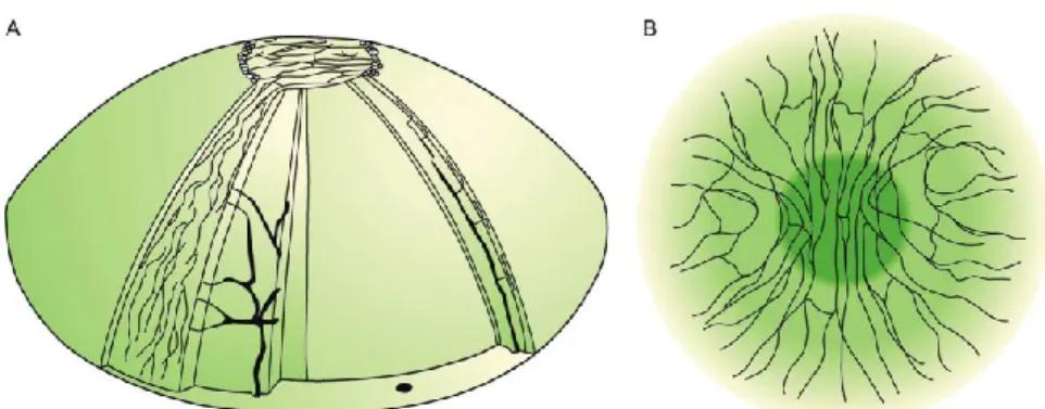
Corneal neurons can be classified as thin myelinated (A-delta type, 30% in the mouse) or unmyelinated (C type, 70% in the mouse), depending on the size and presence of a myelin sheath in the axon (Belmonte and Giraldez 1981). All peripheral axons of corneal neurons lose the myelin sheath when they enter the corneal stroma, mainly grouped in a variable number of radially oriented nerve bundles. They then branch extensively, forming a subepithelial plexus from which thin branches ascend up traversing the Bowman’s layer and enter into the basal layer of the epithelium. There, they run parallel to the corneal surface forming the leashes and terminate in the superficial layers of the corneal epithelium (Fig.1) (ZANDER and WEDDELL 1951;Chan-Ling 1989;Muller et al. 2001;Muller et al. 2003).
12
Fig. 1. (A) Schematic distribution of nerves in the stroma and subbasal plexus
in human corneas. (B) Adapted scheme on the organization of the subbasal plexus. In the apex the nerve bundles show a preferred orientation in the superior-inferior direction and in the surrounding area they tend to be oriented in the nasal-temporal direction. It is estimated that there are approximately 7000 nociceptors per mm2 in the human corneal
epithelium.
The mechanisms by which corneal nerve fibres maintain a healthy cornea and promote wound healing after eye injuries is currently under active research in several laboratories. The results obtained in co-culture studies suggest the existence of a possible crosstalk between neurons and corneal epithelial cells through the mutual release of soluble substances. For example, trigeminal neurons release diffusible factors (e.g. neurotransmitters and neuropeptides) that stimulate corneal epithelial cell growth, proliferation, differentiation, and type VII collagen production (Baker et al. 1993;Garcia-Hirschfeld et al. 1994). Stromal keratocytes also produce neurotrophins (Lambiase et al. 2000) (You et al. 2001), however, the extent to which these substances exert trophic influences on corneal nerve fibres remains to be determined.
The majority of corneal sensory fibers (about 70%), named polymodal nociceptors, are equally activated by near-noxious mechanical energy but they also respond to heat, to exogenous chemical irritants and to a large variety of endogenous chemical mediators released by damage corneal tissue, by resident inflammatory cells or originating from the
13 plasma leaking from limbal vessels (protons, potassium ions, ATP, prostaglandins and other arachidonic acid metabolites, amino acids, amines, cytokines, kynins, growth factor) (Belmonte et al. 2004). Polymodal nociceptors possibly contribute, together with mechano-nociceptors, to the sharp mechanical pain that arises when the cornea is acutely exposed to mechanical force, but they are also the principal source of nerve impulse activity caused by chemical irritation, heat or noxious cold (Belmonte and Giraldez 1981;Beuerman and Rozsa 1985). During inflammation, locally released mediators stimulate polymodal nociceptors, leading to a continuous firing that produces sustained sensations of pain.
Many studies demonstrated that an healthy status of innervations and corneal sensitivity are both essentials for maintaining a healthy ocular surface, mainly because corneal nerves modulate cell proliferation, differentiation and wound healing (Beuerman and Schimmelpfennig 1980;Garcia-Hirschfeld et al. 1994). In addition, corneal nerves have been demonstrated to play a role in ion transport, and collagen expression (Jones and Marfurt 1996;Baker et al. 1993).
Furthermore, it has been shown that corneal denervation induces apoptosis of resident corneal cells (Cortina et al. 2011). However, the mechanisms underlying these effects are not completely understood (Cortina et al. 2011).
3- SURGYCAL PROCEDURESES ON CORNEA WITH OR WITHOUT EPITHELIUM REMOVAL
The corneal refractive surgery is based on “the law of thickness” proposed by Barraquer in 1964 (Barraquer JI 1964) “…changing the
14 thickness of the cornea follows the idea that the cornea is a stable lens, removing tissue in the center or adding tissue on the periphery therefore flattens the cornea.” The argon fluoride (193 nm) excimer laser permits the excision of corneal tissue with minimal damage to the adjacent tissues. It employs an high energy ultraviolet radiation to break the covalent bonds between molecules in the corneal stroma without generating high levels of heat (Krauss et al. 1986). This procedure has been termed photoablative process and is the principal reason making laser refractive surgery a relative predictable and safer procedure. These are the main differences in the method used to remove the epitelial surface:
PRK and LASIK- were the first surgical procedures, in which laser was
used to shape the corneal surface. The most important difference between the two procedures is the way of how the middle layer of the cornea is exposed. During PRK the epithelium is mechanically scraped off using a special instrument and epithelium is not placed back after refractive surgery whereas LASIK, consists in the preparation of a flap and in a replacement of the epithelium back to its position (Lombardo et al. 2011).
LASEK - was a modification of PRK method. During LASEK the epithelium layer is loosen from the tissue below with alcohol and is moved aside for the time of the surgery. The replaced epithelium layer will act as a natural contact lense.
Epi-LASIK (Epi-K) - is the newest method for removing epithelium. Epithelium layer is separated from below layers by using a special microkeratome. As the remaining epithelium cells are not damaged by
15 alcohol or mechanical scraping instrument, the recovering of the eye after Epi-K method is faster and the chance of arising haze is smaller then with using PRK or LASEK method (Reilly et al. 2010).
c-TEN - The cTen system was introduced by an Italian company called iVis Technologies. It includes a 1,000-Hertz excimer laser (iRES) and a software planning system called CIPTA. cTen is an acronym for Customised, TransEpithelial, No-touch surgery. It is indicated for complex cases with irregularity of the cornea (e.g irregular astigmatism). They use the excimer laser to remove the epithelial skin layer and use topography information from a Scheimpflug camera scanner to correct imperfections across the cornea stroma (Thomann and Schipper 2010). 4-PRK AND LASIK COMPLICATIONS
More than 12 million patients have undergone LASIK and PRK since they were approved (see http://www. laser-eye-surgery statistics.com/).
These are the most popular and used techniques to perform experimental corneal surgery. Many studies demonstrated that corneal subbasal nerve density does not recover for up to five years (Erie et al. 2005), and the number of stromal nerves decreases by nearly 90% after LASIK surgery (Erie et al. 2005) then complications, although not frequent, do occur. Complications may arise intraoperatively or during the postoperative period (Filatov et al. 1997;Ghadhfan et al. 2007;Melki and Azar 2001;Stein 2000). Some patients may develop glare, halos, monocular diplopia, changes in contrast sensitivity, and dry eye (Hersh et al. 1997;Hersh et al. 1990;Seiler and McDonnell 1995). Postoperative complications after LASIK include flap striae and folds, dislodging of the flap, interface debris, epithelial ingrowth, diffuse lamellar keratitis, and
16 corneal infections (Davis et al. 2000;Gimbel et al. 1998;Melki and Azar 2001). Instead, postoperative complications after PRK, are related to epithelial debridment and include cronic pain, delayed epithelial healing, infection, and corneal scarring/haze (Alio et al. 2008).
4a- Chronic neurophatic corneal pain, inflammation and impaired wound healing.
The cornea is the most powerful pain generator in the human body. The density of corneal pain receptors has been estimated to be 40 times higher than dental pulp. It is estimated that there are approximately 7000 nociceptors per mm2 in the human corneal epithelium.
Rosenthal et al., suggested that after surgery the damaged corneal nerve fibers (i.e. polymodal nociceptor) could be the cause of all patient symptoms, such as glare, halos, monocular diplopia, sustained pain, impaired wound healing, changes in contrast sensitivity, whether or not the initial disease is severe dry eye or corneal neuropathy. Patients undergoing to this surgery report severe, unremitting, burning pain and photophobia. They also demonstrated using in vivo confocal microscopy that all these patients presented nerve abnormalities and this alteration in corneal nerves (Cruzat et al. 2010) are similar to those reported in skin biopsies of neuropathic pain conditions (Lauria and Devigili 2007;Sommer 2008).
Recently, Cortina et al. (Cortina et al. 2010) confirmed by immunohystochemistry that epithelial removal due to surgical procedures lead to the transection of afferent sensory nerve fibres (Figure 2) and the aberrant regenerated corneal nerves are likely to be among the most important factors associated with impaired wound healing and exaggerated inflammatory response into the corneal layer after surgery
17 (Ambrosio, Jr. et al. 2008). They also demonstrated that the epithelial removal and the impairment of the sub-basal nerve plexus correspond to a lack of sensitivity measured by Coche-Bonnet esthesiometer (Cortina et al. 2010).
Fig.2 Immunohistochemistry of epithelia, subepithelia, and stroma of rabbit
corneal whole mounts stained with anti– β III tubulin antibody 8 weeks after surgery.
Some studies (Tuunanen et al. 1997;Tuisku et al. 2007) suggest that the improvement of corneal wound healing, showed a direct relation
18 with the recover of the corneal subbasal nerve plexus to maintain corneal healing and transparency.
4b- Chronic neurophatic corneal pain treatment and side- effects on epithelial regrowth.
As mentioned above one of the most notable adverse effects of PRK is pain, which typically occurs within the first 72 h after surgery (Assouline et al. 1998;McCarty et al. 1996).
Topical ocular nonsteroidal anti-inflammatory drugs (NSAIDs) have demonstrated efficacy in controlling pain after PRK surgery (Sher et al. 1993;Arshinoff et al. 1996) and they are commonly used during the postoperative period. The best choice of NSAID, however, is yet to be determined.
Topical NSAIDs have been shown to reduce pain after PRK in numerous clinical trials. Some studies, however, report delayed epithelial wound healing, (Assouline et al. 1998;Rajpal and Cooperman 1999) and others have shown no adverse effects on corneal reepithelialization when topical NSAIDs were used (Sher et al. 1993). These conflicting results highlight the need for comparative studies among agents. Durrie et al. (2007) evaluated the efficacy of 3 approved ophthalmic NSAIDs nepafenac 0.1%, ketorolac 0.4%, and bromfenac 0.09% to determine their effects on corneal reepithelialization and postoperative pain control in patients undergoing PRK surgery (Durrie et al. 2007). No difference in time to corneal reepithelialization was noted between nepafenac 0.1% and ketorolac 0.4%. Average time to complete healing was approximately 5.5 d with both regimens; however, patients treated with bromfenac 0.09% demonstrated a delay in corneal healing of approximately 1.5 d (mean healing time, 7 d). In a recent study
19 comparing nepafenac 0.1% with ketorolac 0.4%, the average time to healing was approximately 4 d in both treatment groups (Donnenfeld et al. 2007). It has been postulated that the effects of NSAIDs on the arachidonic pathway, similar to corticosteroids, may play a role in corneal healing (Hersh et al. 1990).
Mechanistically, NSAIDs have varying effects on the cyclooxygenase and lipoxygenase pathways. This makes the comparative studies in this kind of field important in assessing differences between agents. In addition, specific preservatives (eg, thimerosal) have been implicated in delayed healing (Assouline et al. 1998).
The dose and duration of use may play an important role because higher doses and longer duration of use seem to have a greater association with delayed healing.
4c- Biochemical pathway involved in corneal inflammatory response after injury.
Cytokines and growth factors released after injury are the soluble factors mediating the signals and interaction between different cells and components to restore corneal functionality (Bazan 2005).
Inflammation is the first response of corneal tissue to an insult, and during the first hours, the cells of the corneal layers respond by releasing arachidonic acid (AA) (Eakins et al. 1972) from membrane phospholipids and converting this fatty acid into eicosanoids and platelet-activating factor (PAF) (Bazan et al. 1993).
When inflammation is more severe and persists, cells that infiltrate the cornea, mainly neutrophils, amplify the response and contribute to the increase in these lipids. Some of these mediators increase pain, delay wound healing, and promote neovascularization through an induction of
20 overexpression of MMP-1, MMP-9, COX-2 and VEGF in the epithelial cell (Bellner et al. 2011a). PAF also increases keratocyte apoptosis and stimulates myofibroblasts to synthesize furin, a convertase that activates growth factors and receptors (Bazan 2005). Blocking the action of PAF, with PAF-antagonist (LAU-0901), prevents both diffuse lamellar keratitis (DLK) after LASIK surgery and stromal melting after severe alkali burn. On the other hand, two lipoxygenase metabolites, 12(S)-HETE and 15(S)-12(S)-HETE, are activated by growth factors and are involved in proliferation and wound repair (Figure 3) (Gronert et al. 2005).
Growth factors such as epidermal growth factor (EGF) and hepatocyte growth factor (HGF), released in response to corneal injury, utilize signal mechanisms involving lipoxygenase activation to exert their proliferative effects on epithelial cells and contribute to the repair phase. Bazan et al. (Bazan 2005) demonstrated that the omega-3 fatty acid docosahexaenoic acid (DHA) potentiates the corneal nerve regenerative action by nerve growth factor (NGF) after PRK and also stimulation of cyclooxygenase-2 (COX-2) produces a different pattern of prostaglandin synthesis in epithelial and endothelial cells, suggesting different functions; e.g. PGE2 released from epithelial cells has been implicated in pain. In conclusion, lipids play an important role in the complex inflammatory responses that occur after corneal injury (Cortina et al. 2011).
21
Fig.3 Schematic representation of current knowledge on the actions of lipid
mediators in corneal damage and repair (Bazan 2005).
5- HEME OXYGENASE AND ITS ANTI-INFLAMMATORY, ANTIOXIDANT AND NEUROPROTECTIVE PROPERTIES.
HO is the rate-limiting enzyme in heme metabolism cleaving heme, a potent oxidant, into three products: CO, biliverdin and iron. Biliverdin is further reduced by biliverdin reductase to bilirubin. The significance of the HO system extends far beyond its original role in the catabolism of heme in senescent heme proteins (Abraham and Kappas, 2008). In the recent years the heme oxygenase system (HO-1 and HO-2) has emerged as a fundamental endogenous cytoprotective and anti-inflammatory system in many tissues producing compounds that uniquely combine vasodilatory (CO), anti-oxidant (CO and bilirubin) and anti-apoptotic (CO, bilirubin) properties (Abraham and Kappas, 2008).
22 Overall, HO has a well-deserved reputation of a potent endogenous anti-oxidant cell defense system. So far, HO-1, transcriptionally upregulated in response to oxidative stress, was considered the only HO isoform that provided cytoprotection. However, it is becoming clear that, in the brain and in many other tissues, HO-2, rather than HO-1, participates in a multitude of housekeeping functions directed at maintaining tissue homeostasis and protecting against oxidative stress under constantly changing conditions (Chen et al. 2003).
The ability of HO-2 to be post-transcriptionally activated provides immediate actions including blocking oxidant-producing systems, scavenging reactive oxygen species (ROS), and, therefore, preventing and eliminating further damage caused by ROS (Parfenova and Leffler 2008). For example, the cerebroprotective functions of HO-2 include CO-mediated increases in cerebral blood flow in response to seizures, hypoxia, hypotension, and glutamate aimed at providing neurons with nutrients and oxygen (Chen et al. 2003). In addition, CO, via its ability to strongly bind to heme, inhibits heme-containing ROS-generating systems, NADPH oxidase and the mitochondrial respiratory chain, thus reducing oxidative stress (Parfenova and Leffler 2008).
Furthermore, bilirubin, a potent ROS scavenger, eliminates preformed oxidant radicals thus strengthening the anti-oxidant effects of CO. Therefore, HO-2 builds a strong cerebrotective system in the brain and cerebral circulation that, in contrast to HO-1, provides immediate responses to cerebrovascular stress and prevents potential damage to neurons, astrocytes, and cerebral vascular endothelium (Parfenova and Leffler 2008). Chen et al. (Chen et al. 2003) investigated the roles of HO-1 and HO-2 in the homeostasis of a neuronal population using the
23 olfactory system as a model and knock out mice for both HO isoforms. The olfactory system is an important neuronal and developmental model (Graziadei and Monti Graziadei 1980;Calof and Chikaraishi 1989;Hansel et al. 2001), and may be used to delineate the functions of HO in neurons. The olfactory epithelium contains one of the few neuronal populations capable of robust neurogenesis throughout life, and is the only mammalian neuronal population known to be able to replace itself functionally (Graziadei and Monti Graziadei 1980). They provided evidence that HO-1 and HO-2 are both expressed in neurons and in embryonic and adult olfactory epithelium, where they are positioned to mediate autocrine/paracrine functions (Chen et al. 2003).
They demonstrated that loss of HO-1 or HO-2 affects the homeostasis of the neuronal population. Olf-1/early B-cell factor (O/E-1) was used as a marker to identify post-mitotic cells of the olfactory neuronal lineage. O/E-1 labeling was significantly reduced in both HO-1 and HO-2 animals. The numbers of cells labeled with BrdU were also decreased in HO-1 and HO-2 null mice. In contrast, the number of TUNEL-positive cells was only increased in the HO-2 null mouse, indicating that only HO-2 mediated survival cues. The proliferative and survival functions of HO could be mediated by any of the products of HO action then further studies need to be made to evaluate this hypothesis. Thus Chen et al. (2003) were able to show that HO-2 supports neuronal survival even under baseline condition.
24 Aim of the study
The aim of my research has been an extensive characterization of the HO system status in the cornea correlated to the inflammatory/reparative response to injury elicited by corneal epithelial removal. For this purpose, I first used an in vitro model of human corneal epithelial cells and successively I employed an in vivo model in which the corneal epithelial removal was used to mimic the first step of the surgical procedure. Both models were used to correlate spatial/temporal changes of HO expression and activity with inflammatory and reparative response following epithelial removal. Leukocyte infiltration, pro-inflammatory mediators production, and wound closure were evaluated under various experimental conditions in which HO activity is differently upregulated or inhibited.
25
26 Knockdown of Heme Oxygenase-2 Impairs Corneal Epithelial Cell
Wound Healingδ
Adna Halilovic‡, Kiran A Patil‡, Lars Bellner, Giuseppina Marrazzo, Kirkland Castellano, Giuseppe Cullaro, Michael W. Dunn and Michal
Laniado-Schwartzman*
Department of Pharmacology, New York Medical College, Valhalla, NY 10595
δJournal of Cellular Physiology. 2011 Jul;226(7):1732-40. doi:
10.1002/jcp.22502. ABSTRACT
Heme oxygenase (HO) represents an intrinsic cytoprotective system based on its anti-oxidative and anti-inflammatory properties mediated via its products biliverdin/bilirubin and carbon monoxide (CO). We showed that deletion of HO-2 results in impaired corneal wound healing with associated chronic inflammatory complications. This study was undertaken to examine the role of HO activity and the contribution of HO-1 and HO-2 to corneal wound healing in an in vitro epithelial scratch injury model. A scratch wound model was established using human corneal epithelial (HCE) cells. These cells expressed both 1 and HO-2 proteins. Injury elicited a rapid and transient increase in HO-1 and HO activity; HO-2 expression was unchanged. Treatment with biliverdin or CORM-A1, a CO donor, accelerated wound closure by 10% at 24 h. Inhibition of HO activity impaired wound closure by more than 50%. However, addition of biliverdin or CORM-A1 reversed the effect of HO inhibition on wound healing. Moreover, knockdown of HO-2 expression, but not HO-1, significantly impaired wound healing. These results
27 indicate that HO activity is required for corneal epithelial cell migration. Inhibition of HO activity impairs wound healing while amplification of its activity restores and accelerates healing. Importantly, HO-2, which is highly expressed in the corneal epithelium, appears to be critical for the wound healing process in the cornea. The mechanisms by which it contributes to cell migration in response to injury may reside in the cytoprotective properties of CO and biliverdin.
INTRODUCTION
A rapid healing of corneal epithelial wounds is important not only for the maintenance of corneal transparency but also to protect the underlying stroma from further damage and to avoid infection. Repair of corneal surface wounds initiates with migration of existing epithelial cells to cover the wound followed with proliferation of epithelial cells to restore the normal thickness of the cornea and a return to homeostasis. This process is regulated by a multitude of growth factors, cytokines and structural proteins that orchestrate the necessary steps for completion of healing including migration, adhesion, proliferation, differentiation as well as apoptosis (Imanishi et al., 2000; Lu et al., 2001). We have shown that a deficiency in heme oxygenase (HO) activity impairs corneal wound healing whereas amplification of HO activity accelerates epithelial repair suggesting that the HO system is an integral component of the corneal repair system (Patil et al., 2008; Seta et al., 2006).
HO is the rate-limiting enzyme in heme metabolism cleaving heme, a potent oxidant, into three products: CO, biliverdin and iron. Biliverdin is further reduced by biliverdin reductase to bilirubin. The significance of the HO (HO-1 and HO-2) system extends far beyond its
28 original role in the catabolism of heme in senescent heme proteins (Abraham and Kappas, 2008). It is clear that the HO system has potent cytoprotective and anti-inflammatory properties that are critical in many organs including the eye (Bellner et al., 2008; Seta et al., 2006) and are attributed to its products biliverdin/bilirubin and carbon monoxide (CO). At physiologic concentrations bilirubin (and to a lesser extent biliverdin) is a potent and efficient scavenger of reactive oxygen species (Dore et al., 1999; Stocker et al., 1987).
Biliverdin/bilirubin decreases LPS-induced pro-inflammatory transcription factors (NF-κB) and cytokines (IL-6, TNFα) (Foresti et al., 2004; Gibbs and Maines, 2007; Soares et al., 2004) and this may be mediated via its ROS scavenging mechanisms as the expression of many pro-inflammatory genes is redox-sensitive (Chen and Kunsch, 2004). CO, as well as biliverdin, contributes substantially to the anti-inflammatory properties of HO by suppressing pro-anti-inflammatory cytokines and chemokines while increasing expression of the anti-inflammatory mediator interleukin-10 (Kim et al., 2006; Morse et al., 2003; Ollinger et al., 2007; Song et al., 2003; Wegiel et al., 2009). Iron derived from heme via HO is effectively controlled by ferritin, which is readily upregulated by HO, via sequestration and ferroxidase activity (Balla et al., 2007).
HO-1 and HO-2 are expressed in most tissues. HO-1 is induced by numerous factors that increase cellular stress while HO-2, in general, is constitutively expressed and developmentally regulated and is altered in many human pathological conditions (Zenclussen et al., 2003). In terms of mechanisms of heme oxidation, substrate and cofactor requirements and susceptibility to porphyrins, they are alike. But they
29 differ in function; while HO-2 is thought to serve as the constitutive HO activity maintaining cell homeostasis, HO-1 expression is relatively low in most tissues and its expression rises rapidly in response to cellular stress (Abraham and Kappas, 2008). Induction of HO-1 has been shown to attenuate ocular surface inflammation (Conners et al., 1995; Laniado Schwartzman et al., 1997), accelerate corneal wound healing (Patil et al., 2008), attenuate retinal detachment-induced photoreceptor apoptosis (Shyong et al., 2008), protect photoreceptors from light damage (Sun et al., 2007) and prevent retinal ganglion cell death (Hegazy et al., 2000).
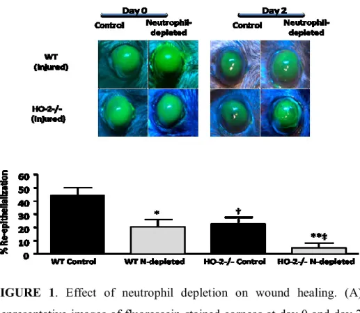
The use of HO-2 null mice has brought new insights into HO research. One such insight is the association of HO-2 deletion with impaired HO-1 expression indicating that HO-2 is critical for HO-1 induction (Seta et al., 2006). The result of this is a mouse deficient in HO activity. A recent study described the altered course of acute inflammation and the reparative response in these HO-2 null mice following mechanical de-epithelialization of their corneas. Wild type (WT) mice heal the epithelial defect in seven days, the acute inflammatory response and the associated inflammatory cytokines and infiltrating neutrophils resolve in a few days and there is no significant neovascularization. In stark contrast to the WT mice, the HO-2 null mice corneas experience an unresolved acute inflammation from the injury, epithelial regeneration is greatly impaired, massive revascularization is evident and, by day seven, all corneas suffer perforation (Seta et al., 2006). Attempts to rescue the HO-2 phenotype in HO-2 null mice have been made using biliverdin. Application of this HO bioactive product rescued the acute inflammatory and reparative response in HO-2 null mice with a two-fold increase in the rate of re-epithelialization reaching
30 that of the WT mice (Seta et al., 2006). In the suture-induced model of corneal inflammatory neovascularization, HO-2 null mice displayed an exaggerated inflammatory and neovascular response that is greatly attenuated with topical application of biliverdin(Bellner et al., 2008).
The present study further explores the role of HO in and the contribution of each isoform (HO-1 and HO-2) to corneal epithelial wound healing using cultured human corneal epithelial cells in a model of epithelial injury. The results indicate that injury activates the HO system and that HO activity is important for re-epithelialization. Moreover, HO-2 and not HO-1 appears to be the critical component of the HO system for epithelial wound healing.
MATERIALS AND METHODS Materials
Biliverdin, heme and metalloporphyrines were from Frontier Scientific Inc. (Logan, UT). Carbon Monoxide releasing molecule (CORM-A1) (Motterlini et al., 2005) was obtained from Dr. John R. Falck (University of Texas Southwestern Medical Center, Dallas, TX). Hydroxyurea was from MP Biochemical (Solon, OH). Silencing inhibitory RNAs (siRNAs) against the human HO-1 and HO-2 as well as non-specific and GFP-conjugated siRNAs were purchased from Santa Cruz Biotech (Santa Cruz, CA). Antibodies against HO-1 and HO-2 were from Assay designs (Ann Arbor, MI) and β-actin from Sigma Aldrich (St Louis, MO).
31 Primary cultures of human corneal epithelial cells (passage 2-3; ScienCell, Carlsbad, CA) and a human corneal epithelial cell line (HCE; passages 29-45) obtained from Dr. Haydee Bazan (LSU Health Science Center) (Sharma et al., 2005), were used in these experiments. Cells were grown in fibronectin-coated T-75 flasks and maintained in serum-free keratinocyte growth medium (KGM, Lonza, USA) supplemented with appropriate growth factors and antibiotics. For all experiments, cells (6-12-well plates) were grown to 60-70% confluency in KGM and then the medium was changed to a growth factor- and supplement-free keratinocyte basal medium (KBM) and cells were incubated for additional 24 h. Scratch injury was performed with a sterile 20 µl pipet tip to remove cells in two perpendicular linear scraps and generate a wound approximately 0.1 cm in width. The medium was discarded and cells were cultured in fresh KBM medium in the presence and absence of hydroxyurea (0.5mM), chromium mesoporphyrin (CrMP, 10 µM), CORM-A1 (100-400 µM) and Biliverdin (5-20 µM). Phase-contrast images of the injured area near the crossing point were acquired at the time of injury and at the indicated time points thereafter with an inverted microscope equipped with a charge-coupled device camera (Axiovert; Carl Zeiss, Thornwood, NY). The wound area (mm2) was determined by computerized planimetry using Axiovision 4.6 software (Zeiss). The extent of healing over time was defined as percent of the denuded area at the time of injury. Cell proliferation and viability was measured using the methyl thiazolyl tetrazolium (MTT) colorimetric assay.
siRNA transfection of HCE cells
HCE cells were grown in 12-well plates to 60-70% confluency. Transfection of siRNA was performed according to the manufacturers’
32 instructions (Santa Cruz). Briefly, cells were incubated with 500 µl of transfection solutions containing a mixture of siRNA (100 nM) and siRNA transfection reagent (5 μl) (Santa Cruz) for 6 h. Afterwards, the transfection solutions were replaced with fresh KGM-2 growth medium for an additional 24 h. Medium was changed to the growth factor- and supplement-free KBM medium and injury performed 24 h later. Cells were also transfected with control siRNA (100 nM) in parallel to ensure specific gene silencing. At the end of the experiment (24 h after injury), cells were collected for Western blot analysis and real time PCR. Transfection efficiency was assessed using GFP-conjugated non-specific siRNA (Santa Cruz).
HO Activity/CO Measurement
HCE cells were collected by trypsnization 4 h after injury. Cells were washed with phosphate buffered saline, pH 7.4 and resuspended in oxygenated Krebs Buffer. HCE cells were subsequently incubated for 1 h in the dark at 37°C with 2 mM hemin in the presence and absence of CrMP (10 µM). CO released in the headspace gas was analyzed by gas chromatography-mass spectrometry (GC-MS, HP5989A interfaced to HP5890; Hewlett Packard, Palo Alto, CA). The amount of CrMP-sensitive CO was calculated from standard curves constructed with an abundance of ions m/z 28 and m/z 29 or m/z 31, as previously described (Bellner et al., 2009).
Real-Time Polymerase Chain Reaction (PCR)
Total RNA was isolated using RNeasy Protect Mini Kit (QIAGEN, Carlsbad, CA) and RNA integrity was verified by agarose gel electrophoresis and quantitated by Nano drop. Reverse transcription
33 reaction of total RNA was performed using the qScript cDNA synthesis kit (Quanta Bioscience, Gaithersburg, MD). Quantitative real-time PCR was performed using PerfeCTa SYBR Green QPCR FastMix (Quanta Bioscience) and the Mx3000 real-time PCR system (Stratagene, La Jolla, CA). Specific primers were designed based on published sequences (GenBank) and were as follows: HO-1 sense,
5'-TGCTCAACATCCAGCTCTTT-3' and anti-sense,
5'-GCAGAATCTTGACTTTGTT-3'; HO-2 sense,
5'-ATGTCAGCGGAAGTGGAAAC-3' and anti-sense,
5'-CGAGAGGTCAGCCATTCTCA-3'; 18S sense,
5'-GATGGGCGGCGGAAAATAG-3' and anti-sense,
5'-GCGTGGATTCTGCATAATGG-3'. PCR efficiency for each primer pair was determined by quantitating amplification with increasing concentrations of template cDNA, and specific amplification was verified by subsequent analysis of melt curve profiles for each amplification. A nontemplate control served as negative control to exclude the formation of primer dimers or any other nonspecific PCR products. RNA expression of target genes was calculated based on the real-time PCR efficiency (E) and the threshold crossing point (CP) and is expressed in comparison to the reference gene 18S as described (Bellner et al., 2009).
Western blot
Western blot analysis, HCE cells were homogenized in T-PER tissue protein extraction reagent containing Halt protease inhibitor cocktail (Pierce Biotechnology, Inc., Rockford, IL). Proteins were separated by gel electrophoresis, and immunoblotting was performed using the following primary antibodies: rabbit anti-human HO-1
34 (1:2,500), rabbit anti-human HO-2 (1:2,500) and β-actin mouse monoclonal antibody (1:10,000). Detection and densitometry analysis was performed as previously described (Bellner et al., 2009).
Immunofluorescence assay
HCE cells were grown in 16-well chamber slides at 37°C to 100% confluence. Scratch injury at different time points was performed with a sterile 0.1-10 µl pipet tip to remove cells in a single linear scrape. Briefly, slides were fixed in 4% paraformaldehyde for 10 minutes at room temperature, washed with PBS and then blocked with 5% goat serum in PBS whereupon the slides were incubated with rabbit anti-human HO-1 antibody (1:400) or with rabbit anti-anti-human HO-2 antibody (1:50) overnight at 4°C, then washed and further incubated with a secondary Cy3 conjugated goat anti-rabbit antibody (1:500, Jackson Immunoresearch, West Grove, PA) for 2 h at room temperature. To further verify cellular entity, the slides were washed and counterstained for nuclei with 4’,6- diamidino-2-phenylindole dihydrochloride (DAPI) for 15 minutes in dark at room temperature. Pictures were taken adjacent to the wound edge and distal to wound edge (≥300 µm) using a Zeiss Axioplan-2 fluorescent microscope. The total fluorescence intensity (normalized to cell number) was measured using AxioVision 2 multi channel image processing software (Zeiss, Göttingen, Germany) and the intensity at the wound edge was compared to the intensity obtained distal to wound edge.
Statistical analysis
Results are the mean±SEM. Significance of difference in mean values was determined using either the Mann-Whitney U-test or one-way
35 ANOVA followed by Newman-Keuls post-hoc test for multiple comparisons. P<0.05 was considered to be significant.
RESULTS
HO-1 and HO-2 expression in Human corneal epithelial cells
Primary cultures of human corneal epithelial cells express HO-1 and HO-2. In response to injury, HO-1 protein levels increased within 1 h and returned to control levels 4 h after injury (Figure 1A). HO-2 expression was not affected by injury (Figure 1B). The HCE cell line has been extensively used to elucidate cellular mechanisms of corneal epithelial wound healing. These cells express HO-1 and HO-2 and oxidative injury elicited a rapid activation of the HO system (Abraham and Kappas, 2008). Similar to primary cultures, HCE cells responded to injury with a transient increase in HO-1 expression with no change in HO-2 expression (Figure 1C and D). The increase in HO-1 expression was accompanied by a 50% increase in HO activity, as measured by the amount of CrMP-sensitive CO (Figure 1E).
The effect of injury on HO-1 and HO-2 expression was also assessed by immunofluorescence imaging of cells at the edge of the wound and at a distance of 1-2 µm from the wound. As seen in Figure 2, HO-1 protein expression 1 h after injury increased by 2.5- and 1.7-fold at the wound edge and distal to the wound. HO-1 protein levels remained elevated for 4 h and returned to control (pre-injury) 8 h after injury. In contrast, HO-2 expression either at the wound edge or far from the wound was not altered after injury (Figure 2).
36 Human Corneal epithelial cells healing enhanced by biliverdin and CORM-A1
Scratch wounds in HCE cells were closed by 50.72± 3.59% and 78.24±2.67% at 12 and 24 h, respectively (Figure 3). Addition of 10 µM of biliverdin enhanced wound healing by 11% (61.54±2.71 % closure) and 10 % (89.77± 2.49 % closure) at 12 and 24 h, respectively. The effect of biliverdin was dose-dependent (Figure 4A). Addition of CORM-A1 which releases CO in aqueous solutions (Motterlini et al., 2005) was less efficacious than biliverdin; at 100 µM CORM-A1 enhances wound healing by 5% (55.59±2.95 % closure) and 10% (87.68%±1.53 % closure) at 12 and 24 h, respectively (Figure 3). CORM-A1 effect was also dose-dependent (Figure 4B). The inactivated CORM (iCORM) at concentrations of 100-400 µM had no significant effect on wound closure (data not shown).
Inhibition of HO activity impairs wound healing
CrMP at the concentration used (10 µM) inhibited HO activity measured as CO production by 82±2 and 92±3% in control and injured cells, respectively (mean±SE, n=3). CrMP had no effect on cell viability at all time points, however, it significantly inhibited the healing of scratch wounds in HCE cells. As seen in Figure 5A-C, in cells treated with CrMP, healing was inhibited by 65%; only 18.21±1.75% and 28.13±3.22% of the wounds were closed at 12 and 24 h, respectively. The inhibitory action of CrMP was partially negated at 12 h by adding back biliverdin (34%±1.97%) or CORM-A1 (36.08±2.33%) (Figure 5). Wound closure at 24 h after injury of cells treated with CrMP and supplemented with either biliverdin or CORM-A1 was not different than
37 that of the control untreated cells, suggesting that HO activity (via production of biliverdin and/or CO) is required for epithelial cell healing. Addition of CrMP, biliverdin or CORM-A1 did not significantly alter the rate of proliferation after injury (Figure 5D).
To differentiate the contribution of HO to cell proliferation and migration in wound closure, the cell cycle blocker hydroxyurea (0.5 mM) was added in the scratch wound model. Assessment of cell viability indicated that this concentration was sufficient to inhibit HCE proliferation without significantly affecting cell viability. As seen in Figure 6, in the presence of hydroxyurea, healing of the wounds at 12 and 24 h after injury amounted to of 29.74 and 63.14%, indicating attenuation of healing by 39% and 20%, respectively. Addition of the HO inhibitor CrMP to cells treated with hydroxyurea further attenuated migration-driven healing at 24 h by 23% when compared to cells treated with hydroxyurea. Addition of biliverdin or CORM-A1, respectively, significantly enhanced migration-driven wound healing by 27 and 42% at 12 h, and 17 and 20% at 24 h (Figure 6).
HO-2 knockdown impairs healing of wounded HCE cells
Knockdown of HO-1 and HO-2 expression was carried out with the use of specific siRNA. Transfection with GFP-conjugated siRNA demonstrated a transfection efficiency of 65±10% (n=4). Scratch injury was performed in cell transfected with HO-1, HO-2 and non-specific siRNAs. As seen in Figure 7, wound closure in cells treated with HO-2 siRNA was significantly impaired; only 50% of the scratch wound closed at 6 and 12 h after injury and 70% at 24 h after injury. In contrast, wound closure in cells treated with HO-1 siRNA was not significantly different
38 than the control untreated or cell treated with non-specific siRNA at all time points (Figure 7). That the siRNAs specifically knockdown their corresponding proteins is seen in Figure 8. Real time PCR of HO-1 and HO-2 mRNA as well as Western blot analysis of HO-1 and HO-2 protein clearly indicated a specific knockdown of HO-1 and HO-2 gene expression by their corresponding siRNA preparations (Figure 8). Densitometry analysis further demonstrated that suppression of HO-1 and HO-2 by their corresponding siRNA was about the same (viz., 72±9 and 84±6%, respectively, when compared to control; 73±13 and 78±12% when compared to control siRNA).
DISCUSSION
This in vitro study provides further evidence for a critical role for HO in corneal epithelial wound healing. Using an in vitro model of epithelial scratch injury in HCE cells, we showed that inhibition of HO activity attenuates wound closure while amplification of HO activity by supplementation of its catalytic products, biliverdin or CO, accelerates wound closure. The beneficial effect of the HO system was further assigned to HO-2, the constitutive isoform of the HO system, based on findings that 2-specific siRNA impaired wound closure whereas HO-1-specific siRNA had no significant effect. The fact that HO-2 is highly expressed in the corneal epithelium (Bellner et al., 2008) together with a previous study demonstrating that its deficiency results in aberrant inflammatory and repair response to epithelial injury typified by perforation, ulceration and neovascularization (Seta et al., 2006) suggest that HO-2 is a critical component for corneal epithelial homeostasis. Several findings in the current study provide additional evidence to
39 support an important role for HO-2 expression and HO activity in corneal epithelial homeostasis.
First is the demonstration that addition of the HO products, biliverdin and CO, significantly accelerated closure of the wound made by scratching HCE monolayers, consistent with previous findings that upregulation of the HO system attenuates the inflammatory response while accelerating the repair of the cornea following injury (Conners et al., 1995; Patil et al., 2008). It is well-documented that biliverdin and CO act as endogenous cytoprotective molecules, each with its specific mode of action. Biliverdin, the final product of HO catalytic activity, is further reduced to bilirubin by biliverdin reductase (BVR) (Abraham and Kappas, 2008). Bilirubin, and to a lesser extent biliverdin, is a powerful endogenous/physiological antioxidant (Stocker et al., 1987). Studies have found that bilirubin at 10 nM was capable of protecting cells from a 10,000 fold increase in oxidative stress generated by hydrogen peroxide and identified an amplification process for bilirubin bioactions whereby bilirubin acting as an antioxidant, is itself oxidized to biliverdin and then recycled by BVR back to bilirubin a process that could readily afford 10,000 fold amplification (Baranano et al., 2002). BVR is widely expressed in most tissues including the cornea (Bellner et al, IOVS, 2008 49, E-Abstract 2378). Hence, administration of water-soluble biliverdin is accompanied with increased tissue/cell levels of bilirubin. The primary mechanism for bilirubin-mediated cytoprotection in various types of stress appears to be due to its potent antioxidant activity. Given that injury by itself evokes in any given tissue or cell a stress response which frequently is accompanied by increased reactive oxygen radicals, i.e., oxidative stress, it is reasonable to assume that the increased healing of
40 wounded HCE in the presence of biliverdin is the consequence of decreasing this stress response. Indeed, application of antioxidants has been shown to increase corneal wound healing in diabetic rats (Hallberg et al., 1996). Moreover, increased reactive oxygen species in high glucose-treated HCE cells is believed to underlie inhibition of the EGFR–phosphatidylinositol 3-kinase/Akt pathway and consequently impairment of wound healing (Xu et al., 2009). Interestingly, BVR/biliverdin has been shown to activate the phosphatidylinositol 3-kinase/Akt pathway and to provide protection against ischemic injury (Pachori et al., 2007). Along with potent antioxidant properties, bilirubin also exerts anti-inflammatory effects including inhibition of NF-κB activation and release of inflammatory cytokines such as IL-6 and TNF-α (Foresti et al., 2004; Nakao et al., 2004; Sarady-Andrews et al., 2005). This may well underlie its beneficial effect in reducing inflammation and increasing healing of the cornea in vivo (Bellner et al., 2008; Seta et al., 2006). It may also contribute to its effect on HCE wound closure since tight coordination and regulation of cytokine production is required for an ordered healing process and for maintaining corneal health (Lu et al., 2001). The other product of the HO catalytic activity, CO, also increased the rate of healing in HCE cells. CO was administered in the form of the CO donor CORM-A1 which has been shown to effectively release CO in aqueous solutions (Motterlini et al., 2005). HO-derived CO has been identified as playing a role in many processes related to tissue viability (Kim et al., 2006). It is anti-inflammatory and cytoprotective and it does so in part through inhibition of pro-apoptotic factors including caspases (Morse et al., 2009) and pro-inflammatory cytokines via activation of the MKK3/p38 MAP kinase pathway (Otterbein et al., 2003). It is interesting to note that p38 kinase has been shown to play an important
41 role in the regulation of cell migration and proliferation in healing corneal epithelium (Saika et al., 2004). Whether this confers the ability of CO to increase the rate of healing is yet to be established.
The second important finding is the observation that inhibition of HO activity greatly impairs wound closure indicating that HO activity is a critical factor in maintaining corneal epithelial renewal. This was further substantiated by the demonstration that adding back the catalytic products of HO, biliverdin or CO, negated the effect of HO inhibition. Epithelial wound healing involves migration (sliding) of surviving epithelium to cover the defect and proliferation of these cells to restore the original number of cells. The in vitro scratch injury used in this study provides a means to separate the relative contributions of HO to these two processes in wound closure. Thus, the use of serum-free medium during the injury and the application of hydroxyurea to inhibit cell proliferation indicate that adequate HO activity is important for epithelial cell migration, the first stage in corneal epithelial wound closure. This was evidenced by the demonstration that treatment with the HO inhibitor altered the rate of wound closure in the presence of hydroxyurea and that migration-driven healing was significantly enhanced by the addition of biliverdin or CORM-A1. Moreover, neither inhibition of HO activity nor addition of the HO catalytic products had a significant effect on the rate of cell proliferation after injury, further suggesting that migration rather than proliferation is the process affected by HO activity. HO activity has been implicated in the regulation of vascular cell migration and proliferation (Abraham and Kappas, 2008). A recent study by Grochot-Przeczek et al (Grochot-Grochot-Przeczek et al., 2009) demonstrated that inhibition of HO activity impaired cutaneous wound closure further supporting a role for the HO system in wound healing process.
42 Lastly is the finding that despite the fact that HO-1 and HO-2 share the same catalytic activity, knockdown of HO-2 but not HO-1 impaired wound healing. This is a seminal observation given that HO-1 is the stress and injury inducible isoform (Abraham and Kappas, 2008) and its upregulation in many tissues including the cornea (Conners et al., 1995; Laniado Schwartzman et al., 1997; Patil et al., 2008) confers cytoprotection while, HO-2 is constitutively expressed. However, even though HO-1 and HO-2 catalyze the same reaction, notable differences between the two exist (Shibahara, 2003). Human HO-1 and HO-2 share 43% amino acid sequence identity and about 80% to their correspondent rat and mice proteins. HO-2 (316 amino acids; 36 kDa) contains 3 cysteine residues while HO-1 (288 amino acids; 33 kDa) contains no cysteine residues. Unlike HO-1, HO-2 contains additional two heme binding sites which are not involved in heme breakdown reaction and are conserved at equivalent positions of human, mouse and rat HO-2 (McCoubrey et al., 1997). HO-2 expresses at relatively constant levels but its activity is regulated by phosphorylation under the control of different protein kinases (Boehning et al., 2003; Dore et al., 1999). All nucleated cells depend on heme for their survival as heme senses or uses oxygen. Heme functions as a prosthetic moiety of various hemoproteins. Hence, heme must be synthesized and degraded within an individual cell because heme cannot be recycled among different cells. HO is the only enzyme that can degrade heme, maintain cellular heme homeostasis and affect hemeprotein levels (Shibahara, 2003). The distinct properties of HO-2 including constitutive expression, activation by phosphorylation and additional binding sites for heme set it apart from HO-1. Moreover, the finding that HO-2 is cytoprotective without degrading heme (Kim and Dore, 2005; Kim et al., 2005) suggests that HO-2 participates not
43 only in maintaining heme homeostasis but also in cellular defense mechanisms against injury. To this end, the healthy corneal epithelium expresses high levels of 2 (Bellner et al., 2008). The absence of HO-2 expression as in the HO-HO-2 null mice leads to an aberrant corneal response to epithelial injury including exaggerated inflammation and lack of healing (Bellner et al., 2008; Seta et al., 2006). Other studies clearly showed that HO-2 deficiency leads to increased oxidative stress and cell apoptotic signaling further supporting a role for HO-2 in cytoprotection (Chang et al., 2003; Goodman et al., 2006; Turkseven et al., 2007).
The demonstration in this study that despite the presence of the two isoforms, HO-1 and HO-2, in HCE cells before and after injury, only a knockdown of HO-2 attenuated wound closure indicates a cytoprotective role for HO-2 not necessarily compensated for by HO-1. Yet, induction of HO-1 increases the rate of re-epithelialization after corneal abrasion injury (Patil et al., 2008) whereas deficiency in HO-1 inducibility is associated with attenuated wound healing response (Biteman et al., 2007; Seta et al., 2006), raising the question of the differential roles of HO-1 and HO-2 in the response of the cornea to injury. In the healthy cornea HO-1 expression is largely absent (Bellner et al., 2008). Injury induces corneal HO-1 expression with most of the expression residing within the inflammatory cell infiltrates (Bellner et al., 2008; Seta et al., 2006). It is well accepted that the wound repair process is closely linked to a complex inflammatory response that must be precisely regulated to ensure proper healing and optimal visual outcome. In fact, early leukocyte migration appears to be important for full corneal re-epithelialization (Li et al., 2006). As the inflammatory and repair
44 responses are intimately linked so is the functional and regulatory relationships between these two isoforms, each provide a distinct contribution to the healing process. The present study together with our previous reports (Bellner et al., 2008; Seta et al., 2006) suggest that HO-2 participates in corneal epithelial homeostasis promoting cell migration whereas HO-1 is primarily involved in the resolution of inflammation (Willis et al., 2000). In all, it appears that both HO-1 and HO-2 are required for controlled corneal inflammatory and repair responses to injury. Amplification of the HO system through induction of HO-1 or supplementation of the HO catalytic products may be beneficial in conditions such as diabetic keratopathy, basement membrane dystrophies, neuropathies and infections where healing is significantly impaired.
Acknowledgment
Dr. Giuseppina Marrazzo was supported by the International Ph.D. Program in Neuropharmacology, University of Catania Medical School, Catania, Italy.
REFERENCES
Abraham NG, Kappas A. 2008. Pharmacological and clinical aspects of heme oxygenase. Pharmacological reviews 60(1):79-127.
Balla J, Vercellotti GM, Jeney V, Yachie A, Varga Z, Jacob HS, Eaton JW, Balla G. 2007. Heme, heme oxygenase, and ferritin: how the vascular endothelium survives (and dies) in an iron-rich environment. Antioxid Redox Signal 9(12):2119-2137.
45 Baranano DE, Rao M, Ferris CD, Snyder SH. 2002. Biliverdin reductase: a major physiologic cytoprotectant. Proc Natl Acad Sci U S A 99(25):16093-16098.
Bellner L, Martinelli L, Halilovic A, Patil K, Puri N, Dunn MW, Regan RF, Schwartzman ML. 2009. Heme oxygenase-2 deletion causes endothelial cell activation marked by oxidative stress, inflammation, and angiogenesis. J Pharmacol Exp Ther 331(3):925-932.
Bellner L, Vitto M, Patil KA, Dunn MW, Regan R, Laniado-Schwartzman M. 2008. Exacerbated corneal inflammation and neovascularization in the HO-2 null mice is ameliorated by biliverdin. Experimental eye research 87:268-278.
Biteman B, Hassan IR, Walker E, Leedom AJ, Dunn M, Seta F, Laniado-Schwartzman M, Gronert K. 2007. Interdependence of lipoxin A4 and heme-oxygenase in counter-regulating inflammation during corneal wound healing. Faseb J 21(9):2257-2266.
Boehning D, Moon C, Sharma S, Hurt KJ, Hester LD, Ronnett GV, Shugar D, Snyder SH. 2003. Carbon monoxide neurotransmission activated by CK2 phosphorylation of heme oxygenase-2. Neuron 40(1):129-137.
Chang EF, Wong RJ, Vreman HJ, Igarashi T, Galo E, Sharp FR, Stevenson DK, Noble-Haeusslein LJ. 2003. Heme oxygenase-2 protects against lipid peroxidation-mediated cell loss and impaired motor recovery after traumatic brain injury. J Neurosci 23(9):3689-3696.
46 Chen XL, Kunsch C. 2004. Induction of cytoprotective genes through Nrf2/antioxidant response element pathway: a new therapeutic approach for the treatment of inflammatory diseases. Curr Pharm Des 10(8):879-891.
Conners MS, Stoltz RA, Dunn MW, Levere RD, Abraham NG, Schwartzman ML. 1995. A closed eye-contact lens model of corneal inflammation. II. Inhibition of cytochrome P450 arachidonic acid metabolism alleviates inflammatory sequelae. Investigative ophthalmology & visual science 36:841-850.
Dore S, Takahashi M, Ferris CD, Zakhary R, Hester LD, Guastella D, Snyder SH. 1999. Bilirubin, formed by activation of heme oxygenase-2, protects neurons against oxidative stress injury. Proc Natl Acad Sci U S A 96(5):2445-2450.
Foresti R, Green CJ, Motterlini R. 2004. Generation of bile pigments by haem oxygenase: a refined cellular strategy in response to stressful insults. Biochem Soc Symp(71):177-192.
Gibbs PE, Maines MD. 2007. Biliverdin inhibits activation of NF-kappaB: reversal of inhibition by human biliverdin reductase. International journal of cancer 121(11):2567-2574.
Goodman AI, Chander PN, Rezzani R, Schwartzman ML, Regan RF, Rodella L, Turkseven S, Lianos EA, Dennery PA, Abraham NG. 2006. Heme oxygenase-2 deficiency contributes to diabetes-mediated increase in superoxide anion and renal dysfunction. J Am Soc Nephrol 17(4):1073-1081.
47 Grochot-Przeczek A, Lach R, Mis J, Skrzypek K, Gozdecka M, Sroczynska P, Dubiel M, Rutkowski A, Kozakowska M, Zagorska A, Walczynski J, Was H, Kotlinowski J, Drukala J, Kurowski K, Kieda C, Herault Y, Dulak J, Jozkowicz A. 2009. Heme oxygenase-1 accelerates cutaneous wound healing in mice. PloS one 4(6):e5803.
Hallberg CK, Trocme SD, Ansari NH. 1996. Acceleration of corneal wound healing in diabetic rats by the antioxidant trolox. Research communications in molecular pathology and pharmacology 93(1):3-12.
Hegazy KA, Dunn MW, Sharma SC. 2000. Functional human heme oxygenase has a neuroprotective effect on adult rat ganglion cells after pressure-induced ischemia. Neuroreport 11(6):1185-1189. Imanishi J, Kamiyama K, Iguchi I, Kita M, Sotozono C, Kinoshita S.
2000. Growth factors: importance in wound healing and maintenance of transparency of the cornea. Progress in retinal and eye research 19(1):113-129.
Kim HP, Ryter SW, Choi AM. 2006. CO as a cellular signaling molecule. Annu Rev Pharmacol Toxicol 46:411-449.
Kim YS, Dore S. 2005. Catalytically inactive heme oxygenase-2 mutant is cytoprotective. Free Radic Biol Med 39(4):558-564.
Kim YS, Zhuang H, Koehler RC, Dore S. 2005. Distinct protective mechanisms of HO-1 and HO-2 against hydroperoxide-induced cytotoxicity. Free Radic Biol Med 38(1):85-92.
48 Laniado Schwartzman M, Abraham NG, Conners MS, Dunn MW, Levere RD, Kappas A. 1997. Heme oxygenase induction with attenuation of experimentally-induced corneal inflammation. BiochemPharmacol 53:1069-1075.
Li Z, Burns AR, Smith CW. 2006. Two waves of neutrophil emigration in response to corneal epithelial abrasion: distinct adhesion molecule requirements. Investigative ophthalmology & visual science 47(5):1947-1955.
Lu L, Reinach PS, Kao WW. 2001. Corneal epithelial wound healing. Experimental biology and medicine (Maywood, NJ 226(7):653-664.
McCoubrey WK, Jr., Huang TJ, Maines MD. 1997. Heme oxygenase-2 is a hemoprotein and binds heme through heme regulatory motifs that are not involved in heme catalysis. J Biol Chem 272(19):12568-12574.
Morse D, Lin L, Choi AM, Ryter SW. 2009. Heme oxygenase-1, a critical arbitrator of cell death pathways in lung injury and disease. Free Radic Biol Med 47(1):1-12.
Morse D, Pischke SE, Zhou Z, Davis RJ, Flavell RA, Loop T, Otterbein SL, Otterbein LE, Choi AM. 2003. Suppression of inflammatory cytokine production by carbon monoxide involves the JNK pathway and AP-1. J Biol Chem 278(39):36993-36998.
Motterlini R, Sawle P, Hammad J, Bains S, Alberto R, Foresti R, Green CJ. 2005. CORM-A1: a new pharmacologically active carbon monoxide-releasing molecule. Faseb J 19(2):284-286.